Pathology & Presentation of Benign Breast Disease Zdenek ...
28
Pathology & Presentation of Benign Breast Disease Zdenek Dubrava - February 2006
Transcript of Pathology & Presentation of Benign Breast Disease Zdenek ...

Pathology & Presentation of Benign Breast DiseaseZdenek Dubrava - February 2006

Presentation
• Lump• Pain• Nipple Discharge• Breast Shape/Size
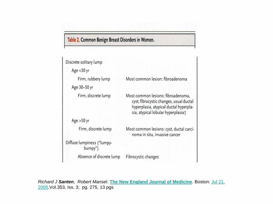
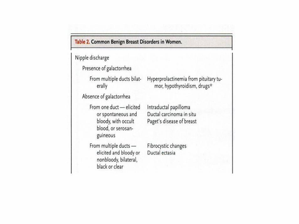
Richard J Santen, Robert Mansel. The New England Journal of Medicine. Boston: Jul 21, 2005.Vol.353, Iss. 3; pg. 275, 13 pgs
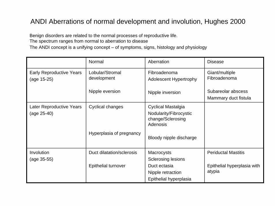
ANDI Aberrations of normal development and involution, Hughes 2000
Benign disorders are related to the normal processes of reproductive life. The spectrum ranges from normal to aberration to disease The ANDI concept is a unifying concept – of symptoms, signs, histology and physiology
Normal Aberration Disease
Early Reproductive Years(age 15-25)
Lobular/Stromaldevelopment
Nipple eversion
FibroadenomaAdolescent Hypertrophy
Nipple inversion
Giant/multiple Fibroadenoma
Subareolar abscessMammary duct fistula
Later Reproductive Years(age 25-40)
Cyclical changes
Hyperplasia of pregnancy
Cyclical MastalgiaNodularity/Fibrocystic change/SclerosingAdenosis
Bloody nipple discharge
Involution(age 35-55)
Duct dilatation/sclerosis
Epithelial turnover
MacrocystsSclerosing lesionsDuct ectasiaNipple retractionEpithelial hyperplasia
Periductal Mastitis
Epithelial hyperplasia with atypia

Fibrocystic Change
• Synonyms: Fibroadenosis, chronic mastitis, cystic hyperplasia, benign mammary dysplasia
• Lumpy-Bumpy breasts• Hormone related (between menarche and menopause• Unilateral or bilateral• Premenstrual pain and swelling, settle after period• Nipple discharge (various colours, mainly green-grey)

• A: Fibrocystic change• B: Lobular hyperplasia without atypia (adenosis)• C: Ductal hyperplasia without atypia• D: Florid ductal hyperplasia without atypia• E: Ductal hyperplasia with atypia [secondary lumens, hyperchromasia]
• F: Lobular hyperplasia with atypia [distended acini, monotonous cells]

Relationship between benign and malignant breast disease
• No increased riskAdenosisDuct ectasiaMild hyperplasia FibroadenomaCysts Apocrine change Apocrine metaplasia
• Slight increased risk (1.5 to 2 times)Moderate or florid hyperplasia Papilloma with fibro vascular core
• Moderate increased risk (4 to 5 times)Atypical ductal hyperplasia Atypical lobular hyperplasia
• High risk (8 to 10 times)Ductal carcinoma in-situLobular carcinoma in-situ
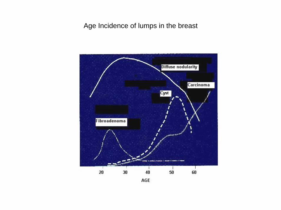
Age Incidence of lumps in the breast

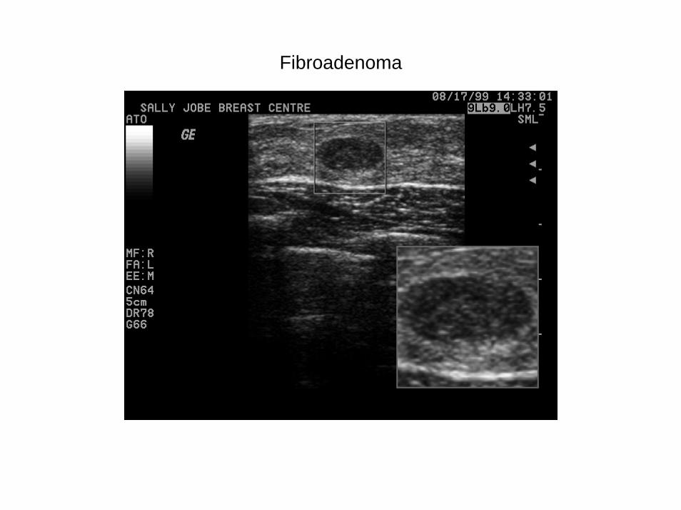
Fibroadenoma

Fibroadenoma
• Fibroadenomas are derived from the breast lobule, pathogenesis is unclear • They have both epithelial and connective tissue elements, therefore can undergo hormonaly
induced changes as in the rest of the breast.• They are not true neoplasms being polyclonal rather than monoclonal • The mass is composed of a proliferation of attenuated ducts in a loose, myxoid connective tissue.• Compress the surrounding breast.

Fibroadenoma
• Freely mobile and smooth (breast mouse)• Usually 2-3 cm in diameter • Usually young women• Decrease incidence approaching the menopause • May present has 'hard' calcified mass in the elderly • Approximately 10% of fibroadenomas are multiple• Half of adenomas resolve if <3cm over 5yrs• Adenomas >4cm should be excised (exclude phyllodes tumor)
Fibroadenoma

Fibroadenoma

Fibroadenoma
• Giant fibroadenomaCan be defined as > 5cmBimodal age presentation - teens & premenopausalRapidly grow to a large size Present with pain, breast distortion No evidence that these tumours recur
• Phyllodes tumourOccur in premenopausal women Wide spectrum of activityBenign to locally aggressive Have cellular fibrous elementIs a separate entity to fibroadenoma

Breast Cysts
• 7% of women will develop a clinically palpable cyst • Usually occurs in perimenopausal women• Due to relative excess oestrogen, usually in 4-5th decades• Fluctuate with menses• Highest prevalence is 45-55 years • 50% of these are solitary, 30% have 2-5 cysts and the rest more than 5 cysts • May appear suddenly and are frequently painful
Types of breast cysts :ApocrineAre lined by secretory epithelium (Na : K <3 ) Likely to have more than 5 cysts5 times likely to develop further cystsFlattenedLined by less active epithelium. (Na : K ratio = greater than 3 ) Fluid resembles plasma

Breast Cysts
• Beware of carcinoma if :The aspirate is blood stained There is a residual mass There is a persistent density in the Mammogram after cyst aspiration The cyst recurs on 3 occasions after aspiration The carcinoma is likely to be a composite of solid and cystic areas Intracystic carcinomas are rare (0.1%)

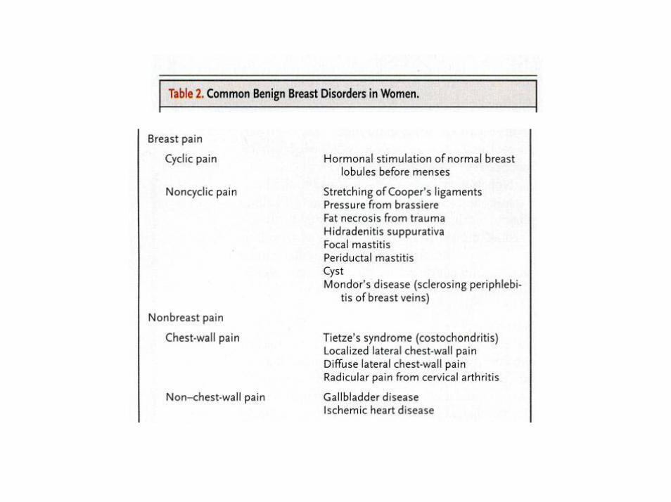
Pain
Cyclical mastalgiaYoung womenIncidence: 58% mild discomfort, 11% mod-severe painUsually bilateral, affects upper outer quadrant No consistent hormonal abnormality PremenstrualResolves at the onset of menses No definite link with caffeine, fat intake, psychological factors
Non cyclical mastalgia• True (breast related): usually unilateral, localized
– Enlarging cyst, ruptured ectatic duct, mastitis – Mondor’s Disease: thrombophlebitis,
spontaneous or following breast augment
• Musculoskeletal : costochondral (Tietze’s syndrome), lateral chest wall
Pain
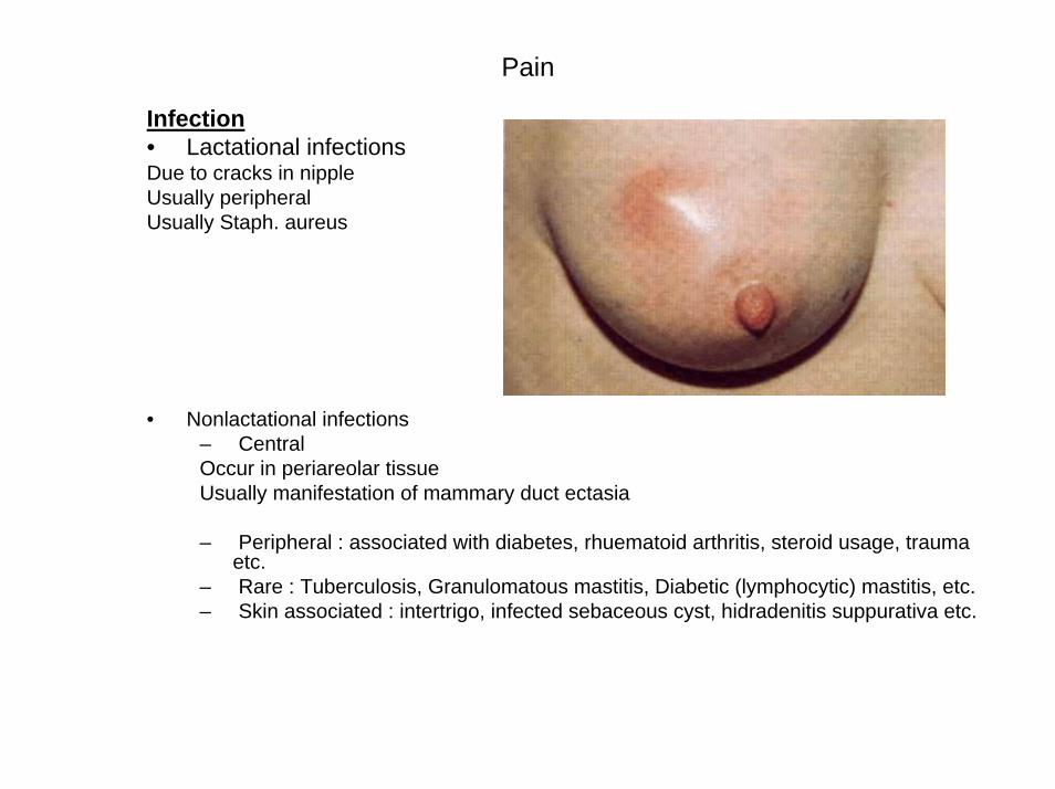
Infection• Lactational infectionsDue to cracks in nipple Usually peripheral Usually Staph. aureus
• Nonlactational infections– Central Occur in periareolar tissue Usually manifestation of mammary duct ectasia
– Peripheral : associated with diabetes, rhuematoid arthritis, steroid usage, trauma etc.
– Rare : Tuberculosis, Granulomatous mastitis, Diabetic (lymphocytic) mastitis, etc.– Skin associated : intertrigo, infected sebaceous cyst, hidradenitis suppurativa etc.
Mammary Duct Ectasia
• Synonyms: periductal mastitis, plasma cell mastitis• Women during or after menopause • More common in smokers • Caused by ducts beneath the nipples becoming dilated or clogged with fatty
material • Firm, tender, poorly defined swelling near the areola• Whole breast quadrant may be involved.• Nipple inversion results from traction on the nipple by ducts that have undergone
fibrosis in about 30% to 40% of cases • Toothpaste like nipple discharge.• Pathogenesis not clear but the primary event seems to be periductal inflammation
and duct ectasia is the ultimate outcome. • Both aerobic and anaerobic bacteria involved.• May be troublesome condition with recurrent abscesses and fistula formation
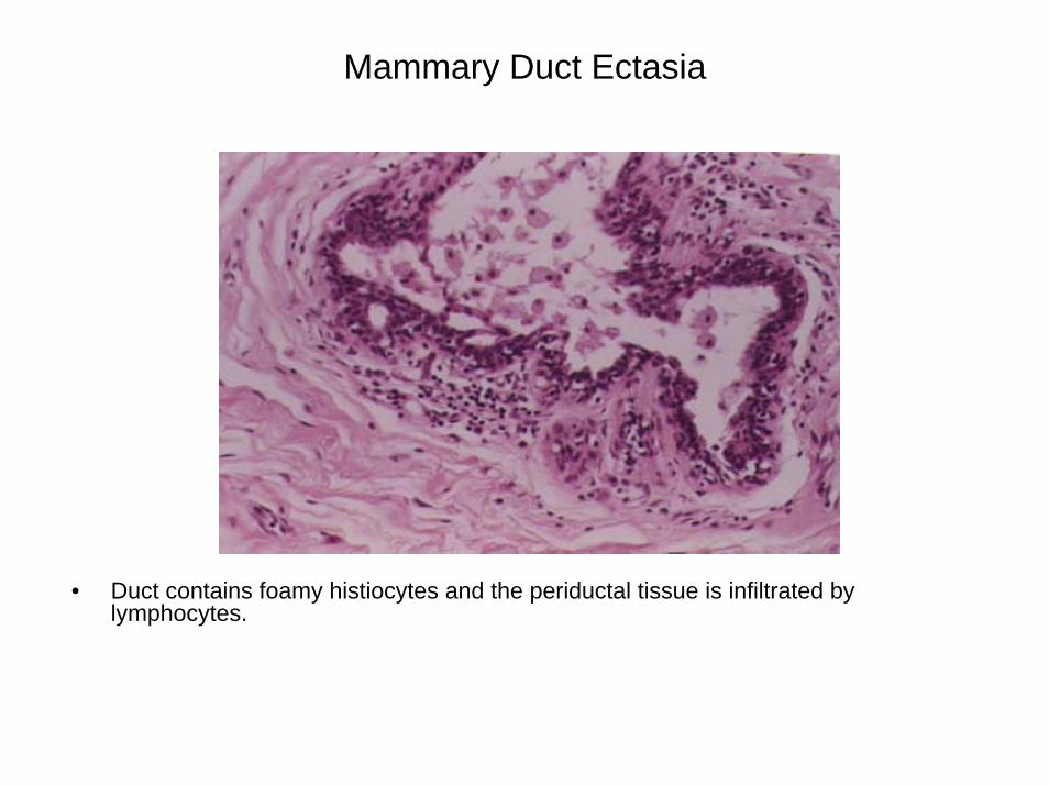
Mammary Duct Ectasia
• Duct contains foamy histiocytes and the periductal tissue is infiltrated by lymphocytes.

Mammary Duct Ectasia
• Fistula formation and scarring

Nipple Discharge
Description of nipple dischargeUnilateral or bilateral Single or multiple ducts ColourBlood-stained Spontaneous or expressed
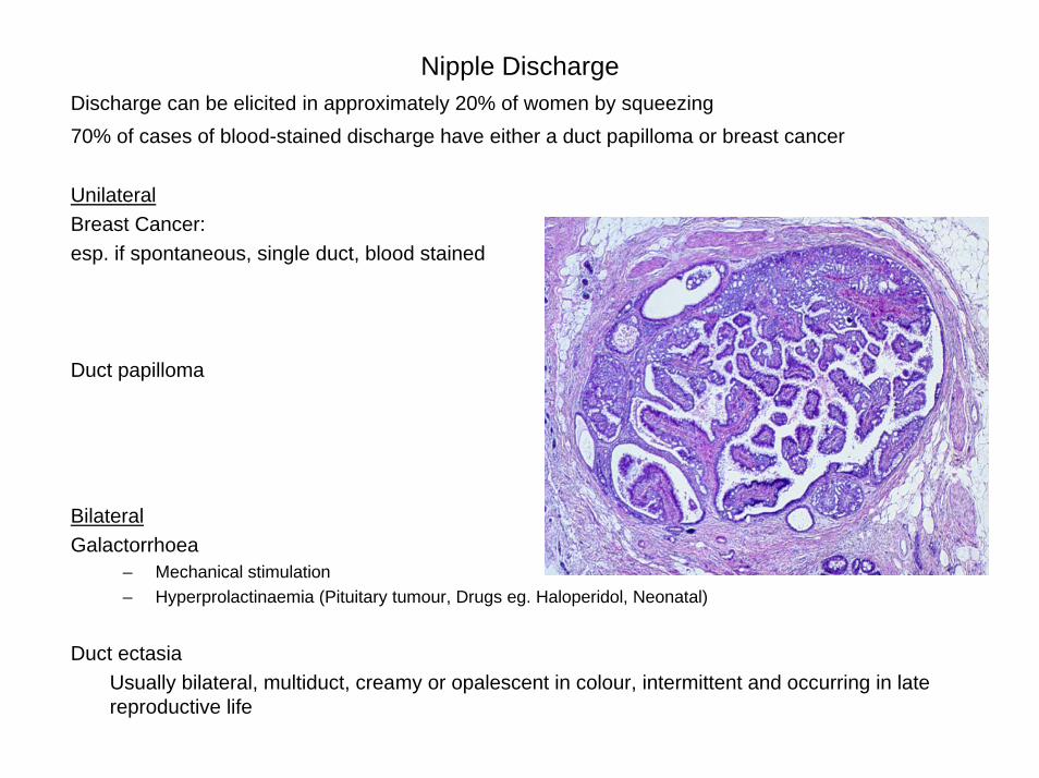
Nipple DischargeDischarge can be elicited in approximately 20% of women by squeezing 70% of cases of blood-stained discharge have either a duct papilloma or breast cancer
UnilateralBreast Cancer: esp. if spontaneous, single duct, blood stained
Duct papilloma
BilateralGalactorrhoea
– Mechanical stimulation– Hyperprolactinaemia (Pituitary tumour, Drugs eg. Haloperidol, Neonatal)
Duct ectasiaUsually bilateral, multiduct, creamy or opalescent in colour, intermittent and occurring in late reproductive life

Gynaecomastia• Aetiology• Most cases are idiopathic • Physiological causes are due to relative oestrogen excess • Physiological causes
– Neonatal – Puberty – Senile
• Pathological causes – Primary Testicular Failure
• Anorchia• Klinefelter's Syndrome • Bilateral Cryptorchidism• Acquired Testicular Failure • Mumps • Irradiation
– Secondary Testicular Failure • Generalised hypopituitarism• Isolated gonadotrophin deficiency
– Endocrine Tumours• Testicular • Adrenal • Pituitary
– Non-Endocrine Tumours• Bronchial carcinoma • Lymphoma • Hypernephroma
– Hepatic Disease • Cirrhosis • Haemochromatosis
– Drugs • Oestrogens and oestrogen agonists - digoxin, spironolactone• Hyperprolactinaemia - methyldopa, phenothiazines• Gonadotrophins• Testosterone target cell inhibitors - cimetidine, cyproterone Acetate

References
• Richard J Santen, Robert Mansel. Benign Breast Disorders. The New England Journal of Medicine. Boston: Jul 21, 2005.Vol.353, Iss. 3; pg. 275, 13 pgs
• Mr. Stephen Parker, Surgical-tutor.org.uk, Postgraduate revision notes 2005.
• Schwartz’s Principles of Surgery, 8th Edition 2005, pp. 453
• S. Lester. The Breast. Robbins Pathologic Basis of Disease 6th Ed. 1999. p.1093

END