PATHOLOGIAL STUDY OF BLADDER CANCER BY MAPPLING OF UROTHELIUM€¦ · CANCER BY MAPPLING OF...
15
Nagoya J. Med. Sci. 45. 1-15. 1982 Pathological Study of Bladder Cancer PATHOLOGIAL STUDY OF BLADDER CANCER BY MAPPLING OF UROTHELIUM TATSURO MURASE Department of Urology (Director: Prof H. Mitsuya) Nagoya University School of Medicine; Nagoya Japan ABSTRACT Using sixty-four cases of bladder cancer, the distribution ofcarcinomatous lesions, carcinoma in situ, and atypism were observed by step section of the entire urothelium. Carcinoma of the bladder was divided into four tumor types in accordance with the mapping of the disease foci, the patterns of tumor growth, and the DNA distribution pattern in the tumor cells. Type I was localized consisting mainly of papillary non-invastive tumors. Atypia was mild around the tumor, and neither atypia nor carcinoma in situ was seen in sites distant from the tumors. Type 2 was multifocal, non-invasive, and consisted mainly of papillary tumors. Type 3 was multifocal, invasive and multiple carcinomata in situ were present at sites distant from the tumors. Type 4 was localized and invasive with no findings suggestive of precancerous lesions in the bladder mucosa. Type 4 tumors grew very rapidly and were highly malignant. According to the retrospective analysis of the clinical course of each type, a plan for reasonable treatment of the bladder cancer was proposed as follows. Type I; This type of tumor may be controlled sufficiently by transurethral resection. In cases of T 2 (Bd or more, dissection of the lymph node is necessary. Type 2; This type may basically be controlled by transurethral resection, but careful follow-up is required because of high incidence of recurrence. When T 2 (Bd or more are diagnosed, total cystectomy is indicated. Type 3; Total cystectomy including the urethra and dissections of the lymph node are necessary. Type 4; Partial. sometimes total, cystectomy and dissection of the regional lymph node at an early stage are necessary. INTRODUCTION Of all tumors treated by urologists, bladder cancer has the highest incidence, but its natural history is not clear and at present no fixed treatment is available. In the case of radical surgery, the patients become urologically crippled due to the diversion of the urinary tract; consequently, great care must be exercised in total cystectomy. However, more than a few cases come to unfortunate results because of insufficient treatment. The prognosis of bladder cancer is customarily based on the assessment of histologic grade, type of tumor, and clinical pathological stage. The aim of this report was to clarify the natural history of bladder tumors and to establish a reasonable approach to the treatment of baldder tumors by type. In recent years, increasing attention has been directed to the potential prognostic importance of abnormality in the bladder epithelium. In 1952, Melicow (I) and Melicowand Hollowell (2) reported that epithelial abnormalities, ranging from hyperplasia to carcinoma in situ, might be observed in incidental biopsy material and by histologic examination of seemingly unaffected bladder epithelium in cystectomy specimens. They pointed out the difficulty in cystoscopic identification of such lesions. Our study demonstrates the distribution of epithelial lesions in surgically-removed cancerous bladders by a complete histologic examination. Melamed (3), Koss (4), Soto (5), Skinner, (6) and Cooper et al. (7) studied the continuity and discontinuity of tumor foci by means of step sectioning of total cystectomy specimens, Received for Publication May 7. 1982
Transcript of PATHOLOGIAL STUDY OF BLADDER CANCER BY MAPPLING OF UROTHELIUM€¦ · CANCER BY MAPPLING OF...

Nagoya J. Med. Sci. 45. 1-15. 1982
Pathological Study of Bladder Cancer
PATHOLOGIAL STUDY OF BLADDERCANCER BY MAPPLING OF UROTHELIUM
TATSURO MURASE
Department of Urology (Director: Prof H. Mitsuya)Nagoya University School of Medicine; Nagoya Japan
ABSTRACT
Using sixty-four cases of bladder cancer, the distribution of carcinomatous lesions, carcinoma in situ,and atypism were observed by step section of the entire urothelium. Carcinoma of the bladder was dividedinto four tumor types in accordance with the mapping of the disease foci, the patterns of tumor growth,and the DNA distribution pattern in the tumor cells. Type I was localized consisting mainly of papillarynon-invastive tumors. Atypia was mild around the tumor, and neither atypia nor carcinoma in situ wasseen in sites distant from the tumors. Type 2 was multifocal, non-invasive, and consisted mainly ofpapillary tumors. Type 3 was multifocal, invasive and multiple carcinomata in situ were present at sitesdistant from the tumors. Type 4 was localized and invasive with no findings suggestive of precancerouslesions in the bladder mucosa. Type 4 tumors grew very rapidly and were highly malignant.
According to the retrospective analysis of the clinical course of each type, a plan for reasonabletreatment of the bladder cancer was proposed as follows. Type I; This type of tumor may be controlledsufficiently by transurethral resection. In cases of T2 (Bd or more, dissection of the lymph node isnecessary. Type 2; This type may basically be controlled by transurethral resection, but careful follow-upis required because of high incidence of recurrence. When T 2 (Bd or more are diagnosed, total cystectomyis indicated. Type 3; Total cystectomy including the urethra and dissections of the lymph node arenecessary. Type 4; Partial. sometimes total, cystectomy and dissection of the regional lymph node at anearly stage are necessary.
INTRODUCTION
Of all tumors treated by urologists, bladder cancer has the highest incidence, but its naturalhistory is not clear and at present no fixed treatment is available. In the case of radicalsurgery, the patients become urologically crippled due to the diversion of the urinary tract;consequently, great care must be exercised in total cystectomy. However, more than a fewcases come to unfortunate results because of insufficient treatment. The prognosis of bladdercancer is customarily based on the assessment of histologic grade, type of tumor, and clinicalpathological stage. The aim of this report was to clarify the natural history of bladder tumorsand to establish a reasonable approach to the treatment of baldder tumors by type.
In recent years, increasing attention has been directed to the potential prognosticimportance of abnormality in the bladder epithelium. In 1952, Melicow (I) and MelicowandHollowell (2) reported that epithelial abnormalities, ranging from hyperplasia to carcinomain situ, might be observed in incidental biopsy material and by histologic examination ofseemingly unaffected bladder epithelium in cystectomy specimens. They pointed out thedifficulty in cystoscopic identification of such lesions. Our study demonstrates thedistribution of epithelial lesions in surgically-removed cancerous bladders by a completehistologic examination.
Melamed (3), Koss (4), Soto (5), Skinner, (6) and Cooper et al. (7) studied the continuityand discontinuity of tumor foci by means of step sectioning of total cystectomy specimens,
Received for Publication May 7. 1982

2 T. MURASE
and showed that the tumors were multifocal intra-epithelial carcinomas. This mappingmethod of disease foci showed the lesions, which multiply with time, at each stage of bladdercarcinoma and therefore provided suitable material for estimating the natural history of thebladder cancer.
In our work, focal mapping of the bladder cancer was performed on 64 of the patients whounderwent radical total cystectomy. The tumors were classified into four types according tothe disease foci conditions as seen from mapping of the growth pattern of the tumors. Some ofthe tissues were subjected to Feulgen's staining. Because Feulgen reagent reacts to nucleardeoxyribonucleic acid (DNA), the DNA content in cell nuclei is easily determined.Microspectrophotometric values obtained by this method are found to be consistent withthose obtained biochemically. Nuclear DNA as well as chromosomal number are constant inthe same animal except for specific tissues (8). A comparative investigation was made of thehistograms showing the quantities of DNA in the nuclei of the tumor cells.
MATERIALS AND METHODS
The patients were selected for cystectomy in the Nagoya University Hospital and NationalCancer Center because they had failed to respond to local treatment and also because biopsyevidence indicated the presence of widespread disease of the bladder epithelium or deepinvasions of the bladder wall.
Table I summarizes the clinical and pathological data on 64 patients prior to and aftercystectomy. There were 53 men and I I women. At the onset, the youngest patient was 33 yearsold, the oldest 75. At the time of cystectomy the youngest was 35 years old, the oldest 75. Theduration of known carcinoma prior to cystectomy ranged from I to 240 months, averaging23.9 months.
Freshly removed bladders were cut open the front surface of the bladders extirpated duringsurgery together with the prostate gland in males and the urethra in females, then spreadstretched, and pinned down on a piece of stiff wooden board, and fixed in 10% bufferedformal dehyde solution. Following a 48-hour fixation, the entire epithelium of the bladderwas cut into appropriately identified tissue blocks measuring about 3.5 x 0.5 em and about Iem in thickness. Approximately 100 blocks were required to process the bladder in toto.Mapping was performed on the parts of the bladder with atypical hyperplasia (hereafterreferred to as ATP) and carcinoma in situ. The criteria of Koss were used in diagnosis of theATP. carcinoma in situ, and grade of cancer (4). The stages of the bladder cancer wereclassified according to Jewett (9) and the UICC (10). A schematic diagram was adopted toshow the distribution of cancer lesions, carcinoma in situ, and atypical hyperplasia. Growthpatterns, of turmor, carcinoma in situ, and non-papillary invasive carcinoma wereinvestigated. The presence or absence of lymph node metastasis was also checked in each case.Analysis of the foregoing pathological study of carcinoma of the bladder was divided into thefollowing four types.
Type I tumors were unifocal, localized and papillary. They were mainly non-invasive, butwhen invasive, they showed a rpedominantly broad fron invasion. ATP was present in thearea surrounding the tumors, but there was no ATP or carcinoma in situ at sites distant fromthe tumors. The grade was I or 2 in almost all cases. Type 2 tumors were multifocal andpapillary. In the mapping, there were many ATP and microscopic papillary projections notonly in the vicinity of the tumors but also at sites distant from them. The grade of malignancywas 1-2 in almost all cases.

PATHOLOGIAL STUDY OF BLADDER CANCER 3
Table I. Clinical and pathological findings before and after cystectomy
Duration of
No. Age Sex Symtoms CystoscopicPast treatment
cystectomyStage rade lymph
findings from first g node metasymptom
61 M Hematuria papillary IBM PT\ A 1-2
2 53 M Burning papillary sessile TUC X 4 51M PT, B, 3Hematuria non papillary
3 58 M Hematuria papillary sessile segmental resection 6M PT, B2 3
4 68 F Burning non papillary 7M PT\ A 2
5 43 M Hematuria papillary IBM PTa 0
6 63 M Hematuria non papillary radiation 7M PT, D 3 +
7 62 M Hematuria non papillary Tumorectomy60M PTD 3 +by section alta
8 74 M Hematuria non papillary TUR 1M PT, B2 3
9 73 M Hematuria non papillary segmental resection 19M PT, B2 3
10 39 F Frequency non papillary 16M PT, B2 3 +
II 57 M Burning papillary 16M PT, B2 3 +
12 46 M Hematuria papillary TUC60M PTa 0 2segmental resection
13 55 M Hematuria papillary 13M PT1 A 2
14 64 M Hematuria papillary TUCX 3 20M PTa 0 2
15 52 M Hematuria papillary TUR X 3 35M PT\ A 2
16 59 M Hematuria papillary TUR X 3 24M PT1A 1-2
17 65 M Hematuria papillary 4M PT2 B1 2
18 66 M Hematuria non papillary segmental resection 13M PT\ A 2
19 66 M Hematuria non papillary Tumorectomy25M PT, B2 2-3by section alta
20 66 M Hematuria papillary TumorectomyIBM PT2 B\by section alta
21 67 M Hematuria papillaryTumorectomy
29M PT, A 3 +by section alta
22 46 M Micro non papillary segmental resection 12M PT, B2 3hematuria
23 75 F Hematuria papillary 2M PT2B\ 2 0
24 72 M T urbia urine non papillary 2M PT, D 3 +

4 T. MURASE
Duration of
No. Age Sex SymtomsCystoscopic Past treatment
cystectomy Stage grade lymphfindings from first node mela
symptom
25 71 M Burning non papillary 8M PT, B2 3 0
26 63 M Hematuria non papillary rediation 36M PT3 D 3 +
27 42 FHematuria papillary 1M PT, A 2-3Burning
28 33 M Hematuria non papillary Tumorectomy 6M PT3 D 3 +by section alta
29 72 F Hematuria non papillary 1M PT, D 3 +
30 71 M Hematuria non papillary 10M PT,D 3 +
31 46 M Micro non papillary radiation 26M PT, D 3hematuria
32 70 FBurning non papillary 36M PT, D 3 +Hematuria
33 52 F Hematuria non papillary 120M PT, D 3 +
34 75 M Hematuria papillary segmental resection 60M PT, D 3 +TUR X4
35 62 M Hematuria non papillary TUC X I 16M PT3 D 3 +
36 68 M Hematuria non papillary 4M PT3 D 2-3 +
37 70 F Hematuria non papillary TUCX4 240M PT,A 2-segmental resection
38 44 M Hematuria non papillary segmental resection 12M PT3 D 3 +
39 72 M Frequency non papillary 24M PTIA 2
40 58 M Frequency non papillary 38M PT3 B2 3
41 65 M Hematuria papillary TUR X I 12M PTa 0 2
42 44 M Hematuria papillary 30M PTa 0 1-2
43 59 M Hematuria papillary 24M PTa 0 2
44 62 M Hematuria papillary 3M PT, A 2
45 70 M Hematuria non papillary 2M PIS 0 3
46 73 M Hematuria papillary TUCX 3 60M P, A 2
47 46 M Frequency non papillary 4M P, B, 3
48 57 M Hematuria non papillary radiation 26M P, C 3

PATHOLOGIAL STUDY OF BLADDER CANCER
Duration of
No. Age Sex SymtomsCystoscopic
Past treatmentcystectomy
Stage grade lymphfindings from first node meta
symptom
49 53 M Burning non papillary TURX I 26M P, C 3
50 58 M Hematuria non papillary 4M P, BI 3
51 35 M Hematuria papillary radiation 20M PTa 0 2
52 46 M Hematuria papillary 40M PT,A 2
53 49 M Frequency papillary TUCX I 8M PTa A 2Burning segmental resection
54 38 M Hematuria papillary 8M PTa 0 2
55 71 M Hematuria papillary 3M PT, A 2
56 69 M Hematuria non papillary 4M PT3C 3 +
57 63 F Hematuria non papillary TURX I 6M PT, BI 3
58 64 M Burning non papillary TURX I 4M PT, B1 3
59 71 M Hematuria papillary TURX I %M PT\ A 2
60 64 F Hematuria non papillary 6M PT3 D 2-3 +
61 63 M Hematuria papillary 10M PT, A 2
62 57 M Hematuria papillary 4M PI A 2
63 67 M Hematuria papillary TURX I 6M PTJ A 2
64 58 M Hematuria non papillary 3M PT, A 3
TUC: transurethral cagulationTUR: transurethral resection
5
Type 3 tumors were non-papillary, invasive, or a mixture of papillary, non-invasive, andinvasive tumors, mostly graded 2 or 3. They were multifocal, with ATP and carcinoma in situfound not only around the tumors but also in sited distant from them. Type 4 consisted oflocalized, non-papillary invasive tumors deeply invading the muscle layer at an early stage.The invasion was tentacular. No carcinoma in situ or ATP was seen at sited distant from thetumor.
Some tissues were subjected to Feuglen's staining and the amount of DNA in 100 cancercells was measured. As a control, lymphocytes in the same tissue were measured. In order todescribe these DNA distribution patterns the following terms have been employed. Themodal value has been called "2C" if it coresponds to a normal diploid population and "4C" ifit corresponds to a normal tetraploid population. The DN A value most frequently foud in thelymphocytes was 2C. Olympus florescent measuring system was used as the measuringequipment. A MH SP-lF green filter was used with a wave length of 610 nm.

6 T. MURASE
RESULTS
Table 2 shows four types of bladder cancer. Only 5 cystectomies were performed for type Itumors, which were mainly of the giant papaillary type. Cases up to B1(PT2) were all that wereobserved and there was no case of lymph node metastasis nor any case of grade 3 malinancy.
Type 2 was observed in 24 cases. They were also cases of early stage up to stage O-stage BI
(PTa-PT2). Most cases were graded I or 2, and no metastasis to the lymph node was observed.Type 3 was present in 27 cases including those from stage 0 (PTIS) to stage D (PT4). Inadvanced stage cases, there was lymph node metastasis. Five cases of recurrence in the urethraafter total cystectomy were found. There were cases with a mixture of grades 2 and 3 or thosewith only grade 3 and some with only grade 2. Tyep 4, though relatively rare, was observed inseven cases. The stage ranged from stage 0 (PTIS) to stage D (PT4), and lymph node metastasiswas seen in advanced stage cases. Figures I, 2, 3, 4, show the mapping from types 1 to 4 witheach starting from the early stage and may serve as a tool to surmised the natural history ofeach type of bladder cancer. The DNA distribution pattern was also obtained.
Figure 1 shows the type 1 cases according to stage. Figure 1-1 illustrates a stage 0 (PTa)tumor with no ATP at other sites. Figure 1-2 shows a case of a giant papillary tumor in whichthe patient was initially diagnosed as having bladder cancer but refused to undergocystectomy. After 8 years, hematuria became severe and total cystectomy was performed. Thestage was A (PT 1). Figure 1-3 shows preservation of the papillary morphology and invasion ofthe tumor up to the middle of the muscle layer. The invasion was of the broad fron invasiontype. In figures 1-2 and 1-3, the ATP and carcinoma in situ in the other epithelium wereobserved only in. the vicinity of the neoplasm. Figure 1-1 represents grade I and the DNAdistribution mode is 2C. Figure 1-2 and 1-3 show modes in 3c. It is possible for type I tumorsto remain at an early stage over long periods of time. Figure 2shows type 2 cases according tostage. Figure 2-1 is a small papillary tumor with multiple ATP. The stage is stage 0 (PTa) andthe grade is 2. Figure 2-1 shows a tumor which may change into one such as presented inFigure 2-2 after a certain degree ofgrowth. The tumor in Figure 2-2 is also graded 2. Figure 23 depicts a giant papillary tumor mixed with a small papillary tumor and ATP being observedextensively. The stage was A (PT1) and the grade 2. Figure 2-4 shows tumors one year and sixmonths after onset. These were multifocal tumors including a 5 x 5 cm giant papillary tumor
Table 2. Post surgical stage and 4 types of bladder cancer
PTa (PTis) PT t PT 2 PT 3 PT.
Type 1 0 3 2 0 0
Type 2 88 15 1 0 0
Type 3 o (1) 12 9 5(3) 1 (1)
Type 4 0 1 2(1) 3(3) 1(1)
) : cases of lymph node metastases.

1 •
PATHOLOGIAL STUDY OF BLADDER CANCER
1 •
2
<:~2" Be
3
7
Fig. I Mapping of bladder cancer and DNA values in Type I
~ ca in situ and atypical hyperplasia_ cancer
Figure I-I A stage 0 (PTa), grade I papillary tumor. No ATP in other sites was found by mapping. TheDNA histogram of the tumor showed a mode in the vicinity of 2C.
Figure 1-2 A stage A (PT,), grade 2 tumor with an 8-year course from the initial symptoms until totalcystectomy. A giant papillary tumor, it has remained at stage A (PT,). There is no ATP or microscopicpapillary elevations anywhere else on the bladder. No carcinoma in situ is noted. The DNA histogram showeda mode near 3C. n = number of cell nuclei measured.
In evaluation of continuous changes estimated from figure I-I to figure 1-3, no cases in Figure I evidencedlymph node metastasis.
with slight invasion of the muscle layer; the stage was B1(PT2) and the grade 2. Type 2tumorswere considered to be a multifocal variation of type I. This kind of tumor can also becomegiant, but even when the entire bladder was occupied by many tumors, there was only slightinvasion of the muscular layer. Such tumors appear to remain at an early stage for a longperiod of time.
The DNA distribution was investigated for the tumors in figures 2-3 and 2-4, but the modewas found to be approximately 3C, which was almost the same as that of type I. There werealso many cells with 4C or more. Type 3 tumors were invasive, multifocal, and sometimesthere was a mixture of invasive and non-invasive tumors. In the mapping, many carcinomasin situ were seen. Figure 3 shows type 3 cases ranging from stage 0 (PTis) to stage D (PT4). Itwas possible to estimate continuous changes from carcinoma in situ to stage D (PT4). Figure3-1 shows the case of a patient who came to the hospital with the symptoms of cystitis.Cystoscopy revealed no tumor. The cytology was continuously positive and total cystectomy

8 T. MURASE
2
~~
3
nl%j
~._.;....-.,~
2 •
4
Fig. 2 Mapping of bladder cancer and DNA values in Type 2
Figure 2-1 A case with many stage 0 (PTa), grade 1-2 multifocal small papillary tumors with considerableATP and many microscopic papillary elevations. The DNA histogram showed a mode near 3C.
Figure 2-2 A tumor type of stage A (PT,) and grade 2considered to be an advanced form of the tumor typein figure 2-1. Note the many papillary tumors.
Figure 2-3 A giant papillary tumor of stage A (PT,) and grade 2 present together with a small papillarytumor. ATP and microscopic papillary elevations were also found at sites distant from the tumor.
Figure 2-4 Bladder carcinoma consisting ofa single stage B, (PT2), grade 2giant papillary tumor and manypapillary tumors. The giant papillary tumor has begun to invade the muscle layer. The mode of DNAdistribution' is near 3C.
Continuous changes are inferred from figure 2-1 - figure 2-2 - figure 2-3 - figure 2-4. No cases in Figure 2indicated lymph node metastasis.
was performed. Multiple carcinoma in situ was observed. Figure 3-2 indicates a small nonpapillary tumor with submucosal invasion. The stage was A (PT I ) with grade 3 malignancy.Figure 3-3 shows multiple carcinoma in situ in a non-papillary tumor' with invasion of thecancer into the lymphatic vessels of the muscle layer. The stage was B1(PT2) with grade 2 andgrade 3.
Figure 3-4 illustrates a non-papillary tumor of stage C (PT3) and grade 3 with a wideranging carcinoma in situ. After total cystectomy, recurrence of the cancer was noted in theurethra. Type 3 was found to be multiple from the focal mapping with the possibility ofcarcinoma in situ existing in the urethra. This type is considered liable to have recurrence inthe urethra. Figure 3-5 represents an advanced case of the tumor shown in Figure 3-4. Cancerwas also found in the fatty tissue surrounding the bladder. Metastasis in the lymph nodes wasalso noted. The stage was D (PT4).

PATHOLOGIAL STUDY OF BLADDER CANCER 9
o'P
;;·Il~'!riIJlI~I~)i7ffiTi:;'
Fig. 3 Mapping of bladder cancer and DNA values in Type 3
Figure 3-1 A case of total cystectomy with many carcinoma in situ.Figure 3-2 Multifocal stage A (PT,), grade 2-3 non-papillary tumors with partial submucosal invasion in
some foci. Carcinoma in situ and ATP are also present at sites distant from the tumor. DNA was in a modenear Sc. The mode was dispersed.
Figure 3-3 A mixed stage B, (PT2), grade 2-3 non-papillary and papillary tumors. There are manycarcinoma in situ. Cancer cells were found in the lymphatic vessels of the muscle layer at the site of the nonpapillary tumor. The DNA showed mode dispersion with a mode near 5C.
Figure 3-4 A stage B2 (PT3), grade 3 non-papillary tumor showing invasion of the muscle layer. There aremany carcinoma in situ. Recurrence in the urethra was observed one year after total cystectomy.
Figure 3-5 A stage C (PT3), grade 3 non-papillary tumor together with many carcinoma in situ. There aremetastases to the lymph nodes of the internal iliac artery.
The DNA histogram of many cells showed 4C or more and a mode of about 5C with thedistribution pattern of tumor cells becoming larger (Figures 3-1 and 3-3). Type 4 was the mostmalignant and rapidly progressing; the grade was 3 in all cases. Figure 4 represents cases fromstage A (PT,) to D (PT4). This type had a localized focus with no carcinoma in situ in the areafar from the tumor. Figure 4-1 shows a non-papillary carcinoma with a small focus. The stagewas A (PT1) and the grade high. Figure 4-2 illustrates a stage B, (PT2) tumor with singlemetastasis to the lung. The lung was partially resected. Figure 4-3 shows tentacular invasionto the fatty tissue around the bladder and lymph node metastasis. The stage was C (PT3).
Figure 4-4 shows a case which reached stage D (PT4) in two years from the initial symptoms.During surgery, tumor cells were found in the ascites. The stage was D (PT4) and the grade 3.There were no stage B2 (PT3) patients who survived for more than five years. The DN Ahistograms in Figures 4-1, 4-3 and 4-4 showed many cells with DNA of 8C or more. Thedispersion of mode was remarkable.
10 T. MURASE
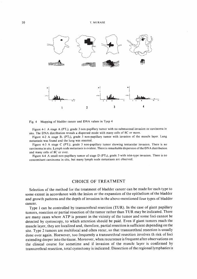
U \5 £=~~ uumrmmtmlll ~ •~~
Fig. 4 Mapping of bladder cancer and DNA values in Tyep 4
Figure 4-1 A stage A CPT,), grade 3 non-papil1ary tumor with no submucosal invasion or carcinoma insitu. The DNA distribution reveals a dispersed mode with many cells of 8C or more.
Figure 4-2 A stage BI (PT2), grade 3 non-papil1ary tumor with invasion of the muscle layer. Lungmetastasis was found and the lung was resected.
Figure 4-3 A stage C (PT l ), grade 3 non-papil1ary tumor showing tentacular invasion. There is nocarcinoma in situ. Lymph node metastasis is evident. There is remarkable dispersion of the DNA distributionand many cells of 8C or over.
Figure 4-4 A smal1 non-papillary tumor of stage D (PT.), grade 3 with islet-type invasion. There is noconcomitant carcinoma in situ, but many lymph node metastases are observed.
CHOICE OF TREATMENT
Selection of the method for the treatment of bladder cancer can be made for each type tosome extent in accordance with the lesion or the expansion of the epithelium of the bladderand growth patterns and the depth of invasion in the above-mentioned four types of bladdercancer.
Type I can be controlled by transurethral resection (TUR). In the case of giant papillarytumors, resection or partial resection of the tumor rather than TUR may be indicated. Thereare many cases where ATP is present in the vicinity of the tumor and some foci cannot bedetected by cystoscopy, to which attention should be paid. Even if giant tumors reach themuscle layer, they are localized and, therefore, partial resection is sufficient depending on thesite. Type 2 tumors are multifocal and often recur, so that transurethral resection is usuallydone over again. Hoewever, too frequently a transurethral resection involves th risk of fociextending deeper into the tissue. Moreover, when recurrence is frequent after observations onthe clinical course for sometime and if invasion of the muscle layer is confirmed bytransurethral resection, total cystectomy is indicated. Dissection ofthe regional lymphatics is

PATHOLOGIAL STUDY OF BLADDER CANCER 11
not always necessary, but resection of the urethra is needed since the tumors are multifocaland there is a possibility of recurrences in the urethra. Type 3 tumors are multiple andinvasive, so that controlling the tumor by transurethral resection is impossible. Totalcystectomy and dissection ofthe regional lymphatics are indicated. Resection of the urethra isalso necessary since the tumors are multifocal and there is a possibility of carcinoma in situ inthe urethra. Since a type 4 tumor progresses rapidly, total cystectomy, in case partialcystectomy should be performed without delay. dissection of the regional lymphatics is alsonecessary.
DISCUSSION
It has been repeatedly stressed that the prognosis of bladder cancer is greatly influenced bythe depth the cancer has reached and the accompanying lymph node metastasis, but thenatural history of the bladder cancer advancing from a surface tumor or carcinoma in situ toan invasive tumor, is not always clear. In clarifying the natural history of bladder cancer,studies on the carcinoma in situ, the original form of bladder cancer, are useful andsignificant.
Melicow (2) was the first to recognize the histological similarities between carcinoma of thebladder and Bowen's disease and to introduce the concept of carcinoma in situ in carcinomaof the bladder as subclinical preinvasive carcinoma and intraurethelial cancer. Heinvestigated the seemingly normal epithelium of the bladder separated from the main tumorin specimens of total cystectomies and frequently found carcinoma in situ. After that,Eisenberg (16) and Simmon et al. (17) reported similar findings. Shade et al. (18) foundprecancerous lesions or severe atypia in 80% of biopsy specimens of bladder mucosa whichwas unrelated to the tumor. Carcinoma in situ delt with in the reports hitherto published wasone accompanying carcinoma of the bladder, clearly observable macroscopically, and notdiscussed in connection with the course of growth of carcinoma of the bladder.
However, Melamed (19) reported cases which were negative for cystoscopy but positive forurinary cytology, subsequently mentioning that cancer became invasive in eight out of 25cases of carcinoma in situ. Farrow et al. (20) also made follow-up investigations ofcarcinomain situ in 69 cases, reporting that the carcinoma became invasive within five years in 37 casesand within three years in most cases. Koss (21) and Yate et al. (22) noted the possibility ofcarcinoma' in situ developing into non-papillary or papillary carcinoma and showed thenatural history of baldder cancer. Althausen et al. (23) reported that 42% of accompanyingcarcinoma in situ developed into invasive carcinoma within 5 years. The reports above haveclarified the history from carcinoma in situ to invasive cancer, but the natural history ofpapillary tumors is not clear. Papillary type cancer does not always follow the growthpatterns as described by WHO (24) or Pugh's diagram (25).
While the above-mentioned reports were taken into consideration, the mapping andgrowth patterns of many foci were investigated simultaneously and the natural hisory ofcarcinoma of the bladder was divided into four types for study. It became clear that the waythe carcinoma spread was not always the same, and that the rate of spread varied withexpansion in the epithelium of the bladder and the growth pattern.
The difference between types I and 2 according to the authors' classification arises from thepresence of unifocallocalized tumors and multifocal tumors. Investigations of the mappingof overall specimens of the multifocal type (type 2) reveal that ATP and low grade carcinomain situ cannot always be confirmed by cystoscopy. This suggests that the ATP and low grade

12 T. MURASE
carcinoma in situ become the focus of "recurrence" even in a tumor-free state. On the otherhand, there are cases which have no recurrence whatsover after one transurethral resectionand the recurrence rate after transurethral resection is reported as 40-70% (26) (27) (28). Theproblem of recurrence appears to depend on whether the bladder cancer is of the localized ormultifocal type.
Types I and 2 are papillary. In almost all of the cases under study, the stage did not advancebeyond A (PI). Four cases advanced to stage BI and no tumor developed to stage B2(P3) orbeyond. Nor was there any lymph node metastasis. Grade 3 papillary tumors existed but wererare. According to Koss (4), Grade 3 papillary tumors is often present as a complication withinvasive cancers in other sites. No grade 3 cases were present among types I and 2 papillarytumors in this study. All of the tumors which had developed to stage BI were cases of broadfront invasion. As the method of treatment, transurethral resection is basically employedboth for types I and 2 tumors. When transurethral resection is difficult, as in the case of agiant tumor occasionally observed with type I, highly radical effects can be obtained bytumor resection or by partial resection of the bladder. Cases of type 2 are similar to type I inthe ON A distribution pattern, are graded I or 2, and are not high in biological potential.Since considerable time is required to reach stage B2, it is preferable to follow such cases withtransurethral resection for a while and then to perform total cystectomy when invasion of themuscle layer has been confirmed. For tyep 2, partial resection runs the risk of the tumor beingembedded in the muscle layer. A detailed examination by multiple biopsies is necessary todifferentiate between types I and 2.
Type 3 is the one most frequently observed in bladder mapping by Melamed, (3) Koss (4)and others. Both type 3 and type 2 are similar in being multifocal but differ in that the formeris invasive. With type 3, there are many carcinomas in situ not only near the invasive tumorbut also at sites distant from it as confirmed in many investigation hitherto made on totalcystectomy specimens. Skinner (6) found carcinoma in situ in.44% of total cystectomyspecimens from 59 patient. Cooper et al. (7) found severe atypia (carcinoma in situ) in 39% ofsites distant from the foci of carcinoma of the bladder. There have also been reports thatcarcinoma in situ and ATP are found in the ureter (29) (30) (31) or urethra (32) (33) as lesionsconcomitant with bladder cancer. Carcinomas of the bladder are basically consideredmultifocal. This has been supported by investigations using mapping or giant sections.However, it does not mean that carcinoma of the bladder on the whole is multifocal since type3 accounts for most of the cases of total cystectoymy.
In the mapping of 64 cases in the present study, 27 were of type 3. There were not so manymultifocal invasive cases as compared with the reports of Koss (4) and others using giantsections. Tyep 3 appeared to represent one type of bladder cancer, which is both multifocaland invasive. Needless to say, multiple biopsies are necessary for diagnosis of type 3.
Generally, type 3 is said to have a poor prognosis; hence, the largest number of cases of totalcystectomies are of type 3. Also, since the possibility of type 3 having recurrences in theurethra is considered to be high, resection of the urethra simultaneously with total cystectomyis indicated. Richies (34) mentioned the presence of intraepithlial carcinoma in the neck ofthebladder and the urethra as an indication for urethral resection in cases of total cystectomy.However, it is difficult to find carcinoma is situ in the urethra preoperatively.
The presence of type 4 was most conspicuous in this focal mapping. This type showed thetentacular invasion described by Mostofi (35) and lymphatic invasion at an early stage. Sotoet al. (5) have reported broad front invasion in papillary tumors and tentacular type invasionin solid types. Types I and 2 show broad front invasion and type 3 shows both broad front andtentacular invasion. Tyep 4 generally shows only tentacular invasion and is considered a type

PATHOLOGIAL STUDY OF BLADDER CANCER 13
of cancer which is concentrated at one point and becomes invasive at an early stage. Therehave been few reports on type 4. Soto (5) has reported a case, although rare, in which thetumors are localized without carcinoma in situ or ATP and in which tentacular invasion isobserved. Mitani (36) has classified morphologically an irregular-type tumor in which deathoccurs within two years. Such a tumorconsiderd to be equivalent to t-ype 4. Most of the type 4tumors are grade 3 or undifferentiated carcinoma.
Various investigators (37) (38) (39) have stated that the bladder epithelial cells of normalhuman subjects and mice are polypoid, consisting of tetraploid and octaploid cells as well asdiploid cells.
In this study DNA values of the normal bladder were diploid. The DNA histogramsrevealed the same dispersion for tyeps I and 2 tumor histograms. Type 3 showed moredispersion, which agrees with reports of various authors (40) (41) (42) that the mode of ahistogram of nuclear DNA values becomes fiat and that the incidence of polypoid cellsbecomes higher with an increase in the grade of cancer. With type 4 the histogram is flatterthan that of type 3 and there is a higher degree of atypia in the cancer cells. This type isdifferent from type 3, likewise an invasive type, in the rate of invasion of the muscle andlymphatic vessels. Treatment requires immediate radical extirpation evenat an early stage.Treatment by partial cystectomy is also possible because the focus is localized. Resnick et al.(43) and Novick et al. (44) have reported that the survival rate after partial cystectomy is highin the case of unifocal tumros.
AKNOWLEDGEMENT
The author wishes to thank Prof. Hideo Mitsuya for his advice and review of themanuscript.
REFERENCES
I) Melicow MM: Histological study of vesical urothelium intervening between gross neoplasms in totalcystectomy. J. Ural, 68,261-278, 1952.
2) Melicow MM, Hollowell JW: Intraurothelial cancer: Carcinom in situ, Bowen's disease of the urinary system:Discussion of thirty cases. J. Ural., 68,763-772, 1952.
3) Melamed, M. R., Grabstald, H. and Whitmore, W. F. Jr.: Carcinoma in situ of bladder: clinico-pathologicstudy of case with a suggested approach to detection J. Ural., 96,466-471, 1966.
4) Koss, L. G., Tiamson, E. M. and Robbins, M. A.: Mapping cancerous and precancerous bladder changes: astudy of the urothelium in ten surgically removed bladders. J. A. M. A .. 227,281-286,1974.
5) Soto, E. A., Friedell, G. H. and Tiltman, A. J.: Bladder cancer as seen in giant histologic sections. Cancer, 39,447-455, 1977.
6) Skinner D. G., Richie, J. P., Cooper, P. H. et 01. The clinical significance of carcinoma in situ of the bladder andits ass9ciation with overt carcinoma. J. Ural., 112,68-71, 1974.
7) Cooper, P. H., Waisman, J. Fohnston, W. H. and el 01.: Severe atypia oftransitional epithelium and carcinomaof the urinary bladder. Cancer, 31, 1055-1060, 1973.
8) Mendelsohn, M. L.: The two-wavelength method of microspectro photometry. II. A set of tables to facilitatethe calculation. J. Biophys, Biochem. Cylal.. 4,415-424, 1958.
9) Jewett, H. J. and Strong, L. G.: Infiltrating carcinoma of the bladder: relation of depth of penetration ofbladder was to incidence of local extension and metastases. J. Ural., 55,366-372, 1946.
10) VICC: TNM Classification of malignant tumors. Bladder (ICD-o 188) 1978.II) Dukes, C. E., and Masina, F.: Classification of epithelial tumors of the bladder. Br. J. Ural, el 01.,21,273
295, 1949.

14 T. MURASE
12) Friedell, G. H., Bell, J. R., Burney, S. W., Soto, E. A.,and Tiltman, A. J.: Histopathologyand classification ofurinary bladder carcinoma. 53-70, 1976.
13) Mostofi, F. K.: Pathological aspects and spread of carcinoma of the bladeer. JAMA 206,1764-1769,1968.14) Dretler, S. P., Ragsdale, B. D., and Leadbetter, W. F.: The value of pelvic Iyumphadenectomy in the surgical
treatment of bladder cancer. J. Ural.. 109,414-417, 1973.15) Matsumoto, K., Fujita, J., Kakizoe, T., et al.: Survival rates ofcases after radical operation for bladder cancer
with lymph node involvement, XVIII Congres Societe International D. urologie. Paris, 1979.16) Eisenberg, R. B., Roth, R., B., and Schweinsberg, M. J.: Bladder tumors and associated proliferative mucosal
lesions. J. Ural.. 84, 544-550, 1960.17) Simon, W., Cordonnier, J. J. and Snordgrass, W. T.: The pathogenesis of bladder carcinoma. J. Ural.. 88,
797-802, 1962.18) Schade, R. O. K. and Swinney, J.: pre-cancerous changes in bladder epithelium. Lancel.. 2,943-946, 1968.19) Melamed, M. R., Voutsa, N. G. and Grabstald, J.: Natual history and clinical behaviourofin situ carcinoma of
the human urinary bladder. Cancer. 17, 1535-1545, 1964.20) Farrow, G. M., el al.: Clinical observations on 69 cases of in situ carcinoma of the urinary blader. Cancer Res.
37, 2794-2798, 1977.21) Koss, L. G., Melamed, M. R. and Kelly, R. W.: Further cytologic and histologic studies of bladder lesions in
workers exposed to paraaminodiphenyl: progress report. J. NaIl. Cancer Insl., 43, 233-241, 1969.22) Yates-Ball, A, J.: Carcinoma in situ of the bladder. Brit. j. surg.. 58, 359-364, 197123) Althausen, A. F., Prout, G. R., J r., and Daly. J. J.: Non-invasive papillary carcinoma of the bladder associated
with carcinom in situ J. Urol.. 116, 575-580, 1976.24) W. H. 0.: International histological classification of tumors-histological typing of urinary bladder tumors. F.
K. Mostofi ed. Geneva, W. H. O. 1973.25) Pugh, R. C. B.: The pathology of cancer of the baldder. Cancer. 32, 1267-1274, 1973.26) Barnes, R. W., Dick, A. L., Hadley, H. L., and Johnston, O. L.: Survival following transurethral resection of
bladder carcinoma. Cancer Res. 37, 2895-2897, 1977.27) Greene, L. E, Hanash, K. A., and Farrow, G. M.: Benign papilloma or papillary carcinoma of the bladder. J.
Ural., 110,205-207, 1973.28) Pyrah, L. N., Raper, F. P.,and Thomas, G. M.: Report ofa follow-up of papillary tumors of the bladder. Br. J.
Ural., 36, 14-25, 1964.29) Culp, O. S., Utz, D. C. and Harrison, E. G. Jr.: Experiences with ureteral carcinoma in situ detected during
operation for vesical neoplasm. J. Ural.. 97,679-782,1967.30) Wallace, D.: Cancer of the bladder. Am. J. Roentogenol. Radium Ther. Nucl. Med.• 102,581-586,1968.31) Schade, R. O. K., Tubingen, Fiac, D. F., Serck-Hanssen, A. and et al. J., Frcs, Durh. :Morhological changes in
the ureter in cases of bladder carcinoma. Cancer, 27,1267-1272,1971.32) Gowing, N. F.: Urethral carcinom associated with cancer of the bladder. Brit. J. Ural, 32,428-439, 1960.33) Gordonnier, J. J. and Spjut, H. J.: Urethral occurrence of bladder carcinoma following cystectomy. J. Urol..
87, 398-403, 196234) Riches, E.: Choice of teratment in carcinoma of the bladder. J. Ural., 84,472-480, 1960.35) Mostofi, F. K.: Pathological aspects and spread of carcinoma of the bladder. J. A. M. A. 206, 1764-1769,
1968.36) Mitani, G.: A histopahological evaluation of malignancy of bladder tumors from stromal proliferation with
reference to prognosis. Jap. J. Clin. Oneal., 19,69-78, 1979.37) Walker, B. E:: Polypolidy and differentiation in the transitional epithelium of mouse urinary bladder.
Chromosoma. 9, 105, 1957.38) Leuchtenberger, c., Leuchtenberger, R. and Davis, A. M.: A microspectro.photometric study of the
desoxyribose uncleic acid (DNA) content in cells of normal and malignant human tissues. Amer. J. Path.. 30,65-85, 1954.
39) Levi, P. E., Cowen, D. M. and Cooper, E. H.: Induction of cell proliferation in the mouse bladder by 4Ethylsulphonyl-Naphthalene-L-Sulphonamide. Cell. Tiss. kjnetics. 2249, 1969.
40) Fossa, S. D., Feulgan-DNA-values in transitional cell carcinoma of the human urinary bladder. Beitr. Path.Bd.. 155,44-55, 1975.
41) Lederer, B., Mikruz, G., Gutter, W. e/ al.: Zystophotometrische Untersuchungen von Tumoren desUbergangsepithels der Harnblase. Vergleich zystophotometrischer Untersuchungsergebnisse mit demhistologischen Granding. Bei/r. Path. Bd.• 1472, 379-389, 1972.
42) Fossa, S. D., Kalhus, O. and Scott-Kundsen, 0.: The clinical and histophthological significance of Feu1genDNA values in transitional cell carcinoma of the human urinarybladederEur. J. Cancer, 13, /155-1162, 1977.

PATHOLOGIAL STUDY OF BLADDER CANCER 15
43) Resnick, M. I. and O'Conor, V. J., Jr.: Segmental resection for carcinoma of the bladder: review of 102patients. J. Uro!., 109, 1007-1010, 1973.
44) Novick, A. C. and Stewart, B. H.: Partial cystectomy in the treatment of primary and secondary carcinoma ofthe bladder J. Uro!., 116, 570-574, 1976.