Pathogenesis of ANCA-Associated Vasculitis of ANCA-Associated... · VASCULITIS (LR ESPINOZA,...
13
VASCULITIS (LR ESPINOZA, SECTION EDITOR) Pathogenesis of ANCA-Associated Vasculitis Rodrigo Cartin-Ceba & Tobias Peikert & Ulrich Specks Published online: 29 August 2012 # Springer Science+Business Media, LLC 2012 Abstract Antineutrophil cytoplasmic autoantibodies (ANCA)- associated vasculitides (AAV) are a group of systemic vasculitis syndromes characterized by inflammation and necrosis of blood vessel walls. Genetic, epigenetic, and environmental factors contribute to the etiology and path- ogenesis of AAV. On the basis of currently available clinical and experimental evidence, it is reasonable to believe that, in predisposed patients, different triggers can lead to the production of autoantibodies (ANCA) that, in the context of an inflammatory environment, can cause tissue inflammation and vascular injury. Several different pathways and mechanisms in the pathogenesis of AAV are described in this contemporary review. Keywords ANCA . ANCA-associated vasculitis . Granulomatosis with polyangiitis . Microscopic polyangiitis . Eosinophilic granulomatosis with polyangiitis . Pathogenesis . Etiology Abbreviations NG Necrotizing granuloma H Histiocyte PMN Polymorphonuclear neutrophil T T Lymphocyte B B Lymphocyte DC Dendritic cell GC Giant cell P Plasma cell Mono Monocyte PR3 Proteinase 3 LPS Lipopolysaccharide ROS Reactive oxygen species ANCA Anti-neutrophil cytoplasmic antibodies HLE Human leukocyte elastase NO Nitric oxide MPO Myeloperoxidase IL-8 Interleukin 8 MCP-1 Monocyte chemoattractant protein-1 TNFα Tumor necrosis factor α IL-1β Interleukin 1β IL-1 Interleukin 1 ICAM Intercellular adhesion molecule VCAM Vascular cell adhesion molecule PGE2 Prostaglandin E2 TxB2 Thromboxane B2 MAC Membrane attack complex TLRs Toll-like receptors Introduction Three systemic autoimmune small-vessel vasculitis syn- dromes are associated with antineutrophil cytoplasmic auto- antibodies (ANCA) and are collectively known as ANCA- associated vasculitides (AAV) [1]. AAV comprises granulo- matosis with polyangiitis (GPA, formerly known as Wege- ner ’ s granulomatosis), microscopic polyangiitis (MPA), and eosinophilic granulomatosis with polyangiitis (EGPA, for- merly known as Churg–Strauss syndrome) [1]. These syn- dromes have distinguishing clinical manifestations and histological findings. GPA and EGPA are characterized by granulomatous inflammation that usually affects the respi- ratory tract; this is—by definition—absent in MPA. The granulomatous inflammation of GPA is predominantly neu- trophilic, whereas that of EGPA is eosinophilic. In contrast, the clinical manifestations caused by necrotizing small- vessel vasculitis and capillaritis, for example diffuse R. Cartin-Ceba : T. Peikert : U. Specks (*) Division of Pulmonary and Critical Care Medicine, Mayo Clinic, 200 First Street SW, Rochester, MN 55905, USA e-mail: [email protected] Curr Rheumatol Rep (2012) 14:481–493 DOI 10.1007/s11926-012-0286-y
Transcript of Pathogenesis of ANCA-Associated Vasculitis of ANCA-Associated... · VASCULITIS (LR ESPINOZA,...

VASCULITIS (LR ESPINOZA, SECTION EDITOR)
Pathogenesis of ANCA-Associated Vasculitis
Rodrigo Cartin-Ceba & Tobias Peikert & Ulrich Specks
Published online: 29 August 2012# Springer Science+Business Media, LLC 2012
Abstract Antineutrophil cytoplasmic autoantibodies (ANCA)-associated vasculitides (AAV) are a group of systemicvasculitis syndromes characterized by inflammation andnecrosis of blood vessel walls. Genetic, epigenetic, andenvironmental factors contribute to the etiology and path-ogenesis of AAV. On the basis of currently availableclinical and experimental evidence, it is reasonable tobelieve that, in predisposed patients, different triggerscan lead to the production of autoantibodies (ANCA)that, in the context of an inflammatory environment, cancause tissue inflammation and vascular injury. Severaldifferent pathways and mechanisms in the pathogenesisof AAV are described in this contemporary review.
Keywords ANCA . ANCA-associated vasculitis .
Granulomatosis with polyangiitis . Microscopicpolyangiitis .Eosinophilic granulomatosiswithpolyangiitis .
Pathogenesis . Etiology
AbbreviationsNG Necrotizing granulomaH HistiocytePMN Polymorphonuclear neutrophilT T LymphocyteB B LymphocyteDC Dendritic cellGC Giant cellP Plasma cellMono MonocytePR3 Proteinase 3LPS Lipopolysaccharide
ROS Reactive oxygen speciesANCA Anti-neutrophil cytoplasmic antibodiesHLE Human leukocyte elastaseNO Nitric oxideMPO MyeloperoxidaseIL-8 Interleukin 8MCP-1 Monocyte chemoattractant protein-1TNFα Tumor necrosis factor αIL-1β Interleukin 1βIL-1 Interleukin 1ICAM Intercellular adhesion moleculeVCAM Vascular cell adhesion moleculePGE2 Prostaglandin E2TxB2 Thromboxane B2MAC Membrane attack complexTLRs Toll-like receptors
Introduction
Three systemic autoimmune small-vessel vasculitis syn-dromes are associated with antineutrophil cytoplasmic auto-antibodies (ANCA) and are collectively known as ANCA-associated vasculitides (AAV) [1]. AAV comprises granulo-matosis with polyangiitis (GPA, formerly known as Wege-ner’s granulomatosis), microscopic polyangiitis (MPA), andeosinophilic granulomatosis with polyangiitis (EGPA, for-merly known as Churg–Strauss syndrome) [1]. These syn-dromes have distinguishing clinical manifestations andhistological findings. GPA and EGPA are characterized bygranulomatous inflammation that usually affects the respi-ratory tract; this is—by definition—absent in MPA. Thegranulomatous inflammation of GPA is predominantly neu-trophilic, whereas that of EGPA is eosinophilic. In contrast,the clinical manifestations caused by necrotizing small-vessel vasculitis and capillaritis, for example diffuse
R. Cartin-Ceba : T. Peikert :U. Specks (*)Division of Pulmonary and Critical Care Medicine, Mayo Clinic,200 First Street SW,Rochester, MN 55905, USAe-mail: [email protected]
Curr Rheumatol Rep (2012) 14:481–493DOI 10.1007/s11926-012-0286-y

alveolar hemorrhage, mononeuritis multiplex, or glomeru-lonephritis, are shared between all three syndromes.
Two different types of ANCA are associated with thesesyndromes [2]. ANCA directed against the neutrophil serineprotease proteinase 3 (PR3) cause a cytoplasmic (C-ANCA)staining pattern on ethanol-fixed neutrophils in indirect im-munofluorescence microscopy. PR3-ANCA are the predomi-nant ANCA type in GPA, less common in MPA, and the rareexception in EGPA [2]. In contrast, ANCA directed againstmyeloperoxidase (MPO) generating a perinuclear (P-ANCA)fluorescence pattern are found in most patients with MPA, butonly in approximately 5–10 % of patients with GPA [2]. InEGPA, ANCA are usually of the MPO-ANCA type and canbe detected in 30–70 % of patients [3–5].
There is likely to be a contribution of genetic, epigenetic,and environmental factors in the etiology and pathogenesisof AAV. In this review, our objective is to summarize anddiscuss current views on and insights into the pathogenesisof AAV with particular focus on GPA and MPA.
Epidemiology of AAV
AAV have a reported incidence of 10–20 cases per millionper year [6]. The distribution of the specific syndromesvaries geographically and with ethnicity [7]. In differentpopulations the annual incidence of GPA is between 4.9and 10.6 cases per million and the reported point prevalencevaries between 24 and 157 cases per million [6, 8–10]. GPAis more common in Northern Europe than in MediterraneanEurope, where MPA is more common [7]. GPA is alsoexceedingly rare in African Americans and in Japan, whereit is almost exclusively encountered on the Northern islandof Hokkaido [11, 12]. A similar latitude-dependent inci-dence gradient has also been documented in New Zealand[13]. Furthermore, in New Zealand and in France the inci-dence of AAV is higher in the populations of Europeanethnicity than among Non-Europeans, Asians, or PacificIslanders [13, 14].
Etiology and Pathogenesis
The etiology of the AAV syndromes is unclear. As for manyother polygenic systemic autoimmune diseases, AAV are inall likelihood the result of complex interactions betweengenetic factors predisposing to loss of self-tolerance andautoimmunity and triggering environmental exposure.These factors induce and maintain inappropriate lympho-cyte activation and autoantibody production which lead totissue injury and predispose to disease relapses. Table 1summarizes different mechanisms and pathways that maycontribute to the development of AAV.
Genetic Predisposition
There are studies of rare cases in which single genedefects can cause AAV-like phenotypes [15–17]; howev-er, the vast majority of genetic association studies ofAAV have been in the context of single nucleotide poly-morphisms (SNPs) with small effect size. A familialaggregation study found a relative risk of 1.56 for GPAamong first-degree relatives of patients with GPA, whichis similar to the risk for rheumatoid arthritis [18]. Famil-ial clustering has also been reported with other AAVsyndromes [19, 20]. Several candidate genes have beeninvestigated in different cohorts of patients with AAV,and SNPs have been found in several genes coding forproteins involved in the immune response [21].
Variants most strongly and most reproducibly associatedwith AAV are found in the human leukocyte antigen (HLA)and PTPN22 genes [21]. HLA genes, located on the shortarm of chromosome 6, have been associated with a varietyof autoimmune disorders, for example rheumatoid arthritis,giant cell arteritis, Behçet’s disease, and antiglomerularbasement membrane disease [22, 23]. Multiple HLA geneshave been confirmed to be prevalent in different AAVpopulations; for example, a group of investigators reportedthat HLA-DRB1*04 was over-represented in a Caucasianpopulation of German patients with GPA [24] whereas an-other group located the risk alleles as HLA-DPB1*0401 in acohort of 282 subjects with GPA [25]. Among Asians,HLA-DRB1*0901 has been found to be associated withMPA and MPO-ANCA-positive vasculitis patients in a Jap-anese population [26, 27].
Most candidate gene studies in AAV comprise only GPAcohorts. MPA and EGPA are usually underrepresented inmany genetic association studies. Nevertheless, interestinggenetic differences are emerging between the different syn-dromes. For instance, the HLA-DPB1*0401 variant is astrong and reproducible genetic risk factor for GPA, butnot for MPA or EGPA [28–30]. Interestingly, African Amer-icans with PR3-ANCA-associated vasculitis had a 36-foldhigher likelihood of having the HLA-DRB1*15 genotypethan community-based controls, and the HLA-DRB*1501allele, which is of Caucasian rather than African descent,resulted in a 73.3-fold higher risk of PR3-ANCA-associatedvasculitis [31].
In contrast, an increase in the PTPN22 620 W allele,which has been linked with other autoantibody-associatedautoimmune diseases and has thus been implicated in theregulation of B lymphocyte activity, was found to be asso-ciated with both GPA and MPA [32–34].
Cytotoxic T lymphocyte antigen 4 (CTLA4), expressedmostly on CD4 positive T lymphocytes, has an inhibitoryeffect on T lymphocytes by binding to CD80 and CD86 onantigen-presenting cells. CTLA4 competes with the co-
482 Curr Rheumatol Rep (2012) 14:481–493

stimulatory molecule CD28, which exerts a stimulatoryeffect on T lymphocytes, for binding to CD80 and CD86.If T lymphocytes are activated via the T cell receptor andCD28, CTLA4 expression is increased, probably as a regu-latory mechanism. Via these mechanisms CTLA4 is thoughtto maintain peripheral self-tolerance [35]. Elevated levels ofCTLA4 have been found in GPA, undoubtedly a reflectionof T lymphocyte activation [36]. CTLA4 is coded for by theCTLA4 gene, and CTLA4 polymorphisms, which may neg-atively affect expression or function of CTLA4, have beenassociated with autoimmune diseases including GPA [33,37, 38]. Recently, a meta-analysis of seven studies revealedincreased susceptibility to AAV associated with CTLA-4polymorphisms in European patients [39].
Another gene of interest is PRTN3, which codes for PR3,the most prominent target antigen for ANCA in GPA. EightSNPs have been identified in the PRTN3 promoter regionand exons, but their function in PR3 expression is unclear.An increased frequency of one promoter polymorphism(A546G) was found among patients with GPA [40]. PR3is not only stored in granules and released during neutrophilactivation, but it is also expressed on the neutrophil mem-brane, where it may be engaged by PR3-ANCA. The mem-brane expression of PR3 varies between individuals,remains constant over time, and seems to be geneticallydetermined [41, 42]. One study found a link between mem-brane PR3 expression and HLA antigens [43]. Individualswith high membrane PR3 expression are significantly more
frequent among patients with GPA than in the normal pop-ulation, and patients with GPA and high membrane PR3expression are at higher risk for relapses than those with lowmembrane PR3 expression [44, 45].
The major natural inhibitor of PR3 is α1-antitrypsin,coded for by the SERPINA1 gene (AAT). Of the many poly-morphisms of AAT, the Z and S alleles are responsible formost α1-antitrypsin deficiency cases. Increased frequencyof heterozygosity for the Z and S alleles in association withGPA, but not MPA, has been reported by several groups insmall studies and recently confirmed in larger studies [46,47]. The clinical significance of this association is not en-tirely clear, but heterozygosity for the Z allele has beenassociated with a worse prognosis of the disease [48]. Thismay be because Z allele carriers, but not S allele carriers,have increased levels of pro-inflammatory polymers circu-lating in their blood [47]. Deposits of these polymers havebeen documented in kidney biopsy specimens from patientswith active GPA, and these polymers can prime neutrophilsand augment the activation of neutrophils by PR3-ANCA invitro [47].
Unconfirmed or conflicting results have been reported forthe association with either GPA or MPA and polymorphismsof other genes, including those coding for the high-affinitysoluble interleukin 2 receptor (IL-2RA), interleukin 10 (IL-10), leukocyte immunoglobulin-like receptor A2 (LILRA2),and CD226 (CD226), and for the Fc receptors FcRIIa andFcRIIIb [21].
Table 1 Different potential factors in the pathogenesis of ANCA-associated vasculitis
Mechanism Evidence
Genetic predisposition Genetic association studies of different SNPs of following genes: HLA, SERPINA1, CTLA 4,PRTN3 and PTPN22
Environmental trigger Silica as inflammasome complex activator, drugs (propylthiouracil, hydralazine, and penicillamine) whichcould have polyclonal B lymphocyte stimulatory properties that may induce the production of ANCA
Infectious trigger Staphylococcus aureus eliciting molecular mimicry and T and B cell activation via superantigens
B cells B cells are precursors of ANCA-forming plasma cells. Increased proportion and total number of B cellshave been identified in AAV patients. Elevated B lymphocyte stimulator factors including BAFF havealso been identified in AAV patients
T cells T cells are found within granulomas and in other lesions present in AAV. Elevated levels of markers of Tcell activity, for example soluble interleukin-2 (IL-2) receptor, neopterin, and soluble CD30, have beenshown to be associated with disease activity Increased T cell number and activity(CD4+CD25+ and CD4+
CD25+CD134+/GITR+) have also been found. Functional defect in Tregs (CD4+CD25high FoxP3+CD127low)have been reported in patients in remission. GPA patients in remission produce increased amounts of Th17cells(CD4+IL-17+) reactive to PR3
Autoantibodies (ANCA) Pathogenic role of ANCA has been supported by several in-vitro and in-vivo animal models. Activationof neutrophils and monocytes by ANCA causes generation of reactive oxygen species, releaseof proteases, cytokine production and NET formation, which all cause or promote inflammation andtissue damage.
Complement pathway Increased neutrophil activity via the alternative complement pathway→C5a primes neutrophilsand enhances ANCA-induced neutrophil activation. Complement activation and its resulting productspromote inflammation and enhance tissue damage.
SNP, single nucleotide polymorphism; HLA, human leukocyte antigen; CTLA4, cytotoxic T-lymphocyte antigen 4; ANCA, anti-neutrophil cytoplasmicantibody; AAV, ANCA-associated vasculitis; BAFF, B cell activating factor; GPA, granulomatosis with polyangiitis; PR3, proteinase 3
Curr Rheumatol Rep (2012) 14:481–493 483
Another, yet unclear, factor to be considered in interpre-tation of genetic SNP association studies is the issue of copynumber variation [49]. Copy number variation as a result ofgene duplication, triplication, or exon shuffling may occur ata higher frequency in the genome than SNPs and may bemore important for evolution [49]. Study of the effects ofcopy number variations on the disease phenotype of com-plex autoimmune diseases including the AAV syndromes isjust beginning. Genome-wide association studies (GWAS)have produced valuable insights into the genetic backgroundof several complex immunologic and non-immunologic dis-orders. Recently, the first GWAS in AAV (GPA and MPA,no EGPA) was performed in the UK in a discovery cohort of1233 AAV patients and 5884 controls, and was replicated in1454 northern European case patients and 1666 controls[50••]. This study confirms previous preliminary data re-vealing heterogenic genetic backgrounds for GPA and MPA,even though the study was relatively underpowered forMPA cases. The strongest associations found in this studywere with the antigenic specificity of ANCA (PR3-ANCAversus MPO-ANCA) rather than with the clinical diagnosis(GPA versus MPA). This GWAS confirmed strong associa-tions with the HLA-DP region, the serpin A1 gene (SER-PINA1, which encodes α1-antitrypsin), and with thePRTN3 gene (encodes proteinase 3) in patients with PR3antibodies, and HLA-DQ in patients with MPO antibodies.Other previously reported associations were not confirmedin this study.
In summary, the predisposing genetic factors in AAV aremost likely to be heterogeneous and differ between patients.Genetic factors may result in alterations in HLA-mediatedantigen presentation, global T and B lymphocyte activation,defective immune regulation, and abnormal target antigenstructure and/or function. Whether any of these predisposinggenetic factors by itself or in combination is sufficient tocause the disease remains to be determined.
Environmental Exposures and Drugs as Triggers
Environmental triggers of the onset of AAV in susceptibleindividuals remain unknown for most patients. A significantassociation between MPA with MPO-ANCA and exposureto silica has been reported [51]. Silica is a potential activatorof the inflammasome complex that generates, among othermediators, interleukin 1 (IL-1) [52].
The onset of AAV (predominantly MPA) has also beenreported after exposure to a variety of therapeutic agentsincluding propylthiouracil, hydralazine, and penicillamine[53, 54]. These agents have polyclonal B lymphocyte stim-ulatory properties, which may induce the production ofANCA. However, several features distinguish drug-induced AAV from typical AAV. First, the ANCA responseoften targets several different antigens at the same time,
whereas in typical AAV ANCA target either PR3 or MPO,but not both, and not other additional antigens, for examplehuman neutrophil elastase or lactoferrin. Second, drug-induced AAV usually subsides after discontinuation of theoffending agent. Thus, the cases of drug-induced AAVsupport the hypothesis of the pathogenic role of ANCA inthe development of vasculitis, but they do not explain theloss of tolerance to ANCA target antigens characterizingGPA and MPA.
Infectious Triggers
Infectious pathogens, for example mycobacterial and fungalorganisms, are the most frequent cause of granulomatousinflammation of the upper respiratory tract. Therefore thepresence of granulomatous inflammation in patients withGPA resulted in an initial extensive search for a causativemicroorganism [55–57]. So far almost all attempts to cul-ture, stain, or treat for infectious pathogens have failed.There are, however, indicators that imply involvement ofmicroorganisms in GPA. Patients with GPA commonly re-port symptoms of upper respiratory tract infections imme-diately preceding their initial presentation or a diseaserelapse [58–61].
Infections have also been implicated as triggers and per-sistent drivers of a variety of autoimmune diseases includingAAV. A variety of different and often interrelated mecha-nisms by which infections trigger and perpetuate the diseasein predisposed patients have been proposed. Many infec-tious agents have been reported to induce an ANCA re-sponse. In most instances these ANCA are directed againsttarget antigens other than MPO or PR3. Furthermore, theseinfection-associated ANCA usually disappear when the in-fection resolves [2, 62, 63]. Thus, it seems that two con-ditions must be met for infections to trigger AAV. First,tolerance to self-antigens must be broken, i.e. host condi-tions enabling the development of autoimmunity must alsoenable the ongoing production of antibodies directed againstself antigens (ANCA). Second, the persistent specific auto-antibodies (ANCA) must have pathogenic potential for thedevelopment of tissue injury characteristic of AAV.
One concept by which infections can elicit an autoim-mune response in susceptible hosts is molecular mimicry[64]. Subsequent diversification of T and B lymphocyteresponses (“epitope spreading”) may lead to reactivity withdifferent epitopes on the same target molecule (intramolec-ular spreading) or even extend to other molecules (intermo-lecular spreading) [65, 66]. The only example of directepitope mimicry leading to ANCA in patients with pauci-immune glomerulonephritis has been reported by Kain et al.[67]. They found ANCA targeting lysosomal membraneprotein-2 (LAMP-2) in most patients with pauci-immunefocal necrotizing glomerulonephritis [67]. These ANCA
484 Curr Rheumatol Rep (2012) 14:481–493

recognized an epitope on LAMP-2 and cross-reacted withthe homologous bacterial adhesin FimH [67]. Antibodies toLAMP-2 transferred to rats caused pauci-immune glomeru-lonephritis in the recipients [67]. Rats immunized withFimH developed antibodies to FimH that cross-reacted withhuman LAMP-2 and also developed pauci-immune glomer-ulonephritis [67]. The frequency of LAMP-2-specificANCA has recently been confirmed in different patientcohorts from Europe [68], but not from the US [69]. It alsoseems that the LAMP-2-specific ANCA disappear quicklyafter initiation of immunosuppressive therapy, and methodsfor their detection are not as robust as those used forthe detection of PR3-ANCA and MPO-ANCA [68]. Conse-quently, the clinical relevance of this finding remainscontroversial.
An indirect mechanism of molecular mimicry leading totypical PR3-ANCA has been proposed by Pendergraft et al.[70]. They demonstrated that in selected patients ANCAmay represent anti-idiotype antibodies [71]. Antibodies areformed against complementary peptides (antisense peptidesequence) to PR3 (cPR3) [70]. The cPR3 peptides, whichmay be mimics of microbial peptide sequences, are thetarget of the primary immune response. Indeed, severalbacterial peptide sequences including Staphylococcus aure-us (S. aureus) sequences were found to have homologieswith cPR3 [70]. True PR3-ANCA are subsequently theresult of a secondary immune response mounted againstthe idiotype of these anti-cPR3 antibodies [70]. Pendergraftet al. were able to show that mice immunized with cPR3developed antibodies against both cPR3 and PR3, and theyfound antibodies against cPR3 in seven of 34 PR3-ANCA-positive patients [70]. However, the same group was unableto demonstrate a similar scenario for MPO-ANCA [72].Furthermore, Tadema et al. were unable to reproduce theincreased frequency of anti-cPR3 antibodies in patients withGPA compared with healthy volunteers or with MPO-ANCA positive patients [73]. Interestingly, portions of thecPR3 sequence have homology with portions of the plas-minogen sequence, and anti-plasminogen antibodies weredetected in some patients with AAV, potentially contribut-ing, among other potential mechanisms, to the well recog-nized increased risk of thromboembolic events [74, 75].
Whereas direct or indirect epitope mimicry may accountfor the development of an antibody response, these phenom-ena do not fully explain the sustained loss of self toleranceobserved in most of these patients. A variety of mechanismsby which infections promote the autoimmune response andthe propensity for chronic relapses in AAV have recently beenstudied. Nearly two thirds of patients presenting with GPA arenasal carriers of S. aureus [76]. This is much higher than in thegeneral population. S. aureus carriers are at higher risk ofrelapse of GPA than non-carriers, and treatment withtrimethoprim-sulfamethoxazole resulted in significant
reduction of relapse rate [76–78]. Multiple mechanisms maycontribute to the increased risk of relapse conveyed by S.aureus. S. aureus produces superantigens known to be pow-erful non-specific (antigen-independent) T and B lymphocyteactivators able to induce significant cell proliferationand cytokine release [79, 80]. Patients colonized withsuperantigen-producing strains of S. aureus are at greater riskof disease flares than those colonized with superantigen-negative strains [81]. Compared with healthy controls,patients with GPA were found to have an expansion of Tlymphocytes expressing Vbeta segments specific for S. aureussuperantigen [82]. However, a direct association between thepresence of S. aureus-producing superantigens and the ex-pansion of T lymphocytes reactive to these superanti-gens in individual patients could not be confirmed [82].
S. aureus-derived superantigens and peptidoglycans andfungal beta-glucans can induce the expansion of IL-17 pro-ducing CD4 positive T cells, so called Th17 cells, in an IL-23dependent manner [83–85]. Th17 cells, are now recognized ascrucial to the development of autoimmunity. They are highlypotent inflammatory cells that initiate and maintain tissueinflammation by recruiting other inflammatory cells whilegenerating a milieu that makes them resistant to control byT-regulatory (Treg) cells [86, 87]. Persistent IL-23 productionby antigen-presenting cells seems necessary to maintain Th17cells at the site of inflammation [87]. Both elevated IL-23 andIL-17 levels have been found in patients with active diseaseand remained elevated despite treatment during clinical remis-sion [88•]. An increased frequency of Th17 cells respondingto staphylococcal enterotoxin B was found in GPA patients inremission compared with healthy controls, irrespective ofANCA status, whereas an increased frequency of PR3-responsive Th17 cells was restricted to PR3-ANCA-positivepatients [89]. Chronic infections may thus induce chronicongoing inflammation and the loss of self tolerance.
Necrotizing granulomatous inflammation consisting ofmonocytes, macrophages, neutrophils, T cells, B cells, andplasma cells, predominantly located in the respiratory tract,sets GPA clinically apart from MPA [1]. Structures resem-bling germinal centers have been documented within thegranulomatous inflammatory tissue of GPA, and the tissuealso stained positive for PR3 [90]. The immunoglobulin(VH) gene mutational patterns obtained from granuloma-tous nasal tissue of patients with GPA suggest that theselection and maturation of PR3-ANCA-producing B lym-phocytes may start within the granulomatous lesions [90].
Cytosine–phosphate–guanine (CPG) motifs are pathogen-associated molecular patterns (PAMPs) recognized by thepattern recognition receptor Toll-like receptor 9 (TLR9).Unmethylated CPG oligodeoxynucleotides (CpG-ODN) arepotent immune-stimulants. B lymphocytes isolated frompatients with AAV with active disease and during remissioncan be induced to produce ANCA when exposed to CpG-
Curr Rheumatol Rep (2012) 14:481–493 485

ODN and IL-2 [91, 92•]. Significantly, more PR3-ANCApatients than MPO-ANCA patients produced ANCA in vitroin response to CpG exposure [92•]. These observations pro-vide another link between S. aureus infection, granulomatousinflammation of the respiratory tract, and the greater incidenceof relapse among patients with PR3-ANCA or GPA comparedwith patients with MPO-ANCA or MPA.
Neutrophils are crucially important in the innate immunedefense against microorganisms. An additional antimicrobialdefense mechanism of neutrophils has recently been described—a unique type of cell death distinct from apoptosis andnecrosis that is associated with the formation and release ofneutrophil extracellular traps (NETs) [93]. NETs are extracel-lular structures containing chromatin and granule proteins(including ANCA target antigens) in which invadingmicrobes are trapped and killed. The formation of NETsdepends on reactive oxygen species generated by NADPHoxidase [94]. ANCA, which are known to induce a respiratoryburst in primed neutrophils, can also induce the formation ofNETs, and ANCA antigens bound to NETs are accessible toANCA [95••]. NETs containing PR3 and MPO are detectablein the kidneys of patients with AAV in the absence of infec-tion. Interestingly, S. aureus can rapidly and strongly induceNETs formation even without causing neutrophil death [96••].Chromatin–immunoglobulin complexes are thought to lead toloss of tolerance and autoantibody (ANCA) production in aTLR9-dependent manner [97]. Taken together, these observa-tions suggest that ANCA may induce a vicious cycle of self-perpetuated NETs formation and more ANCA production,particularly in the presence of S. aureus infection. This hy-pothesis is further supported by observations of increasedTLR9 expression by monocytes of patients with GPA whowere S. aureus nasal carriers [98•].
To summarize, current evidence suggests that infectionsand/or colonization with infectious organisms for exampleS. aureus contribute to the pathogenesis of AAV, especiallyGPA. Pathogens may provide the initial antigen sources(molecular mimicry), pro-inflammatory signals (e.g. stimu-lation of pathogen-associated molecular pattern receptors byCpG, peptidoglycans and fungal beta-glucans), and triggersfor innate (NET formation) and adaptive (superantigens)activation of immune cells. In genetically susceptible indi-viduals and/or in the context of other environmental expo-sures these factors may trigger a break in immune toleranceand the development of sustained or recurrent autoimmunity.
Role of B and T Lymphocytes
The role of B lymphocytes has attracted increased interestafter efficacy in AAVof the B-cell-depleting agent rituximabwas demonstrated [99]. B lymphocytes have been found inaffected tissues including kidneys and, most significantly, ingranulomatous lesions of the respiratory tract, where they are
located in close proximity to numerous PR3-positive cells andwhere the selection and maturation into PR3-ANCA-producing B lymphocytes may occur [90]. Antigen-specificB lymphocytes are the progenitor cells of short-lived plasmacells thought to be the source of autoantibodies includingANCA [100, 101]. The proportion of circulating activated Blymphocytes is increased in patients with GPA compared withhealthy controls, and it is higher in patients with active diseasethan in those in remission, and in patients with generalizeddisease than in those with limited disease [102]. Moreover,elevated serum levels of B lymphocyte stimulator (BLyS, alsoknown as B cell activating factor, BAFF), a cytokine belong-ing to the TNF superfamily and known to promote B cellsurvival, differentiation and proliferation, were found inpatients with active AAV [103, 104•, 105].
T cells are usually found within granulomas and in otherlesions present in AAV. Circulating T cells in AAV have differ-ent abnormalities, and disturbed homeostasis that is skewedtowards memory and pro-inflammatory T-cell types. ANCAare high-affinity class-switched antibodies [106]. This impliesthat ANCA production depends on T lymphocyte help, thatautoreactive T lymphocytes are present, and that there is insuf-ficient counter-regulation by regulatory T cells (Tregs). Indeed,T lymphocyte abnormalities have long been suspected of beingthe main reason for the chronic relapsing nature of GPA [102].Markers of T cell activity, for example soluble IL-2 receptor,neopterin, and soluble CD30, have been shown to be associatedwith disease activity [107, 108]. Patients with GPA in remissionhave an increased percentage of circulating CD4-positive ef-fector memory T lymphocytes and a functional defect of circu-lating CD4-positive CD25 positive Tregs [109, 110, 111••].Moreover, patients with reduced numbers of Tregs requiredmore prolonged treatment to achieve remission, and the inci-dence of relapse among these patients was also higher [111••].
As previously discussed, another T cell subset that hasrecently attracted attention in autoimmunity is the IL-17-producing T cell subset (Th17) [112]. The number of Th17cells reactive to the autoantigen PR3 was found to be increasedin patients with GPA in remission [88•, 113]. Furthermore,Ordonez et al. found that AAV patients have an expandedCD45RC-positive T helper cell population that is a source ofIL-17 [114]. IL-17 acts on endothelial cells, epithelial cells, andantigen-presenting cells, causing release of chemokines thatwill activate neutrophils, for example IL-8 and CXCL-1 [115].IL-17 can also induce IL1-beta and TNF-alpha production andrelease by macrophages, and these cytokines, in turn, primeneutrophils and monocytes resulting in the expression ofANCA-target antigens on their surface [116].
Pathogenic Role of ANCA
Clinical observations and a large amount of experimentaldata suggest pathogenic involvement of ANCA in the
486 Curr Rheumatol Rep (2012) 14:481–493

development of small-vessel vasculitis. The small-vesselinjury of AAV seems to be caused by activated leukocytes.The pro-inflammatory pathogenic effects of ANCA are allcontingent on their interactions with their target antigensexpressed on the surface of primed neutrophils and mono-cytes. When primed with inflammatory cytokines, for ex-ample tumor necrosis factor (TNF)-α or microbial products,in vitro, leukocytes express proteinase 3 (PR3) and myelo-peroxidase (MPO) on their surface [117, 118]. PR3-ANCAand MPO-ANCA can activate primed neutrophils andmonocytes by binding directly to their antigens expressedon the surface or by Fc-receptor engagement; these inter-actions initiate signal-transduction cascades via multiplepathways that are similar in neutrophils and monocytes[119–123].
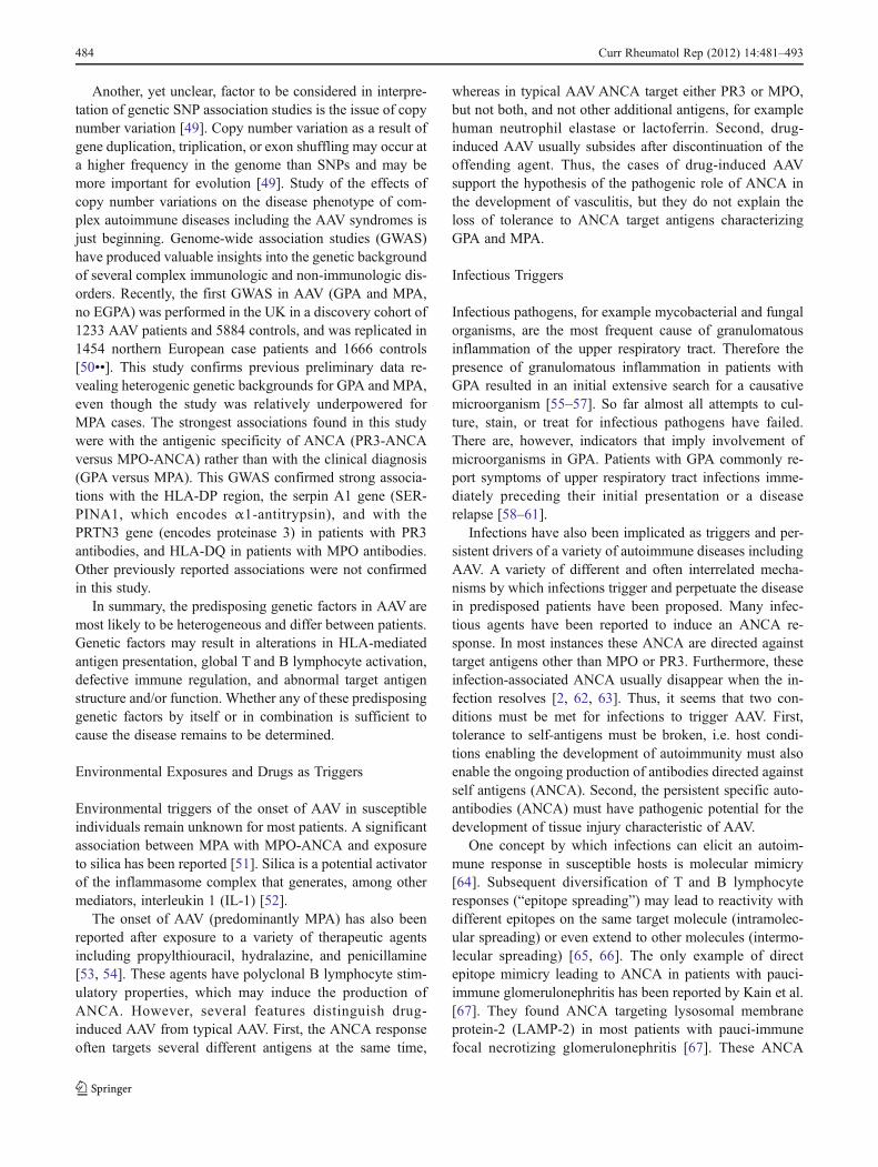
Several pro-inflammatory effects derived from the acti-vation of neutrophils and monocytes by ANCA are respon-sible for the tissue injury in AAV. Fully activated neutrophilsdegranulate and release toxic proteases and enzymes includ-ing elastase, PR3, MPO, and others [117]. ANCA alsoinduce a respiratory burst resulting in the release of oxygenradical species [117, 124]. ANCA also induce expression ofcell-adhesion molecules on neutrophils and endothelial cellsleading to increased adhesion of neutrophils to endothelialcells [125–129]. Moreover, the binding of ANCA to primedleukocytes induces the production and release of chemotac-tic cytokines including IL-1, MCP-1 and IL-8 [130–133].These cytokines attract more neutrophils and monocytes tothe site of inflammation. Thus, when the ANCA-inducedcytokine release occurs at the endothelial interface, thenormal chemotactic gradient that draws neutrophils out ofthe vasculature into the tissues is lost. This causes furtheraccumulation of fully activated neutrophils in the vesselwall, where they cause more injury. Figure 1 summarizesseveral complex interactions between ANCA and inflam-matory and endothelial cells thought to be instrumental inthe formation of granulomatous inflammation and vasculitis.
The ANCA target antigens released from activated or dyingneutrophils can also bind directly to endothelial cells [134].This may result in apoptosis of endothelial cells and in local-ized immune complex formation with circulating ANCA[135]. Low levels of localized immune-complex deposition,which has been documented in early vasculitic skin lesions andin renal lesions, can, in turn, induce localized complementactivation [136, 137].
In-vitro studies have also suggested that ANCAmodify theclearance of apoptotic cells. Opsonization of pre-apoptoticcells by ANCA is associated with increased production ofinflammatory cytokines by phagocytosing macrophages[138]. Moreover, pre-apoptotic cells have reduced cell-surface expression of phosphatidylserine (the recognition sig-nal for macrophages) in the presence of ANCA [139]. Con-sequently, in the presence of ANCA the non-inflammatory
clearance of apoptotic cells by macrophages may be perturbedin favor of inflammation and necrosis.
Pathogenic involvement of ANCA has also been sup-ported by several in-vivo animal models. These models arebased on the transfer of antibodies generated against ANCAtarget antigens to healthy recipient animals. The transfer ofanti-MPO IgG or splenocytes obtained from MPO-knockoutmice, which were immunized with murine MPO, into Rag 2knock-out mice (lacking mature T and B lymphocytes) andinto wild-type mice resulted in pauci-immune crescenticnecrotizing glomerulonephritis similar to that found inhumans [140]. A direct augmenting effect of MPO-ANCAon neutrophil–endothelial interactions causing microvascu-lar injury has been documented in a rat anti-MPO antibodytransfer model [141]. Murine anti-PR3 antibodies generatedin a similar fashion only caused an increased inflammatoryresponse at the site of tissue injury, but not a vasculiticphenotype, when transferred into wild-type mice [142].
The inflammatory lesions of AAVare referred to as “pauci-immune”, implying that only few immune complexes or com-plement factors can be identified by immunofluorescencemicroscopy. Furthermore, in contrast with classic immune-complex mediated disease, patients with active AAV havenormal serum complement levels. Nevertheless, low-gradelocalized immune-complex formation and complement acti-vation may be involved, and there is growing evidence sug-gesting that the complement pathway is involved in thepathogenesis of AAV [137, 143–145].Moreover, complementactivation might also contribute to the increased risk of venousthromboembolism observed in active AAV, because activatedcomplement factors trigger the coagulation cascade [146, 147].
It has recently been recognized that activation of thealternative complement pathway by ANCA may be an im-portant amplification loop of inflammation that contributesto renal (and other tissue) injury in AAV. In the murine anti-MPO antibody transfer model the development of necrotiz-ing glomerulonephritis is dependent on activation of thealternative complement pathway, and the development oflesions can be prevented and treated with an antibody thatinhibits complement factor 5 (C5) activation [148, 149].Mice lacking the receptor for activated C5 on neutrophilsalso do not develop the renal lesions [150]. In-vitro studiesshowed that supernatant from ANCA-activated neutrophilscan cause the production of C5a in normal serum, C5areceptor dependent priming of normal neutrophils, and in-creased neutrophil membrane expression of PR3. Althoughthe renal lesions in humans are called “pauci-immune”,components of the alternative complement pathway can bedetected in patients with AAV, but not in normal controls orin patients with minimum change disease [151].
Despite all this evidence, proof that ANCA alone cancause disease in humans has remained elusive. One casestudy in which an infant was born to a mother with active
Curr Rheumatol Rep (2012) 14:481–493 487

Fig. 1 Pathogenic mechanisms of AAV. (A) Necrotizing granuloma-tous inflammation develops in response to an unknown insult. Thisinflammatory reaction and the original insult result in release of avariety of cytokines, proteinase 3 (PR3), and, potentially, bacterialsuperantigens. Granulomatous inflammation in conjunction with theoriginal insult, secreted cytokines, and possibly bacterial superantigenpromote the production of ANCA by PR3 specific B lymphocytes andthe selection of PR3 specific T cells in predisposed individuals. Thereis a dysbalance between effector T cells (Teffs) and regulatory T cells(Tregs) with further release of proinflammatory cytokines promotingneutrophil priming. (B) ANCA have been shown in vitro to bind toPR3 expressed on the surface of neutrophils and monocytes. ANCAbinding results in cellular activation, release of pro-inflammatory cyto-kines, and neutrophil degranulation. (C) In-vitro ANCA also opsonizeapoptotic neutrophils by binding to PR3 expressed on their cell surface.In addition in the presence of ANCA, reduced phosphatidylserineexpression is observed on the cell surface of apoptotic neutrophils.
Neutrophil opsonization and perturbed phosphatidylserine expressionresult in inappropriate release of pro-inflammatory cytokines by mac-rophages clearing apoptotic neutrophils. (D) Endothelial damage ispromoted by a variety of mediators released by neutrophils. In addi-tion, increased expression of cell-adhesion molecules and the presenceof ANCA facilitate the binding of neutrophils to endothelial cells. PR3is internalized by endothelial cells and promotes apoptosis. (E) Neu-trophil extracellular trap (NET) formation occurs in lesions as a con-sequence of neutrophil apoptosis and degranulation. NET-derivedproducts activate dendritic cells and B cells by sensing via Toll-likereceptors. Interferon (IFN-a) production by dendritic cells might affectlocal immune regulation and has been shown to impair regulatory Tcells function. (F) Endothelial damage and tissue inflammation causedby localized complement activation, predominantly by the alternativepathway, resulting in cleavage of C5 into C5b which causes assemblyof MAC. C5a is able to prime neutrophils to enhance ANCA-inducedneutrophil activation
488 Curr Rheumatol Rep (2012) 14:481–493

MPA is often quoted as such evidence [152]. The infantdeveloped a pulmonary-renal syndrome 48 hours after de-livery and was found to have serum MPO-ANCA titerssimilar to the mother [152]. The child was treated withglucocorticoids and plasma exchange and recovered. How-ever, this observation is countered by another report of acase in which MPO-ANCA were also transferred from themother to the newborn, but the newborn remained perfectlyhealthy despite persistence of the transferred MPO-ANCAin the newborn for several weeks [153]. The two contrastingcase studies are consistent with observations made in largecohort studies. The development of severe vasculitic diseasemanifestations and severe flares usually do not occur in theabsence of ANCA, but not all patients with persistentANCA inevitably suffer such flares [76, 154–156].
Conclusions
On the basis of currently available clinical and experimentalevidence it is reasonable to believe that in predisposedpatients different triggers can lead to the production ofautoantibodies (ANCA) that in the context of an inflamma-tory environment can cause tissue inflammation and vascu-lar injury. However, many of the proposed mechanismsbehind the pathophysiology of AAV may apply solely tospecific clinical subsets of patients. Moreover, despite theseveral different pathways and mechanisms described in thisreview, there is not a one-size-fits all cases, or even most ofthe cases. Despite the substantial advances in our under-standing of the pathogenesis of AAV, many open questionsremain. As our knowledge regarding the pathogenesis ofAAVevolves, new therapeutic targeted strategies are emerg-ing in the continuing quest to control disease activity withthe minimum of adverse effects in individual patients.
Disclosure No potential conflicts of interest relevant to this articlewere reported. Dr. Specks is supported by NIH grant U54 AR057319.
References
Papers of particular interest, published recently, have beenhighlighted as:• Of importance•• Of major importance
1. Jennette JC, Falk RJ, Andrassy K, Bacon BA, Churg J, GrossWL, etal. Nomenclature of systemic vasculitides: the proposal of an inter-national consensus conference. Arthritis Rheum. 1994;37:187–92.
2. Hoffman GS, Specks U. Anti-neutrophil cytoplasmic antibodies.Arthritis Rheum. 1998;41:1521–37.
3. Keogh KA, Specks U. Churg-Strauss syndrome. clinical presen-tation, antineutrophil cytoplasmic antibodies, and leukotrienereceptor antagonists. Am J Med. 2003;115(4):284–90.
4. Sinico RA, Di Toma L, Maggiore U, Bottero P, Radice A, TosoniC, et al. Prevalence and clinical significance of antineutrophilcytoplasmic antibodies in Churg–Strauss syndrome. ArthritisRheum. 2005;52(9):2926–35.
5. Sable-Fourtassou R, Cohen P, Mahr A, Pagnoux C, Mouthon L,Jayne D, et al. Antineutrophil cytoplasmic antibodies and theChurg–Strauss syndrome. Ann Intern Med. 2005;143(9):632–8.
6. Watts RA, Lane SE, Bentham G, Scott DG. Epidemiology ofsystemic vasculitis: a ten-year study in the United Kingdom.Arthritis Rheum. 2000;43(2):414–9.
7. Watts RA, Gonzalez-GayMA, Lane SE, Garcia-Porrua C, BenthamG, Scott DG. Geoepidemiology of systemic vasculitis: comparisonof the incidence in two regions of Europe. Ann Rheum Dis.2001;60(2):170–2.
8. Reinhold-Keller E, Herlyn K, Wagner-Bastmeyer R, Gutfleisch J,Peter HH, Raspe HH, et al. No difference in the incidences ofvasculitides between north and south Germany: first results of theGerman vasculitis register. Rheumatol (Oxford). 2002;41(5):540–9.
9. Watts RA, Lane SE, Scott DG, Koldingsnes W, Nossent H,Gonzalez-Gay MA, et al. Epidemiology of vasculitis in Europe.Ann Rheum Dis. 2001;60(12):1156–7.
10. Watts RA, Scott DG. Epidemiology of the vasculitides. SeminRespir Crit Care Med. 2004;25(5):455–64.
11. Cotch MF, Hoffman GS, Yerg DE, Kaufman GI, Targonski P,Kaslow RA. The epidemiology of Wegener's granulomatosis.Estimates of the five-year period prevalence, annual mortality,and geographic disease distribution from population-based datasources. Arthritis Rheum. 1996;39:87–92.
12. Watts RA, Scott DG, Jayne DR, Ito-Ihara T, Muso E, Fujimoto S,et al. Renal vasculitis in Japan and the UK–are there differencesin epidemiology and clinical phenotype? Nephrol Dial Trans-plant. 2008;23(12):3928–31.
13. O'Donnell JL, Stevanovic VR, Frampton C, Stamp LK, ChapmanPT. Wegener's granulomatosis in New Zealand: evidence for alatitude-dependent incidence gradient. Intern Med J. 2007;37(4):242–6.
14. Mahr A, Guillevin L, Poissonnet M, Ayme S. Prevalences ofpolyarteritis nodosa, microscopic polyangiitis, Wegener's granu-lomatosis, and Churg–Strauss syndrome in a French urban mul-tiethnic population in 2000: a capture-recapture estimate.Arthritis Rheum. 2004;51(1):92–9.
15. Moins-Teisserenc HT, Gadola SD, Cella M, Dunbar PR, Exley A,Blake N, et al. Association of a syndrome resembling Wegener'sgranulomatosis with low surface expression of HLA class-I mol-ecules. Lancet [Res Support, Non-US Gov't]. 1999;354(9190):1598–603.
16. Villa-Forte A, de la Salle H, Fricker D, Hentges F, Zimmer J.HLA class I deficiency syndrome mimicking Wegener's granulo-matosis. Arthritis Rheuma [Case Rep Res Support, Non-USGov't]. 2008;58(8):2579–82.
17. Moins-Teisserenc HT, Gadola SD, Cella M, Dunbar PR, Exley A,Blake N, et al. Association of a syndrome resembling Wegener'sgranulomatosis with low surface expression of HLA class-I mol-ecules. Lancet. 1999;354(9190):1598–603.
18. Knight A, Sandin S, Askling J. Risks and relative risks of Wege-ner's granulomatosis among close relatives of patients with thedisease. Arthritis Rheum. 2008;58(1):302–7.
19. Rottem M, Cotch MF, Fauci AS, Hoffman GS. Familial vasculi-tis: report of 2 families. J Rheumatol. 1994;21(3):561–3.
20. Manganelli P, Giacosa R, Fietta P, Zanetti A, Neri TM. Familialvasculitides: Churg–Strauss syndrome and Wegener's granuloma-tosis in 2 first-degree relatives. J Rheumatol. 2003;30(3):618–21.
Curr Rheumatol Rep (2012) 14:481–493 489

21. Willcocks LC, Lyons PA, Rees AJ, Smith KG. The contributionof genetic variation and infection to the pathogenesis of ANCA-associated systemic vasculitis. Arthritis Res Ther. 2010;12(1):202.
22. Shiina T, Inoko H, Kulski JK. An update of the HLA genomicregion, locus information and disease associations: 2004. TissueAntigens [Review]. 2004;64(6):631–49.
23. Yang R, Cui Z, Zhao J, Zhao MH. The role of HLA-DRB1 alleleson susceptibility of Chinese patients with anti-GBM disease. ClinImmunol [Res Support, Non-US Gov't]. 2009;133(2):245–50.
24. Gencik M, Borgmann S, Zahn R, Albert E, Sitter T, Epplen JT, etal. Immunogenetic risk factors for anti-neutrophil cytoplasmicantibody (ANCA)-associated systemic vasculitis. Clin ExpImmunol. 1999;117(2):412–7.
25. HeckmannM, Holle JU, Arning L, Knaup S, Hellmich B, NothnagelM, et al. The Wegener's granulomatosis quantitative trait locuson chromosome 6p21.3 as characterised by tagSNP genotyp-ing. Ann Rheum Dis. 2008;67(7):972–9.
26. Tsuchiya N, Kobayashi S, Hashimoto H, Ozaki S, Tokunaga K.Association of HLA-DRB1*0901-DQB1*0303 haplotype withmicroscopic polyangiitis in Japanese. Genes Immun [CompStudy Res Support, Non-US Gov't]. 2006;7(1):81–4.
27. Tsuchiya N, Kobayashi S, Kawasaki A, Kyogoku C, Arimura Y,Yoshida M, et al. Genetic background of Japanese patients withantineutrophil cytoplasmic antibody-associated vasculitis: associ-ation of HLA-DRB1*0901 with microscopic polyangiitis. JRheumatol. 2003;30(7):1534–40.
28. Vaglio A, Martorana D, Maggiore U, Grasselli C, Zanetti A, PesciA, et al. HLA-DRB4 as a genetic risk factor for Churg–Strausssyndrome. Arthritis Rheum. 2007;56(9):3159–66.
29. Wieczorek S, Hellmich B, Gross WL, Epplen JT. Associations ofChurg–Strauss syndrome with the HLA-DRB1 locus, and rela-tionship to the genetics of antineutrophil cytoplasmic antibody-associated vasculitides: comment on the article by Vaglio et al.Arthritis Rheum. 2008;58(1):329–30.
30. Arning L, Holle JU, Harper L, Millar DS, Gross WL, Epplen JT,et al. Are there specific genetic risk factors for the different formsof ANCA-associated vasculitis? Ann Rheum Dis. 2011;70(4):707–8.
31. Cao Y, Schmitz JL, Yang J, Hogan SL, Bunch D, Hu Y, et al.DRB1*15 allele is a risk factor for PR3-ANCA disease in AfricanAmericans. J Am Soc Nephrol. 2011;22(6):1161–7.
32. Jagiello P, Aries P, Arning L, Wagenleiter SE, Csernok E,Hellmich B, et al. The PTPN22 620W allele is a risk factorfor Wegener's granulomatosis. Arthritis Rheum. 2005;52(12):4039–43.
33. Carr EJ, Niederer HA, Williams J, Harper L, Watts RA, LyonsPA, et al. Confirmation of the genetic association of CTLA4 andPTPN22 with ANCA-associated vasculitis. BMC Med Genet.2009;10:121.
34. Gregersen PK, Lee HS, Batliwalla F, Begovich AB. PTPN22:setting thresholds for autoimmunity. Semin Immunol. 2006;18(4):214–23.
35. Bluestone JA. Is CTLA-4 a master switch for peripheral T celltolerance? J Immunol. 1997;158(5):1989–93.
36. Steiner K, Moosig F, Csernok E, Selleng K, Gross WL, FleischerB, et al. Increased expression of CTLA-4 (CD152) by T and Blymphocytes in Wegener's granulomatosis. Clin Exp Immunol.2001;126(1):143–50.
37. Gough SC, Walker LS, Sansom DM. CTLA4 gene polymorphismand autoimmunity. Immunol Rev. 2005;204:102–15.
38. Wang XB, Zhao X, Giscombe R, Lefvert AK. A CTLA-4 genepolymorphism at position -318 in the promoter region affects theexpression of protein. Genes Immun. 2002;3(4):233–4.
39. Lee YH, Choi SJ, Ji JD, Song GG. CTLA-4 and TNF-alphapromoter-308 A/G polymorphisms and ANCA-associated
vasculitis susceptibility: a meta-analysis. Mol Biol Rep. 2012;39(1):319–26.
40. Gencik M, Meller S, Borgmann S, Fricke H. Proteinase 3 genepolymorphisms and Wegener's granulomatosis. Kidney Int.2000;58(6):2473–7.
41. Halbwachs-Mecarelli L, Bessou G, Lesavre P, Lopez S, Witko-Sarsat V. Bimodal distribution of proteinase 3 (PR3) surfaceexpression reflects a constitutive heterogeneity in the polymor-phonuclear neutrophil pool. FEBS. 1995;374:29–33.
42. Schreiber A, Busjahn A, Luft FC, Kettritz R. Membrane expres-sion of proteinase 3 is genetically determined. J Am Soc Nephrol.2003;14(1):68–75.
43. von Vietinghoff S, Busjahn A, Schonemann C, Massenkeil G,Otto B, Luft FC, et al. Major histocompatibility complex HLAregion largely explains the genetic variance exercised on neutro-phil membrane proteinase 3 expression. J Am Soc Nephrol.2006;17(11):3185–91.
44. Witko-Sarsat V, Lesavre P, Lopez S, Bessou G, Hieblot C, PrumB, et al. A large subset of neutrophils expressing membraneproteinase 3 is a risk factor for vasculitis and rheumatoid arthritis.J Am Soc Nephrol. 1999;10(6):1224–33.
45. Rarok AA, Stegeman CA, Limburg PC, Kallenberg CG. Neutro-phil membrane expression of proteinase 3 (PR3) is related torelapse in PR3-ANCA-associated vasculitis. J Am Soc Nephrol.2002;13(9):2232–8.
46. Mahr AD, Edberg JC, Stone JH, Hoffman GS, St Clair EW,Specks U, et al. Alpha 1-antitrypsin deficiency-related alleles Zand S and the risk for Wegener's granulomatosis. ArthritisRheum. 8 Sep 2010
47. Morris H, Morgan MD, Wood AM, Smith SW, Ekeowa UI,Herrmann K, et al. ANCA-associated vasculitis is linked tocarriage of the Z allele of alpha antitrypsin and its polymers.Ann Rheum Dis. 2011;70(10):1851–6.
48. Segelmark M, Elzouki AN, Wieslander J, Eriksson S. The PiZgene of alpha 1-antitrypsin as a determinant of outcome in PR3-ANCA-positive vasculitis. Kidney Int. 1995;48(3):844–50.
49. Stankiewicz P, Lupski JR. Structural variation in the humangenome and its role in disease. Annu Rev Med. 2010;61:437–55.
50. •• Lyons PA, Rayner TF, Trivedi S, Holle JU, Watts RA, JayneDR, et al. Genetically Distinct Subsets within ANCA-AssociatedVasculitis. N Engl J Med [Res Support, Non-US Gov't]. 2012;367(3):214–23. First AAV GWAS that confirmed that the pathogen-esis of AAV has a genetic component with genetic distinctionsbetween GPA and MPA that are associated with ANCA specificitywhere the response against the autoantigen PR3 is a centralfeature of PR3 AAV patients.
51. Hogan SL, Cooper GS, Savitz DA, Nylander-French LA, ParksCG, Chin H, et al. Association of silica exposure with anti-neutrophil cytoplasmic autoantibody small-vessel vasculitis: apopulation-based, case-control study. Clin J Am Soc Nephrol.2007;2(2):290–9.
52. Franchi L, Eigenbrod T, Nunez G. Cutting edge: TNF-alphamediates sensitization to ATP and silica via the NLRP3 inflam-masome in the absence of microbial stimulation. J Immunol [ResSupport, NIH, Extramural Research Support, Non-US Gov't].2009;183(2):792–6.
53. Short AK, Lockwood CM. Antigen specificity in hydralazineassociated ANCA positive systemic vasculitis. Q J Med. 1995;88:775–83.
54. Choi HK, Merkel PA, Walker AM, Niles JL. Drug-associatedantineutrophil cytoplasmic antibody-positive vasculitis: preva-lence among patients with high titers of antimyeloperoxidaseantibodies. Arthritis Rheum. 2000;43(2):405–13.
55. Carrington CB, Liebow A. Limited forms of angiitis and gran-ulomatosis of Wegener's type. Am J Med. 1966;41(4):497–527.
490 Curr Rheumatol Rep (2012) 14:481–493

56. Fahey J. E L, J C, GC G. Wegener's Granulomatosis. Am J Med.1954;17:168–79.
57. Walton E. Giant-cell granuloma of the respiratory tract (Wege-ner's granulomatosis). Br Med J. 1958;2:497–527.
58. Fauci AS, Haynes BF, Katz P, Wolff SM. Wegener's granuloma-tosis: prospective clinical and therapeutic experience with 85patients for 21years. Ann Intern Med. 1983;98(1):76–85.
59. Hoffman GS, Kerr GS, Leavitt RY, Hallahan CW, Lebovics RS,Travis WD, et al. Wegener granulomatosis: an analysis of 158patients.[see comment]. Ann Intern Med. 1992;116(6):488–98.
60. Pinching AJ, Rees AJ, Pussell BA, Lockwood CM, MitchisonRS, Peters DK. Relapses in Wegener's granulomatosis: the role ofinfection. Br Med J. 1980;281(6244):836–8.
61. Raynauld JP, Bloch DA, Fries JF. Seasonal variation in the onsetof Wegener's granulomatosis, polyarteritis nodosa and giant cellarteritis. J Rheumatol. 1993;20(9):1524–6.
62. Choi HK, Lamprecht P, Niles JL, Gross WL, Merkel PA. Sub-acute bacterial endocarditis with positive cytoplasmic antineutro-phil cytoplasmic antibodies and anti-proteinase 3 antibodies.Arthritis Rheum. 2000;43(1):226–31.
63. Capizzi SA, Specks U. Does infection play a role in the patho-genesis of pulmonary vasculitis? Semin Respir Infect. 2003;18(1):17–22.
64. Albert LJ, Inman RD. Molecular mimicry and autoimmunity. NEngl J Med. 1999;341(27):2068–74.
65. Craft J, Fatenejad S. Self antigens and epitope spreading insystemic autoimmunity. Arthritis Rheum. 1997;40:1374–82.
66. Vanderlugt CJ, Miller SD. Epitope spreading. Curr Opin Immu-nol. 1996;8:831–6.
67. Kain R, Exner M, Brandes R, Ziebermayr R, Cunningham D,Alderson CA, et al. Molecular mimicry in pauci-immunefocal necrotizing glomerulonephritis. Nat Med. 2008;14(10):1088–96.
68. Kain R, Tadema H, McKinney EF, Benharkou A, Brandes R,Peschel A, et al. High Prevalence of Autoantibodies to hLAMP-2in Anti-Neutrophil Cytoplasmic Antibody-Associated Vasculitis.J Am Soc Nephrol. 2012;23(3):556–66.
69. Roth AJ, Brown MC, Smith RN, Badhwar AK, Parente O, ChungHC, et al. Anti-LAMP-2 Antibodies Are Not Prevalent in PatientsWith Antineutrophil Cytoplasmic Autoantibody Glomerulone-phritis. J Am Soc Nephrol. 2012;23(3):545–55.
70. Pendergraft 3rd WF, Preston GA, Shah RR, Tropsha A, Carter JrCW, Jennette JC, et al. Autoimmunity is triggered by cPR-3(105-201), a protein complementary to human autoantigen proteinase-3. Nat Med. 2004;10(1):72–9.
71. Shoenfeld Y. Idiotypic induction of autoimmunity: a new aspectof the idiotypic network. FASEB J. 1994;8(15):1296–301.
72. Astern JM. Myeloperoxidase in vascular disease and autoimmu-nity. Chapel Hill: University of North Carolina; 2007.
73. Tadema H, Kallenberg CG, Stegeman CA, Heeringa P. Reactivityagainst complementary proteinase-3 is not increased in patientswith PR3-ANCA-associated vasculitis. PLoS One. 2011;6(3):e17972.
74. Bautz DJ, Preston GA, Lionaki S, Hewins P, Wolberg AS, YangJJ, et al. Antibodies with Dual Reactivity to Plasminogen andComplementary PR3 in PR3-ANCAVasculitis. J Am Soc Neph-rol. 13 Aug 2008
75. Merkel PA, Lo GH, Holbrook JT, Tibbs AK, Allen NB, Davis JrJC, et al. High incidence of venous thrombotic events amongpatients with Wegener granulomatosis: the Wegener's ClinicalOccurrence of Thrombosis (WeCLOT) Study. Ann Intern Med.2005;142(8):620–6.
76. Stegeman CA. Cohen Tervaert JW, Sluiter WJ, Manson WL, deJong PE, Kallenberg CGM. Association of chronic nasal carriageof Staphylococcus aureus and higher relapse rates in Wegenergranulomatosis. Ann Intern Med. 1994;120:12–7.
77. Stegeman CA, Cohen Tervaert JW, de Jong PE, Kallenberg CG.Trimethoprim-sulfamethoxazole (co-trimoxazole) for the preven-tion of relapses of Wegener's granulomatosis. N Engl J Med.1996;335(1):16–20.
78. Zycinska K, Wardyn KA, Zielonka TM, Krupa R, Lukas W. Co-trimoxazole and prevention of relapses of PR3-ANCA positivevasculitis with pulmonary involvement. Eur J Med Res. 2009;14Suppl 4:265–7.
79. Proft T, Fraser JD. Bacterial superantigens. Clin Exp Immunol.2003;133(3):299–306.
80. Zouali M. Exploitation of host signaling pathways by B cellsuperantigens–potential strategies for developing targeted thera-pies in systemic autoimmunity. Ann N Y Acad Sci. 2007;1095:342–54.
81. Popa ER, Stegeman CA, Abdulahad WH, van der Meer B,Arends J, Manson WM, et al. Staphylococcal toxic-shock-syndrome-toxin-1 as a risk factor for disease relapse in Wegener'sgranulomatosis. Rheumatol (Oxford). 2007;46(6):1029–33.
82. Popa ER, Stegeman CA, Bos NA, Kallenberg CG, Tervaert JW.Staphylococcal superantigens and T cell expansions in Wegener'sgranulomatosis. Clin Exp Immunol. 2003;132(3):496–504.
83. Li H, Nooh MM, Kotb M, Re F. Commercial peptidoglycanpreparations are contaminated with superantigen-like activity thatstimulates IL-17 production. J Leukoc Biol. 2008;83(2):409–18.
84. Acosta-Rodriguez EV, Rivino L, Geginat J, Jarrossay D, GattornoM, Lanzavecchia A, et al. Surface phenotype and antigenic spec-ificity of human interleukin 17-producing T helper memory cells.Nat Immunol. 2007;8(6):639–46.
85. Gerosa F, Baldani-Guerra B, Lyakh LA, Batoni G, Esin S,Winkler-Pickett RT, et al. Differential regulation of interleukin12 and interleukin 23 production in human dendritic cells. J ExpMed. 2008;205(6):1447–61.
86. Fouser LA, Wright JF, Dunussi-Joannopoulos K, Collins M.Th17 cytokines and their emerging roles in inflammation andautoimmunity. Immunol Rev. 2008;226:87–102.
87. Oukka M. Th17 cells in immunity and autoimmunity. AnnRheum Dis. 2008;67(Suppl 3:iii):26–9.
88. • Nogueira E, Hamour S, Sawant D, Henderson S, Mansfield N,Chavele KM, et al. Serum IL-17 and IL-23 levels andautoantigen-specific Th17 cells are elevated in patients withANCA-associated vasculitis. Nephrol Dial Transplant. 2010;25(7):2209–17. T cells producing IL-17 (Th17) have been implicat-ed in the pathogenesis of several autoimmune diseases and thisstudy identified that serum IL-17 levels and autoantigen-specificTh17 cells were elevated in patients with active AAVas comparedto healthy individuals.
89. Abdulahad WH, Stegeman CA, Limburg PC, Kallenberg CG.Skewed distribution of Th17 lymphocytes in patients with Wege-ner's granulomatosis in remission. Arthritis Rheum. 2008;58(7):2196–205.
90. Voswinkel J, Mueller A, Kraemer JA, Lamprecht P, Herlyn K,Holl-Ulrich K, et al. B lymphocyte maturation in Wegener'sgranulomatosis: a comparative analysis of VH genes from endo-nasal lesions. Ann Rheum Dis. 2006;65(7):859–64.
91. Hurtado PR, Jeffs L, Nitschke J, Patel M, Sarvestani G, Cassidy J,et al. CpG oligodeoxynucleotide stimulates production of anti-neutrophil cytoplasmic antibodies in ANCA associated vasculitis.BMC Immunol. 2008;9:34.
92. • Tadema H, Abdulahad WH, Lepse N, Stegeman CA, KallenbergCG, Heeringa P. Bacterial DNA motifs trigger ANCA productionin ANCA-associated vasculitis in remission. Rheumatol (Oxford).2011;50(4):689–96. This in vitro study showed that bacterialDNA motifs (CpG) trigger the production of ANCA by Blymphocytes in patients with AAV in remission.
93. Brinkmann V, Zychlinsky A. Beneficial suicide: why neutrophilsdie to make NETs. Nat Rev Microbiol. 2007;5(8):577–82.
Curr Rheumatol Rep (2012) 14:481–493 491

94. Fuchs TA, Abed U, Goosmann C, Hurwitz R, Schulze I, Wahn V,et al. Novel cell death program leads to neutrophil extracellulartraps. J Cell Biol. 2007;176(2):231–41.
95. •• Kessenbrock K, Krumbholz M, Schonermarck U, Back W,Gross WL, Werb Z, et al. Netting neutrophils in autoimmunesmall-vessel vasculitis. Nat Med. 2009;15(6):623–5. Neutrophilextracellular traps (NETs) are released by ANCA-stimulated neu-trophils in the absence of microbial infection and containproteinase-3 and myeloperoxidase. Deposition of NETs in in-flamed tissue suggest that NET formation triggers vasculitis.
96. •• Pilsczek FH, Salina D, Poon KK, Fahey C, Yipp BG, SibleyCD, et al. A novel mechanism of rapid nuclear neutrophil extra-cellular trap formation in response to Staphylococcus aureus. JImmunol. 2010;185(12):7413–25. Neutrophils respondeduniquely to Staphylococcus aureus via a novel process of NETformation that did not require neutrophil lysis. S. aureus stronglyinduce NETs and S. aureus infections are linked to relapses ofAAV.
97. Leadbetter EA, Rifkin IR, Hohlbaum AM, Beaudette BC,Shlomchik MJ, Marshak-Rothstein A. Chromatin-IgG complexesactivate B cells by dual engagement of IgM and Toll-like receptors.Nature. 2002;416(6881):603–7.
98. • Tadema H, Abdulahad WH, Stegeman CA, Kallenberg CG,Heeringa P. Increased expression of Toll-like receptors by mono-cytes and natural killer cells in ANCA-associated vasculitis.PLoS One. 2011;6(9):e24315. Toll-like receptors (TLRs) sensepathogen associated patterns and bacterial infections are knownto be associated with AAV. In patients with AAV, monocytes andNK cells were shown to have an increased TLR expressionprobably resulting from increased activation, which could playa role in disease reactivation.
99. Stone JH, Merkel PA, Spiera R, Seo P, Langford CA, HoffmanGS, et al. Rituximab versus cyclophosphamide for ANCA-associated vasculitis. N Engl J Med. 2010;363(3):221–32.
100. Odendahl M, Mei H, Hoyer BF, Jacobi AM, Hansen A,Muehlinghaus G, et al. Generation of migratory antigen-specific plasma blasts and mobilization of resident plasma cellsin a secondary immune response. Blood. 2005;105(4):1614–21.
101. Huang H, Benoist C, Mathis D. Rituximab specifically depletesshort-lived autoreactive plasma cells in a mouse model ofinflammatory arthritis. Proc Natl Acad Sci U S A. 2010;107(10):4658–63.
102. Popa ER, Stegeman CA, Bos NA, Kallenberg CG, Tervaert JW.Differential B- and T-cell activation in Wegener's granulomatosis.J Allergy Clin Immunol. 1999;103(5 Pt 1):885–94.
103. Krumbholz M, Specks U, Wick M, Kalled SL, Jenne D, Meinl E.BAFF is elevated in serum of patients with Wegener's granulo-matosis. J Autoimmun. 2005;25(4):298–302.
104. • Schneeweis C, Rafalowicz M, Feist E, Buttgereit F, RudolphPE, Burmester GR, et al. Increased levels of BLyS and sVCAM-1in anti-neutrophil cytoplasmatic antibody (ANCA)-associatedvasculitides (AAV). Clin Exp Rheumatol. 2010;28(1 Suppl57):62–6. Levels of endothelial cell activation (sVCAM-1) wereelevated in patients with AAV as compared to healthy controls.
105. Bader L, Koldingsnes W, Nossent J. B-lymphocyte activatingfactor levels are increased in patients with Wegener's granuloma-tosis and inversely correlated with ANCA titer. Clin Rheumatol.2010;29(9):1031–5.
106. Mellbye OJ, Mollnes TE, Steen LS. IgG subclass distribution andcomplement activation ability of autoantibodies to neutrophilcytoplasmic antigens (ANCA). Clin Immunol Immunopathol.1994;70(1):32–9.
107. Cohen Tervaert JW, Mulder L, Stegeman C, Elema J, Huitema M,The H, et al. Occurrence of autoantibodies to human leucocyteelastase in Wegener's granulomatosis and other inflammatorydisorders. Ann Rheum Dis. 1993;52:115–20.
108. Caux C, Dezutter–Dambuyant C, Schmitt D, Bandhereau J. GM-CSF and TNF-a cooperate in the generation of dendritic Langer-hans cells. Nature. 1992;360(6401):258–61.
109. Abdulahad WH, van der Geld YM, Stegeman CA, KallenbergCG. Persistent expansion of CD4+ effector memory T cells inWegener's granulomatosis. Kidney Int. 2006;70(5):938–47.
110. Abdulahad WH, Stegeman CA, van der Geld YM, Doornbos-vander Meer B, Meer B, Limburg PC, et al. Functional defect ofcirculating regulatory CD4+ T cells in patients with Wegener'sgranulomatosis in remission. Arthritis Rheum. 2007;56(6):2080–91.
111. ••Morgan MD, Day CJ, Piper KP, Khan N, Harper L, Moss PA, etal. Patients with Wegener's granulomatosis demonstrate a relativedeficiency and functional impairment of T-regulatory cells. Im-munology. 2010;130(1):64–73. Immune balance is important incontrolling autoimmune diseases and in this study the percentageof regulatory T cells (Foxp3 positive cells) were found to bedecreased in patients with GPA as compared to healthy controls.In addition, the percentage of regulatory T cells was found to beinversely related to the rate of disease relapse.
112. Bettelli E, Oukka M, Kuchroo VK. T(H)-17 cells in the circle ofimmunity and autoimmunity. Nature Immunol Res Support, NIH,Extramural Res Support, Non-US Gov't Rev. 2007;8(4):345–50.
113. Abdulahad WH, Stegeman CA, Kallenberg CG. Review article:The role of CD4(+) T cells in ANCA-associated systemic vascu-litis. Nephrol (Carlton). 2009;14(1):26–32.
114. Ordonez L, Bernard I, L'Faqihi-Olive FE, Tervaert JW,Damoiseaux J, Saoudi A. CD45RC isoform expression identifiesfunctionally distinct T cell subsets differentially distributed betweenhealthy individuals and AAV patients. PloS one [Res Support,Non-US Gov't]. 2009;4(4):e5287.
115. Altmann F, Staudacher E, Wilson IB, Marz L. Insect cells as hostsfor the expression of recombinant glycoproteins. Glycoconj J.1999;16(2):109–23.
116. Jovanovic DV, Di Battista JA, Martel-Pelletier J, Jolicoeur FC, HeY, ZhangM, et al. IL-17 stimulates the production and expression ofproinflammatory cytokines, IL-beta and TNF-alpha, by humanmacrophages. J Immunol. 1998;160(7):3513–21.
117. Falk RJ, Terrell RS, Charles LA, Jennette JC. Anti-neutrophilcytoplasmic autoantibodies induce neutrophils to degranulate andproduce oxygen radicals in vitro. Proc Natl Acad Sci USA.1990;87:4115–9.
118. Csernok E, Ernst M, Schmitt W, Bainton DF, Gross WL. Acti-vated neutrophils express proteinase 3 on their plasma membranein vitro and in vivo. Clin Exp Immunol. 1994;95(2):244–50.
119. Franssen CF, Huitema MG, Muller Kobold AC, Oost-Kort WW,Limburg PC, Tiebosch A, et al. In vitro neutrophil activation byantibodies to proteinase 3 and myeloperoxidase from patientswith crescentic glomerulonephritis. J Am Soc Nephrol. 1999;10(7):1506–15.
120. Kettritz R, Jennette JC, Falk RJ. Crosslinking of ANCA-antigensstimulates superoxide release by human neutrophils. J Am SocNephrol. 1997;8:386–94.
121. Weidner S, Neupert W, Goppelt-Struebe M, Rupprecht HD. Anti-neutrophil cytoplasmic antibodies induce human monocytes toproduce oxygen radicals in vitro. Arthritis Rheum. 2001;44(7):1698–706.
122. Hewins P, Williams JM, Wakelam MJ, Savage CO. Activation ofSyk in neutrophils by antineutrophil cytoplasm antibodies occursvia Fcgamma receptors and CD18. J Am Soc Nephrol. 2004;15(3):796–808.
123. van der Veen BS, Chen M, Muller R, van Timmeren MM,Petersen AH, Lee PA, et al. Effects of p38 mitogen-activatedprotein kinase inhibition on anti-neutrophil cytoplasmic autoan-tibody pathogenicity in vitro and in vivo. Ann Rheum Dis. 9Nov 2010
492 Curr Rheumatol Rep (2012) 14:481–493

124. Radford DJ, Lord JM, Savage CO. The activation of the neutro-phil respiratory burst by anti-neutrophil cytoplasm autoantibody(ANCA) from patients with systemic vasculitis requires tyrosinekinases and protein kinase C activation. Clin Exp Immunol.1999;118(1):171–9.
125. Mayet WJ, Schwarting A, Orth T, Duchmann R, BuschenfeldeKH Mz. Antibodies to proteinase 3 mediate expression of vascu-lar cell adhesion molecule-1 (VCAM-1). Clin Exp Immunol.1996;103(2):259–67.
126. De Bandt M, Meyer O, Hakim J, Pasquier C. Antibodies toproteinase-3 mediate expression of intercellular adhesionmolecule-1 (ICAM-1, CD 54). Br J Rheumatol. 1997;36(8):839–46.
127. Muller Kobold AC, van Wijk RT, Franssen CF, Molema G,Kallenberg CG, Tervaert JW. In vitro up-regulation of E-selectin and induction of interleukin-6 in endothelial cells byautoantibodies in Wegener's granulomatosis and microscopicpolyangiitis. Clin Exp Rheumatol. 1999;17(4):433–40.
128. Radford DJ, Savage CO, Nash GB. Treatment of rolling neutro-phils with antineutrophil cytoplasmic antibodies causes conver-sion to firm integrin-mediated adhesion. Arthritis Rheum. 2000;43(6):1337–45.
129. Taekema-Roelvink ME, Kooten C, Kooij SV, Heemskerk E,Daha MR. Proteinase 3 enhances endothelial monocyte chemo-attractant protein-1 production and induces increased adhe-sion of neutrophils to endothelial cells by upregulatingintercellular cell adhesion molecule-1. J Am Soc Nephrol. 2001;12(5):932–40.
130. Casselman BL, Kilgore KS, Miller BF, Warren JS. Antibodies toneutrophil cytoplasmic antigens induce monocyte chemoattrac-tant protein-1 secretion from human monocytes. J Lab Clin Med.1995;126(5):495–502.
131. Berger SP, Seelen MAJ, Hiemstra PS, Gerritsma JSJ, HeemskerkE, van der Woude FJ, et al. Proteinase 3, the major autoantigen ofWegener's granulomatosis, enhances IL-8 production by endothe-lial cells in vitro. J Am Soc Nephrol. 1996;7:694–701.
132. Brooks CJ, King WJ, Radford DJ, Adu D, McGrath M, SavageCOS. IL-1b production by human polymorphonuclear leucocytesstimulated by anti-neutrophil cytoplasmic autoantibodies: rel-evance to systemic vasculitis. Clin Exp Immunol. 1996;106:273–9.
133. Ralston DR, Marsh CB, Lowe MP, Wewers MD. Antineutrophilcytoplasmic antibodies induce monocyte IL-8 release. Role ofsurface proteinase-3, alpha1-antitrypsin, and Fcgamma receptors.J Clin Invest. 1997;100(6):1416–24.
134. Zhou Z, Dionne A, Richard C, Menard HA. On the origin ofsurface proteinase 3 of nonmyeloid cells: evidence favoring anexogenous source. Clin Immunol. 2000;97(2):171–81.
135. Yang JJ, Preston GA, Pendergraft WF, Segelmark M, Heeringa P,Hogan SL, et al. Internalization of proteinase 3 is concomitantwith endothelial cell apoptosis and internalization of myeloper-oxidase with generation of intracellular oxidants. Am J Pathol.2001;158(2):581–92.
136. Brons RH, de Jong MC, de Boer NK, Stegeman CA, KallenbergCG, Cohen Tervaert JW. Detection of immune deposits in skinlesions of patients with Wegener's granulomatosis. Ann RheumDis. 2001;60(12):1097–102.
137. Haas M, Eustace JA. Immune complex deposits in ANCA-associated crescentic glomerulonephritis: a study of 126 cases.Kidney Int. 2004;65(6):2145–52.
138. Harper L, Cockwell P, Adu D, Savage CO. Neutrophil primingand apoptosis in anti-neutrophil cytoplasmic autoantibody-associated vasculitis. Kidney Int. 2001;59(5):1729–38.
139. Harper L, Ren Y, Savill J, Adu D, Savage CO. Antineutrophilcytoplasmic antibodies induce reactive oxygen-dependent
dysregulation of primed neutrophil apoptosis and clearance bymacrophages. Am J Pathol. 2000;157(1):211–20.
140. Xiao H, Heeringa P, Hu P, Liu Z, Zhao M, Aratani Y, et al.Antineutrophil cytoplasmic autoantibodies specific for myeloper-oxidase cause glomerulonephritis and vasculitis in mice. J ClinInvest. 2002;110(7):955–63.
141. Little MA, Smyth CL, Yadav R, Ambrose L, Cook HT, NoursharghS, et al. Antineutrophil cytoplasm antibodies directed againstmyeloperoxidase augment leukocyte-microvascular interactionsin vivo. Blood. 2005;106(6):2050–8.
142. Pfister H, Ollert M, Frohlich LF, Quintanilla-Martinez L, ColbyTV, Specks U, et al. Antineutrophil cytoplasmic autoantibodiesagainst the murine homolog of proteinase 3 (Wegener autoanti-gen) are pathogenic in vivo. Blood. 2004;104(5):1411–8.
143. European Federation of Neurological Societies/Peripheral NerveSociety Guideline on management of multifocal motor neuropa-thy. Report of a joint task force of the European Federation ofNeurological Societies and the Peripheral Nerve Society. JPeripher Nerv Syst. 2006; 11(1):1–8
144. Haas M, Jafri J, Bartosh SM, Karp SL, Adler SG, Meehan SM.ANCA-associated crescentic glomerulonephritis with mesangialIgA deposits. Am J Kidney Dis. 2000;36(4):709–18.
145. Mentzel HJ, Neumann T, Fitzek C, Sauner D, Reichenbach JR,Kaiser WA. MR Imaging in Wegener granulomatosis of the spinalcord. AJNR Am J Neuroradiol. 2003;24(1):18–21.
146. Stassen PM, Derks RP, Kallenberg CG, Stegeman CA. Venousthromboembolism in ANCA-associated vasculitis–incidence andrisk factors. Rheumatol (Oxford). 2008;47(4):530–4.
147. Markiewski MM, Nilsson B, Ekdahl KN, Mollnes TE, LambrisJD. Complement and coagulation: strangers or partners in crime?Trends Immunol [Res Support, NIH, Extramural Research Sup-port, Non-US Gov't Rev]. 2007;28(4):184–92.
148. Xiao H, Schreiber A, Heeringa P, Falk RJ, Jennette JC. Alterna-tive complement pathway in the pathogenesis of disease mediatedby anti-neutrophil cytoplasmic autoantibodies. Am J Pathol.2007;170(1):52–64.
149. Huugen D, van Esch A, Xiao H, Peutz-Kootstra CJ, BuurmanWA, Tervaert JW, et al. Inhibition of complement factor C5protects against anti-myeloperoxidase antibody-mediated glomer-ulonephritis in mice. Kidney Int. 2007;71(7):646–54.
150. Schreiber A, Xiao H, Jennette JC, SchneiderW, Luft FC, Kettritz R.C5a receptor mediates neutrophil activation and ANCA-inducedglomerulonephritis. J Am Soc Nephrol. 2009;20(2):289–98.
151. Xing GQ, Chen M, Liu G, Heeringa P, Zhang JJ, Zheng X, et al.Complement activation is involved in renal damage in humanantineutrophil cytoplasmic autoantibody associated pauci-immune vasculitis. J Clin Immunol. 2009;29(3):282–91.
152. Schlieben DJ, Korbet SM, Kimura RE, Schwartz MM, Lewis EJ.Pulmonary-renal syndrome in a newborn with placental transmis-sion of ANCAs. Am J Kidney Dis. 2005;45(4):758–61.
153. Silva F, Specks U, Sethi S, Irazabal MV, Fervenza FC. SuccessfulPregnancy and Delivery of a Healthy Newborn Despite Transpla-cental Transfer of Antimyeloperoxidase Antibodies From a Moth-er With Microscopic Polyangiitis. Am J Kidney Dis. 2009
154. Sinico RA, Di Toma L, Maggiore U, Tosoni C, Bottero P, SabadiniE, et al. Renal involvement in Churg–Strauss syndrome. Am JKidney Dis. 2006;47(5):770–9.
155. Finkielman JD, Lee AS, Hummel AM, Viss MA, Jacob GL,Homburger HA, et al. ANCA are detectable in nearly all patientswith active severe Wegener's granulomatosis. Am J Med.2007;120(7):643–14. e9.
156. Finkielman JD, Merkel PA, Schroeder D, Hoffman GS, Spiera R,St Clair EW, et al. Antiproteinase 3 Antineutrophil CytoplasmicAntibodies and Disease Activity in Wegener Granulomatosis.Ann Intern Med. 2007;147(9):611–9.
Curr Rheumatol Rep (2012) 14:481–493 493