Pathobiology of Hodgkin lymphoma - academia.cat · Pathobiology of Hodgkin lymphoma. Ralf Küppers....
43
Pathobiology of Hodgkin lymphoma Ralf Küppers Institute of Cell Biology (Cancer Research) University of Duisburg-Essen, Essen, Germany
-
Upload
phungkhuong -
Category
Documents
-
view
220 -
download
0
Transcript of Pathobiology of Hodgkin lymphoma - academia.cat · Pathobiology of Hodgkin lymphoma. Ralf Küppers....

Pathobiology of Hodgkin lymphoma
Ralf KüppersInstitute of Cell Biology (Cancer Research)
University of Duisburg-Essen, Essen, Germany

Hodgkin lymphoma
• One of the most frequent lymphomas in Western world
• Tumor cells are called Hodgkin and Reed/Sternberg (HRS)
cells in classical HL
• Derivation of HRS cells from germinal center B cells
• HRS cells usually represent <1% of cells in tissue
• HRS cells always CD30-positive
• Activation of numerous signaling
pathways
CD30

DCmemory
B cell
SELECTION
DIFFERENTIATION
T
CLONAL EXPANSIONSOMATIC HYPERMUTATION
disadvantageousmutations:
- nonsense mutation- reduced affinity- gain of autoreactivity
HRScell
apoptoticB cell
additionaltransforming
events
rescue fromapoptosis
advantageousmutations
Scenario for HRS cell generation from “crippled“ GC B cells
Küppers & Rajewsky; Annu Rev Immunol. 1998

Topics
• Lost B cell phenotype
• Genetic lesions
• Generation of multinuclear cells
• Normal CD30+ B cells and their relationship to HRS cells
• The role of BATF3 in Hodgkin lymphoma and ALCL
The lost B cell identity of HRS cells of
classical HL:
Deregulated transcription factor networks

Lost B-cell identity of HRS cells
• Global gene expression analysis ofHRS cell lines with microarrays
• Nearly complete loss of B cellspecific gene expression program
• Retained expression of severalB cell transcription factors (PAX5, E2A, EBF) and genes involved in interaction with CD4+ T cells
• Validated by immunohistochemistryand GEP of primary HRS cells
RP105BCMACD20BLNKOBF
LckTOSOSpi-BBlk
SykCXCR5GrapMD-1PLCgLyl-1PTP1C Spi-1SHIPVav
B cell-specific
lymphocyte-specific
hematopoietic-specific
Schwering et al., Blood, 2003; Tiacci et al., Blood 2012
HL lines Normal B cells

Factors contributing to the downregulation of B cell genesin HRS cells
Küppers, Nature Rev Cancer, 2009Kreher et al., PNAS, 2014
NK cellfactor
T cellfactor
Myeloidreceptor
PAX5
EBF
E2A
Oct-2 Pu.1
BoB1NOTCH1-IC
LMP1
STAT5
?
Deltex1
BMI-1?
CSF1R
B cell gene
B cell gene
B cell gene
E2A
ABF-1
M M M
ID2
IRF5
E2A

The lost B cell phenotype of HRS cells- conclusions-
• Multiple factors contributing to lost B cell program:- downregulation of key B cell transcription factors- aberrant expression of master regulators of non-B cells- epigenetic silencing of B cell genes
• Reexpression of B cell program toxic for HRS cellsFOXO1: Xie, Blood 2012; PU.1: Yuki, Blood 2013; E2A: Guan, Oncotarget 2016

The lost B cell phenotype of HRS cells- conclusions-
• Multiple factors contributing to lost B cell program:- downregulation of key B cell transcription factors- aberrant expression of master regulators of non-B cells- epigenetic silencing of B cell genes
• Reexpression of B cell program toxic for HRS cells
Loss of B cell phenotype critical pathogenetic eventNeeded for HRS precursor cells to escape fromapoptosis as crippled germinal center B cells?

Genetic lesions in HRS cells

NF-kB pathway

Multiple genetic lesions in the NF-kB pathway in HRS cells

Genetic lesions in the JAK-STAT pathway in HRS cells
JAKJAK
PP
P P
P P
P
P
P
PDNA
STAT
SOCS1
JAK
H3P
PTPN1

Genetic lesions in the JAK-STAT pathway in HRS cells
JAKJAK
PP
P P
P P
P
P
P
PDNA
STAT
SOCS1Inactivating mutations in SOCS1 (40% of cases)
Gains in JAK2 (30% ofcases)
JAK
H3P
PTPN1Inactivating mutations in PTPN1 (20% of cases)
Gains of JAK2 often affect further pathogenetically relevant genes: PD-1 ligands 1 and 2 and JMJD2C.
Gains of STAT6 (30% of cases)

Further recently identified genetic lesions in HRS cells
• Tanslocations affecting the MHC class II transactivator in
ca. 15% of cHL (Steidl et al., 2011)
• Mutations in the MHC class I component b2 microglobulin
in >50% of cHL (Reichel et al., 2015)
• Frequent mutations in CD58 gene in HL cell lines (3/7 )
and deletions in primary cHL cases (3/13) (Schneider et al., 2015)
-> Immune evasion strategies of HRS cells?

Generation of bi- und multinuclear Reed-Sternberg cells from mononuclear Hodgkin
cells

Relationship between Hodgkin and Reed-Sternberg cells
Key feature of classical HL:Tumor cell population composed of mononuclear Hodgkin and bi- or multinucleated Reed-Sternberg cells
Mechanism?- Fusion of two independent
cells unlikely (Küppers et al., Blood 2000;Re et al., 2001)
- Mitosis without cell division (acytokinetic mitosis)?
- Other mechanism?
Experiment:long-term time-lapse microscopy of HL cell lines
CD30

Time lapse microscopy of HL cell lines
Rengstl, .., Rieger, Hansmann, PNAS, 2014
50 µm*
*
*
*
*
**
*
*
50 µm
* ** *
*
** *
D
C

Complete or incomplete cytokinesis before refusion?
time-lapse experiment with tubulin-RFP labeled HL cell lines
83% of refused cells with detectable persistent microtubule bonds

RS cell generation- Summary and Conclusions -
• Refusion as major route of RS cell generation from Hodgkin cells
• Refusion mostly if not always based on incomplete cytokinesis
• Mechanisms for failure to complete cytokinesis unknown

Gene expression profiling analysis of CD30-positive B cells and their relationship to HRS
cells of Hodgkin lymphoma

J Mol Histol 2005;36:249blue = CD30
Extrafollicular and GC CD30-positive lymphocytes

Human CD30+ B cells: open questions
memoryB cell
SELECTION
DIFFERENTIATION
CLONALEXPANSION
SOMATIC HYPERMUTATION
naiveB cell
apoptosis
HRS cell
Cellular origin of CD30+ NGC B cells?
Close relationship of CD30+ B cells
to HRS cells?
CD30+ B cells distinct B cell
subsets ?
?
?
?
CD30+B cell
CD30+B cell
Normal GC reactionof CD30+ GC B cells?

VH gene mutation analysis of CD30+ GC and non-GC B cells
*of mutated sequences; #of productive rearrangements
Donor CD30+ cells
Mutated sequences (%)
Aver. mutation frequency* (%)
R/S ratiosFRs#
1 GC 30 / 30 (100) 5.9 1.4
2 GC 23 / 23 (100) 8.2 1.9
3 GC 28 / 28 (100) 4.6 2.3
4 GC 38 / 41 (93) 6.2 1.5
3 non-GC 16 / 20 (84) 3.9 1.6
4 non-GC 36 / 41 (88) 5.6 1.6
5 non-GC 22 / 26 (79) 8.6 1.4
6 non-GC 9 / 20 (42) 2.2 1.9

Naive Memory Plasma CD30+ CD30+ Bulk GCB cells B cells cells GC B non-GC B cells
cells B cells
Unsupervised hierarchical clustering of normal B cell subsets
Manhatten distanceAverage linkageSD > 1407 probesets

High MYC activity in CD30+ GC B cells
CD30+ GC B cells versus bulk GC B cells
CD30+
GC Bcells
Bulk GCB cells
Gene setMYC targets Seitz; 2011PLoS One
NES 2.105
Nominal p-value 0.0
FDR q-value 0.0
26 MYC gene sets among most significantly enriched gene sets in normal CD30+ vs. bulk GC B cells
CD30+
bulk

Differential gene expression between CD30+ B cells and HRS cells
Higher expression in CD30+ B cells- typical B cell genes- signatures for strong proliferation (MYC, E2F)- genes with roles in regulation of mitosis and DNA stability
Higher expression in HRS cells- regulators of extracellular matrix- many chemokines and cytokines- non-B-cell genes (CD3D, granzyme B, ID2,..)- several transcription factors: MAF, MAFB, STAT1, BATF3
Key features of HRS cells are disease-associated

CD30+ GC HRS cells DLBCL FLB cells
Potential mechanisms for genetic instability and disturbed cytokinesis in HRS cells
Supervised analysis of genesdifferentially expressed betweenHRS cells and CD30+ GC B cells
Among 207 genes downregulated at least 5-fold in HRS cells, therewere 41 genes with functionsin mitosis, cytokinesis, genomic stability, DNA repair
Such a downregulation is lesspronounced in DLBCL and FL
Potential cause for the genomic in-stability of HRS cells and generationof multinucleated RS cells
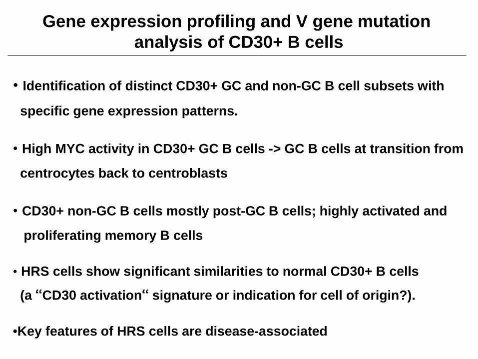
Gene expression profiling and V gene mutationanalysis of CD30+ B cells
• Identification of distinct CD30+ GC and non-GC B cell subsets with
specific gene expression patterns.
• High MYC activity in CD30+ GC B cells -> GC B cells at transition from
centrocytes back to centroblasts
• CD30+ non-GC B cells mostly post-GC B cells; highly activated and
proliferating memory B cells
• HRS cells show significant similarities to normal CD30+ B cells
(a “CD30 activation“ signature or indication for cell of origin?).
•Key features of HRS cells are disease-associated

The AP-1 transcription factor BATF3 is constitutively expressed in classical Hodgkin
lymphoma and contributes to tumor cell survival and proliferation

Gene expression profiling studies of HRS cells revealed high expression of BATF3 in primary HRS cells
Schwering et al, Mol Med, 2003
RT-PCR of microdissected or sorted cells
Samples BATF3 positive
HRS cells
case 1case 2case 3case 4
3/32/33/32/3
Non-HRS cells 0/12
GC B cells 0/6
Affymetrix HG-U133-Plus2.0Tiacci et al, Blood, 2012

Frequent BATF3 protein expression in classical HL, PMBCL and ALCL
• Frequent BATF3 protein expression in all three types of lymphomas
• EBV+ HRS cells rarely positive
• Lymphomas share CD30 positivity of tumor cells & JAK/STAT activity
Lymphoma BATF3 positive
classical Hodgkinlymphoma
EBV- 12 /20
EBV+ 1 / 10
PMBCL 8 / 9
ALCL* 40 / 40
Hodgkin lymphoma, BATF3 staining *Eckerle et al., Leukemia, 2009

331 aabasic leucine zipper
317 aa
125 aa
Transactivation, DNA binding and dimerization domains
127 aa
Basic facts about BATFs
TAD
TAD
Proposed inhibitory function of BATF Interaction at AP-1-IRF composite elements
JunJunB
BATFBATF3
- Members of the AP-1 family- form heterodimers with otherAP-1 family members
- form composite elements withIRFs
- Can have inhibitory and activating functions- BATF3 mainly expressed in TH1 cells and subsets of dendritic cells
Murphy, Nat Rev Immunol, 2013

HL cell linesALCLcHLNLP
HL
DEV
HD
LM2
KM
H2
L123
6
L428
SUPH
D1
UH
O1
K29
9
SR78
6
SUD
HL1
NHL cell linesGCBBLcHL PMBLAB
CMM
DLBCL
L428
UH
O1
Dau
di
Raj
i
SUD
HL4
SUD
HL6
OC
I-Ly3
K11
06P
Med
B-1
U26
6
IRF4IRF4
α-Tub
IRF8
α-Tub
JunB
c-Jun
IRF8
JunB
c-Jun
BATF3 BATF3
High expression of AP-1 and IRF factors in classical HL and ALCL cell lines

L-428
pSTAT6
µM
BATF3
ß-actin
0 5 10 20 40 0 5 10 20 40 0 5 10 20 40 0 5 10 20 40 0 5 10 20 40
Inhibition of JAK2 leads to downregulation of BATF3in 4/5 cHL cell lines
STAT6
c-MYC
U-HO1 HDLM-2 SUP-HD1 L-1236
TG101348/ Fedratinib (Jak2 inhibitor)
BATF3 downregulation upon JAK2 inhibition in a PMBCL line (Rui et al., Cancer Cell 2010)

STAT factors bind to the BATF3 promoter
Chromatin immunoprecipitation and quantitative PCR to study binding of pSTAT3, pSTAT5 and pSTAT6 to a putative STAT binding site in the BATF3 promoter in the cHL cell lines L-428 and U-HO1.
Binding of pSTAT3 and pSTAT6 to the BATF3 promoter in cHL cell lines
AP-1 sites
AP-1 site
AP-1 sites
STAT sites STAT sitesBATF3 gene

IB – Immunoblot
The AP-1 family members JUN and JUNB form heterodimers with BATF3 (co-immunoprecipitation studies)
Immunoprecipitation:U-HO1
IB: BATF3
IB: JUNIB: JUNB
IB: BATF3
IB: JUNB
IB: JUN
Immunoprecipitation:SR-786
K-299
IB: BATF3IB: JUN
IB: JUNB
Immunoprecipitation:
HDLM-2
IB: BATF3
IB: JUNB
IB: JUN
Immunoprecipitation:

A)
L-428
U-HO1
cHL ALCL
shRNA-mediated down-regulation of BATF3 leads to growth disadvantages of HRS and ALCL cells
B)
BATF3
b-actin
BATF3
b-actin
SR-786
K-299
BATF3
b-actin
BATF3
b-actin

Downregulation of positive MYC target genes in L428 HRS cells upon downregulation of BATF3

β-actin
shN
T
sh#4
L-428 (cHL)
Myc
sh#1
BATF3
BATF3
SR-786 (ALCL)
shN
T
sh#4sh#1BATF3
Downregulation of BATF3 causes downregulation of MYC
sh#5sh#4MYC
Myc
BATF3

BATF3 and JUN bind to the MYC promoter,indicating direct regulation of MYC expression
JUN and JUND binding known(Iavarone et al., J Biol Chem 2003)
Chromatin immunoprecipitation for BATF3 and JUN
UHO-1
PCR
00,20,40,60,8
11,2
Rel
ativ
e C
hIP
sign
al
(nor
mal
ized
to In
put)qPCR
MYC
L-428
UHO1
L-428
AP-1 motif
ChIP PCR product

BATF3 in Hodgkin lymphoma and ALCL- Summary and Conclusions -
BATF3 geneSTAT3/6
JAK2
STAT3/6
IL13
BATF3 MYC geneBATF3 JUN
BATF3 target genesBATF3 JUN(B)
Proliferation, survivalupregulated by STAT3/6 activity
highly expressed in cHL and ALCL
binds to MYC promoter and upregulates MYC
expression
forms heterodimers with JUN and JUNB
promotes survival and proliferation of HRS and
ALCL cells
(cHL)

Institute of Cell Biology(Cancer Research), EssenStefanie SchneiderSabrina RüschenbaumJanine DuppachMarc WenigerAnna LolliesMarc SeifertMarkus Schneider
Institute of Pathology, FrankfurtClaudia DöringSylvia HartmannBenjamin RengstlSebastian NewrzelaMartin-Leo Hansmann
Acknowledgements
Institute of Hematology, Perugia, ItalyEnrico Tiacci
Clinic for Otorhinolaryngology, EssenJudith Arnolds
Department of Hematology/Oncology, FrankfurtMichael Rieger