Parts of the Neuron - University of Northern Iowa · Parts of the Neuron Although blue in this...
7
1/22/2010 1 • What is the difference between “gray matter” and “white matter”? Book Fig.1.1 Parts of the Neuron Although blue in this figure, the myelin sheath is actually a white, fatty insulating coating on many axons. Any part of the CNS with lots of axons looks white as a result, in contrast to areas with lots of cell bodies. Gray & White Matter • Brain areas with lots of neuron cell bodies/dendrites look darker (“gray matter”) and function like information processors –receiving & combining input • Areas with lots of myelinatedaxons appear lighter (“white matter”) and function like cables connecting regions • A group of neuron cell bodies = “nucleus” (in CNS) or “ganglion” (in PNS) • A group of axons = “tract” or “pathway” (in CNS) or “nerve” (in PNS) Cortex (the outer layer) is gray matter. Beneath cortex is dense white matter – all the axons carrying messages to and away from cortex. Cortex is Gray Matter Book Fig. 1.22 5 levels of spinal cord & column with 31 pairs of spinal nerves Side view View from belly side Back side
Transcript of Parts of the Neuron - University of Northern Iowa · Parts of the Neuron Although blue in this...

1/22/2010
1
• What is the difference
between “gray matter”
and “white matter”?
Book Fig.1.1
Parts of the Neuron
Although blue in this figure, the myelin sheath is actually a white,
fatty insulating coating on many axons. Any part of the CNS with lots of
axons looks white as a result, in contrast to areas with lots of cell bodies.
Gray & White Matter
• Brain areas with lots of neuron cell bodies/dendrites look
darker (“gray matter”) and function like information
processors – receiving & combining input
• Areas with lots of myelinated axons appear lighter (“white
matter”) and function like cables connecting regions
• A group of neuron cell bodies = “nucleus” (in CNS) or
“ganglion” (in PNS)
• A group of axons = “tract” or “pathway” (in CNS) or “nerve”
(in PNS)
Cortex (the outer layer) is
gray matter. Beneath
cortex is dense white
matter – all the axons
carrying messages to and
away from cortex.
Cortex is Gray Matter
Book Fig. 1.22
5 levels of spinal
cord
& column with 31
pairs of spinal
nerves
Side view View from belly side
Back side
1/22/2010
2
Cross Section of Cord
Book Fig. 1.4 and 1.16
Afferent
(Sensory)
Efferent
(Motor)
Directional Terms
Horizontal
Or Frontal 5 Chunks of
of Brain
Telencephalon – outer part of forebrain
Cerebral hemispheres
Diencephalon – inner part of forebrain
Thalamus & Hypothalamus
Mesencephalon - midbrain
Metencephalon – upper part of hindbrain
Pons & Cerebellum
Myelencephalon – lower part of hindbrain
Medulla oblongata
Book Fig. 1.10 and 1.12
5 Vesicles that
will develop into
the 5 chunks of
the adult brain
are visible very
early in the
Embryonic Brain
Our authors use the term
brain stem to refer to the
last 3 divisions (midbrain,
pons, medulla) but you
may find other sources
that include the
diencephalon
(hypothalamus &
thalamus) in their
definition of the stalk-like
brain stem
Brain stem
& Cerebellum
These evolutionarily
ancient regions are
sometimes called our
reptilian brain

1/22/2010
3
The Brain is Like a Tootsie PopLimbic System
Part of the
‘middle layer’
– wraps
around the
brainstem
core.
Basal Ganglia
Also part of the ‘middle layer’
Cortex – the outer layer
Protection of the CNS
Bones, Meninges & Cerebrospinal Fluid (CSF)
do a good job under normal everyday
conditions.
Looking Into the Bottom of the Skull
Anterior Fossa�
Middle Fossa ->
Posterior Fossa
Foramen
Magnum >
Book Fig 5.1

1/22/2010
4
The Meninges
• 3 layers of connective tissue enclosing brain & spinal cord
• Starting from the outside, the layers are:
– dura mater
– arachnoid mater
– pia mater
– Meninges mnemonic (from the inside ����out) = PAD (the meninges PAD the outside of the brain)
The Dura Mater
(“Tough Mother”
Or
“Tough Matter”)
Dura Mater (“tough mother”)
• Actually has 2 layers which run close together in
most locations
– outer layer is anchored to skull bone in certain places
– inner layer forms folds that partition skull cavity into
compartments
• one between R & L hemispheres: falx cerebri
• one between occipital lobe & cerebellum: tentorium cerebelli
• spaces between layers at those folds form “dural
venous sinuses” for blood leaving brain
Falx is Latin for “sickle”
A sickle
Falx cerebri – sickle shaped membrane of the cerebrum between R and L
hemispheres
Falx cerebri model Falx Cerebri – dural
partition dividing upper
cranium in half �
Cross-Section of �
Tentorium cerebelli between
hemispheres
& cerebellum
(the “tent” over the cerebellum)
Book Fig. 5.3

1/22/2010
5
Fold of dura
between hemispheres
and cerebellum
Tentorium seen from above
(Hemispheres removed from skull)
A frontal cross-
section thru the
Falx cerebri
falx
Dural sinus between dural layers)
Arachnoid Mater(“spiderlike”)
• Thinner layer loosely enclosing CNS
• Space beneath arachnoid is filled with
cerebrospinal fluid (CSF)
• Spider-like filaments cross this
“subarachnoid space” to the inner most
layer of meninges, the pia mater
Book Fig. 1.13
Blood vessels supplying brain also travel in the subarachnoid space
(SAS)
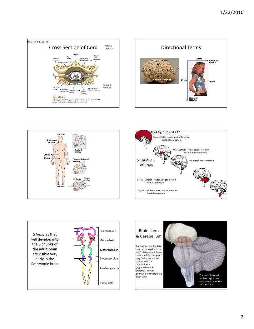
Pia
1/22/2010
6
The spidery filaments spanning the
subarachnoid space between arachnoid and pia.
Pia Mater (“tender matter”)
• Very thin layer that tightly follows brain
surface
• Contains lots of small capillaries supplying
blood to the CNS
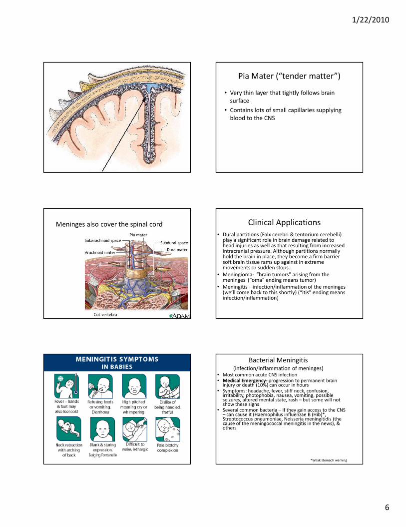
Meninges also cover the spinal cord Clinical Applications
• Dural partitions (Falx cerebri & tentorium cerebelli) play a significant role in brain damage related to head injuries as well as that resulting from increased intracranial pressure. Although partitions normally hold the brain in place, they become a firm barrier soft brain tissue rams up against in extreme movements or sudden stops.
• Meningioma- “brain tumors” arising from the meninges (“oma” ending means tumor)
• Meningitis – infection/inflammation of the meninges(we’ll come back to this shortly) (“itis” ending means infection/inflammation)
Bacterial Meningitis(infection/inflammation of meninges)
• Most common acute CNS infection• Medical Emergency- progression to permanent brain
injury or death (10%) can occur in hours• Symptoms: headache, fever, stiff neck, confusion,
irritability, photophobia, nausea, vomiting, possible seizures, altered mental state, rash – but some will not show these signs
• Several common bacteria – if they gain access to the CNS – can cause it (Haemophilus influenzae B (Hib)*, Streptococcus pneumoniae, Neisseria meningitidis (the cause of the meningococcal meningitis in the news), & others
*Weak stomach warning

1/22/2010
7
The Glass Test – “rash” doesn’t
disappear when pressed on
Bacterial Meningitis continued
• Infection may get to CNS 1) via blood, 2) spread from nearby ear or sinus infections, or 3) through congenital or acquired defects in protective coverings of CNS
• Bacteria release toxins damaging capillaries & causing dangerous cerebral edema (swelling) and increased intracranial pressure. Can also trigger hydrocephalus, increasing the rapid rise in pressure. Antibiotics do not decrease edema but corticosteroids help.
• Causes lasting deficits in 20-30% (impaired hearing, vision or movement, retardation, epilepsy, hydrocephalus) of survivors, especially in neonatal cases or if treatment is delayed.
• http://www.pbs.org/wgbh/nova/meningitis/
• (click on news minute on right)
Tests
• CT scan can show swollen meninges
• Lumbar puncture (spinal tap) to identify infection
• Kernig’s sign
• Brudzinki’s sign
• Now vaccines for 2 varieties available: Hib and
Meningococcal (Menomune and Menactra for
Neisseria strains A,C,Y)) No vaccine for for the
strain B. Menomune lasts 3-5 yrs, Menactra up to
10 years.
Resistance and Pain when
leg is extended
Discomfort and reflex flexing of legs when neck flexed
Viral Meningitis Less Serious
• Initial symptoms similar but mental status and
brain usually unaffected. Excellent prognosis.
• More serious risks if a virus affects the brain
itself (“viral encephalitis”).
• Sometimes drug reactions can cause a similar
syndrome (ibuprofen, naproxen,
trimethoprim, carbamazepine)
• Fungal infection of meninges can occur in
those with compromised immune systems
(like in AIDS)