PART9:SPECIFICCLINICALPROBLEMShealth.utah.gov/epi/diseases/TB/resources/aap_guidelines.pdf · From...
15
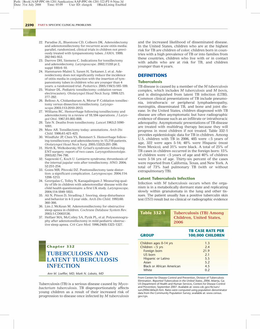
Path: {Book}AAP-PPC-06-1201/Application/AAP-PPC-06-1201-322-PT-9-Nine.3d Date: 31st July 2008 Time: 05:09 User ID: elangok 1BlackLining Enabled 22. Paradise JL, Bluestone CD, Colborn DK. Adenoidectomy and adenotonsillectomy for recurrent acute otitis media: parallel, randomized, clinical trials in children not previ- ously treated with tympanostomy tubes. JAMA. 1999; 282:945-953. 23. Darrow DH, Siemens C. Indications for tonsillectomy and adenoidectomy. Laryngoscope. 2002;112(8 pt 2, suppl 100):6-10. 24. Hammaren-Malmi S, Saxen H, Tarkanen J, et al. Ade- noidectomy does not significantly reduce the incidence of otitis media in conjunction with the insertion of tym- panostomy tubes in children who are younger than 4 years: a randomized trial. Pediatrics. 2005;116(1):185-189. 25. Walner DL. Pediatric tonsillectomy: coblation versus electrocautery. Otolaryngol Head Neck Surg. 1999;121: 277-282. 26. Belloso A, Chidambaram A, Morar P. Coblation tonsillec- tomy versus dissection tonsillectomy. Laryngo- scope.2003;113:2010-2013. 27. Williams RC. Hemorrhage following tonsillectomy and adenoidectomy in a review of 18,184 operations. J Laryn- gol Otol. 1967;81:805-808. 28. Tate N. Deaths from tonsillectomy. Lancet.1963;2:1090- 1091. 29. Maw AR. Tonsillectomy today: annotations. Arch Dis Child. 1986;61:421-423. 30. Windfuhr JP, Chen YS, Remmert S. Hemorrhage follow- ing tonsillectomy and adenoidectomy in 15,218 patients. Otolaryngol Head Neck Surg. 2005;132(2):281-286. 31. Hirth K, Welkoborsky HJ. Grisel’s syndrome following ENT-surgery: report of two cases. Laryngothinootolgie. 2003;82:794-798. 32. Sagowski C, Koch U. Lemierre syndrome; thrombosis of the internal jugular vein after tonsillectomy. HNO. 2004; 52:251-254. 33. Goins MR, Pitovski DZ. Posttonsillectomy taste distor- tion: a significant complication. Laryngoscope. 2004;114: 1206-1213. 34. Georgalas C, Tolley N, Kanagalingam J. Measuring qual- ity of life in children with adenotonsillar disease with the child health questionnaire: a first UK study. Laryngoscope. 2004;114:1849-1855. 35. Ali N, Pitson D, Stradling J. Snoring, sleep disturbance and behavior in 4-5 year olds. Arch Dis Child. 1993;68: 360-366. 36. Lim J, McKean M. Adenotonsillectomy for obstructive sleep apnea in children. Cochrane Database System Rev. 2003;1:CD003136. 37. Helfaer MA, McColley SA, Pyzik PL, et al. Polysomnogra- phy after adenotonsillectomy in mild pediatric obstruc- tive sleep apnea. Crit Care Med. 1996;24(8):1323-1327. Chapter 332 TUBERCULOSIS AND LATENT TUBERCULOSIS INFECTION Ann M. Loeffler, MD; Mark N. Lobato, MD Tuberculosis (TB) is a serious disease caused by Myco- bacterium tuberculosis. TB disproportionately affects young children as a result of their increased risk of progression to disease once infected by M tuberculosis and the increased likelihood of disseminated disease. In the United States, children who are at the highest risk for TB are children of color, children born in coun- tries with a high prevalence of TB or into families from these countries, children who live with or in contact with adults who are at risk for TB, and children younger than 4 years. DEFINITIONS Tuberculosis TB disease is caused by a member of the M tuberculosis complex, which includes M tuberculosis and M bovis, and is distinguished from latent TB infection (LTBI). Common clinical presentations of TB include pneumo- nia, intrathoracic or peripheral lymphadenopathy, meningitis, disseminated TB, and bone and joint dis- ease. In the United States, children diagnosed with TB disease are often asymptomatic but have radiographic evidence of disease such as an infiltrate or intrathoracic adenopathy. Asymptomatic presentations of TB disease are treated with multidrug therapy because they will progress in most children if not treated. Table 332-1 provides epidemiologic data for TB in children. Among U.S. children with TB in 2006, 485 were <5 years of age, 322 were ages 5-14; 48% were Hispanic (most from Mexico), and 31% were black. A total of 25% of TB cases in children occurred in the foreign born: 15% of children were <5 years of age and 40% of children were 5-14 yrs of age. Thirty-six percent of the cases were reported from California, Texas, and New York. A total of 73% had pulmonary TB (with or without extrapulmonary TB). Latent Tuberculosis Infection Infection with M tuberculosis occurs when the orga- nism is in a metabolically dormant state and replicating slowly within granulomata in the lung and other tis- sues. The patient usually has a positive tuberculin skin test (TST) result but no clinical or radiographic evidence Table 332-1 Tuberculosis (TB) Among Children, United States, 2006 GROUP TB CASE RATE PER 100,000 CHILDREN Children ages 0-14 yrs 1.3 Children <5 yrs 2.4 Foreign born 21.9 US born 2.1 Hispanic or Latino 5.5 Asian 5.2 Black or African American 4.5 White 0.2 From Centers for Disease Control and Prevention, Division of Tuberculosis Elimination. Reported Tuberculosis in the United States, 2006. Atlanta, Ga: US Department of Health and Human Services, Centers for Disease Control and Prevention; September 2007. Available at: www.cdc.gov/tb/surv/ surv2006/default.htm. Rates were computed using population denominator data from the Community Population Survey; available at: www.census. gov/cps. 2590 PART 9: SPECIFIC CLINICAL PROBLEMS
Transcript of PART9:SPECIFICCLINICALPROBLEMShealth.utah.gov/epi/diseases/TB/resources/aap_guidelines.pdf · From...
Path: {Book}AAP-PPC-06-1201/Application/AAP-PPC-06-1201-322-PT-9-Nine.3dDate: 31st July 2008 Time: 05:09 User ID: elangok 1BlackLining Enabled
22. Paradise JL, Bluestone CD, Colborn DK. Adenoidectomyand adenotonsillectomy for recurrent acute otitis media:parallel, randomized, clinical trials in children not previ-ously treated with tympanostomy tubes. JAMA. 1999;282:945-953.
23. Darrow DH, Siemens C. Indications for tonsillectomyand adenoidectomy. Laryngoscope. 2002;112(8 pt 2,suppl 100):6-10.
24. Hammaren-Malmi S, Saxen H, Tarkanen J, et al. Ade-noidectomy does not significantly reduce the incidenceof otitis media in conjunction with the insertion of tym-panostomy tubes in children who are younger than 4years: a randomized trial. Pediatrics. 2005;116(1):185-189.
25. Walner DL. Pediatric tonsillectomy: coblation versuselectrocautery. Otolaryngol Head Neck Surg. 1999;121:277-282.
26. Belloso A, Chidambaram A, Morar P. Coblation tonsillec-tomy versus dissection tonsillectomy. Laryngo-scope.2003;113:2010-2013.
27. Williams RC. Hemorrhage following tonsillectomy andadenoidectomy in a review of 18,184 operations. J Laryn-gol Otol. 1967;81:805-808.
28. Tate N. Deaths from tonsillectomy. Lancet.1963;2:1090-1091.
29. Maw AR. Tonsillectomy today: annotations. Arch DisChild. 1986;61:421-423.
30. Windfuhr JP, Chen YS, Remmert S. Hemorrhage follow-ing tonsillectomy and adenoidectomy in 15,218 patients.Otolaryngol Head Neck Surg. 2005;132(2):281-286.
31. Hirth K, Welkoborsky HJ. Grisel’s syndrome followingENT-surgery: report of two cases. Laryngothinootolgie.2003;82:794-798.
32. Sagowski C, Koch U. Lemierre syndrome; thrombosis ofthe internal jugular vein after tonsillectomy. HNO. 2004;52:251-254.
33. Goins MR, Pitovski DZ. Posttonsillectomy taste distor-tion: a significant complication. Laryngoscope. 2004;114:1206-1213.
34. Georgalas C, Tolley N, Kanagalingam J. Measuring qual-ity of life in children with adenotonsillar disease with thechild health questionnaire: a first UK study. Laryngoscope.2004;114:1849-1855.
35. Ali N, Pitson D, Stradling J. Snoring, sleep disturbanceand behavior in 4-5 year olds. Arch Dis Child. 1993;68:360-366.
36. Lim J, McKean M. Adenotonsillectomy for obstructivesleep apnea in children. Cochrane Database System Rev.2003;1:CD003136.
37. Helfaer MA, McColley SA, Pyzik PL, et al. Polysomnogra-phy after adenotonsillectomy in mild pediatric obstruc-tive sleep apnea. Crit Care Med. 1996;24(8):1323-1327.
Chapter 332
TUBERCULOSIS ANDLATENT TUBERCULOSISINFECTIONAnn M. Loeffler, MD; Mark N. Lobato, MD
Tuberculosis (TB) is a serious disease caused by Myco-bacterium tuberculosis. TB disproportionately affectsyoung children as a result of their increased risk ofprogression to disease once infected by M tuberculosis
and the increased likelihood of disseminated disease.In the United States, children who are at the highestrisk for TB are children of color, children born in coun-tries with a high prevalence of TB or into families fromthese countries, children who live with or in contactwith adults who are at risk for TB, and childrenyounger than 4 years.
DEFINITIONS
TuberculosisTB disease is caused by a member of the M tuberculosiscomplex, which includes M tuberculosis and M bovis,and is distinguished from latent TB infection (LTBI).Common clinical presentations of TB include pneumo-nia, intrathoracic or peripheral lymphadenopathy,meningitis, disseminated TB, and bone and joint dis-ease. In the United States, children diagnosed with TBdisease are often asymptomatic but have radiographicevidence of disease such as an infiltrate or intrathoracicadenopathy. Asymptomatic presentations of TB diseaseare treated with multidrug therapy because they willprogress in most children if not treated. Table 332-1provides epidemiologic data for TB in children. AmongU.S. children with TB in 2006, 485 were <5 years ofage, 322 were ages 5-14; 48% were Hispanic (mostfrom Mexico), and 31% were black. A total of 25% ofTB cases in children occurred in the foreign born: 15%of children were <5 years of age and 40% of childrenwere 5-14 yrs of age. Thirty-six percent of the caseswere reported from California, Texas, and New York. Atotal of 73% had pulmonary TB (with or withoutextrapulmonary TB).
Latent Tuberculosis InfectionInfection with M tuberculosis occurs when the orga-nism is in a metabolically dormant state and replicatingslowly within granulomata in the lung and other tis-sues. The patient usually has a positive tuberculin skintest (TST) result but no clinical or radiographic evidence
Table 332-1 Tuberculosis (TB) AmongChildren, United States,2006
GROUPTB CASE RATE PER100,000 CHILDREN
Children ages 0-14 yrs 1.3Children <5 yrs 2.4
Foreign born 21.9US born 2.1Hispanic or Latino 5.5Asian 5.2Black or African American 4.5White 0.2
From Centers for Disease Control and Prevention, Division of TuberculosisElimination. Reported Tuberculosis in the United States, 2006. Atlanta, Ga:US Department of Health and Human Services, Centers for Disease Controland Prevention; September 2007. Available at: www.cdc.gov/tb/surv/surv2006/default.htm. Rates were computed using population denominatordata from the Community Population Survey; available at: www.census.gov/cps.
2590 PART 9: SPECIFIC CLINICAL PROBLEMS
Path: {Book}AAP-PPC-06-1201/Application/AAP-PPC-06-1201-322-PT-9-Nine.3dDate: 31st July 2008 Time: 05:09 User ID: elangok 1BlackLining Enabled
of TB disease. Until new diagnostic tests become betterstudied in children and are more readily available, apositive TST result is used to define LTBI. An interferon-gamma release assay such as the QuantiFERON-TB testmay also diagnose LTBI in children. Patients should betreated with isoniazid (isonicotinyl hydrazine [INH])monotherapy daily for 9 months, unless they have amedical contraindication (including infection witha known INH-resistant strain). Because LTBI is not areportable condition in most states, the number ofchildren who have LTBI is unknown.
Tuberculosis ExposureA person exposed to TB is one who has spent time inclose proximity to a potentially contagious patientwith TB disease. The exposed individual may or maynot be infected. Young children can progress rapidlyto TB once infected; thus they should be quickly eval-uated and treated prophylactically while awaitingcompletion of the evaluation if they are exposed to TB.
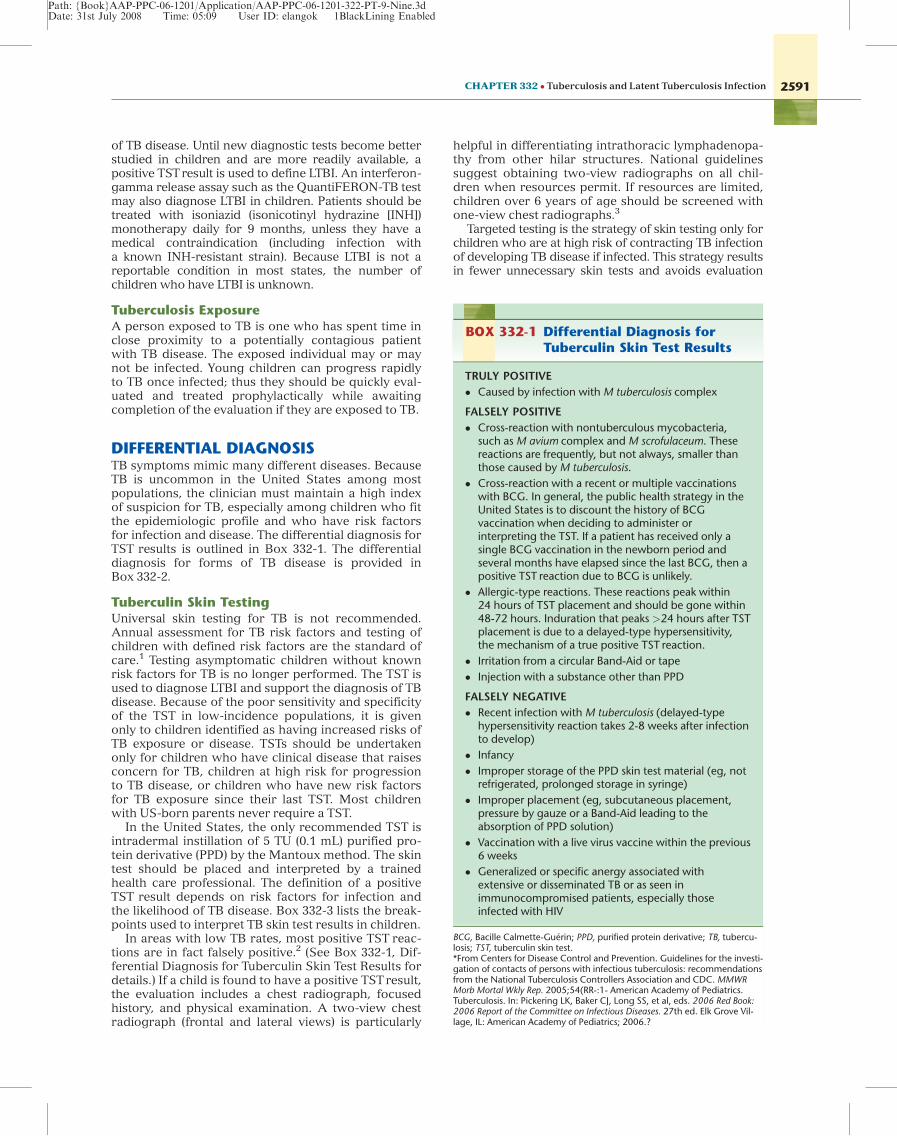
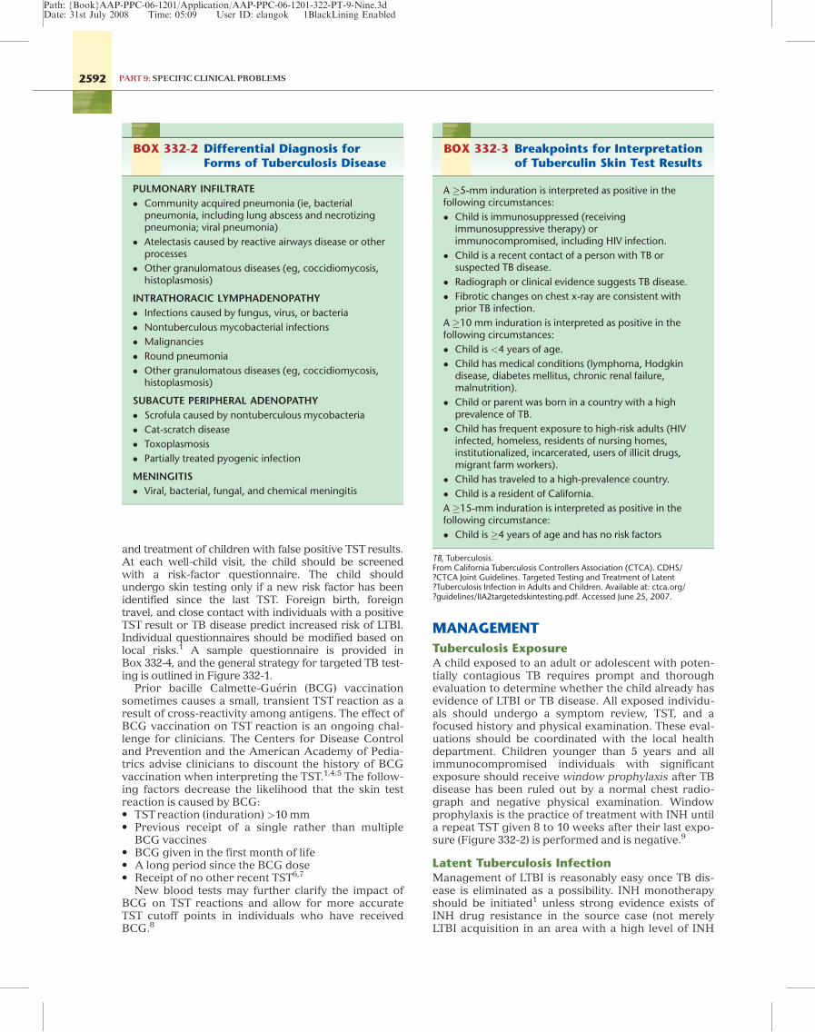
DIFFERENTIAL DIAGNOSISTB symptoms mimic many different diseases. BecauseTB is uncommon in the United States among mostpopulations, the clinician must maintain a high indexof suspicion for TB, especially among children who fitthe epidemiologic profile and who have risk factorsfor infection and disease. The differential diagnosis forTST results is outlined in Box 332-1. The differentialdiagnosis for forms of TB disease is provided inBox 332-2.
Tuberculin Skin TestingUniversal skin testing for TB is not recommended.Annual assessment for TB risk factors and testing ofchildren with defined risk factors are the standard ofcare.1 Testing asymptomatic children without knownrisk factors for TB is no longer performed. The TST isused to diagnose LTBI and support the diagnosis of TBdisease. Because of the poor sensitivity and specificityof the TST in low-incidence populations, it is givenonly to children identified as having increased risks ofTB exposure or disease. TSTs should be undertakenonly for children who have clinical disease that raisesconcern for TB, children at high risk for progressionto TB disease, or children who have new risk factorsfor TB exposure since their last TST. Most childrenwith US-born parents never require a TST.
In the United States, the only recommended TST isintradermal instillation of 5 TU (0.1 mL) purified pro-tein derivative (PPD) by the Mantoux method. The skintest should be placed and interpreted by a trainedhealth care professional. The definition of a positiveTST result depends on risk factors for infection andthe likelihood of TB disease. Box 332-3 lists the break-points used to interpret TB skin test results in children.
In areas with low TB rates, most positive TST reac-tions are in fact falsely positive.2 (See Box 332-1, Dif-ferential Diagnosis for Tuberculin Skin Test Results fordetails.) If a child is found to have a positive TST result,the evaluation includes a chest radiograph, focusedhistory, and physical examination. A two-view chestradiograph (frontal and lateral views) is particularly
helpful in differentiating intrathoracic lymphadenopa-thy from other hilar structures. National guidelinessuggest obtaining two-view radiographs on all chil-dren when resources permit. If resources are limited,children over 6 years of age should be screened withone-view chest radiographs.3
Targeted testing is the strategy of skin testing only forchildren who are at high risk of contracting TB infectionof developing TB disease if infected. This strategy resultsin fewer unnecessary skin tests and avoids evaluation
BOX 332-1 Differential Diagnosis forTuberculin Skin Test Results
TRULY POSITIVE
� Caused by infection with M tuberculosis complex
FALSELY POSITIVE
� Cross-reaction with nontuberculous mycobacteria,such as M avium complex and M scrofulaceum. Thesereactions are frequently, but not always, smaller thanthose caused by M tuberculosis.
� Cross-reaction with a recent or multiple vaccinationswith BCG. In general, the public health strategy in theUnited States is to discount the history of BCGvaccination when deciding to administer orinterpreting the TST. If a patient has received only asingle BCG vaccination in the newborn period andseveral months have elapsed since the last BCG, then apositive TST reaction due to BCG is unlikely.
� Allergic-type reactions. These reactions peak within24 hours of TST placement and should be gone within48-72 hours. Induration that peaks >24 hours after TSTplacement is due to a delayed-type hypersensitivity,the mechanism of a true positive TST reaction.
� Irritation from a circular Band-Aid or tape
� Injection with a substance other than PPD
FALSELY NEGATIVE
� Recent infection with M tuberculosis (delayed-typehypersensitivity reaction takes 2-8 weeks after infectionto develop)
� Infancy
� Improper storage of the PPD skin test material (eg, notrefrigerated, prolonged storage in syringe)
� Improper placement (eg, subcutaneous placement,pressure by gauze or a Band-Aid leading to theabsorption of PPD solution)
� Vaccination with a live virus vaccine within the previous6 weeks
� Generalized or specific anergy associated withextensive or disseminated TB or as seen inimmunocompromised patients, especially thoseinfected with HIV
BCG, Bacille Calmette-Gu�erin; PPD, purified protein derivative; TB, tubercu-losis; TST, tuberculin skin test.*From Centers for Disease Control and Prevention. Guidelines for the investi-gation of contacts of persons with infectious tuberculosis: recommendationsfrom the National Tuberculosis Controllers Association and CDC. MMWRMorb Mortal Wkly Rep. 2005;54(RR-:1- American Academy of Pediatrics.Tuberculosis. In: Pickering LK, Baker CJ, Long SS, et al, eds. 2006 Red Book:2006 Report of the Committee on Infectious Diseases. 27th ed. Elk Grove Vil-lage, IL: American Academy of Pediatrics; 2006.?
2591CHAPTER 332 � Tuberculosis and Latent Tuberculosis Infection
Path: {Book}AAP-PPC-06-1201/Application/AAP-PPC-06-1201-322-PT-9-Nine.3dDate: 31st July 2008 Time: 05:09 User ID: elangok 1BlackLining Enabled
and treatment of children with false positive TST results.At each well-child visit, the child should be screenedwith a risk-factor questionnaire. The child shouldundergo skin testing only if a new risk factor has beenidentified since the last TST. Foreign birth, foreigntravel, and close contact with individuals with a positiveTST result or TB disease predict increased risk of LTBI.Individual questionnaires should be modified based onlocal risks.1 A sample questionnaire is provided inBox 332-4, and the general strategy for targeted TB test-ing is outlined in Figure 332-1.
Prior bacille Calmette-Gu�erin (BCG) vaccinationsometimes causes a small, transient TST reaction as aresult of cross-reactivity among antigens. The effect ofBCG vaccination on TST reaction is an ongoing chal-lenge for clinicians. The Centers for Disease Controland Prevention and the American Academy of Pedia-trics advise clinicians to discount the history of BCGvaccination when interpreting the TST.1,4,5 The follow-ing factors decrease the likelihood that the skin testreaction is caused by BCG:� TST reaction (induration) >10 mm� Previous receipt of a single rather than multiple
BCG vaccines� BCG given in the first month of life� A long period since the BCG dose� Receipt of no other recent TST6,7
New blood tests may further clarify the impact ofBCG on TST reactions and allow for more accurateTST cutoff points in individuals who have receivedBCG.8
MANAGEMENT
Tuberculosis ExposureA child exposed to an adult or adolescent with poten-tially contagious TB requires prompt and thoroughevaluation to determine whether the child already hasevidence of LTBI or TB disease. All exposed individu-als should undergo a symptom review, TST, and afocused history and physical examination. These eval-uations should be coordinated with the local healthdepartment. Children younger than 5 years and allimmunocompromised individuals with significantexposure should receive window prophylaxis after TBdisease has been ruled out by a normal chest radio-graph and negative physical examination. Windowprophylaxis is the practice of treatment with INH untila repeat TST given 8 to 10 weeks after their last expo-sure (Figure 332-2) is performed and is negative.9
Latent Tuberculosis InfectionManagement of LTBI is reasonably easy once TB dis-ease is eliminated as a possibility. INH monotherapyshould be initiated1 unless strong evidence exists ofINH drug resistance in the source case (not merelyLTBI acquisition in an area with a high level of INH
BOX 332-2 Differential Diagnosis forForms of Tuberculosis Disease
PULMONARY INFILTRATE
� Community acquired pneumonia (ie, bacterialpneumonia, including lung abscess and necrotizingpneumonia; viral pneumonia)
� Atelectasis caused by reactive airways disease or otherprocesses
� Other granulomatous diseases (eg, coccidiomycosis,histoplasmosis)
INTRATHORACIC LYMPHADENOPATHY
� Infections caused by fungus, virus, or bacteria
� Nontuberculous mycobacterial infections
� Malignancies
� Round pneumonia
� Other granulomatous diseases (eg, coccidiomycosis,histoplasmosis)
SUBACUTE PERIPHERAL ADENOPATHY
� Scrofula caused by nontuberculous mycobacteria
� Cat-scratch disease
� Toxoplasmosis
� Partially treated pyogenic infection
MENINGITIS
� Viral, bacterial, fungal, and chemical meningitis
BOX 332-3 Breakpoints for Interpretationof Tuberculin Skin Test Results
A �5-mm induration is interpreted as positive in thefollowing circumstances:
� Child is immunosuppressed (receivingimmunosuppressive therapy) orimmunocompromised, including HIV infection.
� Child is a recent contact of a person with TB orsuspected TB disease.
� Radiograph or clinical evidence suggests TB disease.
� Fibrotic changes on chest x-ray are consistent withprior TB infection.
A �10 mm induration is interpreted as positive in thefollowing circumstances:
� Child is <4 years of age.
� Child has medical conditions (lymphoma, Hodgkindisease, diabetes mellitus, chronic renal failure,malnutrition).
� Child or parent was born in a country with a highprevalence of TB.
� Child has frequent exposure to high-risk adults (HIVinfected, homeless, residents of nursing homes,institutionalized, incarcerated, users of illicit drugs,migrant farm workers).
� Child has traveled to a high-prevalence country.
� Child is a resident of California.
A �15-mm induration is interpreted as positive in thefollowing circumstance:
� Child is �4 years of age and has no risk factors
TB, Tuberculosis.From California Tuberculosis Controllers Association (CTCA). CDHS/?CTCA Joint Guidelines. Targeted Testing and Treatment of Latent?Tuberculosis Infection in Adults and Children. Available at: ctca.org/?guidelines/IIA2targetedskintesting.pdf. Accessed June 25, 2007.
2592 PART 9: SPECIFIC CLINICAL PROBLEMS

Path: {Book}AAP-PPC-06-1201/Application/AAP-PPC-06-1201-322-PT-9-Nine.3dDate: 31st July 2008 Time: 05:09 User ID: elangok 1BlackLining Enabled
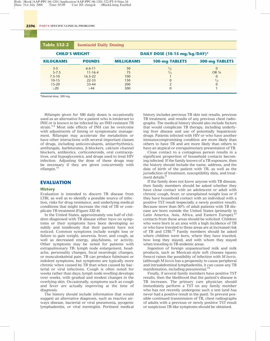
resistance). Dosing is 270 daily doses (or twice a weekadministered by directly observed therapy [DOT]within a 12-month period). Table 332-2 lists INHdoses by weight. When a prolonged break occursafter a short initial treatment period, then therapyshould be restarted, but short lapses are tolerated,especially if the regimen is well underway. If inter-ruption of therapy is greater than 2 months, then thechild should be reevaluated for possible TB disease
before restarting INH. Vitamin-B6 (pyridoxine) sup-plementation is indicated only for exclusivelybreastfed infants, children and adolescents on milk-and meat-deficient diets, children who experienceparesthesias while receiving isoniazid therapy, andthose with HIV infection.3
INH is available as 100-mg and 300-mg scored tab-lets and as a liquid suspended in sorbitol. The liquidformulation causes cramping and diarrhea in more
BOX 332-4 Tuberculosis Risk Assessment Questionnaire
Name: ________________________ DOB: _____________________
Last tuberculin skin test (TST) date: _________ Results: ___________ mm induration OR ______ not read by health careprofessional
If positive TST result in the past: Chest radiograph date and result: _______________
1. Was your child born outside the United States? _______Yes _______No
Country: _________________________________
2. Since the last TB skin test, has your child traveled outside the United States? _______Yes _______No
Country or countries visited: ______________________________________
Dates of travel and how long did they travel? ______________________________________
Where did they stay (hotel, family, resort)? ______________________________
3. Since the last TB skin test, has your child been exposed to anyone with TB disease? _______Yes _______No
Name of their disease? _____________________________
Positive TST result with normal chest taking one medicine or no treatment OR TB disease taking many pills and differentkinds of medicine?
Name of person: ______________________________ DOB: ____________________
Where is the person being treated? ______________________________________________
4. Since the child’s last skin test, has your child had close contact with a person who has a positive TST result? _______Yes_______No
Nature of their disease? _________________________________________________________________________
Positive TST result with normal chest radiograph taking medicine or no treatment OR TB disease taking many pills anddifferent kinds of medicine?
Name of person: ___________________________ DOB: _____________________
Where is the person being treated? _______________________________________________
Optional questions depending on local epidemiology:
Since the last skin test, has your child consumed unpasteurized milk or cheese (from Mexico or Central America)? _______Yes_______No
Since the last skin test, has your child been around people in jail, homeless or in shelters, people who have HIV, or use illegaldrugs? _______Yes _______No
Since the last skin test, has your child lived with a new person who was born or traveled outside the US? _______Yes_______No
INSTRUCTIONS FOR PROVIDERS: Test only children who have a new risk factor since their last TST.
If the child has previously had a positive TST result, then do not administer another TST.
Significant travel is considered travel to a country with a high prevalence of TB (eg, in Africa, Asia, Latin American, andEastern Europe) for >1 week AND had a substantial contact with indigenous people from such countries (did not stay in aresort).
From Pediatric Tuberculosis Collaborative Group. Targeted tuberculin skin testing and treatment of latent tuberculosis infection in children and adolescents.Pediatrics. 2004;114:1175-1201.
2593CHAPTER 332 � Tuberculosis and Latent Tuberculosis Infection

Path: {Book}AAP-PPC-06-1201/Application/AAP-PPC-06-1201-322-PT-9-Nine.3dDate: 31st July 2008 Time: 05:09 User ID: elangok 1BlackLining Enabled
than one half of children because of its osmotic load.The tablet can be crushed and mixed with or layeredinto a strong-flavored semisoft food in a spoon. Chil-dren should be examined monthly and questionedabout symptoms of toxicity. INH-related transientincrease of transaminases has been noted in children,with the effects increasing with increasing age; how-ever, INH rarely causes clinical hepatotoxicity in chil-dren.10 Routine monitoring of liver transaminases isnot indicated for asymptomatic children who do nothave underlying liver disease and who are not receiv-ing other hepatotoxic drugs.1 Families should be thor-oughly educated about and instructed on recognizingsymptoms of hepatotoxicity (eg, anorexia, malaise,abdominal pain, vomiting) and to stop the therapy andreturn to the clinic if these symptoms arise. Lack ofassociation with other viral symptoms and lack of
improvement after a few days should suggest the pos-sibility of hepatotoxicity rather than an intercurrentillness. Patients receiving antiepileptic drugs (particu-larly phenytoin and carbamazepine) should have thesedrug levels monitored.
Parents should be asked about the child’s adherenceto therapy and results of skin testing of family mem-bers and other contacts. Figure 332-3 shows an exam-ple of a flow sheet for monitoring LTBI treatment.Every effort should be made to promote and facilitateadherence through enablers such as walk-in visits forrefills (nurse visits) or school-based dosing or monthlymonitoring. Incentives such as stickers and calendars,prizes, and end-of-treatment rewards can also be usedto promote adherence (for an example, see www.maine.gov/dhhs/boh/ddc/treasure_chest_program_tb.htm).Children receiving antiepileptic drugs should be
No Yes
Assess all children for TBrisk factors by using a
questionnaire at each wellchild visit (see Box 332-4)
TB Risk Factor?
No TST – screenagain next visit
• Place TST• Reading by trained provider in 48–72 hours • Record results in mm induration
TST results negative basedon breakpoints (Box 332-3)
TST results positive basedon breakpoints (Box 332-3)
No further evaluation –screen again next visit
Monitor monthly foradherence to therapy;
drug toxicity and symptomsof TB disease
If suspicious of TB disease:• Report to local health department• Consult with a TB expert • Collect cultures• Consider multidrug TB therapy by DOT • Notify infection control (hospital)
Focused historyand physical examination;chest radiograph frontal
and lateral
If no evidence of TBdisease on exam or chest
radiograph (ie, LTBI),initiate isoniazid
monotherapy (unlessexposure to resistant
case known)
Figure 332-1 General strategy for targeted tuberculosis testing. LTBI, Latent tuberculin infection; TST, tuberculin skin test;DOT, directly observed therapy.
2594 PART 9: SPECIFIC CLINICAL PROBLEMS

Path: {Book}AAP-PPC-06-1201/Application/AAP-PPC-06-1201-322-PT-9-Nine.3dDate: 31st July 2008 Time: 05:09 User ID: elangok 1BlackLining Enabled
monitored closely because INH affects the drug levelsof some of these medications.
After completing the 270 doses of INH for LTBI,the family should be provided with a card or letter
documenting completion of therapy and remindedthat the child should not undergo tuberculin testing inthe future. An end-of-completion radiograph is notnecessary.1
No Yes
Child deemed to havesignificant contact to
contagious case
Child �5 years old,immunocompromised
or symptomatic
Child exposed to aperson with contagious
tuberculosis
• TST• Focused history and physical examination • Chest radiograph, frontal and lateral (regardless of TST results)
Confer with the localhealth department andassess:• Degree of contagion of the source case• Duration of contact• Intimacy of contact
No evaluation of childRevisit if symptoms
arise or new informationbecomes available
Asymptomatic child�5 years old
Abnormal radiographTreat for TB
(See Table 332-3)
Abnormal radiographTreat for TB
(See Table 332-3)
Abnormal radiographTreat for TB
(See Table 332-3)
TST, focused history andphysical examination
If TST is �5 mm and noevidence of TB disease—consider INH “windowprophylaxis” with local
health department
Repeat TST 8-10 weeksafter contact with sourcecase ends, or source case
is noncontagious
Repeat TST 8-10 weeksafter contact with sourcecase ends, or source case
is noncontagious
If TST is �5 mm—obtainfrontal and lateral chest
radiograph
If TST is �5 mm—obtainfrontal and lateral chest
radiographs
If TST is �5 mm but noevidence of active TB—
treat with 9 monthsof INH
If is TST �5 mm—finish9 months of INH
If TST is �5 mm andradiograph is normal—
begn INH “windowprophylaxis”
If TST is still �5 mm—nofurther treatments;
revisit if symptoms ariseor new informationbecomes available
If TST is still �5 mm—nofurther treatments;
revisit if symptoms ariseor new informationbecomes available
No evidence of TBdisease
Finish 9 months of INH
No evidence of TB diseaseFinish 9 months of INH
Figure 332-2 Evaluation of a child exposed to a person with contagious tuberculosis. INH, Isoniazid; TST, tuberculin skin test.
2595CHAPTER 332 � Tuberculosis and Latent Tuberculosis Infection
Path: {Book}AAP-PPC-06-1201/Application/AAP-PPC-06-1201-322-PT-9-Nine.3dDate: 31st July 2008 Time: 05:09 User ID: elangok 1BlackLining Enabled
Rifampin given for 180 daily doses is occasionallyused as an alternative for a patient who is intolerant toINH or is known to be infected by an INH-resistant TBstrain.1,3 Most side effects of INH can be overcomewith adjustments of timing or symptomatic manage-ment. Rifampin may accelerate the metabolism orhave other interactions with several important classesof drugs, including anticonvulsants, antiarrhythmics,antifungals, barbiturates, b-blockers, calcium channelblockers, antibiotics, corticosteroids, oral contracep-tives, oral hypoglycemics, and drugs used to treat HIVinfection. Adjusting the dose of these drugs maybe necessary if they are given concurrently withrifampin.11
EVALUATION
HistoryEvaluation is intended to discern TB disease fromLTBI, as well as to identify a possible source of infec-tion, risks for drug resistance, and underlying medicalconditions that might increase the risk of TB or com-plicate TB treatment (Figure 332-4).
In the United States, approximately one half of chil-dren diagnosed with TB disease either have no symp-toms or their symptoms have been developing sosubtly and insidiously that their parents have notnoticed. Common symptoms include weight loss orfailure to gain weight, anorexia, fever, and cough, aswell as decreased energy, playfulness, or activity.Other symptoms may be noted for patients withextrapulmonary TB: lymph node enlargement, head-ache, personality changes, focal neurologic changes,or musculoskeletal pain. TB can produce fulminant orindolent symptoms, but symptoms are typically morechronic when caused by TB than when caused by bac-terial or viral infections. Cough is often noted forweeks rather than days; lymph node swelling developsover weeks, with gradual and modest changes in theoverlying skin. Occasionally, symptoms such as coughand fever are actually improving at the time ofdiagnosis.
The history should include information that wouldsuggest an alternative diagnosis, such as reactive air-ways disease, bacterial or viral pneumonia, pyogeniclymphadenitis, or viral meningitis. Pertinent medical
history includes previous TB skin test results, previousTB treatment, and results of any previous chest radio-graphs. The medical history should also include factorsthat would complicate TB therapy, including underly-ing liver disease and use of potentially hepatotoxicdrugs. Patients infected with HIV or who have anotherimmunocompromising condition are more likely thanothers to have TB and are more likely than others tohave an atypical or extrapulmonary presentation of TB.
Close contact to a contagious person results in asignificant proportion of household contacts becom-ing infected. If the family knows of a TB exposure, thenthe history should include the name, address, and thedate of birth of the patient with TB, as well as thejurisdiction of treatment, susceptibility data, and treat-ment details.9
If the family does not know anyone with TB disease,then family members should be asked whether theyhave close contact with an adolescent or adult withchronic cough, fever, or unexplained weight loss or ifthey have household contact with an individual with apositive TST result (especially a newly positive result).Because more than 50% of adult patients with TB dis-ease are born outside the United States (primarily inLatin America, Asia, Africa, and Eastern Europe),12
contacts from these areas should be solicited. Childrenwho were born in an area with a high incidence of TBor who have traveled to these areas are at increased riskof TB and LTBI.13 Family members should be askedwhere children were born, where they have traveled,how long they stayed, and with whom they stayedwhen traveling in TB-endemic areas.
Ingestion of foreign unpasteurized milk and milkproducts, such as Mexican-style soft cheeses (quesofresco) raises the possibility of infection with M bovis.(although M bovis has a propensity to cause peripheraland intraabdominal lymphadenitis, it can cause any TBmanifestation, including pneumonia).14
Finally, if several family members have positive TSTresults, then the likelihood that the patient’s disease isTB increases. The primary care physician shouldimmediately perform a TST on any family memberwho has not recently undergone such a test (and hasnever had a positive result in the past). To prevent pos-sible continued transmission of TB, chest radiographsof adults with a previous or newly positive TST resultor suspicious TB-like symptoms should be obtained.
Table 332-2 Isoniazid Daily Dosing
CHILD’S WEIGHT DAILY DOSE (10-15 mg/kg/DAY)*
KILOGRAMS POUNDS MILLIGRAMS 100-mg TABLETS 300-mg TABLETS
3-5 6.6-11 50 1=2 05-7.5 11-16.4 75 3=4 OR ¼
7.5-10 16.5-22 100 1 010-15 22-33 150 0 1=215-20 33-44 200 2 0>20 >44 300 0 1
*Maximal dose, 300 mg.
2596 PART 9: SPECIFIC CLINICAL PROBLEMS

Path: {Book}AAP-PPC-06-1201/Application/AAP-PPC-06-1201-322-PT-9-Nine.3dDate: 31st July 2008 Time: 05:09 User ID: elangok 1BlackLining Enabled
Physical ExaminationThe focused physical examination should emphasizevital signs, growth parameters, conjunctival examina-tion, neck flexion, lymph node palpation, auscultationof heart and lungs, abdomen and flank palpation,spine and bone palpation, brief skin examination, andneurologic examination (depending on concerns for
TB of the central nervous system). Children diagnosedas having LTBI will have no examination abnormalitiesthat suggest TB disease.1,3 Even children with pulmo-nary TB may have no findings at physical examination.The findings on chest radiograph are frequently moreuseful than those found by physical examination orhistory.
TUBERCULOSIS MANAGEMENT RECORD
Name: ________________________________________ Parent name: __________________________________
DOB: _________________________________________ Parent telephone: (_____) _______________________
Language spoken by parent: _____________________
VISIT DATE:
PATIENT WEIGHT:
MEDICATION: Isoniazid (IHN) dose in milligrams:
Bottle number:
Date on current bottle:
Number of pills in bottle:
DRUG SCREEN (yes or no answers): Taking medications regularly?
Fatigue?
Loss of appetite?
Rash or itching?
Nausea or vomiting?
Tingling of fingers or toes?
Color change in skin or eyes?
Tender abdomen?
See progress note?
FOLLOW-UP: Tuberculois education:
Return appointment:
Provider’s initials:
Phramacy name: _______________________________ Pharmacy telephone: (_____) ____________________
Prescription number: ____________________________
Prescribe 1 bottle of 30 doses each visit. When 9 bottles (270 doses) are consumed, therapy is complete.
Recalculate dose if weight increases significantly (10-15 mg/kg/dose)
Remind the family during each visit to stop medication and call if concerning side effects(3 days of anorexia or malaise that does not improve)
Figure 332-3 Tuberculosis medication flow sheet.
2597CHAPTER 332 � Tuberculosis and Latent Tuberculosis Infection

Path: {Book}AAP-PPC-06-1201/Application/AAP-PPC-06-1201-322-PT-9-Nine.3dDate: 31st July 2008 Time: 05:09 User ID: elangok 1BlackLining Enabled
Laboratory EvaluationRoutine testing for children suspected of having TBincludes HIV serologic testing and mycobacterial cul-tures. Sputum specimens are challenging to collectfrom young children, but they can be collected by gas-tric aspiration (Box 332-5),15 induction, or bronchoal-veolar lavage. Gastric aspirates are typically collectedon three consecutive mornings after an overnightfast.16 Historically, yields are between 30% and 50%,with the highest yields being in the youngest infantsand from the initial sample collected. If the patient is
not otherwise ill enough to require inpatient manage-ment, then gastric aspirates can be collected in theoutpatient setting.17
In older children, sputum induction with hypertonicsaline can be attempted; inducing sputum in infants isdifficult.18 Bronchoalveolar lavage is used primarilywhen diagnostic possibilities other than TB are beingstrongly considered. Yield for bronchoalveolar lavagein culturing M tuberculosis in children is between 10%and 21%, and yields are less than that for gastriclavage in children.15
Positive TST resultbased on breakpoints
(see Box 332-3)
Collect culturesNotify local health departmentStart 3 or 4 drug TB therapy
Collect culturesNotify local health departmentStart 3 or 4 drug TB therapy
Collect culturesNotify local health departmentStart 3 or 4 drug TB therapy
No clinical or radiographic findings Clinical and radiographic findings
No Yes
Treat for LTBI
Treat for LTBI
Other diagnosisconfirmed
Course inconsistent with TB disease
TB still possible?
Reassess weekly
More consistent withanother diagnoisis
Consistent with TB
Patient very stable?
Consider culturecollection (no INH
until TB disease ruledout). Treat alternate
diagnosis: pneumonia,asthma, adenitis
• Focused history and physical examination: signs and symptoms of TB disease• Chest radiograph; frontal and lateral• Evaluate for possible source of infection
Figure 332-4 Evaluation of a child with a positive TST result. LTBI, Latent tuberculin infection; TST, tuberculin skin test.
2598 PART 9: SPECIFIC CLINICAL PROBLEMS

Path: {Book}AAP-PPC-06-1201/Application/AAP-PPC-06-1201-322-PT-9-Nine.3dDate: 31st July 2008 Time: 05:09 User ID: elangok 1BlackLining Enabled
Guided by the physical examination and clinical sce-nario, other specimens may be collected, includingcerebrospinal fluid (CSF). CSF culture has a 50% to75% yield in diagnosis of TB meningitis. Acid-fastbacillus (AFB) smear has an even lower yield, but itcan be improved by centrifugation of large volumes ofCSF.19 The use of the polymerase chain reaction tech-nique has been disappointing, but it may play a role asan adjunct diagnostic method.19 AFB smear and
culture of other tissues should be undertaken as indi-cated for lymph node tissue, abscess drainage, boneor synovial fluid, urine, blood, bone marrow, or othertissue. Specimens for AFB smear and culture shouldbe submitted in a sterile cup (rather than on a swab)and without formalin preservative.
Regardless of the culture-collection method or spec-imen being collected, culture for M tuberculosis inchildren has suboptimal yields. Families should under-stand that AFB smears are not usually positive fromspecimens from children, that cultures must be incu-bated for several weeks before any results are avail-able, and that cultures have less than 50% yield inmost situations.15 Specimens are collected so that ifcultures are positive and yield susceptibility data, thenthe treatment regimen can be optimized. In mostcases, the diagnosis of TB in a child is a clinical diagno-sis, influenced by the probability of exposure to a per-son with infectious TB, TST results, clinical symptomsand signs, and results of imaging tests. Although noneof these elements is diagnostic for TB disease, theexperienced TB clinician weighs all these factors alongwith the risk to the child of not treating TB when con-sidering whether to begin treatment. Unless an alter-native diagnosis is established, most often, once TBtherapy is begun, the course should be completed.
Other laboratory evaluations should be consideredbased on individual circumstances. Patients who haveTB-HIV coinfection, severe TB disease, symptoms orsigns of hepatitis, or known underlying liver diseaseor those who are receiving other hepatotoxic medica-tions should have liver transaminase levels measured.
The QuantiFERON-TB Gold test is an in vitro diag-nostic test for detecting interferon gamma (IFN-g)when a patient’s whole blood is incubated with spe-cific TB proteins and controls. The detection of IFN-gindicates a T-cell response by the patient’s lympho-cytes and probable infection with M tuberculosis.8
Few data support its applicability to children, althoughstudies are underway.20
Imaging StudiesAny child whose TST result is positive or who is sus-pected of having pulmonary or extrapulmonary TBshould have a chest radiograph performed. For thebest-quality radiograph, the child should be in fullinspiration and should not be rotated. For childrenyounger than 8 years, both frontal and lateral viewsshould be obtained.1,21 The lateral view is particularlyhelpful in distinguishing other central shadows fromintrathoracic lymph nodes, which are spherical andcan frequently be seen on both views.15 Ideally thefilms should be interpreted by a clinician or radiologistexperienced in pediatric TB. Computed tomography isnot indicated in the evaluation of an asymptomaticchild with a normal chest radiograph and a positiveTST result. A computed tomography scan can be help-ful when the radiograph is equivocal and when look-ing for other causes of lung disease is necessary.22
Findings on chest radiographs of children with TBare variable. Enlarged intrathoracic lymph nodes andinfiltrate are the most common abnormalities. Intra-thoracic adenopathy is frequently seen in children andis reported to be present in up to 85% of children
BOX 332-5 Gastric Aspirate Procedurefor Culture of Mycobacteriumtuberculosis
� For health care workers present during gastric aspirateprocedures of a patient with suspected or confirmedinfectious TB disease, at least N-95 disposablerespirators should be worn.
� Collect all supplies and have everything ready: N-95respirators, papoose board or sheet, No. 10 or largerFrench nasogastric or suction tube, 30-mL syringe withappropriate connector for tube; pen; sterile water;specimen cup or laboratory-preprepared tubecontaining bicarbonate for bedside neutralization;requisition and label; helper.
� Child should not take anything by mouth for at least6 hours before the procedure.
� Immobilize the child with a sheet with or without apapoose board.
� Measure the distance from the nose to the stomach.
� Insert a No. 10 French nasogastric tube through thenose into the stomach.
� Puff in the child’s face as the tube enters the throat toelicit a swallow reflex.
� Gently aspirate the tube with an appropriately fitted30- to 60-mL syringe.
� If no significant yield, then advance and withdraw thetube slightly while aspirating.
� If yield is still less than 5 to 10 mL, then place anycollected mucus into a container.
� Check tube position by auscultating the stomach whilepushing air from the syringe into tube.
� Instill 20 mL of sterile water into the stomach andquickly aspirate again.
� If yield is less than 5 to 10 mL, then roll the child on theside, advance the tube, aspirating continuously to findthe pool of mucus in the stomach.
� As tube is withdrawn, continuously aspirate thesyringe.
� Place any yield, including any spontaneously vomitedemesis, in the specimen container.
� Label the specimen and order AFB smear and culture.
� Promptly transport the specimen to the laboratory forprocessing (tell the laboratory if the specimen hasalready been neutralized).
AFB, Acid-fast bacillus; TB, tuberculosis.From Francis J. Curry National Tuberculosis Center. Pediatric Tuberculosis: AGuide to the Gastric Aspirate Procedure. 2006. Available at: www.nationaltbcenter.edu/catalogue/epub/index.cfm?tableName=GAP.Accessed March 7, 2007.
2599CHAPTER 332 � Tuberculosis and Latent Tuberculosis Infection

Path: {Book}AAP-PPC-06-1201/Application/AAP-PPC-06-1201-322-PT-9-Nine.3dDate: 31st July 2008 Time: 05:09 User ID: elangok 1BlackLining Enabled
under 3 years of age.21,23 Hilar, mediastinal, paratra-cheal, and subcarinal nodes may be seen and are mostoften found on the right side. Isolated adenopathyshould be treated as TB disease.3 Figure 332-5 showsthe radiograph of a child with typical intrathoracicadenopathy caused by TB.
An infiltrate may be seen in any lung field and isseen in multiple lobes in one quarter of children.Parenchymal disease may be caused by several pro-cesses. A larger consolidation may be associatedwith advancement of the infection—the so-called pro-gressive primary process—or it may be due to atelecta-sis or collapse-consolidation that results from lymphnode obstruction. Lymph node obstruction can alsocause air trapping behind the node with resultantwheezing and hyperinflation. Older children, espe-cially adolescents, may have radiographic findings thatare consistent with adult reactivation (postprimary)TB, including upper lobe disease with fibronodularinfiltrates, volume loss, hilar retraction, and cavities.
Infection is sometimes spread to other parenchymallocations after erosion of a lymph node with spillingof infectious material (bronchogenic spread). Thissituation can cause a segmental lesion when the mate-rial is limited to one bronchus, or it may result in dif-fuse bronchopneumonia when the organism spreadsthroughout the lung.
Distribution of M tuberculosis via hematogenousdissemination that causes disease to the lung andother organs is termed disseminated disease, althoughthe term miliary disease was formerly used because ofthe small, round, millet-like appearance of the diffuselesions. Figure 332-6 shows the radiograph of aninfant with disseminated TB. Primary bacillemiaoccurs during the initial process of the proximal lymphnodes draining into the thoracic duct. The infectionmay also be disseminated secondarily if a necrotizinglymph node or airspace focus erodes into a blood ves-sel. These disseminated processes do not alwaysappear radiographically in the classic disseminatedpattern. Larger, patchy, reticulonodular lesions may
A B
Figure 332-6 Chest radiograph of infant with disseminated tuberculosis.
Figure 332-5 Chest radiograph of a child with enlargedintrathoracic lymph nodes.
2600 PART 9: SPECIFIC CLINICAL PROBLEMS
Path: {Book}AAP-PPC-06-1201/Application/AAP-PPC-06-1201-322-PT-9-Nine.3dDate: 31st July 2008 Time: 05:09 User ID: elangok 1BlackLining Enabled
Tab
le3
32
-3T
reatm
ent
Reg
imen
sfo
rT
ub
ercu
losi
sin
Ch
ild
ren
*
TB
MA
NIF
ESTA
TIO
NM
INIM
AL
DU
RA
TIO
NO
FT
HER
AP
YIN
ITIA
LR
EG
IMEN
FO
LLO
W-U
PR
EG
IMEN
CO
MM
EN
TS
Pulm
onar
yTB
6m
oIs
onia
zid,
rifam
pin,
pyra
-zi
nam
ide,
and
etha
mbu
-to
ldai
lyfo
r2
wk
to2
mo
(thr
ee-d
rug
ther
apy
only
ifno
risk
ofre
sist
ance
)
Stop
etha
mbu
tola
sso
onas
the
patie
ntor
relia
ble
sour
ceca
seis
olat
eis
foun
dto
bedr
ugsu
scep
tible
.D
ocum
enta
follo
w-u
pch
estr
adio
-gr
aph
2m
oin
toth
erap
y.If
the
isol
ate
isse
nsiti
vean
dth
epa
tient
iscl
inic
ally
wel
land
radi
o-gr
aphi
cally
impr
ovin
gor
stab
le,t
hen
chan
geto
ison
iazi
dan
drif
ampi
nat
2m
oto
com
plet
ea
6-m
oco
urse
;tw
ice-
wee
kly
ther
apy
can
bepr
o-vi
ded
bydi
rect
lyob
serv
edth
erap
y.D
ocum
entc
hest
radi
ogra
phat
end
oftr
eatm
ent—
freq
uent
lyno
tqui
teno
rmal
.
Four
-dru
gin
itial
ther
apy
ispr
ovid
edif
any
risks
exis
tsof
drug
resi
stan
ce,
incl
udin
gpr
evio
usTB
trea
tmen
tor
expo
sure
toa
pers
onw
ithkn
own
drug
-res
ista
ntTB
.Ifa
cavi
tary
lesi
onw
aspr
esen
ton
the
ches
trad
iogr
aph
and
sput
umcu
lture
ispo
sitiv
eaf
ter
2m
oof
trea
tmen
t,th
enth
eto
tal
trea
tmen
tsho
uld
beex
tend
edto
9ra
ther
than
6m
o.
Extr
apul
mon
ary
(men
ingi
tis,
bone
orjo
int,
diss
emin
ated
)
9-12
mo
Sam
eas
pulm
onar
ydi
seas
e7-
10m
oof
ison
iazi
dan
drif
ampi
n,ei
ther
daily
ortw
ice
aw
eek
bydi
rect
lyob
serv
edth
erap
y.
Som
ecl
inic
ians
use
anin
ject
able
drug
(eg,
amik
acin
,kan
amyc
in)f
orin
itial
trea
tmen
tofd
isse
min
ated
orm
enin
-ge
aldi
seas
e.St
rong
lyco
nsid
erco
rtic
oste
roid
ther
-ap
yfo
rsom
ety
pes
ofex
trap
ulm
onar
ydi
seas
e(e
g,m
enin
gitis
,per
icar
ditis
).O
ther
extr
apul
mon
ary
(cer
vica
lade
nopa
thy)
Sam
eas
pulm
onar
ydi
seas
eSa
me
aspu
lmon
ary
dise
ase
Sam
eas
pulm
onar
ydi
seas
eex
cept
none
edto
follo
wch
estr
adio
grap
hsif
initi
ally
norm
al
Sam
eas
pulm
onar
ydi
seas
e
TB,T
uber
culo
sis.
*Dire
ctly
obse
rved
ther
apy
bya
trai
ned
heal
thca
rew
orke
ris
the
stan
dard
ofca
refo
ral
lchi
ldre
nw
ithTB
.
2601CHAPTER 332 � Tuberculosis and Latent Tuberculosis Infection
Path: {Book}AAP-PPC-06-1201/Application/AAP-PPC-06-1201-322-PT-9-Nine.3dDate: 31st July 2008 Time: 05:09 User ID: elangok 1BlackLining Enabled
be present and difficult to distinguish from other dif-fuse lung infections.
Pleural effusion and empyema are less common inchildren with TB compared with adults. Isolated,dense nodules with calcification, nonenlarged calcifiedlymph nodes, and isolated pleural thickening are con-sidered signs of healed M tuberculosis infection andare not considered to be TB disease. Peribronchialcuffing or thickening is commonly associated withreactive airway disease and viral infection and, in iso-lation, is not consistent with TB.
The clinician should obtain a chest radiograph2 months after therapy for TB disease has begun andagain when therapy has ended. Radiographic abnor-malities in children with TB resolve slowly, and enlarge-ment of lymph nodes may persist for a long period. Thechest radiograph is not normal in more than one half ofchildren at the end of therapy. However, they continueto improve gradually. The radiograph at the completionof therapy should be greatly improved compared withthe original radiograph, which will serve as a baselinefor monitoring future changes. The chest radiographneed not be repeated for children receiving or complet-ing LTBI treatment unless they develop symptoms com-patible with TB disease.
Treatment of Tuberculosis DiseaseIn all states, Puerto Rico, and US territories, providersare legally mandated to report persons suspected ofhaving or confirmed to have TB to the local healthdepartment. Reporting is an important public healthfunction because the health department assumesresponsibility for collaboration in case management,provides DOT, and tests exposed contacts.
Children with TB disease should be managed in adedicated TB clinic or by the most experienced pediat-ric TB clinician available. In areas where this treatmentis not feasible, close and ongoing consultation with anexperienced clinician should be sought.
Children with clinical or radiographic evidence ofactive TB, regardless of the TST result, should be eval-uated immediately, as outlined in Figure 332-4. Speci-mens for AFB smear and culture should be collected.3
TB disease is hard to diagnose definitively in childrenbecause culture confirmation is frequently lacking orcan be delayed for several weeks. Children who have apositive TST result, who have known exposure to TB orrisk factors for TB exposure, who have radiographicchanges consistent with TB, or who have relatively fewsymptoms compared with their radiographic changesare more likely to have TB as opposed to alternativediagnoses such as community-acquired pneumonia orreactive airways disease.
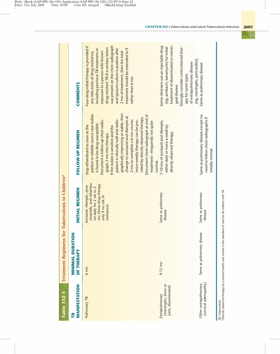
Table 332-3 shows recommended treatment regi-mens for TB disease in children. A four-drug empiricregimen (INH, rifampin, pyrazinamide, and ethambu-tol) is recommended for individuals who are at higherrisk for having INH-resistant TB, including exposureto an individual from an area of high prevalence ofdrug-resistant TB, with known drug-resistant TB, orprevious treatment for TB.24 DOT by a health care pro-fessional (not parents) is recommended for treatmentof TB in children and adolescents.24 After 2 months oftreatment, a repeat chest radiograph should be per-formed. For children from whom sputum can be
obtained, follow-up sputum should be obtained todocument culture conversion. If the patient has beenadherent to therapy, is clinically well, has an improv-ing or at least stable radiograph, and no reason existsto suspect drug resistance, the regimen can then bechanged to two drugs (INH and rifampin) after com-pleting 2 months of pyrazinamide so as to complete a6-month course. Twice-weekly dosing by DOT can beused after the induction phase of daily treatment if thechild is tolerating the regimen well and has shownconsiderable clinical improvement. The number ofdoses actually observed should be counted when con-sidering whether a patient has completed therapy.Patients receiving daily doses for the first 2 monthswill typically receive 40 observed doses (Mondaythrough Friday for approximately 8 weeks) followedby 36 twice-weekly doses in the following 18 weeks.24
Treatment of INH-monoresistant TB disease requiresat least 6 months of rifampin, pyrazinamide, andethambutol. Treatment of drug-resistant TB should beperformed in consultation with an expert in this area.(See Farhart M et al5 and Menzies6 for details.)
The most important element of TB therapy is theactual ingestion of the drugs. Children are difficult todose with TB drugs, given that the formulations arenot particularly child friendly. See dosing suggestionsin the previous section on Latent Tuberculosis Infec-tion.25 The parents and public health staff should bewarned that they might have to endure a several-weekperiod of trial and error. Patients should be monitoredmonthly during therapy. Routine laboratory evaluationneed not be performed unless the patient has symp-toms of toxicity or underlying liver disease or unlessthe patient is taking other medications, which mightinterfere with the TB drugs or cause similar toxicities.
An end-of-therapy chest radiograph should beobtained. Most children do not have a normal radio-graph at the end of therapy, but significant improve-ment is expected.
Corticosteroids have been shown to be beneficial incentral nervous system disease, particularly stage 2and 3 (altered mental status).19,24 Some clinicianswould use steroids for any child with symptomatic TBmeningitis. Steroids are also frequently used for TBpericarditis.24 Two reports support the use of steroidsin children with symptomatic airways compressioncaused by lymphatic disease.15 Prednisone is generallyused at a dose of 1 to 2 mg/kg/day given for 4 to8 weeks and then tapered over several weeks.
CONCLUSIONTB is a focal problem in the United States, dispropor-tionately affecting immigrant, Hispanic, and blackpopulations. TB risk assessments at well-child andother visits have replaced universal screening of chil-dren by TST.26 Only children who have a new risk forTB exposure since the last TST or who have featuressuggestive of TB disease should undergo the TST. Allchildren diagnosed as having LTBI should be treatedand closely monitored for adherence and toxicity.Clinics should develop or modify systems to removebarriers to completion of therapy, including walk-innurse visits, minimal paperwork, easy chart forms,and incentives. Many children with TB in the UnitedStates are asymptomatic at the time of diagnosis. TB
2602 PART 9: SPECIFIC CLINICAL PROBLEMS

Path: {Book}AAP-PPC-06-1201/Application/AAP-PPC-06-1201-322-PT-9-Nine.3dDate: 31st July 2008 Time: 05:09 User ID: elangok 1BlackLining Enabled
disease in children is diagnosed clinically and radio-graphically, often without culture confirmation. Expe-rienced TB clinicians are best qualified to manage TBdisease, but ongoing consultation should be soughtwhen local resources are limited.
Prevention of TB includes identification of childrenat risk for exposure to TB, aggressive evaluation andtreatment of children exposed to potentially contagiousadolescents and adults with TB (young children aregenerally not contagious), treatment of LTBI, andprompt treatment of contagious TB patients.
The authors thank Phil LoBue, MD, Ann Lanner, andMichael Iademarco, MD, Centers for Disease Controland Prevention, for their input on this chapter.
The findings and conclusions in this report are thoseof the authors and do not necessarily represent the viewsof the Centers for Disease Control and Prevention,Agency for Toxic Substances and Disease Registry.
WHEN TO REFER
� All patients suspected of having active TBshould be reported to the local health depart-ment according to state statute (eg, within 1working day).
� In many jurisdictions, young children with LTBIshould be reported to the local health depart-ment, according to local regulations.
� Ideally, an experienced pediatric TB clinicianshould manage children with TB disease. If localresources are not available, then close andongoing consultation with a pediatric TB expertshould be established.
WHEN TO ADMIT
� Children should be admitted to the hospital forculture collection if local resources are not avail-able for outpatient culture collection.
� Few children require admission to the hospitalbased on clinical severity of TB disease. Patientswith increased work of breathing, meningitis,or complicating simultaneous conditions orpatients who require diagnostic evaluationshould be admitted.
TOOLS FOR PRACTICEEngaging Patient and Family
� Tuberculosis (fact sheet), American Academy of Pedia-
trics (www.aap.org/publiced/BK0_Tuberculosis.htm).
� Tuberculosis: General Information (fact sheet), Centers
for Disease Control and Prevention (www.cdc.gov/tb/pubs/
tbfactsheets/tb.htm).
Medical Decision Support
� Division of Tuberculosis Elimination (Web page), Centers
for Disease Control and Prevention (www.cdc.gov/tb/
default.htm).
� Interactive Core Curriculum on Tuberculosis (other), Cen-
ters for Disease Control and Prevention (www.cdc.gov/
tb/webcourses/CoreCurr/index.htm).
� Red Book: 2006 Report of the Committee on Infectious
Diseases, 27th edition, American Academy of Pediatrics
(www.aap.org/bookstore).
� TB Education and Training Resources (interactive tool),
Centers for Disease Control and Prevention and the
National Prevention Information Network (www.findt-
bresources.org/scripts/index.cfm).
� TB Guidelines (Web page), Centers for Disease Control
and Prevention (www.cdc.gov/tb/pubs/mmwr/Maj_
guide/List_date.htm).
AAP POLICY STATEMENTSAmerican Academy of Pediatrics, Committee on Community
Health Services. Providing care for the immigrant, home-less, and migrant children. Pediatrics. 2005;115(4):1095-1100.(aappolicy.aappublications.org/cgi/content/full/pediatrics;115/4/1095)
REFERENCES1. Pediatric Tuberculosis Collaborative Group. Targeted
tuberculin skin testing and treatment of latent tuberculo-sis infection in children and adolescents. Pediatrics. 2004;114:1175-1201.
2. Huebner RE, Schein MF, Bass JB Jr. The tuberculin skintest. Clin Infect Dis. 1993;17:968-975.
3. American Academy of Pediatrics. Tuberculosis. In: Pick-ering LK, Baker CJ, Long SS, et al, eds. 2006 Red Book:2006 Report of the Committee on Infectious Diseases.27th ed. Elk Grove Village, IL: American Academy ofPediatrics; 2006.
4. Centers for Disease Control and Prevention. Targetedtuberculin testing and treatment of latent tuberculosisinfection. MMWR Morb Mortal Wkly Rep. 2000;49 (RR-6):1-51.
5. Farhat M, Greenaway C, Pai M, et al. False-positivetuberculin skin test: what is the absolute effect of BCGand non-tuberculous mycobacteria? Int J Tuberc LungDis. 2006;10:1192-1204.
6. Menzies D. What does tuberculin reactivity after bacilleCalmette-Gu�erin vaccination tell us? Clin Infect Dis. 2000;31:S71-S74.
7. Bozaykut A, Ozahi Ipek I, Ozkars MY, et al. Effect of BCGvaccine on tuberculin skin tests in 1-6-year-old children.Acta Paediatr. 2002;91:235-238.
8. Mazurek GH, Jereb J, Lobue P, et al. Guidelines for usingthe QuantiFERON-TB Gold Test for detecting Mycobac-terium tuberculosis infection, United States. MMWRRecomm Rep. 2005;54(RR-15):49-55. Erratum in MMWRMorb Mortal Wkly Rep. 2005;54:1288.
9. Centers for Disease Control and Prevention. Guidelinesfor the investigation of contacts of persons with infec-tious tuberculosis: recommendations from the NationalTuberculosis Controllers Association and CDC. MMWRMorb Mortal Wkly Rep. 2005;54(RR-15):1-37.
10. Palusci VJ, O’Hare D, Lawrence RM. Hepatotoxicity andtransaminase measurement during isoniazid chemopro-phylaxis in children. Pediatr Infect Dis J. 1995;14:144-148.
11. Centers for Disease Control and Prevention, Division ofTuberculosis Elimination. TB/HIV drug interactions.Updated Guidelines for the Use of Rifamycins for theTreatment of Tuberculosis Among HIV-Infected PatientsTaking Protease Inhibitors or Nonnucleoside ReverseTranscriptase Inhibitors. Updated January 20, 2004.Available at: www.cdc.gov/tb/TB_HIV_Drugs/default.htm. Accessed June 25, 2007.
12. Centers for Disease Control and Prevention. Trends intuberculosis—United States, 2005. MMWR Morb MortalWkly Rep. 2006;55:305-308.
2603CHAPTER 332 � Tuberculosis and Latent Tuberculosis Infection

Path: {Book}AAP-PPC-06-1201/Application/AAP-PPC-06-1201-322-PT-9-Nine.3dDate: 31st July 2008 Time: 05:09 User ID: elangok 1BlackLining Enabled
13. Lobato MN, Hopewell PC. Mycobacterium tuberculosisinfection after travel to or contact with visitors fromcountries with a high prevalence of tuberculosis. Am RevRespir Dis Crit Care Med. 1998;158:1871-1875.
14. LoBue PA, Betacourt W, Peter C, et al. Epidemiology ofMycobacterium bovis disease in San Diego County,1994-2000. Int J Tuberc Lung Dis. 2003;7:180-185.
15. Loeffler AM. Pediatric tuberculosis. Semin Respir Infect.2003;18:272-291.
16. Francis J. Curry National Tuberculosis Center. PediatricTuberculosis: A Guide to the Gastric Aspirate Procedure,2006. Available at: www.nationaltbcenter.edu/catalogue/epub/index.cfm?tableName=GAP. Accessed June 25,2007.
17. Lobato MN, Loeffler AM, Furst K, et al. Detection ofMycobacterium tuberculosis in gastric aspirates col-lected from children: hospitalization is not necessary.Pediatrics. 1998;102:e40.
18. Zar HJ, Hanslo D, Apolles P, et al. Induced sputum versusgastric lavage for microbiological confirmation of pul-monary tuberculosis in infants and young children: aprospective study. Lancet. 2005;365:130-134.
19. Starke JR. Tuberculosis of the central nervous system inchildren. Semin Pediatr Neurol. 1999;6:318-331.
20. Starke JR. Interferon-gamma release assays for diagno-sis of tuberculosis infection in children. Pediatr Infect DisJ. 2006;25(10):941-942.
21. Smuts NA, Beyers N, Gie RP, et al. Value of the lateralchest radiograph in tuberculosis in children. PediatrRadiol. 1994;24:478-480.
22. Neu N, Saiman L, San Gabriel P, et al. Diagnosis of pedi-atric tuberculosis in the modern era. Pediatr Infect Dis J.1999;18:122-126.
23. Cremin BJ, Jamieson DH. Childhood tuberculosis. In:Modern Imaging and Clinical Concepts. London, UK:Springer-Verlag; 1995.
24. American Thoracic Society, Centers for Disease Controland Prevention, Infectious Diseases Society of America.Treatment of tuberculosis. MMWR Recomm Rep. 2003;52(RR-11):1-77.
25. Francis J. Curry National Tuberculosis Center. PediatricTuberculosis, 2007. Available at: www.nationaltbcenter.edu/pediatric_tb. Accessed May 22, 2007.
26. Bright Futures at Georgetown University. Appendices.In: Bright Futures: Guidelines for Health Supervision ofInfants, Children, and Adolescents. 2nd rev ed.Washing-ton, DC: National Center for Education in Maternal andChild Health; 2002. Available at: www.brightfutures.org/bf2/pdf/pdf/Appendices.pdf. Accessed June 26, 2007.
Chapter 333
UMBILICAL ANOMALIES
Robert W. Marion, MD
‘‘It [the umbilicus] is all that remains of the stemthat bound us to the parental stalk. It is a reminderthat we have been plucked and must sooner orlater die. It might be said that when the stem issevered, we cease to live in any true sense. Wemay be ornamental like roses or useful like cab-bages but only for a little while. Our dissolutionhas begun.’’1
Despite its essential role in the survival of the fetusduring prenatal life, the umbilicus, the external vestigeof the umbilical cord, is frequently ignored or over-looked by the pediatric primary care physician. How-ever, aberrations in either the formation or theposition of this structure can offer helpful clues tounderlying disease in the young child. Major congeni-tal anomalies of the ventral abdominal wall, such asomphalocele, gastroschisis, and exstrophies of thebladder and cloaca, are described in detail elsewhere.This chapter deals with minor anomalies in configura-tion, placement, and formation of the umbilicus. Inaddition to the conditions described here, the umbili-cus can be the site of both tumors (either vascular orteratomatous neoplasms) and infections (omphalitis).2
To understand the causes and significance ofanomalies of the umbilicus, a review of some basic fun-damentals of the embryologic development of theumbilical cord is necessary.
EMBRYOLOGIC DEVELOPMENTOF THE UMBILICAL CORDAppearing within the first 6 weeks of gestation, theumbilical cord is derived from the fusion of 3 separateembryonic structures: (1) the primitive or primary yolksac, which contains the allantois and a portion of thevitelline duct, transient structures that ultimately formthe central portion of the embryonic gut, the urinarybladder, the urachus, and the umbilical blood vessels(usually 2 arteries and 1 vein); (2) the secondary yolksac, composed of the remainder of the vitelline duct;and (3) the mesenchyme of the connecting body stalkof the embryo, the tissue that produces Wharton jelly,which is the packing substance that holds the cordtogether. After fusion is complete, these unified struc-tures become covered by the amnion and are ulti-mately surrounded by amniotic fluid.3
Many of these embryonic structures that form theumbilical cord are present for only brief periods dur-ing embryogenesis. After the 7th week of gestation,the vitelline duct regresses and is ultimately com-pletely resorbed. Similarly the allantois, which is con-tiguous with the urinary bladder, degenerates,forming a fibrous cord called the urachus, which con-nects the apex of the bladder with the umbilicus.Anomalies may result when these structures fail toundergo normal regression, causing them to persistinto postnatal life.
ANOMALIES
Abnormalities of Position and MorphologyAnatomically, the level of the umbilicus is usually atthe top of the iliac crest ventral to the 3rd or 4th lum-bar vertebra.4 Variations in the position of the umbili-cus can result from abnormalities in the way in whichthe abdominal wall itself has formed and, as such, maybe a clue to the diagnosis of specific dysmorphic syn-dromes. For example, as described in Table 333-1, theumbilicus has been noted to be low set in achondro-plasia (in which disproportionate growth of the trunkaccounts for the aberration in position), in bladder and
2604 PART 9: SPECIFIC CLINICAL PROBLEMS