PART V THE ORIGIN AND CLASSIFICATION OF LIFE The...
40
CHAPTER 21 The Nature of Microorganisms D ead zones are areas in the world’s oceans where the oxygen level is so low that most organisms die. In 2006, the United Nations published a list of over 200 places in the world’s oceans where dead zones developed for at least part of each year. Similar dead zones occur in some lakes. Dead zones normally develop at the mouths of rivers as a result of human action and the activity of various kinds of microorganisms. One of the largest dead zones is in the Gulf of Mexico off the mouth of the Mississippi River. Fertilizer from agricultural runoff, waste from large industrial livestock operations, and the effluent from poorly controlled municipal and industrial sewage flow into rivers that ulti- mately empty into an ocean (see photo). These nutrients stimulate the growth of single-celled and multi-cellular algae during the warmer months of the year. Eventually, these organisms die and sink to the bottom, where decomposer bacteria use oxygen in the process of aerobic respiration to break down the dead organic matter. This lowers the amount of dissolved oxygen in the water, causing the death of bottom- dwelling animals. • Why are bacteria able to live in regions of low oxygen when animals cannot? • Why do dead zones occur off the mouths of rivers? • Should the amount of fertilizer farmers apply to their fields to increase yields be regulated? 455 PART V THE ORIGIN AND CLASSIFICATION OF LIFE CHAPTER OUTLINE 21.1 What Are Microorganisms? 456 21.2 The Domains Bacteria and Archaea 456 The Domain Bacteria The Domain Archaea 21.3 The Kingdom Protista 464 Algae Protozoa Funguslike Protists 21.4 Multicellularity in the Protista 472 21.5 The Kingdom Fungi 472 The Taxonomy of Fungi The Significance of Fungi HOW SCIENCE WORKS 21.1: How Many Microbes Are There? 457 HOW SCIENCE WORKS 21.2: Bioremediation 458 OUTLOOKS 21.1: Food Poisoning/Foodborne Illness/ Stomach Flu 462 OUTLOOKS 21.2: The Microbial Ecology of a Cow 464 OUTLOOKS 21.3: The Marine Microbial Food Web 466 HOW SCIENCE WORKS 21.3: Penicillin 475 Are We Killing the Oceans? Fertilizer may be the cause. www.aswarphysics.weebly.com
Transcript of PART V THE ORIGIN AND CLASSIFICATION OF LIFE The...

CHAPTER
21 The Nature of Microorganisms
D ead zones are areas in the world’s oceans where the oxygen level is so low that most organisms die. In 2006, the United Nations published a list of over
200 places in the world’s oceans where dead zones developed for at least part of each year. Similar dead zones occur in some lakes. Dead zones normally develop at the mouths of rivers as a result of human action and the activity of various kinds of microorganisms. One of the largest dead zones is in the Gulf of Mexico off the mouth of the Mississippi River. Fertilizer from agricultural runoff, waste from large industrial livestock operations, and the effluent from poorly controlled municipal and industrial sewage flow into rivers that ulti-mately empty into an ocean (see photo). These nutrients stimulate the growth of single-celled and multi-cellular algae during the warmer months of the year. Eventually, these organisms die and sink to the bottom, where decomposer bacteria use oxygen in the process of aerobic respiration to break down the dead organic matter. This lowers the amount of dissolved oxygen in the water, causing the death of bottom-dwelling animals.
• Why are bacteria able to live in regions of low oxygen when animals cannot?
• Why do dead zones occur off the mouths of rivers?
• Should the amount of fertilizer farmers apply to their fields to increase yields be regulated?
455
PART V THE ORIGIN AND CLASSIFICATION OF LIFE
CHAPTER OUTLINE
21.1 What Are Microorganisms? 456
21.2 The Domains Bacteria and Archaea 456 The Domain Bacteria The Domain Archaea
21.3 The Kingdom Protista 464 Algae Protozoa Funguslike Protists
21.4 Multicellularity in the Protista 472
21.5 The Kingdom Fungi 472 The Taxonomy of Fungi The Significance of Fungi
HOW SCIENCE WORKS 21.1: How Many Microbes
Are There? 457
HOW SCIENCE WORKS 21.2: Bioremediation 458
OUTLOOKS 21.1: Food Poisoning/Foodborne Illness/
Stomach Flu 462
OUTLOOKS 21.2: The Microbial Ecology of a Cow 464
OUTLOOKS 21.3: The Marine Microbial Food Web 466
HOW SCIENCE WORKS 21.3: Penicillin 475
Are We Killing the Oceans?
Fertilizer may be the cause.
eng03466_ch21_455-478.indd 455eng03466_ch21_455-478.indd 455 15/10/10 3:05 PM15/10/10 3:05 PM
www.aswarp
hysic
s.wee
bly.co
m

456 PART V The Origin and Classification of Life
Background Check Concepts you should already know to get the most out of this chapter: • The processes of natural selection and evolution (chapter 13) • Prokaryotic organisms have a simpler cellular structure than eukaryotic organisms (chapter 4) • How structural and life history characteristics are used to classify organisms (chapter 20)
21.1 What Are Microorganisms? A microorganism, or microbe, is a tiny organism that usually cannot be seen without the aid of a microscope. These are terms of convenience for a wide variety of organisms, includ-ing the domains Bacteria and Archaea and the kingdoms Protista and Fungi in the domain Eucarya. Often, tiny ani-mals, particularly those that cause disease (mites, worms, etc.), are considered microorganisms as well. Viruses are also treated as microbes. However, we will not consider animals or viruses in this chapter. Viruses were discussed in chapter 20 and animals are discussed in chapter 23.
In a very general sense, microorganisms share several characteristics. These organisms generally consist of cells, which function independently. Many are single-celled organ-isms, although some single-celled microbes form loose aggre-gations, called colonies. Others are multicellular and have some specialization of cells for certain functions. Their pri-mary method of reproduction is asexual reproduction, in which one cell divides to become two cells, although most kinds are also capable of sexual reproduction. Many have special structures involved in the production of gametes. Many microbes, particularly Bacteria, have the ability to exchange pieces of genetic material, which creates new combinations of genes. In some cases this even involves gene exchange between organisms that are considered to be of different species.
Microbes are extremely common organisms. It is estimated that the total biomass of microbes is larger than the biomass of all other kinds of organisms combined. They live in any aquatic or moist environment and occur in huge numbers in the oceans and other bodies of water and in soil. It has been suggested that if one were able to instantly remove all living things from Earth except microbes, everything on Earth would still be visible in outline form because microbes cover all surfaces, including living things. Because they are small, their moist habitat does not need to be large. Microbes can maintain huge populations in places such as human skin or intestine, temporary puddles, and soil. Your skin, mouth, and gut each contains trillions of microbes. Most die if they dry out, but some have the special ability to become dormant and survive long periods without water. When moistened, they become actively growing cells again.
21.2 The Domains Bacteria and Archaea
At one time, all prokaryotic organisms were lumped into one group of microorganisms called bacteria. Today, scientists rec-ognize that there are two, very different kinds of prokaryotic organisms: the domains Bacteria and Archaea. The Bacteria and Archaea differ in several ways: Bacteria have a compound, called peptidoglycan, in their cell walls, which Archaea do not have. The chemical structure of the cell membranes of Archaea is different from that of all other kinds of organisms. When the DNA of Archaea is compared with that of other organisms, it is found that a large proportion of their genes are unique.
Today, most scientists still use the terms bacterium and bacteria. However, they are used in a restricted sense to refer to members of the domain Bacteria. The term archeon is fre-quently used to refer to members of the Domain Archaea.
The Domain Bacteria The Bacteria are an extremely diverse group of organisms. Although only about 2,000 species of Bacteria have been named, most biologists feel that there are probably millions still to be identified (How Science Works 21.1). They occupy every conceivable habitat and have highly diverse metabolic abilities. They are typically spherical, rod-shaped, or spiral-shaped. They are often identified by the characteristics of their metabolism or the chemistry of their cell walls. Many have a kind of flagellum, which rotates and allows for move-ment. Figure 21.1 shows the general structure of a bacterium. Some form resistant spores, which can withstand dry or other harsh conditions. Bacteria play several important ecological roles and interact with other organisms in many ways.
Decomposers Many kinds of bacteria are heterotrophs that are saprophytes. They break down organic matter to provide themselves with energy and raw materials for growth. Therefore, they function as decomposers in all ecosystems. Decomposers are a diverse group and use a wide variety of metabolic processes. Some are anaerobic and break down complex organic matter to simpler organic compounds. Others are aerobic and degrade organic matter to carbon dioxide and water. In nature, this decomposi-tion process is important in the recycling of carbon, nitrogen, phosphorus, and many other elements.
The actions of decomposers have been harnessed for human purposes. Sewage treatment plants rely on bacteria and other organisms to degrade organic matter ( figure 21.2 ) (How Science Works 21.2).
1. What taxonomic groups are included in the cate-gory known as microorganisms?
2. List three general characteristics the various kinds of microbes share.
21.1 CONCEPT REVIEW
eng03466_ch21_455-478.indd 456eng03466_ch21_455-478.indd 456 15/10/10 3:05 PM15/10/10 3:05 PM
www.aswarp
hysic
s.wee
bly.co
m

CHAPTER 21 The Nature of Microorganisms 457
capsulegel-like coating outsidethe cell wall
chromosomelocation of the bacterialDNA
sex piluselongated, hollow appendage used to transferDNA to other cells
flagellumrotating filament that propels the cell
ribosomesite of protein synthesis
plasma membranesheet that surrounds the cytoplasm and regulates entrance and exit of molecules
cell wallstructure that provides support and shapes the cell
fimbriae hairlike bristles that allow adhesion to surfaces
cytoplasmsemifluid solution surroundedby the plasma membrane;contains nucleoid andribosomes
FIGURE 21.1 Bacteria Cell Structure The plasma membrane regulates the movement of material between the cell and its environment. A rigid cell wall protects the cell and determines its shape. Some bacteria, usually pathogens, have a capsule to protect them from the host’s immune system. The genetic material consists of a loop of DNA.
HOW SCIENCE WORKS 21.1
How Many Microbes Are There? Biologists have long suspected that there are large numbers of undiscovered species of microbes in the world. One of the major problems associated with identifying microbes is that they must be isolated and grown to be characterized. Unfortunately, it appears that most microbes cannot be grown in the lab and therefore cannot be studied in detail.
However, the technology of DNA sequencing has provided a better estimate of the number of kinds of microbes in our world. J. Craig Venter, one of the scientists who developed techniques for sequencing the human genome, has applied the DNA sequencing techniques to the ocean ecosystem. Water samples were collected from many parts of the ocean. The
samples were filtered to collect the microbes. The DNA from these mixtures of organisms was then sequenced. The result was a “metagenome,” a picture of the DNA of an ecosystem.
Once this composite of DNA was known, pieces of it could be compared to known genes and new, unique sequences could be identified. The result was the identification of 1.2 million new genes and a doubling of the number of kinds of proteins pro-duced from those genes. Many new genes appear to be related to molecules responsible for trapping sunlight by autotrophic microbes. The identification of new genes and the proteins they produce implies that there are many new species in the ocean responsible for their production.
eng03466_ch21_455-478.indd 457eng03466_ch21_455-478.indd 457 15/10/10 3:05 PM15/10/10 3:05 PM
www.aswarp
hysic
s.wee
bly.co
m

458 PART V The Origin and Classification of Life
HOW SCIENCE WORKS 21.2
Bioremediation Bioremediation involves the use of naturally occurring microbes to break down unwanted or dangerous materials. In many ways we have been using bioremediation for centuries. Composting, sewage treatment plants, and the activities of soil bacteria to break down animal manure are common examples of how microbes break down unwanted organic matter. However, mod-ern society has invented other kinds of pollution that are more resistant to the activities of microbes. Oil spills and the release of synthetic organic compounds such as polychlorinated biphe-nyls (PCBs), trichloroethylene (TCE), and many other persistent organic molecules have created a new kind of pollution that also can be treated with microbes.
Several types of activities are commonly involved when bio-remediation is attempted. In order to find the microbes with
the desired abilities, scientists screen many kinds. In some cases, genetic engineering techniques have been used to intro-duce genes into microbes that allow the microbes to survive in toxic situations. When bioremediation is to be attempted, several actions are commonly taken. Specific microbes with desirable properties may be added to break down the pollut-ant. Nutrients such as nitrogen or phosphorus may be added to stimulate the growth of microorganisms already present. In some cases, the concentration of the pollutant may be diluted so that the pollutant will not kill the microbes that will eventu-ally metabolize it.
Bioremediation has been used to clean up oil spills, degrade pesticides, detoxify metallic contaminants, and in many other ways.
FIGURE 21.2 Decomposers in Sewage A sewage treatment plant is designed to encourage the growth of bacteria and other microorganisms that break down organic matter. The tank in the foreground contains a mixture of sewage and microorganisms, which is being agitated to assure the optimal growth of microbes.
The food industry uses lactic acid fermentation by certain bacteria to produce cheeses, yogurt, sauerkraut, and many other foods. Alcohols, acetones, acids, and other chemicals are produced by bacterial cultures. Some bacteria can even metabolize oil and are used to clean up oil spills.
Unfortunately, decomposer bacteria do not distinguish between items that we want to decompose and those that we don’t want to decompose. Bacteria in food can cause milk to turn sour or vegetables and meat to spoil. Thus, it is often necessary to control the populations of some decomposer
bacteria, so that foods and other valuable materials are not destroyed by rotting or spoiling.
Commensal Bacteria Many kinds of bacteria have commensal relationships with other organisms. They live on the surface or within other organ-isms and cause them no harm, but neither do they perform any valuable functions. Most organisms are lined and covered by populations of bacteria called normal flora ( table 21.1 ). In fact, if an organism lacks bacteria, it is considered abnormal. The bacterium Escherichia coli (commonly called E. coli ) is com-mon in the intestinal tract of humans, other mammals, and birds. A large proportion of human feces is composed of E. coli and other bacteria. Many of the odors humans produce from the skin and gut are the result of commensal bacteria.
Photosynthetic Bacteria Several kinds of Bacteria carry on a form of photosynthesis. A group called the cyanobacteria carries out a form of pho-tosynthesis that is essentially the same as that in plants and algae. They use carbon dioxide and water as raw materials and release oxygen. In fact, the chloroplasts of eukaryotic organisms are assumed to be cyanobacteria that, in the past, formed an endosymbiotic relationship with other cells. Cyanobacteria are thought to be the first oxygen-releasing organisms; thus, their activities led to the presence of oxygen in the atmosphere and the subsequent evolution of aerobic respiration. Cyanobacteria are extremely com-mon and are found in fresh and marine waters and soil and other moist environments. When conditions are favorable, asexual reproduction can result in what is called a bloom —a rapid increase in the population of microorganisms in a body of water ( figure 21.3 ). Many cyanobacteria form fila-ments or other kinds of colonies, which produce large masses when a bloom occurs. Some species of cyanobacte-ria produce toxins. When blooms occur, the levels of toxins
eng03466_ch21_455-478.indd 458eng03466_ch21_455-478.indd 458 15/10/10 3:05 PM15/10/10 3:05 PM
www.aswarp
hysic
s.wee
bly.co
m
CHAPTER 21 The Nature of Microorganisms 459
TABLE 21.1 Common Bacteria in or on Humans
Skin Corynebacterium sp., Staphylococcus sp., Streptococcus sp., Escherichia coli, Mycobacterium sp.
Eye Corynebacterium sp., Neisseria sp., Bacillus sp., Staphylococcus sp., Streptococcus sp.
Ear Staphylococcus sp., Streptococcus sp., Corynebacterium sp., Bacillus sp.
Mouth Streptococcus sp., Staphylococcus sp., Lactobacillus sp., Corynebacterium sp., Fusobacterium sp., Vibrio sp., Haemophilus sp.
Nose Corynebacterium sp., Staphylococcus sp., Streptococcus sp.
Intestinal tract Lactobacillus sp., Escherichia coli, Bacillus sp., Clostridium sp., Pseudomonas sp., Bacteroides sp., Streptococcus sp.
Genital tract Lactobacillus sp., Staphylococcus sp., Streptococcus sp., Clostridium sp., Peptostreptococcus sp., Escherichia coli
Escherichia coli LactobacillusStaphylococcus
FIGURE 21.3 Bloom of Cyanobacteria Many kinds of cyanobacteria reproduce rapidly in nutrient-rich waters and produce masses of organisms known as a bloom.
in the water may be high enough to poison humans and other animals.
Within the filaments of many cyanobacteria are special-ized, larger cells capable of nitrogen fixation which converts atmospheric nitrogen, N 2 , to ammonia, NH 3 . This provides a form of nitrogen usable to other cells in the colony—an example of division of labor.
Two kinds of Bacteria, known as purple and green bacte-ria, carry on different forms of photosynthesis that do not release oxygen. Many of these organisms release sulfur as a result of their photosynthesis.
Mutualistic Bacteria Mutualistic relationships occur between bacteria and other organisms. Some intestinal bacteria benefit humans by produc-ing antibiotics, which inhibit the development of disease-causing bacteria. They also compete with disease-causing bacteria for nutrients, thereby helping keep them in check. They aid digestion by releasing various nutrients. They pro-duce and release vitamin K. Mutualistic bacteria establish this symbiotic relationship when humans ingest them along with food or drink. When people travel, they consume local bacteria with their food and drink and may have problems establishing a new symbiotic relationship with these foreign bacteria. Both the host and the symbionts must adjust to their new environ-ment, which can result in a very uncomfortable situation for both. Some people develop traveler’s diarrhea as a result.
There are many other examples of mutualistic relation-ships between bacteria and other organisms. Many kinds of plants have nitrogen-fixing bacteria in their roots in a symbi-otic relationship. Some fish and other aquatic animals have bioluminescent bacteria in their bodies, allowing them to produce light. Many kinds of lichens contain cyanobacteria as symbionts with their fungal cells.
Bacteria and Mineral Cycles Many different bacteria are involved in the nitrogen cycle. In addition to symbiotic nitrogen-fixing bacteria, free-living nitrogen-fixing bacteria in the soil convert N 2 to NH 3 . Other bacteria convert ammonia to nitrite and nitrate. These bacte-ria are chemoautotrophs that use inorganic chemical reactions involving nitrogen to provide themselves with energy. All of
Pseudomonas
eng03466_ch21_455-478.indd 459eng03466_ch21_455-478.indd 459 15/10/10 3:05 PM15/10/10 3:05 PM
www.aswarp
hysic
s.wee
bly.co
m

460 PART V The Origin and Classification of Life
these bacteria are extremely important ecologically, because they are ultimately the source of nitrogen for plant growth. Finally, some bacteria convert nitrite to atmospheric nitrogen.
In addition to nitrogen; iron, sulfur, manganese, and many other inorganic materials are cycled by chemoautotrophic bacteria with specialized metabolic abilities. Some of these are important ecologically, because they produce acid mine drain-age or convert metallic mercury to methylmercury, which can enter animals and cause health problems.
Disease-Causing Bacteria Disease-causing bacteria are heterotrophs that use the organic matter of living cells as food. Bacteria and other kinds of organisms that are capable of causing harm to their host are called pathogens . Only a small minority of bacteria fall into this category; however, because historically they have been responsible for huge numbers of deaths and continue to be a serious problem, they have been studied intensively and many pathogens are well understood.
Pathogenic bacteria can cause disease in several ways. Many are normally harmless commensals but cause disease when their populations increase to excessively high numbers. For example, Streptococcus pneumoniae can grow in the throats of healthy people without any pathogenic effects. But if a person’s resistance is lowered, as after a bout with viral flu, Streptococcus pneumoniae can invade the lungs and reproduce rapidly, causing pneumonia. The relationship changes from commensalistic to parasitic.
Other bacteria invade the healthy tissue of their host and cause disease by altering the tissue’s normal physiology. Bacteria living in the host release a variety of enzymes that cause the destruction of tissue. The disease ends when the pathogens are killed by the body’s defenses or an outside agent, such as an antibiotic. Examples are the infectious diseases strep throat, syphilis, anthrax, pneumonia, tuberculosis, and leprosy.
Many other illnesses are caused by toxins or poisons produced by bacteria. Some of these bacteria release toxins that may be consumed with food or drink. In this case, dis-ease can be caused even though the pathogens never enter the host. For example, botulism is a deadly disease caused by bacterial toxins in food or drink. Other bacterial diseases are the result of toxins released from bacteria growing inside the host tissue; tetanus and diphtheria are examples. In general, toxins cause tissue damage, fever, and aches and pains.
Bacterial pathogens are also important factors in certain plant diseases. Bacteria cause many types of plant blights, wilts, and soft rots. Apples and other fruit trees are susceptible to fire blight, a disease that lowers the fruit yield because it kills the tree’s branches. Citrus canker, a disease of citrus fruits that causes can-cerlike growths on stems and lesions on leaves and fruit, can generate widespread damage. Federal and state governments have spent billions of dollars controlling this disease ( figure 21.4 ).
Probably all species of organisms have bacterial patho-gens. Plants and animals get sick and die all the time. However, scientists are not likely to spend time and money studying these diseases unless the organisms have economic value to us.
Therefore, scientists know much about bacterial diseases in humans, domesticated animals, and crop plants but know very little about the diseases of jellyfish, squid, or most plants.
Control of Bacterial Populations The diseases and many kinds of environmental problems caused by bacteria are actually population control problems. Small numbers of bacteria cause little harm. However, when the population increases, their negative effects are multiplied. Despite large investments of time and money, scientists have found it difficult to control bacterial populations. Three factors operate in favor of the bacteria: their reproductive rate, their ability to form resistant stages, and their ability to mutate and produce strains that resist antibiotics and other control agents.
Under ideal conditions, some bacteria can grow and divide every 20 minutes. If one bacterial cell and all its off-spring were to reproduce at this ideal rate, in 48 hours there would be 2.2 � 10 43 cells. In reality, bacteria cannot achieve such incredibly large populations, because they would eventu-ally run out of food and be unable to dispose of their wastes. However, many of the methods used to control pathogenic bacteria are those that control their numbers by interfering with their ability to reproduce. Many antibiotics interfere with a certain aspect of bacterial physiology so that the bacteria are killed or become unable to divide and reproduce. This allows the host’s immune system to gain control and destroy the disease-causing organism. Without the antibiotic, the immune system may be overwhelmed and the person may die.
Although antibiotics can save lives, they don’t always work because bacteria mutate and produce individuals that
FIGURE 21.4 A Bacterial Plant Disease Citrus canker is a disease of citrus trees caused by the bacterium Xanthomonas axonopodis. This photograph shows the typical lesions on the fruit and leaves of an orange tree.
eng03466_ch21_455-478.indd 460eng03466_ch21_455-478.indd 460 15/10/10 3:05 PM15/10/10 3:05 PM
www.aswarp
hysic
s.wee
bly.co
m

CHAPTER 21 The Nature of Microorganisms 461
are resistant to the effects of an antibiotic. Because bacteria reproduce so rapidly, a few antibiotic-resistant cells in a bac-terial population can increase to dangerous levels in a very short time. This requires the use of stronger doses or new types of antibiotics to bring the bacteria under control. Furthermore, these resistant strains can be transferred from one host to another, making it difficult to control the spread of disease. For example, sulfa drugs and penicillin, once widely used to fight infections, are now ineffective against many strains of pathogenic bacteria. Methicillin has been a valuable antibiotic for many years. However, some strains of Staphylococcus aureus , a common skin bacterium, have become resistant to methicillin. As a result, common skin infections that should be controlled easily have become life-threatening. These strains have become known as methicillin-resistant Staphylococcus aureus (MRSA). As with methicillin, when any new antibiotic is developed, natural selection encourages the development of resistant bacterial strains. Therefore, humans are constantly waging battles against new strains of resistant bacteria.
In addition to antibiotics, various kinds of antiseptics are used to control the numbers of pathogenic bacteria. Antiseptics are chemicals able to kill or inhibit the growth of microbes. They can be used on objects or surfaces that have colonies of potentially harmful bacteria. Certain antiseptics are used on the skin or other tissues of people who are receiving injections or undergoing surgery. Reducing the numbers of bacteria lessens the likelihood that the microbes on the skin will be carried into the body, causing disease. We all are constantly in contact with pathogenic bacteria; however, as long as their numbers are controlled, they do not become a problem.
Another factor that enables some bacteria to survive a hostile environment is their ability to form endospores. An endospore is a unique bacterial structure with a low meta-bolic rate that can withstand hostile environmental condi-tions and germinate later, when there are favorable conditions to form a new, actively growing cell ( figure 21.5 ). Endospores thought to be Bacillus sphaericus and estimated to be 25 million to 40 million years old have been isolated from the intestinal tract of a bee fossilized in amber. When placed in an optimum growth environment, they have germinated and grown into numerous colonies.
Some spore-forming bacteria are important disease-causing organisms. People who preserve food by canning often boil the food in the canning jars to kill the bacteria, but not all are killed by boiling, because some form endospores. The endo-spores of Clostridium botulinum, the bacterium that causes botulism, can withstand boiling and remain for years in the endospore state. However, endospores do not germinate and produce botulism toxin if the pH of the canned goods is in the acid range; in that case, the food remains preserved and edi-ble. If conditions become favorable for Clostridium endo-spores to germinate, they become actively growing cells and produce toxin. Using a pressure cooker and heating the food to temperatures higher than 121°C for 15 to 20 minutes destroys both the botulism toxin and the endospores.
FIGURE 21.5 Bacterial Endospore (a) The body at the top end of the cell is an endospore. It contains the bacterial DNA, as well as a concentration of cytoplasmic material surrounded and protected by a thick wall. (b) The photo shows a Bacillus bacterium that has formed endospores in some cells.
Endospore
(a) Bacillus thuringiensis
(b) Bacillus bacteria with endospores
Anthrax is an acute infectious disease caused by the spore-forming bacterium Bacillus anthracis. Anthrax spores can live in the soil as spores for long periods and cause disease when they are inhaled, are swallowed, or invade the skin. Because anthrax spores can survive dry conditions, they were used to contaminate mail as an agent of bioterrorism.
Contaminated food and water are common ways that people encounter bacteria that cause them harm. These dis-ease episodes are commonly referred to as food poisoning or stomach flu (Outlooks 21.1).
eng03466_ch21_455-478.indd 461eng03466_ch21_455-478.indd 461 15/10/10 3:06 PM15/10/10 3:06 PM
www.aswarp
hysic
s.wee
bly.co
m

462 PART V The Origin and Classification of Life
contaminated food. Rotavirus is the most common cause of severe diarrhea in infants and young children. Nearly every child in the world has been infected with rotavirus at least once, but they develop partial immunity following infection and subsequent infections tend to be mild. Many kinds of bac-teria are involved in cases of food poisoning: Salmonella, Escherichia coli, Shigella , and Staphylococcus are examples.
An additional cause that leads to similar symptoms involves changes in the kinds and numbers of bacteria normally found in your intestine. Your gut is an ecosystem in which there are many different kinds of Bacteria, Archaea, and protozoa. Each has specific metabolic requirements and produces specific kinds of metabolic waste products. Some of these products may be gases. If you change the kind of food you eat, or if the water you drink has different kinds of minerals in it, some of your intestinal microbes may experience population increases that lead to symptoms similar to those of food poisoning.
Treatment usually does not involve medication. One simply waits until the illness runs its course. The most serious health concern is dehydration from vomiting and diarrhea. Consequently, providing liquids is important and, in severe cases, intravenous fluids may be required.
OUTLOOKS 21.1
Food Poisoning/Foodborne Illness/Stomach Flu Many people talk about a disease experience they call stom-ach flu but it is not caused by the influenza virus. The disease usually involves nausea, vomiting, and diarrhea. It may also involve headache, fever, and abdominal cramping. The Centers for Disease Control and Prevention estimates that food poi-soning causes about 75 million cases of illness in the United States each year. The U.S. population is about 310 million, so we all have about a 1 in 4 chance of having this uncomfortable experience each year.
Food poisoning is not caused by the virus that causes influ-enza and should more properly be called gastroenteritis. In addition, it is not a single disease but is caused by a variety of organisms and mechanisms. The typical way of contracting the disease is through food or water contaminated with viruses, bacteria, or protozoa that, when ingested, multiply and cause the symptoms—hence the name food poisoning . Furthermore, these diseases are usually contagious because those who are sick pass the organism in their feces and can transmit it to those around them if those infected do not practice good hygiene.
Norovirus is responsible for about 50% of cases in United States and generally is contracted as a result of fecally
The Domain Archaea The Archaea are distinct from the Bacteria. They differ from Bacteria in the nature of their cell walls, cell membranes, DNA, and other details of structure and physiology. In addition to the spherical, rod-shaped, and spiral-shaped forms found in the Bacteria, some Archaea are lobed, platelike, or irregular in shape. Like the Bacteria, the Archaea are extremely diverse and extremely common. Only a couple hundred species have been described, but DNA sampling of the ocean and soil suggests that there is a huge number of undescribed species. Some spe-cies are found in extreme habitats—high temperature, high acid, high salt—and are referred as extremophiles (lovers of extremes).Others are very common in the ocean, freshwater, soil, and the digestive tract of animals where they play a variety of ecological roles. To date only one archeon has been identified as a parasite and it is a parasite on other Archaea.
Extreme Halophiles Extreme halophiles (salt lovers) are Archaea that can live only in extremely salty environments—such as the Great Salt Lake in Utah and the Dead Sea, located between Israel and Jordan. They require a solution of at least 8% salt and grow best in solutions that are about 20% salt. The Atlantic Ocean is about 3.5% salt. The Dead Sea is about 15% salt. They also live in artificial salt ponds used to evaporate seawater to pro-duce salt. Because they contain the reddish pigment carotene, they color these salt ponds pinkish or red.
Most of these organisms are aerobic heterotrophs. They use organic matter from their environment as a source of food. Some have been found growing on food products, such as salted fish, causing spoilage. However, some of them are also photosynthetic autotrophs that have a carotene-containing pigment, called bacteriorhodopsin, which absorbs sunlight and allows the cells to make ATP.
Thermophiles The thermophiles (heat lovers) are a diverse group of the Archaea that live in extremely hot environments, such as the hot springs found in Yellowstone National Park and hydrothermal vents on the ocean floor ( figure 21.6 ). All require high environ-mental temperatures—typically, above 50°C (122°F)—and some grow well at temperatures above 100°C (212°F). They are diverse metabolically; some are aerobic whereas others are anaerobic. Some can reduce sulfur or sulfur-containing com-pounds by attaching hydrogen to sulfur (S � 2H → H 2 S). Thus, they release hydrogen sulfide gas (H 2 S). Some live in extremely acidic conditions, with a pH of 1–2 or even less.
Acidophiles and Alkaliphiles Some Archaea live at extreme pHs. One acidophile is known to live at a pH of 0. Another acidophile has been identified as impor-tant in the acid drainage from abandoned mines where they oxidize iron. Alkalophiles live in lakes with basic pHs of 9–11 and maintain a near normal internal pH by pumping hydrogen ions from their environment into their cells.
eng03466_ch21_455-478.indd 462eng03466_ch21_455-478.indd 462 15/10/10 3:06 PM15/10/10 3:06 PM
www.aswarp
hysic
s.wee
bly.co
m

CHAPTER 21 The Nature of Microorganisms 463
Methane is a greenhouse gas tied to the problem of global warming, so scientists have tried to characterize the role of Archaea as producers of methane. However, because they are involved as components of important agricultural activities (rice growing and cattle raising), it is not likely that this source of methane will be controlled.
Non-Extremophiles Although at one time it was thought that the Archaea were all extremophiles, that impression is changing. It is becoming clear that archeons are common in most environments where there is moisture, not just in extreme environments. A major problem with characterizing the roles played by Archaea is that they are difficult to isolate and grow in captivity. However, when environments such as the ocean and soil are sampled for DNA, large amounts of Archaea DNA are found. They appear to be extremely common in the ocean, in freshwater, and in the soil, where they perform a variety of functions (Outlooks 21.3). Many are heterotrophs that degrade organic material and thus are decomposers. Some of these decomposers are aerobic whereas others are anaerobic heterotrophs. Other Archaea are chemoautotrophs that use inorganic chemical reactions to make organic matter. In addition, it appears that there are many archeons that are photoautotrophs that use light to produce organic molecules. In the ocean it appears that these two kinds of autotrophs are important contributors to the base of the marine food web. Because of their small size, they are referred to as picoplankton. These autotrophic archeons are eaten by bacteria, protozoa, and other organisms. In the ocean it is becoming clear that some of chemoautotrophic Archaea are involved in several steps in the nitrogen cycle. Since Archaea are also common in the soil, it is likely that they are also involved in the terrestrial part of the nitrogen cycle as well.
FIGURE 21.6 Hydrothermal Vents Extremely hot, mineral-rich water enters the ocean from hydrothermal vents on the ocean floor. Many kinds of specialized Archaea live in these places, where they use sulfur as a source of energy. These archeons are, in turn, eaten by other organisms that live in the vicinity.
Methanogens Methanogens are members of the Archaea that are strict anaer-obes (do not live where there is oxygen) and release methane as a waste product of cellular metabolism. Most are chemo-synthetic autotrophs that produce methane by transferring hydrogen to carbon dioxide (4H 2 � CO 2 → CH 4 � 2H 2 O). Others are heterotrophic decomposers that break down simple organic molecules, such as acetate, to produce methane (CH 3 COOH → CO 2 � CH 4 ). They live in a variety of envi-ronments where oxygen is absent. Many live in mud at the bottom of lakes and swamps, and some live in the intestinal tracts of animals, including humans, where they generate meth-ane gas. The digestive system of cattle and some other organ-isms involves a complex mixture of microorganisms. Some are Bacteria that break down cellulose to simpler molecules, such as acetate, and release hydrogen. Others are methane-producing Archaea that convert the breakdown products of the Bacteria to methane (Outlooks 21.2). Methanogens are also present in certain kinds of waste treatment systems used to manage animal and human waste. Anaerobic digesters con-taining methanogens can be used to produce methane from human or animal waste. Methanogens are also common in flooded rice paddies. The two most common sources of methane released to the atmosphere are rice paddies and the digestive tracts of animals.
21.2 CONCEPT REVIEW 3. List three ways that Bacteria and Archaea differ. 4. Give an example of a member of the Bacteria that is a. photosynthetic. b. involved in the nitrogen cycle. c. mutualistic. d. commensal. 5. Give two examples of how humans use Bacteria as
decomposers. 6. What is meant by the term bloom? 7. What is a pathogen? Give two examples. 8. Define the term saprophyte. 9. What is a bacterial endospore? 10. What are methanogens? 11. Describe how members of the Archaea are involved
in the nitrogen cycle. 12. Describe three roles played by Archaea in the
ocean. 13. What is a thermophile? A halophile?
eng03466_ch21_455-478.indd 463eng03466_ch21_455-478.indd 463 15/10/10 3:06 PM15/10/10 3:06 PM
www.aswarp
hysic
s.wee
bly.co
m
464 PART V The Origin and Classification of Life
21.3 The Kingdom Protista The kingdom Protista is a taxonomic category of convenience. Scientists do not actually think that the organisms in this group are closely related to one another. The only characteristic they share is that they are the simplest eukaryotic organisms. Many of them are single-celled, but others are multicellular and show a degree of cellular specialization. The ancestors of this group are thought to have formed about 2 billion years ago.
There is great diversity among the more than 250,000 species. Because of this diversity, it is a constant challenge to separate the kingdom Protista into meaningful subgroups. Furthermore, research continually reveals new evidence about
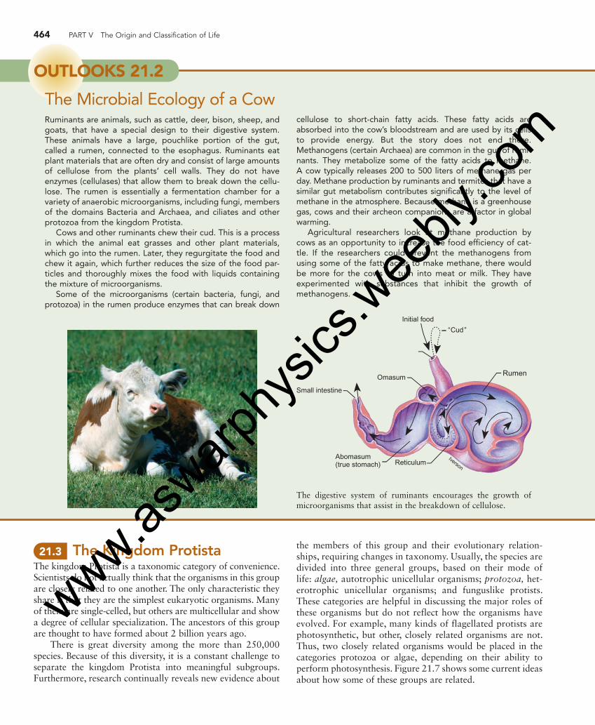
the members of this group and their evolutionary relation-ships, requiring changes in taxonomy. Usually, the species are divided into three general groups, based on their mode of life: algae, autotrophic unicellular organisms; protozoa, het-erotrophic unicellular organisms; and funguslike protists. These categories are helpful in discussing the major roles of these organisms but do not reflect how the organisms have evolved. For example, many kinds of flagellated protists are photosynthetic, but other, closely related organisms are not. Thus, two closely related organisms would be placed in the categories protozoa or algae, depending on their ability to perform photosynthesis. Figure 21.7 shows some current ideas about how some of these groups are related.
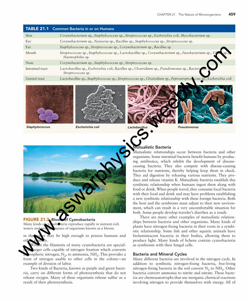
cellulose to short-chain fatty acids. These fatty acids are absorbed into the cow’s bloodstream and are used by its cells to provide energy. But the story does not end there. Methanogens (certain Archaea) are common in the gut of rumi-nants. They metabolize some of the fatty acids to methane. A cow typically releases 200 to 500 liters of methane gas per day. Methane production by ruminants and termites that have a similar gut metabolism contributes significantly to the level of methane in the atmosphere. Because methane is a greenhouse gas, cows and their archeon companions are a factor in global warming.
Agricultural researchers look at methane production by cows as an opportunity to increase the food efficiency of cat-tle. If the researchers could prevent the methanogens from using some of the fatty acids to make methane, there would be more for the cows to turn into meat or milk. They have experimented with substances that inhibit the growth of methanogens.
OUTLOOKS 21.2
The Microbial Ecology of a Cow Ruminants are animals, such as cattle, deer, bison, sheep, and goats, that have a special design to their digestive system. These animals have a large, pouchlike portion of the gut, called a rumen, connected to the esophagus. Ruminants eat plant materials that are often dry and consist of large amounts of cellulose from the plants’ cell walls. They do not have enzymes (cellulases) that allow them to break down the cellu-lose. The rumen is essentially a fermentation chamber for a variety of anaerobic microorganisms, including fungi, members of the domains Bacteria and Archaea, and ciliates and other protozoa from the kingdom Protista.
Cows and other ruminants chew their cud. This is a process in which the animal eat grasses and other plant materials, which go into the rumen. Later, they regurgitate the food and chew it again, which further reduces the size of the food par-ticles and thoroughly mixes the food with liquids containing the mixture of microorganisms.
Some of the microorganisms (certain bacteria, fungi, and protozoa) in the rumen produce enzymes that can break down
Initial food
Abomasum(true stomach) Reticulum
Iverson
“Cud”
Rumen
Small intestine
Omasum
The digestive system of ruminants encourages the growth of microorganisms that assist in the breakdown of cellulose.
eng03466_ch21_455-478.indd 464eng03466_ch21_455-478.indd 464 15/10/10 3:06 PM15/10/10 3:06 PM
www.aswarp
hysic
s.wee
bly.co
m
CHAPTER 21 The Nature of Microorganisms 465
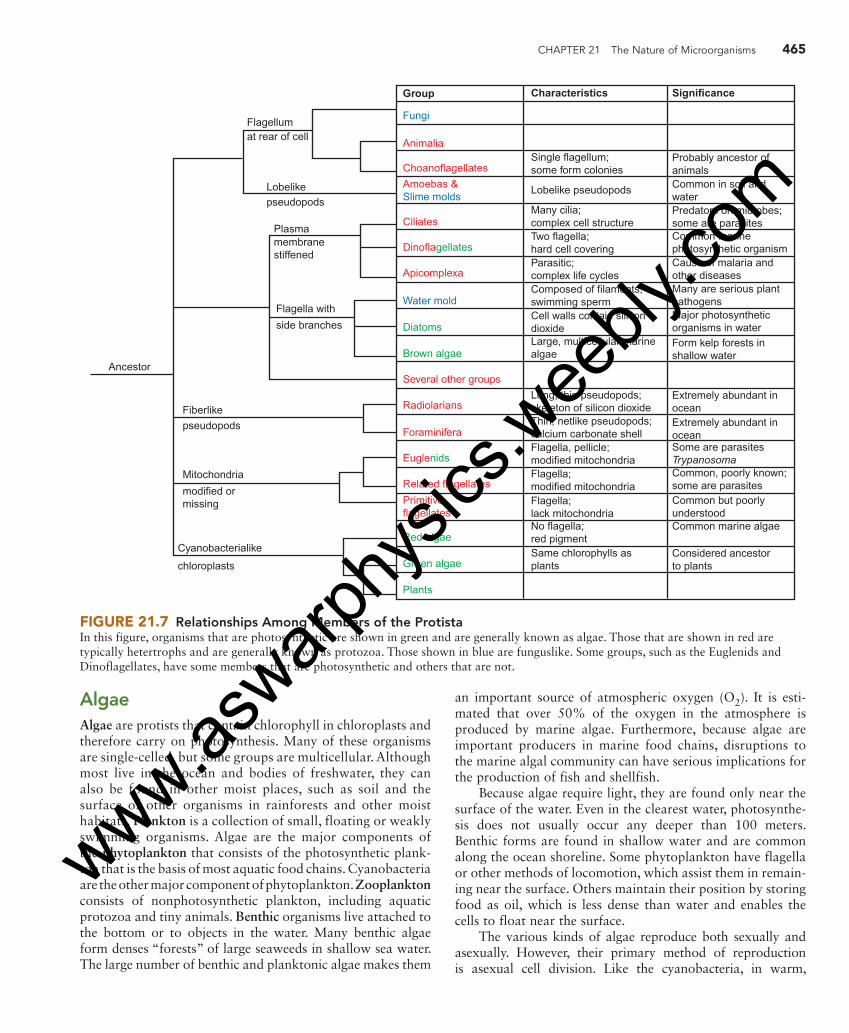
Algae Algae are protists that contain chlorophyll in chloroplasts and therefore carry on photosynthesis. Many of these organisms are single-celled, but some groups are multicellular. Although most live in the ocean and bodies of freshwater, they can also be found in other moist places, such as soil and the surface of other organisms in rainforests and other moist habitats. Plankton is a collection of small, floating or weakly swimming organisms. Algae are the major components of the phytoplankton that consists of the photosynthetic plank-ton that is the basis of most aquatic food chains. Cyanobacteria are the other major component of phytoplankton. Zooplankton consists of nonphotosynthetic plankton, including aquatic protozoa and tiny animals. Benthic organisms live attached to the bottom or to objects in the water. Many benthic algae form denses “forests” of large seaweeds in shallow sea water. The large number of benthic and planktonic algae makes them
an important source of atmospheric oxygen (O 2 ). It is esti-mated that over 50% of the oxygen in the atmosphere is produced by marine algae. Furthermore, because algae are important producers in marine food chains, disruptions to the marine algal community can have serious implications for the production of fish and shellfish.
Because algae require light, they are found only near the surface of the water. Even in the clearest water, photosynthe-sis does not usually occur any deeper than 100 meters. Benthic forms are found in shallow water and are common along the ocean shoreline. Some phytoplankton have flagella or other methods of locomotion, which assist them in remain-ing near the surface. Others maintain their position by storing food as oil, which is less dense than water and enables the cells to float near the surface.
The various kinds of algae reproduce both sexually and asexually. However, their primary method of reproduction is asexual cell division. Like the cyanobacteria, in warm,
Group
FungiFlagellumat rear of cell
Lobelikepseudopods
Plasma
Flagella with
side branches
Fiberlike
Ancestor
pseudopods
Mitochondria
modified ormissing
Cyanobacterialike
chloroplasts
membranestiffened
AnimaliaSingle flagellum;some form colonies
Probably ancestor ofanimalsCommon in soil andwaterPredators on microbes;some are parasitesCommon marinephotosynthetic organismCause of malaria andother diseasesMany are serious plantpathogensMajor photosyntheticorganisms in waterForm kelp forests inshallow water
Extremely abundant inoceanExtremely abundant inoceanSome are parasitesTrypanosomaCommon, poorly known;some are parasitesCommon but poorlyunderstoodCommon marine algae
Considered ancestorto plants
Lobelike pseudopods
Many cilia;complex cell structureTwo flagella;hard cell coveringParasitic;complex life cyclesComposed of filaments;swimming spermCell walls contain silicondioxideLarge, multicellular marinealgae
Long, thin pseudopods;skeleton of silicon dioxideThin, netlike pseudopods;calcium carbonate shellFlagella, pellicle;modified mitochondriaFlagella;modified mitochondriaFlagella;lack mitochondriaNo flagella;red pigmentSame chlorophylls asplants
Choanoflagellates
Amoebas &Slime molds
Ciliates
Dinoflagellates
Apicomplexa
Water mold
Diatoms
Brown algae
Several other groups
Radiolarians
Foraminifera
Euglenids
Related flagellates
Primitiveflagellates
Red algae
Green algae
Plants
Characteristics Significance
FIGURE 21.7 Relationships Among Members of the Protista In this figure, organisms that are photosynthetic are shown in green and are generally known as algae. Those that are shown in red are typically hetertrophs and are generally known as protozoa. Those shown in blue are funguslike. Some groups, such as the Euglenids and Dinoflagellates, have some members that are photosynthetic and others that are not.
eng03466_ch21_455-478.indd 465eng03466_ch21_455-478.indd 465 15/10/10 3:06 PM15/10/10 3:06 PM
www.aswarp
hysic
s.wee
bly.co
m

466 PART V The Origin and Classification of Life
nutrient-rich waters, various kinds of algae can reproduce rapidly by asexual reproduction and cause an algal bloom. The population can become so large that clumps of algae float on the surface or in the case of single-celled algae, the water may become colored or murky.
Single-Celled Algae There are several common kinds of single-celled algae. Euglenids are single-celled algae that move by flagella. They have an outer covering, called a pellicle, which gives them shape but is flexible. Euglenids vary in terms of their metabo-lism. Some lack chloroplasts and are heterotrophs. Others have chloroplasts and are autotrophs. However, even among those that have chloroplasts, many are able to consume food and behave as heterotrophs, particularly when light levels are low. Most of them, like the common Euglena, are found in freshwater. They are widely studied because they are easy to culture.
Diatoms are extremely common single-celled algae found in freshwater, marine, and soil environments. They are
a major component of the phytoplankton of the oceans and serve as a food source for zooplankton and many kinds of filter-feeding organisms, such as whales, clams, and barnacles. A few species are heterotrophs or parasites. They are typically brownish in color. Although they do not have cilia or flagella, they are able to move with a sort of gliding motion. They are unique because their cell walls contain silicon dioxide (silica). The walls fit together like the lid and bottom of a shoe box; the lid overlaps the bottom. The silica-containing walls have many pores, which form interesting patterns. Because their cell walls contain silicon dioxide, they readily form fossils. The fossil cell walls have many tiny holes and can be used in a number of commercial processes. They are used as filters for liquids and as abrasives in specialty soaps, toothpastes, and scouring powders.
Dinoflagellates, along with diatoms, are important food producers in the ocean’s ecosystem. They are also very com-mon in freshwater and brackish water. All members of this group of algae have two flagella, which is the reason for their name ( di � two), and an outer cellulose covering made up of
autotrophs. Flagellates, ciliates, fungi, and bacteria consume other organisms. Finally, we arrive at the animals that are filter-feeding animals, such as sponges, corals, and crustaceans that sift a mixture of organisms from water. These become food for larger animals such as crabs, snails, fish, and squid.
There is an important subplot to this microbial food web picture. Much of the organic matter never reaches animals at higher trophic levels. It is trapped in a microbial loop that involves many kinds of microbes that simply recycle organic mat-ter. It is thought that bacteria alone process more than half of all the carbon involved in metabolism in the oceans. Dissolved organic carbon is an important part of the microbial food web. Autotrophic microbes produce organic molecules from inorganic molecules. Dissolved organic carbon enters the water as a result of leakage and waste products from microbes (both autotrophs and heterotrophs) and the death and decay of organisms. It is becoming clear that viruses have an important role to play in this process. Studies that sample the DNA in the ocean identify a large virus component. Many scientists estimate that there are millions of viruses per milliliter of seawater. Viruses infect and kill their hosts: Bacteria, Archaea, and eukaryotic microbes. The disintegration of their host cells releases organic matter into the water, which, in turn, becomes food for saprophytic microbes.
These studies are significant to understanding the basis for ecologic interactions in the ocean. These interactions can impact fisheries biology and human health when there are huge increases in the population of certain toxic marine microbes that restrict the use of fish for food and cause the closure of beaches to prevent illness.
OUTLOOKS 21.3
The Marine Microbial Food Web The analysis of terrestrial ecosystems typically involves the cat-egorization of organisms into functional groups based on their metabolic abilities and their position in food chains. Plants are identified as producers, animals as consumers, and fungi and bacteria as decomposers. Scientists have long known that micro-organisms were important in marine food webs but have been hampered in their study by the nature of the organisms involved.
There are significant problems in studying microbes. Their small size makes it difficult to identify organisms. A new term, picoplankton, is used to describe aquatic organisms that are in a size range between 0.2 and 2 μm. In addition, once organisms are detected it is often difficult or impossible to grow microbes to study their metabolic abilities and determine how they con-tribute to food webs.
However, by using new techniques for identifying organisms and indirect methods to get an idea of their metabolic abilities, it is becoming clear that the ocean is dominated by a microbial food web. As with all ecosystems, the base consists of auto-trophs that use a source of energy to manufacture organic matter. The majority of photosynthesis in the ocean is the result of cyanobacteria and the eukaryotic dinoflagellates and diatoms. The photosynthetic cyanobacteria are the most com-mon bacteria in the ocean. The Archaea also appear to be important as autotrophs, although many are chemoautotrophs that use inorganic chemical reactions to provide themselves with energy. They are extremely common, particularly in deep ocean waters where sunlight does not penetrate.
Once we get beyond the producer level in the ocean, vari-ous kinds of microbes are first in line to consume the cells of
eng03466_ch21_455-478.indd 466eng03466_ch21_455-478.indd 466 15/10/10 3:06 PM15/10/10 3:06 PM
www.aswarp
hysic
s.wee
bly.co
m

CHAPTER 21 The Nature of Microorganisms 467
plates. Although many dinoflagellates are photosynthetic, some are heterotrophs and others are parasites. Some change from autotroph to heterotroph, depending on environmental conditions.
Some species of dinoflagellates have symbiotic relation-ships with marine animals, such as the reef corals; the dino-flagellates provide a source of nutrients for the reef-building coral. Corals that live in the light and contain dinoflagellates grow 10 times faster than corals without this symbiont. Thus, in coral reef ecosystems, dinoflagellates form the foundation of the food chain.
Some forms of dinoflagellates produce toxins. Because many of the toxin-producing dinoflagellates are reddish in color, a bloom of these organisms is called a red tide. Often, fish and other vertebrates such as birds and mammals are killed by exposure to the toxins. Although the toxins do not seem to harm shellfish, such as oysters, consuming the toxin along with the shellfish can cause sickness or death. Red tides usually occur in the warm months and are more common in tropical and semitropical waters. During red tide episodes, people are warned not to swim in areas that have a red tide or harvest fish or shellfish for food. Commercially available shellfish are tested for toxin content; if they are toxic, they are not marketed.
The dinoflagellate Pfiesteria piscidia has been responsi-ble for the death of millions of fish in estuaries of the eastern United States. These dinoflagellates release toxins that para-lyze fish. The dinoflagellates then feed on the fish. They have also been responsible for human and wildlife poisoning. It appears that blooms of these organisms may be triggered by high amounts of nutrients in the water as a result of runoff from feedlots and agricultural land.
Many marine forms of dinoflagellates are biolumines-cent; they are responsible for the glow seen at night in ocean waves or in a boat’s wake. Figure 21.8 shows examples of euglenids, diatoms, and dinoflagellates.
Multicellular Algae Many kinds of algae are multicellular and can be quite large, with some specialization of cells and body parts. These algae are commonly known as seaweed. They are found in shallow water attached to objects. Two types, red algae and brown algae, are mainly marine forms. The green algae are primarily freshwater species.
Red algae live in warm oceans and attach to the ocean floor by means of a holdfast structure. They are found from the splash zone, the area where waves are breaking, to depths of 100 meters. Some red algae become encrusted with calcium carbonate and are important in reef building. Other species are commercially important, because they produce agar and carrageenin. Agar is widely used as a jelling agent for growth media in microbiology. Carrageenin is a gelatinous material used in paints, cosmetics, and baking. It is also used to make gelatin desserts harden faster and ice cream smoother. In Asia and Europe, some red algae are harvested and used as food.
Brown algae are found in cooler marine environments. Most species of brown algae have a holdfast organ. Colonies of these algae can reach 100 meters in length. Brown algae produce alginates, which are widely used as stabilizers in frozen desserts, as emulsifiers in salad dressings, and as thick-eners to give body to foods such as chocolate milk and cream cheeses; they are also used to form gels in such products as fruit jellies.
Reservoir
Pellicle
Basal bodies
Contractilevacuole
Secondflagellum
Stigma
Flagellum
Nucleus
Chloroplast
Paramylongranule
Transverseflagellum
Longitudinalflagellum
(a) Euglenid (b) Dinoflagellate (c) Diatoms
FIGURE 21.8 Single-Celled Algae Three very common kinds of single-celled algae are the euglenids, dinoflagellates, and diatoms.
eng03466_ch21_455-478.indd 467eng03466_ch21_455-478.indd 467 15/10/10 3:06 PM15/10/10 3:06 PM
www.aswarp
hysic
s.wee
bly.co
m

468 PART V The Origin and Classification of Life
The Sargasso Sea is a large mat of free-floating brown algae between the Bahamas and the Azores. It is thought that this huge mass (as large as the European continent) is the result of brown algae that have become detached from the ocean bottom, have been carried by ocean currents, and have accumulated in this calm region of the Atlantic Ocean. This large mass of floating algae provides a habitat for a large number of marine animals, such as marine turtles, eels, jelly-fish, and innumerable crustaceans. Figure 21.9 shows exam-ples of red and brown algae.
Green algae are found primarily in freshwater ecosys-tems, although a few kinds live in oceans. Some are single-celled and have flagella; some lack flagella and form strings, which either float in the water or grow on surfaces. The members of this group can also be found growing on trees, in the soil, and even on snowfields in the mountains. Like land plants, green algae have cellulose cell walls and store food as starch. Green algae also have the same types of chlorophyll as
do plants. Biologists believe that land plants evolved from the green algae. Figure 21.10 shows a variety of green algae.
Protozoa Protozoa are members of the kingdom Protista; they are eukary-otic, heterotrophic, single-celled organisms that lack cell walls. Generally, protozoa lack all types of chlorophyll, but some organisms may contain chloroplasts at some times in their lives and lack them at others. One common way to classify the pro-tozoa into subgroups is by their method of locomotion. Although this is a convenient way to subdivide the organisms for the pur-poses of discussion, it is clearly not a valid phylogenic grouping.
Flagellates Flagellates are an extremely diverse group of organisms that have flagella and lack cell walls and chloroplasts. They live in any moist environment, including marine waters and freshwater,
Macrasterias Volvox Ulothrix
FIGURE 21.10 Green Algae Some green algae are single-celled; others form colonies.
Red algae
FIGURE 21.9 Red and Brown Algae Red and brown algae are primarily marine organisms. Most of them grow attached to the ocean bottom or other organisms in their environment.
Brown algae
eng03466_ch21_455-478.indd 468eng03466_ch21_455-478.indd 468 15/10/10 3:06 PM15/10/10 3:06 PM
www.aswarp
hysic
s.wee
bly.co
m
CHAPTER 21 The Nature of Microorganisms 469
moist soil, and as parasites or symbionts. Some flagellates have an extremely simple structure, suggesting that they may be the most primitive of all eukaryotic organisms. Some feed by absorbing simple organic molecules through their cell membranes; others engulf food particles or other organisms.
Many kinds of flagellates are mutualistic or parasitic. Termites are insects that eat wood but cannot digest it. Their guts contain mutualistic flagellated protozoa capable of digesting cellulose. Thus, the termite benefits from a food source and the flagellate benefits from a good place to live and a continuous supply of food.
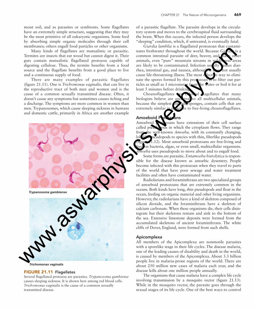
There are many examples of parasitic flagellates ( figure 21.11 ). One is Trichomonas vaginalis, that can live in the reproductive tract of both men and women and is the cause of a common sexually transmitted disease. Often, it doesn’t cause any symptoms but sometimes causes itching and a discharge. The symptoms are more common in women than men. Trypanosomes, which cause sleeping sickness in humans and domestic cattle, primarily in Africa are another example
Trypanosoma gambiense
Trichomonas vaginalis
FIGURE 21.11 Flagellates Several flagellated protozoa are parasites. Trypanosoma gambiense causes sleeping sickness. It is shown here among red blood cells. Trichomonas vaginalis is the cause of a common sexually transmitted disease.
of a parasitic flagellate. The parasite develops in the circula-tory system and moves to the cerebrospinal fluid surrounding the brain. When this occurs, the infected person develops the “sleeping” condition, which, if untreated, is eventually fatal.
Giardia lamblia is a flagellated protozoan that contami-nates freshwater throughout the world. Because Giardia is a common intestinal parasite of deer, beaver, and many other animals, even “pure” mountain streams in wilderness areas are likely to be contaminated. Infection usually causes diar-rhea, intestinal gas, and nausea, although it does not usually cause life-threatening illness. The most effective way to elimi-nate the spores formed by this protozoan is to filter out par-ticles as small as 1 micrometer from the water or boil it for at least 5 minutes before drinking.
Choanoflagellates are colonial flagellates that many biologists believe are ancestral to all multicellular animals, because the simplest animals, sponges, contain cells that are extremely similar in structure to free-living choanoflagellates.
Amoeboid Protozoans Amoeboid protozoans have extensions of their cell surface called pseudopods in which the cytoplasm flows. They range from the well-known Amoeba, with its constantly changing, lobelike pseudopods to species with thin, fiberlike pseudopods ( figure 21.12 ). Most amoeboid protozoans are free-living and feed on bacteria, algae, or even small, multicellular organisms. Amoeba uses pseudopods to move about and to engulf food.
Some forms are parasitic. Entamoeba histolytica is respon-sible for the disease known as amoebic dysentery. People become infected with this protozoan when they travel to parts of the world that have poor sewage and water treatment facilities and often have contaminated water.
Radiolarians and foraminiferans are two specialized groups of amoeboid protozoans that are extremely common in the oceans. Both kinds have long, thin pseudopods and float in the ocean, feeding on organic material and other living organisms. However, the radiolarians have a kind of skeleton composed of silicon dioxide, and the foraminiferans have a skeleton of calcium carbonate. When these organisms die, their cells disin-tegrate but their skeletons remain and sink to the bottom of the sea. Extensive limestone deposits were formed from the accumulated skeletons of ancient foraminiferans. The white cliffs of Dover, England, were formed from such shells.
Apicomplexa All members of the Apicomplexa are nonmotile parasites with a sporelike stage in their life cycles. The disease malaria, one of the leading causes of disability and death in the world, is caused by members of the Apicomplexa. About 3.3 billion people live in malaria-prone regions of the world. There are about 250 million new cases of malaria each year, and the disease kills about one million people annually.
The organisms that cause malaria have a complex life cycle involving transmission by a mosquito vector ( figure 21.13 ). While in the mosquito vector, the parasite goes through the sexual stages of its life cycle. One of the best ways to control
eng03466_ch21_455-478.indd 469eng03466_ch21_455-478.indd 469 15/10/10 3:06 PM15/10/10 3:06 PM
www.aswarp
hysic
s.wee
bly.co
m

470 PART V The Origin and Classification of Life
HumanMosquito
To salivarygland
Ruptureof oocyst
Growthof oocyst
Cyst formationin stomach ofmosquito
Sporozoites
Fertilization
ZygoteFemalegametocyte
Malegametocyte
Red bloodcells
O’Keefe
Merozoites
Liver cells
FIGURE 21.13 The Life Cycle of Plasmodium vivax Plasmodium vivax is one of the members of the Apicomplexa that causes malaria. The life cycle requires two hosts, the Anopheles mosquito and the human. Humans get malaria when they are bitten by a mosquito carrying the larval stage of Plasmodium. The larva undergoes asexual reproduction and releases thousands of individuals, which invade the red blood cell. Their release from massive numbers of infected red blood cells causes the chills, fever, and headache associated with malaria. Inside the red blood cell, more reproduction occurs to form male gametocytes and female gametocytes. When the mosquito bites a person with malaria, it ingests some gametocytes. Fertilization occurs and zygotes develop in the stomach of the mosquito. The resulting larvae are housed in the mosquito’s salivary gland. Then, when the mosquito bites someone, some saliva containing the larvae is released into the person’s blood and the cycle begins again.
FIGURE 21.12 Amoeboid Protozoa Amoeboid protozoa have extensions of their cell surface called pseudopods. Pseudopods contain moving cytoplasm. Some, such as Amoeba, have large, lobelike pseudopods, which change shape as the cell moves and feeds. Others have long, filamentous pseudopods that trap organisms and transport food molecules to the central cell from the objects they feed on.
eng03466_ch21_455-478.indd 470eng03466_ch21_455-478.indd 470 15/10/10 3:06 PM15/10/10 3:06 PM
www.aswarp
hysic
s.wee
bly.co
m

CHAPTER 21 The Nature of Microorganisms 471
this disease is to eliminate the vector, which usually involves using a pesticide. Many of us are concerned about the harm-ful effects of pesticides in the environment. However, in the parts of the world where malaria is common, the harmful effects of pesticides are of less concern than the harm gener-ated by the disease. Many diseases of insects, birds, and mammals are also caused by the members of this group.
Ciliates Ciliates are a group of protozoans with a complex cellular structure and numerous short, flexible extensions from the cell called cilia ( figure 21.14 ). The cilia move in an organized, rhythmic manner and propel the cell through the water. Some types of ciliates, such as Paramecium, have nearly 15,000 cilia per cell and move at a rapid speed of 1 millimeter per second. Most ciliates are free-living cells found in freshwater and salt water or damp soil, where they feed on bacteria and other small organisms. Ruminant animals have large numbers of ciliates in their digestive systems, where they are part of the complex ecology of the ruminant gut (see Outlooks 21.2).
Ciliates have a complex cellular structure with two kinds of nuclei. Most have a macronucleus and one or more micronu-clei. The macronucleus is involved in the day-to-day running of the cell, whereas the micronuclei are involved in sexual repro-duction. Sexual reproduction involves a process called conjuga-tion, in which two cells go through a series of nuclear divisions equivalent to meiosis and exchange some of their nuclear mate-rial. Although the exchange does not result in additional cells, it does result in cells that have a changed genetic mixture.
Funguslike Protists Funguslike protists have a motile reproductive stage but they do not have chitin in their cell walls, which differentiates them from true fungi. There are two kinds of funguslike pro-tists: slime molds and water molds.
Anterior contractile vacuole
Micronucleus
Macronucleus
Pellicle
Posteriorcontractilevacuole
Food vacuole
Gullet
Cilia
Cytoproct
FIGURE 21.14 Ciliates Ciliates, such as Paramecium, have a complex cell structure and a large number of cilia on their surface, which propel them through the water. They feed on a variety of organisms.
Slime Molds Slime molds are amoeba-like organisms that crawl about and digest dead organic matter. Some slime molds look like giant amoebae. They are essentially a large mass several centimeters across, in which the nucleus and other organelles have divided repeatedly within a single large cell ( figure 21.15 ). No cell membranes partition this mass into separate segments. They vary in color from white to bright red or yellow, and they can reach relatively large sizes (45 centimeters in length) when in an optimum environment.
Other kinds of slime mold exist as large numbers of indi-vidual, amoeba-like cells. These haploid cells get food by engulfing microorganisms. They reproduce by mitosis. When their environment becomes dry or otherwise unfavorable, the cells come together into an irregular mass. This mass glides along rather like an ordinary garden slug and is labeled the sluglike stage. This sluglike form may flow about for hours before it forms spores. When the mass gets ready to produce spores, it creates a stalk with cells that have cell walls. At the top of this specialized structure, cells are modified to become haploid spores. When released, these spores may be carried by the wind and, if they land in a favorable place, may develop into new amoeba-like cells.
Water Molds Water molds were once thought to be fungi. However, they differ from fungi in two fundamental ways. Their cell walls are made of cellulose, not chitin, and water molds have a flagellated reproductive stage. Thus, they are con-sidered to be more closely related to the diatoms and brown algae than to fungi. Although called water molds, they live in many moist environments, not just in bodies of water ( figure 21.16 ).
Water molds are important saprophytes and parasites in aquatic ecosystems. They are often seen as fluffy growths on
FIGURE 21.15 Slime Mold Slime molds grow in moist conditions and are important decomposers. As slime molds grow, additional nuclei are produced by mitosis, but there is no cytoplasmic division. Thus, at this stage, a slime mold is a single mass of cytoplasm with many nuclei.
eng03466_ch21_455-478.indd 471eng03466_ch21_455-478.indd 471 15/10/10 3:06 PM15/10/10 3:06 PM
www.aswarp
hysic
s.wee
bly.co
m

472 PART V The Origin and Classification of Life
dead fish or other organic matter floating in water. A parasitic form of water mold is well known to people who rear tropical fish; it causes a cottonlike growth on the fish. Although these organisms are usually found in aquatic habitats, they are not limited to this environment. Some species cause downy mil-dew on plants such as grapes. In the 1880s, this mildew almost ruined the French wine industry when it spread throughout the vineyards. A copper-based fungicide called Bordeaux mixture —the first chemical used against plant diseases—was used to save the vineyards. A water mold was also responsi-ble for the Irish potato blight. In the nineteenth century, potatoes were the staple of the Irish diet. Cool, wet weather in 1845 and 1847 damaged much of the potato crop, and more than a million people died of starvation. Nearly one-third of the survivors left Ireland and moved to Canada or the United States.
21.4 Multicellularity in the Protista The three major types of organisms in the kingdom Protista (algae, protozoa, and funguslike protists) include both single-celled and multicellular forms. Biologists believe that there has been a similar type of evolution in all three of these groups. The most primitive organisms in each group are thought to have been single-celled and to have given rise to the more advanced, multicellular forms. Most protozoan organisms are single-celled however, some ciliates are colonial. The multicel-lular forms of funguslike protists are the slime molds, which have both single-celled and multicellular stages.
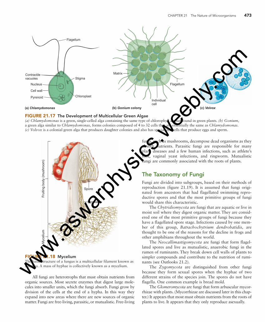
Perhaps the most widely known example of this trend from a single-celled to a multicellular condition is found in the green algae. A very common single-celled green alga is Chlamydomonas, which has a cell wall and two flagella. It looks just like the individual cells of the colonial green algae Volvox. Some species of Volvox have as many as 50,000 cells ( figure 21.17 ). All the flagella of each cell in the colony move in unison, allowing the colony to move in one direction. In some Volvox species, certain cells have even specialized to produce sperm or eggs. Biologists believe that the division of labor seen in colonial protists represents the beginning of the specialization that led to the development of true multicellular organisms with many kinds of specialized cells. Three types of multicellular organisms—fungi, plants, and animals—eventually developed.
21.5 The Kingdom Fungi The members of the kingdom Fungi are nonphotosynthetic, eukaryotic organisms with cell walls. The structure of the cell wall differs from that of other organisms because fungal cell walls contain chitin along with other compounds. Some are single-celled, but most are multicellular organisms composed of filaments of cells joined end-to-end. Each filament is known as a hypha. The hyphae form a network known as a mycelium ( figure 21.18 ).
Even though fungi are nonmotile, they are easily dis-persed, because they form huge numbers of spores. A spore is a cell with a tough, protective cell wall that can resist extreme conditions. Fungi have a variety of kinds of spores. Some spores are produced by sexual reproduction, others by asex-ual reproduction. An average-sized mushroom can produce over 20 billion spores; a good-sized puffball can produce as many as 8 trillion spores. When released, the spores are trans-ported by wind or water. Because of their small size, spores can remain in the atmosphere a long time and travel thou-sands of kilometers. Fungal spores have been collected as high as 50 kilometers above Earth.
FIGURE 21.16 Water Mold Rapidly reproducing water molds quickly produce a large mass of filaments. These filaments cause the fuzzy growth often seen on dead fish and other dead material in the water.
21.3 CONCEPT REVIEW 14. Why is the kingdom Protista not considered a valid
phylogenetic group? 15. What is phytoplankton? 16. List three different categories of organisms that are
considered algae. 17. List two major kinds of marine phytoplankton. 18. List the two major kinds of multicellular marine
algae. 19. Describe a characteristic for each of the following: a. apicomplexa b. ciliates c. flagellates d. foraminifera 20. Why are water molds and slime molds not consid-
ered to be Fungi?
21.4 CONCEPT REVIEW 21. Why do biologists think that the ancestors of
plants, animals, and fungi could have been Protista?
eng03466_ch21_455-478.indd 472eng03466_ch21_455-478.indd 472 15/10/10 3:06 PM15/10/10 3:06 PM
www.aswarp
hysic
s.wee
bly.co
m
CHAPTER 21 The Nature of Microorganisms 473
(a) Chlamydomonas
Pyrenoid
Cell wall
Nucleus
Contractilevacuoles Stigma
Flagellum
Chloroplast
(b) Gonium colony
Matrix
Individualcell
Flagellum
(c) Volvox
FIGURE 21.17 The Development of Multicellular Green Algae (a) Chlamydomonas is a green, single-celled alga containing the same type of chlorophyll as that found in green plants. (b) Gonium, a green alga similar to Chlamydomonas, forms colonies composed of 4 to 32 cells that are essentially the same as Chlamydomonas. ( c ) Volvox is a colonial green alga that produces daughter colonies and also has specialized cells that produce eggs and sperm.
Myc
eliu
mFr
uitin
g bo
dy (
mus
hroo
m)
Spore
Hyphae
FIGURE 21.18 Mycelium The basic structure of a fungus is a multicellular filament known as a hypha. A mass of hyphae is collectively known as a mycelium.
All fungi are heterotrophs that must obtain nutrients from organic sources. Most secrete enzymes that digest large mole-cules into smaller units, which the fungi absorb. Fungi grow by division of the cells at the end of a hypha. In this way they expand into new areas where there are new sources of organic matter. Fungi are free-living, parasitic, or mutualistic. Free-living
fungi, such as mushrooms, decompose dead organisms as they absorb nutrients. Parasitic fungi are responsible for many plant diseases and a few human infections, such as athlete’s foot, vaginal yeast infections, and ringworm. Mutualistic fungi are commonly associated with the roots of plants.
The Taxonomy of Fungi Fungi are divided into subgroups, based on their methods of reproduction ( figure 21.19 ). It is assumed that fungi origi-nated from ancestors that had flagellated swimming repro-ductive spores and that the most primitive groups of fungi would share this characteristic.
The Chytridiomycota are fungi that are aquatic or live in moist soil where they digest organic matter. They are consid-ered one of the most primitive groups of fungi because they have a flagellated spore stage. Infections caused by one mem-ber of this group, Batrachochytrium dendrobatidis, are thought to be one of the reasons for the decline in frogs and other amphibians throughout the world.
The Neocallimastigomycota are fungi that form flagel-lated spores and live as mutualistic, anaerobic fungi in the rumen of ruminants. They break down cell walls of plants to simpler compounds and contribute to the nutrition of rumi-nants (see Outlooks 21.2).
The Zygomycota are distinguished from other fungi because they form sexual spores when the hyphae of two different strains of the species join. The spores do not have flagella. One common example is bread mold.
The Glomeromycota are fungi that form arbuscular mycor-rhizae with plants. (Mycorrhizae are discussed later in this chap-ter.) It appears that most must obtain nutrients from the roots of plants to live. It appears that they only reproduce asexually.
eng03466_ch21_455-478.indd 473eng03466_ch21_455-478.indd 473 15/10/10 3:06 PM15/10/10 3:06 PM
www.aswarp
hysic
s.wee
bly.co
m

474 PART V The Origin and Classification of Life
The Ascomycota are distinguished from other fungi because their cells contain two distinct nuclei and they pro-duce spores by sexual reproduction in a saclike structure. They also produce spores asexually. About 75% of all fungi are in this group. Molds, mildews, yeasts, morel mushrooms, and truffles are examples. In recent years a fungus in this group, Geomyces destructans, has been identified as the cause of white-nose syndrome in bats. Millions of bats have died because of infections with this fungus.
The Basidiomycota are unique in having cells that con-tain two distinct nuclei and sexual spores produced in sets of four on a club-shaped structure. Bracket fungi, most mush-rooms, and puffballs are common examples. Many are para-sites on plants; rusts and smuts are examples.
The term yeast is not a taxonomic term. A yeast is any fungus that is single-celled and generally reproduces by asexual reproduction. One common method of asexual repro-duction is budding in which one cell grows a lobe that eventu-ally pinches off to become a separate organism. Yeasts are found in the Basidiomycota and Ascomycota. The yeasts involved in brewing and baking are in the Ascomycota.
Mold and mildew are other terms associated with fungi. They are not taxonomic terms but simply describe growths of
a fungus that forms a fuzzy growth or discolors a surface. They can be from many different taxonomic categories, not just fungi. They are common everywhere there is a source of organic molecules and moisture.
The Significance of Fungi Fungi play many significant roles in ecosystems. They are also economically important to humans in many ways.
Decomposers Because fungi produce so many spores, any dead organism is likely be colonized by a fungus. Because all fungi are capable of breaking down organic matter, fungi along with bacteria are the major decomposers of organic matter in ecosystems. This decomposer function recycles elements such as carbon, nitrogen, and phosphorus. However, fungi also colonize and break down organic matter that we do not want to have recycled and cause billions of dollars of damage each year. Clothing, wood, leather, and all types of food are susceptible to damage by fungi. One of the best ways to protect against such damage is to keep the material dry, because fungi grow best in a moist environment.
Fungi as Food Fungi and their by-products have been used as sources of food for centuries. When we think of fungi and food, mushrooms usually come to mind. The common mushroom found in the grocer’s vegetable section is grown in many countries and has an annual market value in the billions of dollars. But there are other uses for fungi as food. Shoyu (soy sauce) was originally made by fermenting a mixture of wheat, soybeans, and an ascomycote fungus for a year. Most of the soy sauce used today is made by a cheaper method of processing soybeans with hydrochloric acid. True connoisseurs still prefer soy sauce made the original way. Another mold is important to the soft-drink industry. The citric acid that gives a soft drink its sharp taste was originally produced by squeezing juice from lemons and purifying the acid. Today, however, a mold is grown on a nutrient medium with table sugar (sucrose) to produce great quantities of citric acid at a low cost.
The primary flavors of blue cheeses, such as Danish, American, and the original Roquefort, are produced because the cheeses have been aged with the mold Penicillium roque-fortii. Another species of Penicillium produces the antibiotic penicillin (How Science Works 21.3).
Yeasts are important organisms in the production of alco-holic beverages and in the making of bread. It is difficult to imag-ine a world in which these two food materials are not important. Mycorrhizae Mycorrhizae are associations between certain fungi and the roots of plants. There are two kinds of mycorrhizae. In one kind, arbuscular mycorrhiza, the cells of fungi actually penetrate the cells of the plant roots. Members of the Glomeromycota are always found as arbuscular mycorrhizae. In the other kind,
Zygomycota
Ancestral
fungus
Basidiomycota
Cells withtwo nuclei
No flagellated
Flagellated
spores
spores
Ascomycota
Glomeromycota
Other groups withflagellated spores
Chytridiomycota
Neocallimastigomycota
FIGURE 21.19 Fungus Taxonomy There are several subgroups within the kingdom Fungi. They are distinguished from one another primarily by their methods of reproduction.
eng03466_ch21_455-478.indd 474eng03466_ch21_455-478.indd 474 15/10/10 3:06 PM15/10/10 3:06 PM
www.aswarp
hysic
s.wee
bly.co
m

CHAPTER 21 The Nature of Microorganisms 475
ectomycorrhiza, the fungal cells surround the root cells but do not invade them. Ectomycorrhizae are usually formed by members of the Ascomycota and Basidiomycota ( figure 21.20 ). Mycorrhizal fungi are found in 80–90% of all plants, and many plants cannot live without their mycorrhizal fungi. The addition of these fungi to the roots of plants increases the roots’ surface area for absorption. Plants with mycorrhizal fungi can absorb as much as 10 times more minerals than those without the fungi. Some types of fungi also supply plants with growth hormones, whereas the plants supply car-bohydrates and other organic compounds to the fungi.
Lichens Lichens are organisms that consist of a symbiotic relationship between a fungus and either an alga or a cyanobacterium. The alga or cyanobacterium does photosynthesis and pro-vides the fungus with organic molecules for food, while the fungus provides the moist environment required by the alga or cyanobacterium.
Because the fungi provide a damp environment and the algae produce the food, lichens require no soil for growth. For this reason, many kinds of lichens are commonly found growing on bare rock and are the pioneer organisms in the process of succession. Some lichens grow as fluffy growths on trees or as fleshy structures on the soil ( figure 21.21 ). Lichens are important in the process of soil formation. They secrete an acid, which weathers the rock and makes minerals avail-able for use by plants. When lichens die and decompose, their organic matter mixes with rock particles to form soil.
HOW SCIENCE WORKS 21.3
Penicillin In 1928, Dr. Alexander Fleming was working at St. Mary’s Hospital in London. As he sorted through some old petri dishes on his bench, he noticed something unusual. The mold Penicillium notatum was growing on some of the petri dishes. Apparently, the mold had found its way through an open win-dow and onto a bacterial culture of Staphylococcus aureus. The bacterial colonies that were growing at a distance from the fungus were typical, but there was no growth close to the mold. Fleming isolated the agent responsible for this destruction of the bacteria and named it penicillin .
Through Fleming’s research efforts and those of several colleagues, the chemical was identified and used for about 10 years in microbiological work in the laboratory. Many sus-pected that penicillin could be used as a drug, but the fungus could not produce enough of the chemical to make it worthwhile. When World War II began, and England was being firebombed, there was an urgent need for a drug that would control bacterial infections in burn wounds.
Two scientists from England were sent to the United States to begin research into the mass production of penicillin.
Their research in isolating new forms of Penicillium and purifying the drug were so successful that cultures of the mold now produce over 100 times more of the drug than the original
mold discovered by Fleming. In addition, the price of the drug dropped considerably—from a 1944
price of $20,000 per kilogram to a current price of less than $250.00. The species of Penicillium used to produce penicillin today is P. chrys-ogenum, which was first isolated in Peoria, Illinois, from a mixture of molds found growing on a cantaloupe. The species name, chrysoge-
num, means golden and refers to the golden-yellow droplets of antibiotic that the mold
produces on the surface of its hyphae. The spores of this mold were isolated and irradiated with high
dosages of ultraviolet light, which caused mutations to occur in the genes. When some of these mutant spores were germinated, the new hyphae were found to produce much greater amounts of the antibiotic.
Lichens are found in a wide variety of environments, ranging from the frigid Arctic to the scorching desert. Reindeer moss of cold northern regions is actually a lichen. One reason for this success is their ability to withstand
Ectomycorrhiza
Root cells of plants(cross section)
Arbuscular mycorrhiza
FIGURE 21.20 Mycorrhizae Mycorrhizae are associations between fungi and the roots of plants. Both organisms benefit. The plants have increased absorption of water and minerals, and the fungi receive organic molecules as food from the plants.
eng03466_ch21_455-478.indd 475eng03466_ch21_455-478.indd 475 15/10/10 3:06 PM15/10/10 3:06 PM
www.aswarp
hysic
s.wee
bly.co
m

476 PART V The Origin and Classification of Life
drought conditions. Some lichens can survive with only 2% water by weight. In this condition, they stop photosynthesis and go into a dormant stage until water becomes available and photosynthesis begins again.
Another factor in the success of lichens is their ability to absorb minerals. However, because air pollution increases the amounts of minerals in the air, many lichens are damaged. Some forms of lichen absorb concentrations of sulfur 1,000 times greater than those found in the atmosphere. This increases the amount of sulfuric acid in the lichen, resulting in damage or death. For this reason, areas with heavy air pollution are “lichen deserts.” Because they can absorb minerals, certain forms of lichen have been used to monitor the amount of various pollut-ants in the atmosphere.
Pathogenic Fungi Many kinds of fungi are important pathogens of plants, and a few are human pathogens. Chestnut blight and Dutch elm disease almost caused these two species of trees to become extinct. Wheat rust gets its common name because infected wheat plants look as if they are covered with rust. Corn smut is also due to a fungal pathogen ( figure 21.22 ).
A fungal disease known as sudden oak death was identi-fied in Germany, the Netherlands, California, and Oregon. It is caused by the fungus Phytophthora ramorum, which also infects rhododendrons, redwoods, and fir trees. Once a tree is infected, an oozing canker develops on the lower trunk; a few weeks later, its leaves turn yellow to brown. Control of the disease will require spending millions of dollars for fungi-cides. It will also be expensive and difficult to dispose of the millions of infected plants to prevent the spread of the fungus.
Pathogenic fungi that affect domestic crops cost billions of dollars yearly. Farmers and fruit growers must use large amounts of fungicides to control the spread of fungal disease in their fields and orchards.
The most common sites of human fungal infections are the skin, mouth, and lungs. For example, the organism Pneumocystis is present in the lungs of most people. However, in people with impaired immune systems, such as AIDS patients, the popula-tions of this organism can increase and cause a form of pneu-monia that is often fatal.
(a) Lichens on a stump (b) Lichens on tree bark
FIGURE 21.21 Lichens Lichens grow in a variety of habitats. ( a ) There are several kinds of shrubby lichens growing on this stump in Alaska. ( b ) There are several kinds of lichen growing on the bark of this tree. The different colors are due to the different species of algae or cyanobacteria in the lichens.
FIGURE 21.22 Corn Smut Most people who raise corn have seen corn smut. Besides being unsightly, it decreases the corn yield.
eng03466_ch21_455-478.indd 476eng03466_ch21_455-478.indd 476 15/10/10 3:06 PM15/10/10 3:06 PM
www.aswarp
hysic
s.wee
bly.co
m

CHAPTER 21 The Nature of Microorganisms 477
The growth of many kinds of molds releases large num-bers of spores that are easily inhaled. These can lead to aller-gic reactions that cause serious illness. The flooding of homes on the Gulf Coast of the United States as a result of hurri-canes resulted in many homes developing mold problems. Rehabilitation of these homes requires removal of plaster board to allow the underlying wood structures to dry out and to permit spraying with disinfectants. This costs several thou-sand dollars.
Toxic Fungi A number of fungi produce deadly poisons called mycotoxins . The most deadly of these fungi is Amanita verna, known as “the destroying angel”; it can be found in woodlands dur-ing the summer ( figure 21.23 ). Mushroom hunters must learn to recognize this deadly species. It is believed to be so danger-ous that food accidentally contaminated by its spores can cause illness and possibly death.
The mushroom Psilocybe mexicana has been used for centuries in religious ceremonies by certain Mexican tribes because of the hallucinogenic chemical it produces. These mushrooms have been grown in culture, and the drug psilo-cybin has been isolated. In the past, it was used experimen-tally to study schizophrenia.
Claviceps purpurea is a parasite on rye and other grains that affects the flowers of the grasses. It is commonly called ergot. The infection produces a small, hard structure similar
Summary
Organisms in the domains Archaea and Bacteria and in the kingdoms Protista and Fungi rely mainly on asexual reproduc-tion, and each cell usually satisfies its own nutritional needs. The Bacteria are prokaryotic and perform a wide variety of functions in ecosystems. Cyanobacteria are major photosyn-thesizers. Other functions include decomposer, mineral cycling, parasitism, and mutualism. Only a few species are pathogenic.
The Archaea are prokaryotic organisms that differ from Bacteria in the structure of cells and DNA. They are extremely common in aquatic habitats and the soil. Many live in extreme environments. Some live at high temperatures, some in high-salt environments. Many produce methane. Some are chemoautotrophs while others are decomposers. They are involved in the nitrogen cycle. None are pathogenic.
The members of the kingdom Protista are one-celled eukary-otic organisms. In some species, there is minimal cooperation between cells. The protists include: (1) algae with cells that have a cell wall and carry on photosynthesis; (2) protozoa, which lack cell walls and cannot carry on photosynthesis; and (3) funguslike protists that lack chitin and are thus distinguished from true fungi. Some species of Protista are multicellular and have cells specialized for particular functions. It is thought that the multi-cellular fungi, plants, and animals had Protista-like ancestors.
The kingdom Fungi consists of nonphotosynthetic, eukaryotic organisms with cell walls that contain chitin. Most species are multicellular. Fungi are nonmotile organisms that disperse by producing spores. Lichens are organisms that consist of a combination of organisms involving a mutualistic relationship between a fungus and an algal protist or a cya-nobacterium. Mycorrhizae are associations between fungi and plant roots that are beneficial to both organisms.
FIGURE 21.23 Poisonous Amanita Mushroom The Amanita mushroom is deadly poisonous. It destroys the liver cells of those who eat it, causing death.
21.5 CONCEPT REVIEW 22. Name two beneficial results of fungal growth and
activity. 23. Why are spores important in the life cycle of fungi? 24. Describe the role of fungi in lichens and mycorrhizae. 25. How do fungi obtain food? 26. What kinds of organisms are most affected by par-
asitic fungi?
in appearance to a seed. The metabolic activity of C. pur-purea produces a toxin that can cause hallucinations, muscle spasms, insanity, and even death. However, it is also used in controlled doses to treat high blood pressure, to stop bleeding after childbirth, and to treat migraine headaches.
eng03466_ch21_455-478.indd 477eng03466_ch21_455-478.indd 477 15/10/10 3:06 PM15/10/10 3:06 PM
www.aswarp
hysic
s.wee
bly.co
m

478 PART V The Origin and Classification of Life
9. Benthic algae live attached to objects. (T/F)
10. Lichens and mycorrhizae are similar in that they both involve symbiotic relationships with fungi. (T/F)
11. Both Bacteria and Archaea are involved in the nitrogen cycle. (T/F)
12. The major kind of Bacteria that carries on photosynthesis is _____ .
13. Methanogens are in the domain _____ .
14. Which of the following does not have pseudopods?
a. Amoeba
b. foraminifera
c. radiolarians
d. dinoflagellates
15. A mushroom is composed of hyphae. (T/F)
Answers 1. a 2. T 3. T 4. c 5. diatoms 6. spores 7. F 8. b 9. T 10. T 11. T 12. cyanobacteria 13. Archaea 14. d 15. T
Key Terms
Use the interactive flash cards on the Concepts in Biology, 14/e website to help you learn the meaning of these terms.
algae 465 benthic 465 bloom 458 endospore 461 lichens 475 microorganism (microbe) 456 mycorrhizae 474
mycotoxins 477 pathogens 460 phytoplankton 465 plankton 465 protozoa 468 spore 472 zooplankton 465
Basic Review
1. The domains Bacteria and Archaea differ from the domain Eucarya in that the Eucarya
a. are eukaryotic and the Bacteria and Archaea are prokaryotic.
b. are single-celled but the others are not.
c. carry on photosynthesis but the others do not.
d. are simpler organisms than the others.
2. Some Archaea are found living in extremely salty or hot environments. (T/F)
3. The Apicomplexa are parasitic species of protozoa. (T/F)
4. Which one of the following groups does not have members that carry on photosynthesis?
a. the kingdom Protista
b. the domain Bacteria
c. the kingdom Fungi
d. diatoms
5. The primary organisms that make up marine phyto-plankton are cyanobacteria, dinoflagellates and _____ .
6. Fungi are primarily dispersed by tiny structures called _____ .
7. Methanogens are members of the domain Bacteria. (T/F)
8. The Chytridiomycota differ from Zygomycota in that Chytridiomycota
a. do not have chitin in their cell walls.
b. have spores with flagella.
c. do not cause disease.
d. form mushrooms.
Thinking Critically
A Mushroom Decline Throughout much of Europe, there has been a severe decline in the mushroom population. On study plots in Holland, data collected since 1912 indicate that the number of mushroom species has dropped from 37 to 12 per plot in recent years. Along with the reduction in the number of species, there has been a parallel decline in the number of individual plants; moreover, the surviving plants are smaller. The phenomenon of the disappearing mushrooms is also evident in England. One study noted that, in 60 fungus species, 20 exhibited declining populations. Mycologists are also concerned about a decline in the United States; however, there are no long-term studies, such as those in Europe, to provide evidence for such a decline. Consider the niche of fungi in the ecosystem. How would an ecosystem be affected by a decline in fungi numbers?
eng03466_ch21_455-478.indd 478eng03466_ch21_455-478.indd 478 15/10/10 3:06 PM15/10/10 3:06 PM
www.aswarp
hysic
s.wee
bly.co
m
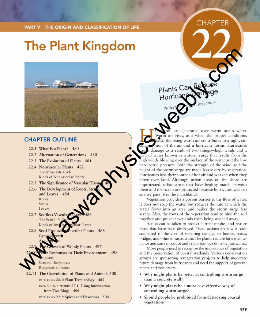
CHAPTER
22 The Plant Kingdom
H urricanes are generated over warm ocean water. Warm air rises, and when the proper conditions exist, the rising warm air contributes to a tight, cir-
cular motion of the air and a hurricane forms. Hurricanes cause damage as a result of two things—high winds and a wall of water known as a storm surge that results from the high winds blowing over the surface of the water and the low barometric pressure. Both the strength of the wind and the height of the storm surge are made less severe by vegetation. Hurricanes lose their source of hot air and weaken when they move over land. Although urban areas on the shore are unprotected, urban areas that have healthy marsh between them and the ocean are protected because hurricanes weaken as they pass over the marshlands.
Vegetation provides a porous barrier to the flow of water. It does not stop the water, but reduces the rate at which the water flows into an area and makes the storm surge less severe. Also, the roots of the vegetation tend to bind the soil together and prevent wetlands from being washed away.
Action can be taken to protect current mashes and restore those that have been destroyed. These actions are low in cost compared to the cost of repairing damage to homes, roads, bridges, and other infrastructure. The plants require little mainte-nance and can reproduce and repair damage done by hurricanes.
More people need to recognize the importance of vegetation and the preservation of coastal wetlands. Various conservation groups are sponsoring revegetation projects to help moderate future damage from hurricanes and need the support of govern-ments and volunteers.
• Why might plants be better at controlling storm surge than a concrete wall?
• Why might plants be a more cost-effective way of controlling storm surge?
• Should people be prohibited from destroying coastal vegetation?
479
PART V THE ORIGIN AND CLASSIFICATION OF LIFE
CHAPTER OUTLINE
22.1 What Is a Plant? 480
22.2 Alternation of Generations 480
22.3 The Evolution of Plants 481
22.4 Nonvascular Plants 482 The Moss Life Cycle Kinds of Nonvascular Plants
22.5 The Significance of Vascular Tissue 483
22.6 The Development of Roots, Stems, and Leaves 484 Roots Stems Leaves
22.7 Seedless Vascular Plants 488 The Fern Life Cycle Kinds of Seedless Vascular Plants
22.8 Seed-Producing Vascular Plants 488 Gymnosperms Angiosperms
22.9 The Growth of Woody Plants 497
22.10 Plant Responses to Their Environment 498 Tropisms Seasonal Responses Responses to Injury
22.11 The Coevolution of Plants and Animals 500
OUTLOOKS 22.1: Plant Terminology 485
HOW SCIENCE WORKS 22.1: Using Information
from Tree Rings 498
OUTLOOKS 22.2: Spices and Flavorings 500
Plants Can Reduce
Hurricane Damage
Protect our coastal vegetation.
eng03466_ch22_479-502.indd 479eng03466_ch22_479-502.indd 479 18/11/10 1:07 PM18/11/10 1:07 PM
www.aswarp
hysic
s.wee
bly.co
m

480 PART V The Origin and Classification of Life
22.2 Alternation of Generations Plants have a life cycle that involves two distinctly different generations: the sporophyte generation and the gametophyte generation. The sporophyte generation is diploid ( 2n ) and has plant parts in which meiosis takes place to produce haploid ( n ) spores. The word spore is used several different ways. There are structures called spores in bacteria, algae, protozoa, fungi, and plants. Each is distinct from the others. In our discussion of plants, the word spore refers to a haploid cell produced by meiosis that germinates to give rise to a multicellular haploid
22.1 What Is a Plant? Plants are eukaryotic, multicellular organisms that have chlorophyll a and chlorophyll b and carry on photosynthe-sis. Their cells have cellulose cell walls. They also have specializations for life on land. Plants are, for the most part, terrestrial organisms that can live in just about any environ-ment, including deserts, arctic regions, and swamps ( figure 22.1 ). Many live in shallow freshwater (e.g., water lilies, cattails), and a few live in shallow oceans (e.g., eel-grass, mangroves). Within the estimated 300,000 species, plants show a remarkable variety of form, function, and activity. Plants range in size from tiny, floating duckweed the size of a pencil eraser to redwood trees over 100 meters tall. There are some that lack chlorophyll and are parasites, some that digest animals, and some that have mutualistic relation-ships with animals.
Background Check Concepts you should already know to get the most out of this chapter: • The processes of natural selection and evolution (chapter 13) • Structural and life history characteristics are used to classify organisms (chapter 20) • Plant cells differ from animal cells (chapter 4)
Desert plants Arctic plants Aquatic plants
FIGURE 22.1 Plant Diversity Plants are adapted to a wide range of terrestrial habitats, including such extremes as deserts and arctic regions. Many live in freshwater and a few live in shallow marine environments.
1. List three characteristics shared by all plants.
22.1 CONCEPT REVIEW
eng03466_ch22_479-502.indd 480eng03466_ch22_479-502.indd 480 15/10/10 12:47 PM15/10/10 12:47 PM
www.aswarp
hysic
s.wee
bly.co
m

CHAPTER 22 The Plant Kingdom 481
was a freshwater member of the green algae. The strongest evidence for this is that green algae have the same kinds of chlorophyll (chlorophyll a and chlorophyll b ) and the same kinds of chloroplasts as plants. Comparison of the DNA of plants and green algae also supports this conclusion. The evolution of plants shows two general trends. One is toward greater specialization for living in a dry environment; the other is toward a more prominent role for the sporophyte generation in the life cycle.
The most primitive plants lack vascular tissue to help carry water. Vascular tissue consists of specialized cells arranged end to end that carry water and nutrients from one place to another. Because nonvascular plants lack vascular tissue, most are limited to moist habitats. The more advanced vascular plants have specialized cells that help transport water and other materials throughout the plant. A second major development that allowed advanced plants to exploit terrestrial habitats was the evolution of seeds that could resist drying.
In the more primitive plants, the gametophyte gen-eration is the more conspicuous, and long-lived (domi-nant) generation, whereas, in more advanced groups, the sporophyte generation is dominant. The taxonomy of plants is based on these trends ( figure 22.3 ). Subsequent sections will discuss the major groups of plants and show how life cycles and the capacity to live in a dry environ-ment evolved.
generation known as the gametophyte generation. The game-tophyte generation is haploid and develops structures that produce gametes: eggs and sperm. Because the gametophyte is already haploid, eggs and sperm are produced by mitosis. When haploid gametes unite, a diploid zygote is formed. The zygote is the first cell in a new sporophyte generation. The zygote divides by mitosis, and a new multicellular sporophyte generation results. Recall that the term alternation of genera-tions is used to describe this kind of life cycle, in which plants cycle between two stages in their life—the diploid sporophyte and the haploid gametophyte ( figure 22.2 ).
FERTILIZATION MEIOSIS
MitosisMitosis
Mitosis
diploid (2n)
haploid (n)
spore (n)
(n)gametophyte
sporophyte(2n)
zygote (2n) sporangium (2n)
gametes (n)
FIGURE 22.2 Alternation of Generations Plants go through two distinctly different generations during their life cycle. The sporophyte generation is diploid and the gametophyte generation is haploid.
2. Which generation in the plant life cycle is haploid and which is diploid?
3. What reproductive cells do sporophytes produce? Are these structures haploid or diploid?
4. What reproductive cells do gametophytes produce? Are these structures haploid or diploid?
22.2 CONCEPT REVIEW
22.3 The Evolution of Plants At one time, all life was aquatic. Therefore, it is logical to look for the ancestors of plants among the aquatic, photosyn-thetic algae. Most scientists feel that the ancestor of plants
Algalancestor
Embryo
Green Algae
Liverworts
Hornworts
Mosses
Club mossesand relatives
Horsetails
Ferns
Whisk ferns
Vasculartissue
Seeds Gymnosperms
Angiosperms
FIGURE 22.3 The Evolution of Plants The evolution of plants involves specializations for living on land and changes in the way they reproduce. Plants differ from their algal ancestors in that they have a multicellular immature stage known as an embryo. Vascular tissue and seeds were important evolutionary steps that allowed plants to be successful away from moist habitats.
eng03466_ch22_479-502.indd 481eng03466_ch22_479-502.indd 481 15/10/10 12:47 PM15/10/10 12:47 PM
www.aswarp
hysic
s.wee
bly.co
m

482 PART V The Origin and Classification of Life
tubular channel leading to the egg at its base. There is usually only one egg cell in each archegonium. When the sperm are mature, the outer jacket of the antheridium splits open, releasing the flagellated sperm. The sperm swim through a film of dew or rainwater to the archegonium and continue down the channel of the archegonium to fertilize the egg. When the sperm and egg nuclei fuse, a diploid zygote is pro-duced. The zygote is the first cell of the sporophyte genera-tion. The zygote divides by mitosis and the sporophyte generation begins to grow within the archegonium and even-tually develops into a mature sporophyte, which grows out of the top of the gametophyte.
The sporophyte consists of a long stalk with a capsule on the end of it. Within the capsule, meiosis takes place, produc-ing haploid spores. These tiny spores are released and distrib-uted by wind and water. When they germinate, they give rise to a multicellular filament known as a protonema. The pro-tonema develops into a mature gametophyte plant. Thus, in mosses and other nonvascular plants, the gametophyte gen-eration is the dominant stage and the sporophyte is depen-dent on the gametophyte.
22.4 Nonvascular Plants The nonvascular plants, which include the mosses, hornworts, and liverworts, are commonly known as bryophytes. These three kinds of plants share the following characteristics.
1. They lack vascular tissue. 2. They do not have true roots or leaves. 3. The gametophyte generation is the most prominent part
of the life cycle. 4. The sperm swim to the egg.
Because they lack vascular tissue and roots for the absorption and transportation of water, they must rely on the physical pro-cesses of diffusion and osmosis to move dissolved materials through their bodies. They have sperm that swim to the egg, so they are similar to their algal ancestors and must have water to reproduce sexually. Thus, most of these organisms are small and live in moist habitats. In other words, they are only minimally adapted to a ter-restrial environment.
The Moss Life Cycle Although there are differences in details of the life cycle of nonvascular plants, we will use the moss life cycle to represent the group. The moss plant that you commonly recognize is the gametophyte generation. Recall that the gametophyte generation is haploid and is the gamete-producing stage in the plant life cycle. Although the cells of the gametophyte have the haploid number of chromosomes (the same as gametes), not all of them function as gametes. At the top of the moss gametophyte are two kinds of structures that produce gametes: the antheridium and the archegonium ( figure 22.4 ).
The antheridium is a sac-like structure that consists of the developing sperm sur-rounded by a layer of cells called jacket cells. The archegonium is a flask-shaped structure that produces the egg; it has a
5. Why are green algae considered to be the ancestors of plants?
6. How does the significance of the sporophyte gener-ation change with the evolution of advanced plants?
22.3 CONCEPT REVIEW
Sperm
FERTILIZATION
Zygote
Developingsporophyte inarchegonium
Maturesporophyte
Parentgametophyte
Capsule
Antheridium
Egg
Archegonium
Male Female
Gametophytes
Protonema
Germinatingspores Spores
MEIOSIS
Mitosis
Haploid(n)
Diploid(2n)
FIGURE 22.4 The Life Cycle of a Moss The haploid gametophyte generation produces eggs in a structure called an archegonium and sperm in a structure called an antheridium. Sperm swim to the egg and fertilization occurs within the archegonium. The zygote is diploid and is the first stage of the sporophyte generation. The sporophyte grows and protrudes from the top of the gametophyte. Meiosis takes place in the capsule of the sporophyte, producing haploid spores. The spores are released and give rise to new gametophyte plants.
eng03466_ch22_479-502.indd 482eng03466_ch22_479-502.indd 482 15/10/10 12:47 PM15/10/10 12:47 PM
www.aswarp
hysic
s.wee
bly.co
m
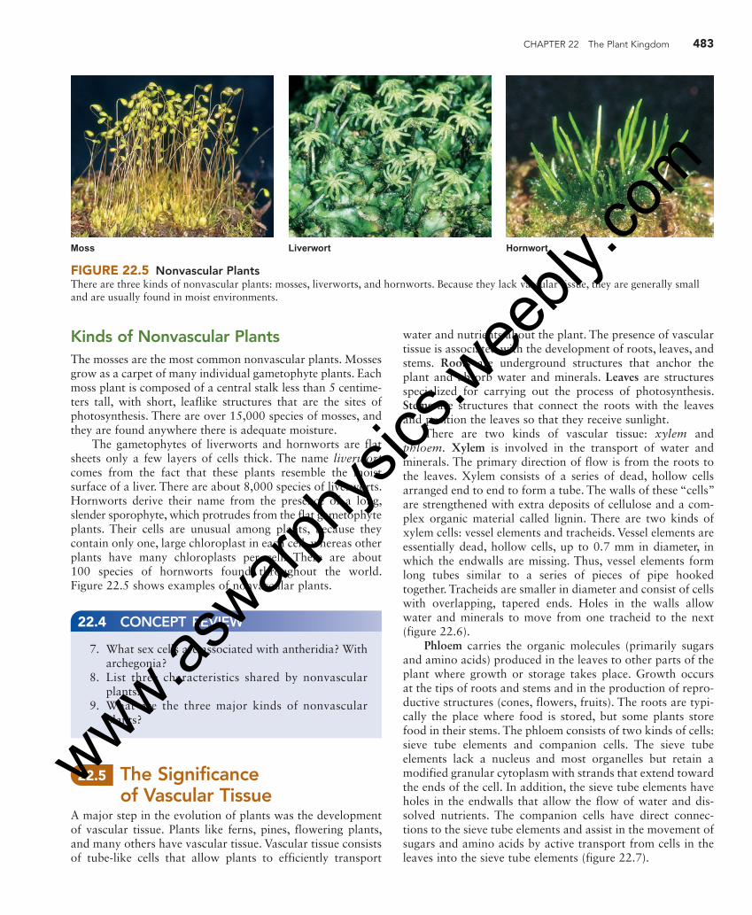
CHAPTER 22 The Plant Kingdom 483
water and nutrients about the plant. The presence of vascular tissue is associated with the development of roots, leaves, and stems. Roots are underground structures that anchor the plant and absorb water and minerals. Leaves are structures specialized for carrying out the process of photosynthesis. Stems are structures that connect the roots with the leaves and position the leaves so that they receive sunlight.
There are two kinds of vascular tissue: xylem and phloem. Xylem is involved in the transport of water and minerals. The primary direction of flow is from the roots to the leaves. Xylem consists of a series of dead, hollow cells arranged end to end to form a tube. The walls of these “cells” are strengthened with extra deposits of cellulose and a com-plex organic material called lignin. There are two kinds of xylem cells: vessel elements and tracheids. Vessel elements are essentially dead, hollow cells, up to 0.7 mm in diameter, in which the endwalls are missing. Thus, vessel elements form long tubes similar to a series of pieces of pipe hooked together. Tracheids are smaller in diameter and consist of cells with overlapping, tapered ends. Holes in the walls allow water and minerals to move from one tracheid to the next ( figure 22.6 ).
Phloem carries the organic molecules (primarily sugars and amino acids) produced in the leaves to other parts of the plant where growth or storage takes place. Growth occurs at the tips of roots and stems and in the production of repro-ductive structures (cones, flowers, fruits). The roots are typi-cally the place where food is stored, but some plants store food in their stems. The phloem consists of two kinds of cells: sieve tube elements and companion cells. The sieve tube elements lack a nucleus and most organelles but retain a modified granular cytoplasm with strands that extend toward the ends of the cell. In addition, the sieve tube elements have holes in the endwalls that allow the flow of water and dis-solved nutrients. The companion cells have direct connec-tions to the sieve tube elements and assist in the movement of sugars and amino acids by active transport from cells in the leaves into the sieve tube elements ( figure 22.7 ).
Kinds of Nonvascular Plants The mosses are the most common nonvascular plants. Mosses grow as a carpet of many individual gametophyte plants. Each moss plant is composed of a central stalk less than 5 centime-ters tall, with short, leaflike structures that are the sites of photosynthesis. There are over 15,000 species of mosses, and they are found anywhere there is adequate moisture.
The gametophytes of liverworts and hornworts are flat sheets only a few layers of cells thick. The name liverwort comes from the fact that these plants resemble the moist surface of a liver. There are about 8,000 species of liverworts. Hornworts derive their name from the presence of a long, slender sporophyte, which protrudes from the flat gametophyte plants. Their cells are unusual among plants, because they contain only one, large chloroplast in each cell, whereas other plants have many chloroplasts per cell. There are about 100 species of hornworts found throughout the world. Figure 22.5 shows examples of nonvascular plants.
Moss Liverwort Hornwort
FIGURE 22.5 Nonvascular Plants There are three kinds of nonvascular plants: mosses, liverworts, and hornworts. Because they lack vascular tissue, they are generally small and are usually found in moist environments.
7. What sex cells are associated with antheridia? With archegonia?
8. List three characteristics shared by nonvascular plants.
9. What are the three major kinds of nonvascular plants?
22.4 CONCEPT REVIEW
22.5 The Significance of Vascular Tissue
A major step in the evolution of plants was the development of vascular tissue. Plants like ferns, pines, flowering plants, and many others have vascular tissue. Vascular tissue consists of tube-like cells that allow plants to efficiently transport
eng03466_ch22_479-502.indd 483eng03466_ch22_479-502.indd 483 15/10/10 12:47 PM15/10/10 12:47 PM
www.aswarp
hysic
s.wee
bly.co
m
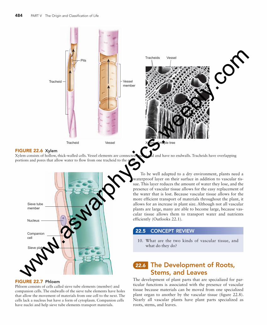
484 PART V The Origin and Classification of Life
Tracheid
Tracheid Vessel
Pits
Vesselmember
T Vracheids essel
Xylem of maple tree
FIGURE 22.6 Xylem Xylem consists of hollow, thick-walled cells. Vessel elements are connected end to end and have no endwalls. Tracheids have overlapping portions and pores that allow water to flow from one tracheid to the next.
Sieve plate
Sieve tubemember
Nucleus
Companioncell
FIGURE 22.7 Phloem Phloem consists of cells called sieve tube elements (member) and companion cells. The endwalls of the sieve tube elements have holes that allow the movement of materials from one cell to the next. The cells lack a nucleus but have a form of cytoplasm. Companion cells have nuclei and help sieve tube elements transport materials.
To be well adapted to a dry environment, plants need a waterproof layer on their surface in addition to vascular tis-sue. This layer reduces the amount of water they lose, and the presence of vascular tissue allows for the easy replacement of the water that is lost. Because vascular tissue allows for the more efficient transport of materials throughout the plant, it allows for an increase in plant size. Although not all vascular plants are large, many are able to become large, because vas-cular tissue allows them to transport water and nutrients efficiently (Outlooks 22.1).
10. What are the two kinds of vascular tissue, and what do they do?
22.5 CONCEPT REVIEW
22.6 The Development of Roots, Stems, and Leaves
The development of plant parts that are specialized for par-ticular functions is associated with the presence of vascular tissue because materials can be moved from one specialized plant organ to another by the vascular tissue ( figure 22.8 ). Nearly all vascular plants have plant parts specialized as roots, stems, and leaves.
eng03466_ch22_479-502.indd 484eng03466_ch22_479-502.indd 484 15/10/10 12:47 PM15/10/10 12:47 PM
www.aswarp
hysic
s.wee
bly.co
m

CHAPTER 22 The Plant Kingdom 485
Perennial plants live many years. Most kinds of plants are perennials, including mosses, ferns, horsetails, pines, and most flowering plants. Many perennial plants are woody, such as various kinds of trees. Other perennial plants produce above-ground parts that die back at the end of a growing season. The plant regrows above-ground parts from the roots each year, as do tulips, daffodils, rhubarb, and ferns.
Trees, Shrubs, and Herbs A tree is a large, woody plant that usually has a single main stem with branches. There are tree ferns, most gymnosperms are trees, and many flowering plants are trees. A shrub is a peren-nial woody plant that generally has several main stems and is relatively short. Herbs are generally small, herbaceous (non-woody) plants. The word herb is also used to indicate plants that have specific food, flavoring, or medicinal value.
OUTLOOKS 22.1
Plant Terminology There are several sets of terminology used to discuss plants that describe their nature but do not have any relationship to their taxonomy or phylogeny.
Herbaceous and woody Herbaceous plants have few cells with thick cell walls and, thus, are generally small and easily damaged. Mosses, grasses, and many garden plants are herbaceous. Woody plants have a great deal of tissue with thick cell walls. The xylem and associ-ated cells are important in strengthening woody plants. There are ferns, gymnosperms, and angiosperms that are woody.
Annual and perennial Annual plants live one year. They germinate from seeds, grow, produce flowers and seeds, and die within 1 year. The seeds produced germinate the following year and the cycle contin-ues. Most annual plants are angiosperms (flowering plants).
Roots Roots are the underground parts of plants that anchor them in the soil and absorb water and nutrients, such as nitrogen, phosphorus, potassium, and other inorganic molecules from the soil. The xylem allows these materials to be distributed for use by other parts of the plant. The phloem transports organic molecules from the leaves and stem to the roots.
Roots grow from their tips. By growing constantly, roots explore new territory for available nutrients and water. As a plant becomes larger, it needs more root surface to absorb water and nutrients and to hold the plant in place. The actively growing portions of the root near the tips have many small, fuzzy, hairlike cell extensions called root hairs, which provide a large surface area for the absorption of nutrients and water.
Most roots are important storage places for the food produced by the above-ground parts of the plant. Food is stored in their roots during the growing season and used to stay alive during the winter. The food also provides the raw materials necessary for growth for the next growing season. Although humans do not eat the roots of plants such as maple trees, rhubarb, and grasses, their roots are as important to them in food storage as are those of carrots, turnips, and radishes ( figure 22.9 ).
Stems Stems are, in most cases, the above-ground structures of plants that support the light-catching leaves in which photo-synthesis occurs. However, they can vary considerably. Trees have stems that support large numbers of branches; vines
Root hair
XylemPhloem
H O2
FIGURE 22.8 Vascular Tissue and the Specialization of Plant Parts Vascular tissue allows for the specialization of leaves for photosynthesis, roots for the absorption of water and minerals, and stems for the positioning of the leaves. The xylem carries water and minerals from the roots to the leaves. The phloem transports organic molecules to places they are needed for growth or storage.
eng03466_ch22_479-502.indd 485eng03466_ch22_479-502.indd 485 15/10/10 12:47 PM15/10/10 12:47 PM
www.aswarp
hysic
s.wee
bly.co
m

486 PART V The Origin and Classification of Life
deposited in the cell walls that provides strength and binds cell walls to one another. This combination of thick cell walls with strengthening lignin is such an effective support mecha-nism that large trees and bushes are supported against the pull of gravity and can withstand strong winds for centuries. Some of the oldest trees on Earth have been growing for sev-eral thousand years.
Another important function of the stem is to transport materials between the roots and the leaves ( figure 22.10 ). A cross section of a stem examined under a microscope reveals that a large proportion of the stem consists of vascular tissue.
In addition to support and transport, the stems of some plants have additional functions. Some stems store food. This is true of sugar cane, yams, and potatoes. (Yams and potatoes are actually highly modified underground stems.) Many plant stems are green and, therefore, are involved in photosynthesis. Cacti and many herbaceous plants are examples. Stems also have a waterproof layer on the outside. In the case of herbaceous plants, it is usually a waxy layer. In the case of woody plants, there is a tough, outer, water-proof bark.
Leaves Leaves are the specialized parts of plants that are the major sites of photosynthesis. (See chapter 7 for a discussion of photosynthesis.) To carry out photosynthesis, leaves must have certain characteristics ( figure 22.11 ). Because it is a solar collector, a leaf should have a large surface area. Also,
have stems that require support; some plants, such as dande-lions, have very short stems, with their leaves flat against the ground; and some stems are actually underground. Stems have two main functions:
1. They support the leaves. 2. They transport raw materials from the roots
to the leaves and manufactured food from the leaves to the roots.
The support that stems provide is possible because of the nature of plant cell walls. First, all plant cells are surrounded by a cell wall made of cellulose fibers interwoven to form a box, within which the plant cell is contained. Because the cell wall consists of fibers, it is like a wicker basket. There are spaces between cellulose fibers through which materials pass relatively easily. However, the cellulose fibers do not stretch; if the cell is full of water and other cellular materials, it becomes quite rigid. Remember that the process of osmosis results in cells having an internal pressure. It is this pressure against the nonstretchable cell wall that makes the cell rigid. Many kinds of small plants, called herbaceous plants, rely primarily on this mechanism for support.
The second way stems provide support involves the thickening of cell walls. Woody plants have especially thick cell walls. The xylem of woody plants has thick cell walls and lignin
Fibrous root Tap root
FIGURE 22.9 Kinds of Roots The roots of grasses are often in the upper layers of the soil and form a dense network. This form of root is called a fibrous root and is an adaptation to relatively dry conditions. In grasslands, rainfall is infrequent and rarely penetrates very deeply into the soil. The network of roots near the surface efficiently captures the water when it is available. The roots of trees and many other plants typically extend deep into the soil, where they obtain moisture and serve as anchors to hold large plants upright. This kind of root is called a tap root.
Bark
Phloem
Annual growthrings
Xylem(wood)
Cross section of basswood stem
0.5 mm
FIGURE 22.10 Cross Section of a Stem This photo shows the cross section of a 3-year-old basswood tree. On the outside is the bark, which contains the phloem. Inside the bark are three layers of xylem tissue. Each layer of xylem constitutes 1 year’s growth.
eng03466_ch22_479-502.indd 486eng03466_ch22_479-502.indd 486 15/10/10 12:47 PM15/10/10 12:47 PM
www.aswarp
hysic
s.wee
bly.co
m
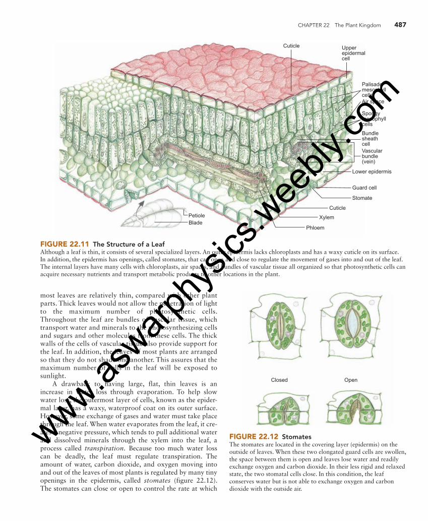
CHAPTER 22 The Plant Kingdom 487
most leaves are relatively thin, compared with other plant parts. Thick leaves would not allow the penetration of light to the maximum number of photosynthetic cells. Throughout the leaf are bundles of vascular tissue, which transport water and minerals to the photosynthesizing cells and sugars and other molecules from these cells. The thick walls of the cells of vascular tissue also provide support for the leaf. In addition, the leaves of most plants are arranged so that they do not shade one another. This assures that the maximum number of cells in the leaf will be exposed to sunlight.
A drawback to having large, flat, thin leaves is an increase in water loss through evaporation. To help slow water loss, the outermost layer of cells, known as the epider-mal layer, has a waxy, waterproof coat on its outer surface. However, some exchange of gases and water must take place through the leaf. When water evaporates from the leaf, it cre-ates a negative pressure, which tends to pull additional water and dissolved minerals through the xylem into the leaf, a process called transpiration. Because too much water loss can be deadly, the leaf must regulate transpiration. The amount of water, carbon dioxide, and oxygen moving into and out of the leaves of most plants is regulated by many tiny openings in the epidermis, called stomates ( figure 22.12 ). The stomates can close or open to control the rate at which
PetioleBlade
Cuticl
Xylem
Phloem
e
Lower epidermis
Guard cell
Stomate
Vascularbundle(vein)
PalisademesophyllcellsAir space
Spongymesophyllcells
Bundlesheathcell
Upperepidermalcell
Cuticle
FIGURE 22.11 The Structure of a Leaf Although a leaf is thin, it consists of several specialized layers. An outer epidermis lacks chloroplasts and has a waxy cuticle on its surface. In addition, the epidermis has openings, called stomates, that can open and close to regulate the movement of gases into and out of the leaf. The internal layers have many cells with chloroplasts, air spaces, and bundles of vascular tissue all organized so that photosynthetic cells can acquire necessary nutrients and transport metabolic products to other locations in the plant.
Closed Open
FIGURE 22.12 Stomates The stomates are located in the covering layer (epidermis) on the outside of leaves. When these two elongated guard cells are swollen, the space between them is open and leaves lose water and readily exchange oxygen and carbon dioxide. In their less rigid and relaxed state, the two stomatal cells close. In this condition, the leaf conserves water but is not able to exchange oxygen and carbon dioxide with the outside air.
eng03466_ch22_479-502.indd 487eng03466_ch22_479-502.indd 487 15/10/10 12:47 PM15/10/10 12:47 PM
www.aswarp
hysic
s.wee
bly.co
m
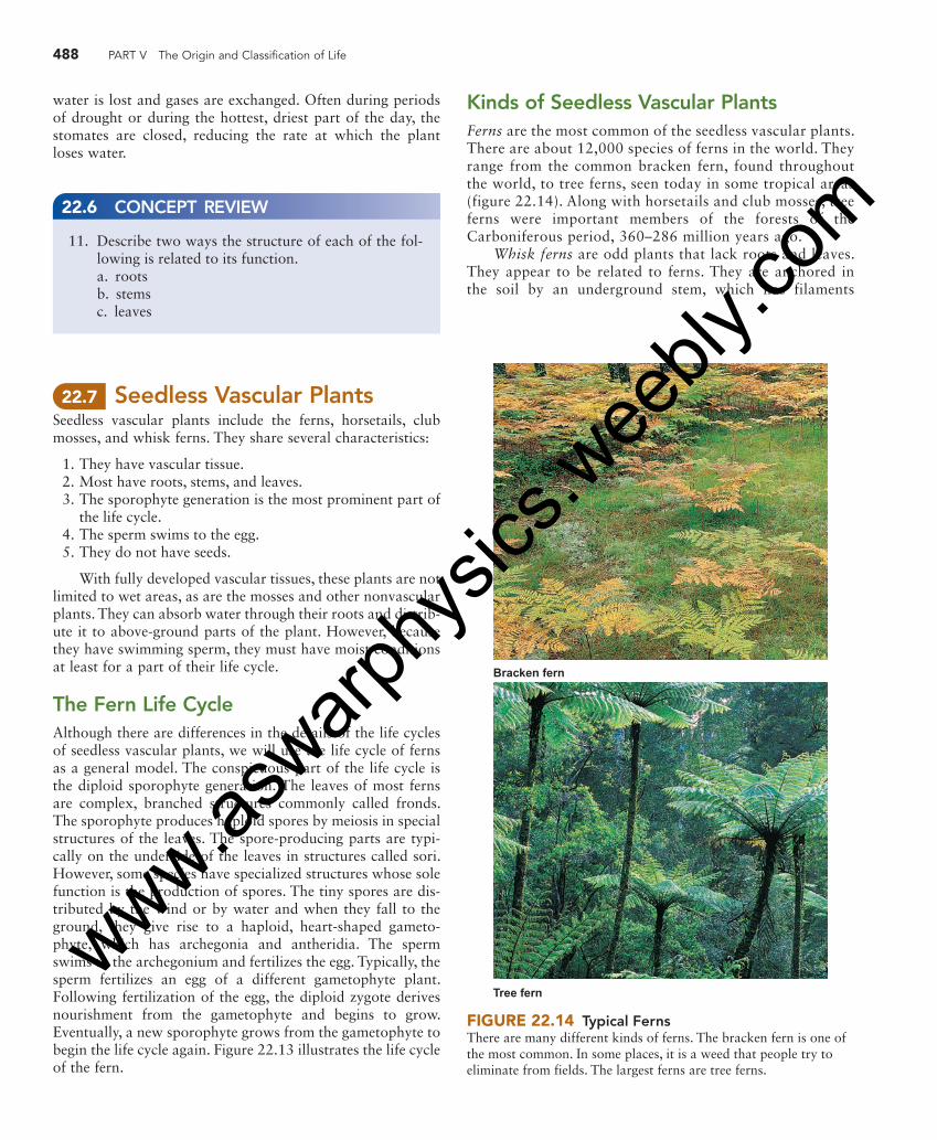
488 PART V The Origin and Classification of Life
Kinds of Seedless Vascular Plants Ferns are the most common of the seedless vascular plants. There are about 12,000 species of ferns in the world. They range from the common bracken fern, found throughout the world, to tree ferns, seen today in some tropical areas ( figure 22.14 ). Along with horsetails and club mosses, tree ferns were important members of the forests of the Carboniferous period, 360–286 million years ago.
Whisk ferns are odd plants that lack roots and leaves. They appear to be related to ferns. They are anchored in the soil by an underground stem, which has filaments
water is lost and gases are exchanged. Often during periods of drought or during the hottest, driest part of the day, the stomates are closed, reducing the rate at which the plant loses water.
11. Describe two ways the structure of each of the fol-lowing is related to its function.
a. roots b. stems c. leaves
22.6 CONCEPT REVIEW
22.7 Seedless Vascular Plants Seedless vascular plants include the ferns, horsetails, club mosses, and whisk ferns. They share several characteristics:
1. They have vascular tissue. 2. Most have roots, stems, and leaves. 3. The sporophyte generation is the most prominent part of
the life cycle. 4. The sperm swims to the egg. 5. They do not have seeds.
With fully developed vascular tissues, these plants are not limited to wet areas, as are the mosses and other nonvascular plants. They can absorb water through their roots and distrib-ute it to above-ground parts of the plant. However, because they have swimming sperm, they must have moist conditions at least for a part of their life cycle.
The Fern Life Cycle Although there are differences in the details of the life cycles of seedless vascular plants, we will use the life cycle of ferns as a general model. The conspicuous part of the life cycle is the diploid sporophyte generation. The leaves of most ferns are complex, branched structures commonly called fronds. The sporophyte produces haploid spores by meiosis in special structures of the leaves. The spore-producing parts are typi-cally on the underside of the leaves in structures called sori. However, some species have specialized structures whose sole function is the production of spores. The tiny spores are dis-tributed by the wind or by water and when they fall to the ground, they give rise to a haploid, heart-shaped gameto-phyte, which has archegonia and antheridia. The sperm swims to the archegonium and fertilizes the egg. Typically, the sperm fertilizes an egg of a different gametophyte plant. Following fertilization of the egg, the diploid zygote derives nourishment from the gametophyte and begins to grow. Eventually, a new sporophyte grows from the gametophyte to begin the life cycle again. Figure 22.13 illustrates the life cycle of the fern.
Bracken fern
Tree fern
FIGURE 22.14 Typical Ferns There are many different kinds of ferns. The bracken fern is one of the most common. In some places, it is a weed that people try to eliminate from fields. The largest ferns are tree ferns.
eng03466_ch22_479-502.indd 488eng03466_ch22_479-502.indd 488 15/10/10 12:47 PM15/10/10 12:47 PM
www.aswarp
hysic
s.wee
bly.co
m

CHAPTER 22 The Plant Kingdom 489
Sorus (clusterof sporangia)
Archegonium
Egg
Antheridium
Sperm
FERTILIZATION
Embryo
Leaf of youngsporophyte
Gametophyte
Rhizome
Maturefrond
Maturesporangium
MEIOSIS
Spore
Rhizoids
Gametophyte
Haploid (n)
Diploid (2n)
Adultsporophyte
Mitosis
FIGURE 22.13 The Life Cycle of a Fern The sporophyte of a fern consists of an underground stem (rhizome) and roots and above-ground leaves. The leaves are known as fronds. On the back of some fern leaves are small dots called sori. Each sorus contains structures that produce spores by meiosis. These haploid spores develop into the heart-shaped gametophyte. The gametophyte has archegonia, which produce eggs, and antheridia, which produce sperm. The sperm swim to the egg. The fertilized egg (zygote) is diploid and grows into a new sporophyte plant.
growing from it that absorb water and soil nutrients. It has flattened structures similar to leaves, but they are not considered leaves because they lack vascular tissue. Whisk ferns are most common in warm, moist environments. There are about 15 species. The spores are produced at
the ends of branches and produce a gametophyte in the soil.
Horsetails are low-growing plants with jointed stems. The leaves are tiny and encircle the stem at the joints. Much of the photosynthesis actually occurs in the green stem.
eng03466_ch22_479-502.indd 489eng03466_ch22_479-502.indd 489 15/10/10 12:48 PM15/10/10 12:48 PM
www.aswarp
hysic
s.wee
bly.co
m
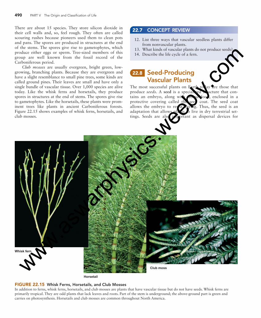
490 PART V The Origin and Classification of Life
FIGURE 22.15 Whisk Ferns, Horsetails, and Club Mosses In addition to ferns, whisk ferns, horsetails, and club mosses are plants that have vascular tissue but do not have seeds. Whisk ferns are primarily tropical. They are odd plants that lack leaves and roots. Part of the stem is underground; the above-ground part is green and carries on photosynthesis. Horsetails and club mosses are common throughout North America.
Horsetail
Club moss
Whisk fern
There are about 15 species. They store silicon dioxide in their cell walls and, so, feel rough. They often are called scouring rushes because pioneers used them to clean pots and pans. The spores are produced in structures at the end of the stems. The spores give rise to gametophytes, which produce either eggs or sperm. Tree-sized members of this group are well known from the fossil record of the Carboniferous period.
Club mosses are usually evergreen, bright green, low-growing, branching plants. Because they are evergreen and have a slight resemblance to small pine trees, some kinds are called ground pines. Their leaves are small and have only a single bundle of vascular tissue. Over 1,000 species are alive today. Like the whisk ferns and horsetails, they produce spores in structures at the end of stems. The spores give rise to gametophytes. Like the horsetails, these plants were prom-inent trees like plants in ancient Carboniferous forests. Figure 22.15 shows examples of whisk ferns, horsetails, and club mosses.
12. List three ways that vascular seedless plants differ from nonvascular plants.
13. What kinds of vascular plants do not produce seeds? 14. Describe the life cycle of a fern.
22.7 CONCEPT REVIEW
22.8 Seed-Producing Vascular Plants
The most successful plants on Earth today are those that produce seeds. A seed is a specialized structure that con-tains an embryo, along with stored food, enclosed in a protective covering called the seed coat. The seed coat allows the embryo to resist drying. Thus, the seed is an adaptation that allows plants to live in dry terrestrial set-tings. Seeds are also important as dispersal devices for
eng03466_ch22_479-502.indd 490eng03466_ch22_479-502.indd 490 15/10/10 12:48 PM15/10/10 12:48 PM
www.aswarp
hysic
s.wee
bly.co
m

CHAPTER 22 The Plant Kingdom 491
The Pine Life Cycle The dominant portion of the pine life cycle is the large, dip-loid sporophyte generation. Pines produce two kinds of cones, which are part of the sporophyte generation but pro-duce separate haploid male and female gametophytes. The large cones on pines are the female cones, which produce the female gametophyte generation. The female gametophyte consists of several thousand cells and produces several arche-gonia, each of which contains an egg.
The small male cones are located on the ends of branches. The male cones produce pollen. Each of these small, dustlike pol-len grains is a male gametophyte plant that contains haploid nuclei. Some of these nuclei function as sperm. In order to obtain a fertilized egg, two separate events must occur: pollination and fertilization. Pollination is the process of getting the pollen from the male cone to the female cone. Conifers are wind pollinated. Pollen is released in such large quantities that clouds of pollen can be seen in the air when sudden gusts of wind shake the branches of the trees. Once the pollen grain (male gametophyte) is in the vicinity of the female gametophyte in the female cone , it begins to grow a tubelike structure—the pollen tube. The pollen tube enters the archegonium of the female gametophyte and releases a sperm nucleus, which fertilizes the egg. The processes
of pollination and fertilization are separate events. Pollination occurs with the transfer of pollen to the female cone. Fertilization occurs when the sperm nucleus from the pollen unites with the egg cell in the arche-gonium. Fertilization in gymnosperms may occur months or even years following pollination.
The fertilized egg (zygote) is diploid and develops into an embryo within a seed. Many kinds of birds and mammals use the seeds of pines and related trees for food. In most pines, the seeds are released from the cone when the scales of the cone fold back. The winged seeds are carried by wind and fall to the ground. The seed germinates and gives rise to a new sporophyte plant, and the life cycle continues. Figure 22.16 reviews the life cycle of the pine.
plants. Two major groups of plants produce seeds: the gymnosperms (conifers and their relatives) and the angio-sperms (flowering plants). There were also extinct fernlike plants that had a kind of seed.
Another important innovation of seed-producing plants is the development of a life cycle in which pollen is produced. Pollen is the miniaturized male gametophyte generation. Pollen can be carried from one plant to another by wind or animals, which makes the presence of water for fertilization unnecessary.
Gymnosperms Several kinds of woody plants produce seeds that are not enclosed. Typically, their seeds are produced in woody struc-tures called cones. However, the seeds are not totally enclosed but are produced on the surface of flat, woody parts of the cone. Because the seeds are not enclosed, they are said to be naked. Thus, these cone- producing plants, such as conifers, are called gymnosperms, which means naked seed plants. However, there are several distinct kinds of plants in this group: the cycads, ginkgos, and conifers. All are woody perennial plants. We will examine the life cycle of the pine tree to illustrate the life cycle of a gymnosperm.
Longisection ofseed, showingembryo
FERTILIZATION(15 months afterpollination)
Ovulate (seed-bearing)cone
Megasporemother cell
Microsporemother cell
Diploid(2n)
Haploid(n)
Microspores
Pollen
Pollen-bearingcone
PollentubePollination
Megaspore
MEIOSIS
Sporophyte
Seedling
Pineseed
Mature seedcone (2nd year)
Scale
Embryo
Scale
Mitosis
MitosisMitosis
Mitosis FIGURE 22.16 The Life Cycle of a Pine The pine tree is the sporophyte plant. It produces two kinds of cones: small, male cones, which produce pollen (the male gametophyte), and larger, female cones, which contain the female gametophytes that produce eggs. The pollen is carried to the female cone by wind. After pollination, fertilization of the egg occurs when a pollen tube grows to the vicinity of the egg. The fertilized egg develops into an embryo enclosed within a seed. The seeds are eventually released from the female cone.
eng03466_ch22_479-502.indd 491eng03466_ch22_479-502.indd 491 15/10/10 12:48 PM15/10/10 12:48 PM
www.aswarp
hysic
s.wee
bly.co
m
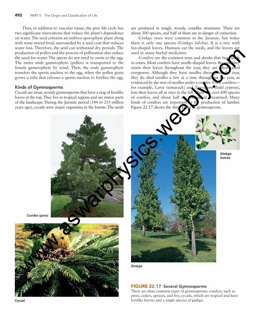
492 PART V The Origin and Classification of Life
Thus, in addition to vascular tissue, the pine life cycle has two significant innovations that reduce the plant’s dependence on water. The seed contains an embryo sporophyte plant along with some stored food, surrounded by a seed coat that reduces water loss. Therefore, the seed can withstand dry periods. The production of pollen and the process of pollination also reduce the need for water. The sperm do not need to swim to the egg. The entire male gametophyte (pollen) is transported to the female gametophyte by wind. Then, the male gametophyte transfers the sperm nucleus to the egg, when the pollen grain grows a tube that releases a sperm nucleus to fertilize the egg.
Kinds of Gymnosperms Cycads are stout, woody gymnosperms that have a ring of fernlike leaves at the top. They live in tropical regions and are minor parts of the landscape. During the Jurassic period (144 to 213 million years ago), cycads were major organisms in the forests. The seeds
Conifer (pine)
Cycad
Ginkgo
Ginkgoleaves
FIGURE 22.17 Several Gymnosperms There are three common types of gymnosperms: conifers, such as pines, cedars, spruces, and firs; cycads, which are tropical and have fernlike leaves; and a single species of ginkgo.
are produced in tough, woody, conelike structures. There are about 300 species, and half of them are in danger of extinction.
Ginkgo trees were common in the Jurassic, but today there is only one species ( Ginkgo biloba ). It is a tree with fan-shaped leaves. Humans eat the seeds, and the leaves are used in many herbal medicines.
Conifers are the common trees and shrubs that bear seeds in cones. Most conifers have needle-shaped leaves. Because they retain their leaves throughout the year, they are often called evergreens. Although they have needles throughout the year, they do shed needles a few at a time throughout the year, as evidenced by the mat of needles under a conifer. A few conifers—for example, Larix (tamarack) and Taxodium (bald cypress), lose their leaves all at once in the fall. There are over 600 species of conifers, and about half are considered threatened. Many kinds of conifers are important in the production of lumber. Figure 22.17 shows the three types of gymnosperms.
eng03466_ch22_479-502.indd 492eng03466_ch22_479-502.indd 492 15/10/10 12:48 PM15/10/10 12:48 PM
www.aswarp
hysic
s.wee
bly.co
m

CHAPTER 22 The Plant Kingdom 493
Angiosperms Angiosperms are plants that produce flowers and have their seeds enclosed in a fruit. A fruit is a modification of the ovary wall into a special structure that contains the seeds. Like the gymnosperms, the flowering plants are well adapted to ter-restrial life with vascular tissue, seeds, and pollen.
Flower Structure A flower is the structure, composed of highly modified leaves, that is responsible for sexual reproduction. At the center of a typical flower is the pistil, which is composed of the stigma, style, and ovary. The ovary produces the female gametophyte plant. Surrounding the pistil are the stamens, which consist of a long filament with anthers at the top. The anther produces the pollen (male gametophyte plant). Petals are a whorl of modified leaves surrounding the stamens and pistil. In many flowering plants, the petals are large and showy. Outside the petals is another whorl of modified leaves, known as sepals. Many kinds of flowers produce odors and a sugary liquid, known as nectar. Figure 22.18 shows the arrangement of parts in a typical flower.
There is a great degree of specialization of flowers. Some are large and showy, such as the flowers of roses and magno-lias. Others are small and inconspicuous, such as the flowers of grasses and birch trees. In addition, some plants have two kinds of flowers. One kind has a pistil, whereas a different flower has the stamens. Any flower that has both pistil and stamens is called a perfect flower; a flower containing just a pistil or just stamens is called an imperfect flower.
The Life Cycle of a Flowering Plant The life cycle of a flowering plant has both sporophyte and gametophyte generations. The sporophyte is the dominant
stage of the life cycle, and the male and female gametophytes are produced within the flower. As in gymnosperms, the male gametophyte is the pollen. The female gametophyte is found within the ovary of the pistil and consists of only eight cells. One of the cells is an egg cell.
Pollination occurs when pollen is transferred from an anther to the stigma of the pistil. In some cases, pollination must be between flowers of different plants of the same species; this is called cross-pollination. However, some spe-cies of plants are able to pollinate themselves; this is called self- pollination.
The pollen grain germinates and produces a pollen tube, which grows down through the tissue of the style to the ovary, where the female gametophyte is located. The pollen tube releases two sperm. One fertilizes the egg nucleus and gives rise to the zygote. The other sperm nucleus combines with two other nuclei in the female gametophyte and pro-duces a triploid nucleus, which develops into endosperm. The endosperm is the stored food of the seed. Because two sperm nuclei are involved, each fertilizing different cells, this is often called double fertilization. Both the embryo and the endosperm grow, and a seed coat develops around them to produce the mature seed. The wall of the ovary develops into a specialized seed-containing structure known as a fruit ( figure 22.19 ).
Pollination Strategies Plants use several strategies to ensure pollination. Most plants that have inconspicuous flowers are wind pollinated. They produce large numbers of flowers and huge amounts of pollen. This is necessary because the distribution of pollen by wind is a random process. By producing large amounts of pollen, the plant increases the chances that pollen will be transferred suc-cessfully. Grasses, sedges, and some other herbaceous plants are wind pollinated. Many trees, such as aspens, birches, and oaks, are also wind pollinated.
It is the pollen from wind-pollinated plants that is responsible for “hay fever,” an allergic reaction to the pres-ence of airborne pollen. Most tree species produce pollen in the spring, so people who have hay fever symptoms in the spring are usually reacting to the pollen of certain trees. Grasses and other herbaceous plants typically produce their pollen in late summer or fall. Ragweed is a common plant that produces pollen late in the growing season and is a com-mon cause of hay fever.
Plants with large, showy flowers are typically polli-nated by animals. Although many showy flowers are pol-linated by insects, such as bees, others are constructed to be pollinated by birds or small mammals. The animals feed on the nectar produced by the flower; in the process, they are dusted with pollen and carry it to another flower of the same species. Many flowers produce odors to help the ani-mals find the plants. This is a mutualistic relationship between the flowers, which are pollinated, and the ani-mals, which receive food in the form of nectar or pollen ( figure 22.20 ).
stamen
petal
sepal receptacle
ovule
ovary
style
stigma
pistil
anther
filament
FIGURE 22.18 Flower Structure The flower is the structure in angiosperms that produces gameophyte plants, which produce sex cells. The female gametophyte is produced within the ovary. The pollen (male gametophyte) is produced within the anther. Following fertilization of the egg, the ovary modifies to form the fruit.
eng03466_ch22_479-502.indd 493eng03466_ch22_479-502.indd 493 15/10/10 12:48 PM15/10/10 12:48 PM
www.aswarp
hysic
s.wee
bly.co
m

494 PART V The Origin and Classification of Life
Haploid(n)
Diploid(2n)
Generative cell
8-nucleateembryo sac
(megagametophyte) (n)Formation ofpollen tube (n)
MEIOSIS
MEIOSIS Tube nucleus
Tubenucleus
Sperm
Style
Sperm
Egg
Polarnuclei
Seed coat
Embryo (2n)
Cotyledons
Seed (2n)
Endosperm (3n)
Youngsporophyte
(2n)
Adultsporophytewith flower
(2n)
Anther
Ovary
Stigma
Anther (2n)
Microsporemother cells (2n)
Microspores (n) Pollen grains(microgametophytes) (n)
Megaspore (n)
Megasporemother cell (2n) Pollen tube
Ovule
DOUBLE FERTILIZATION
Germination
Egg
Mitosis
Mitosis
FIGURE 22.19 Life Cycle of an Angiosperm Angiosperms have flowers, which produce eggs in the ovary and pollen in the anther. Pollen is carried to the pistil by wind or animals. A pollen tube releases haploid nuclei, fertilizing the egg. The fertilized egg develops into the embryo, and the remaining cells associated with the egg develop into a food storage material called endosperm. A seed coat develops around the embryo and endosperm. The ovary enlarges and becomes the fruit, which contains the seeds. The seed germinates to produce the next generation of flowering plant.
Insect pollinated
FIGURE 22.20 Wind and Insect Pollinated Flowers The flowers of wind-pollinated plants, such like this oak, are small and inconspicuous. The flowers of plants that are pollinated by animals are typically large and showy, as are these California poppies and lupines. Wind pollinated
eng03466_ch22_479-502.indd 494eng03466_ch22_479-502.indd 494 15/10/10 12:48 PM15/10/10 12:48 PM
www.aswarp
hysic
s.wee
bly.co
m