Part V: Modeling Neurotransmission - A Dopaminergic Synapse · Part V: Modeling Neurotransmission -...
4
Copyright 3D Molecular Designs All Rights Reserved - 2016 3dmoleculardesigns.com Part V: Modeling Neurotransmission - A Dopaminergic Synapse Student Handout Page 1 1a. Model the metabotropic dopaminergic synapse shown below. Be sure to label all of the following: voltage-gated sodium channel, voltage-gated potassium channel, voltage-gated calcium channel, synaptotagmin, dopamine, synaptic vesicle, presynaptic cell, postsynaptic cell, potassium leak channel, sodium-potassium pump, synaptic cleft, G protein-coupled receptor, SNAP/SNARE proteins, dopamine reuptake transporter, and vesicular transporter When a nerve impulse (action potential) reaches the axon terminal, it sets into motion a chain of events that triggers the release of neurotransmitter. You will next model the events of neurotransmission at a dopaminergic synapse. Student Handout © ...where molecules become real TM Part V: Modeling Neurotransmission - A Dopaminergic Synapse
Transcript of Part V: Modeling Neurotransmission - A Dopaminergic Synapse · Part V: Modeling Neurotransmission -...

Copyright 3D Molecular DesignsAll Rights Reserved - 2016
3dmoleculardesigns.com Part V: Modeling Neurotransmission- A Dopaminergic Synapse
Student Handout Page 1
1a. Model the metabotropic dopaminergic synapse shown below. Be sure to label all of the following: voltage-gated sodium channel, voltage-gated potassium channel, voltage-gated calcium channel, synaptotagmin, dopamine, synaptic vesicle, presynaptic cell, postsynaptic cell, potassium leak channel, sodium-potassium pump, synaptic cleft, G protein-coupled receptor, SNAP/SNARE proteins, dopamine reuptake transporter, and vesicular transporter
When a nerve impulse (action potential) reaches the axon terminal, it sets into motion a chain of events that triggers the release of neurotransmitter. You will next model the events of neurotransmission at a dopaminergic synapse.
Student Handout
©...where molecules become real TM
Part V: Modeling Neurotransmission - A Dopaminergic Synapse
Copyright 3D Molecular DesignsAll Rights Reserved - 2016
3dmoleculardesigns.com Part V: Modeling Neurotransmission- A Dopaminergic Synapse
Student Handout Page 2
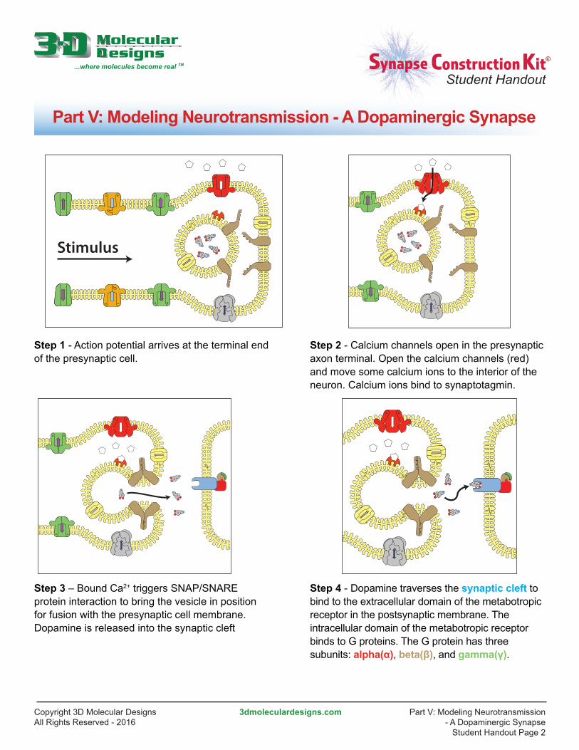
Step 1 - Action potential arrives at the terminal end of the presynaptic cell.
Step 3 – Bound Ca2+ triggers SNAP/SNARE protein interaction to bring the vesicle in position for fusion with the presynaptic cell membrane. Dopamine is released into the synaptic cleft
Step 4 - Dopamine traverses the synaptic cleft to bind to the extracellular domain of the metabotropic receptor in the postsynaptic membrane. The intracellular domain of the metabotropic receptor binds to G proteins. The G protein has three subunits: alpha(α), beta(β), and gamma(γ).
Step 2 - Calcium channels open in the presynaptic axon terminal. Open the calcium channels (red) and move some calcium ions to the interior of the neuron. Calcium ions bind to synaptotagmin.
Stimulus
Student Handout
©...where molecules become real TM
Part V: Modeling Neurotransmission - A Dopaminergic Synapse
Copyright 3D Molecular DesignsAll Rights Reserved - 2016
3dmoleculardesigns.com Part V: Modeling Neurotransmission- A Dopaminergic Synapse
Student Handout Page 3
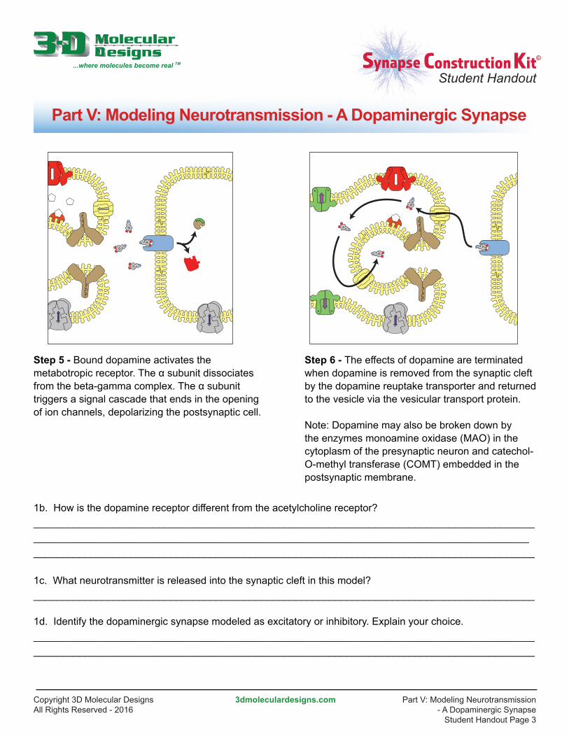
Step 5 - Bound dopamine activates the metabotropic receptor. The α subunit dissociates from the beta-gamma complex. The α subunit triggers a signal cascade that ends in the opening of ion channels, depolarizing the postsynaptic cell.
Step 6 - The effects of dopamine are terminated when dopamine is removed from the synaptic cleft by the dopamine reuptake transporter and returned to the vesicle via the vesicular transport protein.
Note: Dopamine may also be broken down by the enzymes monoamine oxidase (MAO) in the cytoplasm of the presynaptic neuron and catechol-O-methyl transferase (COMT) embedded in the postsynaptic membrane.
1b. How is the dopamine receptor different from the acetylcholine receptor?_______________________________________________________________________________________________________________________________________________________________________________________________________________________________________________________________________
1c. What neurotransmitter is released into the synaptic cleft in this model?________________________________________________________________________________________
1d. Identify the dopaminergic synapse modeled as excitatory or inhibitory. Explain your choice.________________________________________________________________________________________________________________________________________________________________________________
Student Handout
©...where molecules become real TM
Part V: Modeling Neurotransmission - A Dopaminergic Synapse

Copyright 3D Molecular DesignsAll Rights Reserved - 2016
3dmoleculardesigns.com Part V: Modeling Neurotransmission- A Dopaminergic Synapse
Student Handout Page 4
1e. Describe how an ionotropic receptor differs from a metabotropic receptor.________________________________________________________________________________________________________________________________________________________________________________________________________________________________________________________________________________________________________________________________________________________________________________________________________________________________________________________________________________________________________________________________________________
1f. Do ions move through this metabotropic receptor? ________________________________________________________________________________________
1g. Identify the subunits of the G-protein. Which of these triggers the opening of ion channel in the postsynaptic cell?________________________________________________________________________________________
1h. Propose a molecular mechanism for how the postsynaptic cell is depolarized after activation of the G protein-coupled receptor.________________________________________________________________________________________ ________________________________________________________________________________________ ________________________________________________________________________________________ ________________________________________________________________________________________
1i. How is termination of neuronal signaling different in this dopaminergic synapse model compared to the cholinergic synapse model?________________________________________________________________________________________ ________________________________________________________________________________________ ________________________________________________________________________________________________________________________________________________________________________________
Student Handout
©...where molecules become real TM
Part V: Modeling Neurotransmission - A Dopaminergic Synapse