PARIPEX - worldwidejournals.com · We have reviewed all orbital pseudotumor cases with intracranial...
3
ORIGINAL RESEARCH PAPER Neurosurgery RIGHT ORBITAL PSEUDOTUMOR WITH INTRACRANIAL EXTENSION- A CASE REPORT KEY WORDS: Introduction- pseudotumor is a non granulomatous inflammatory processin the orbit or eye in which a local or systemic cause cannot be found. It constitutes the third most common ophthalmic disorder after graves disease and lymphoproliferative disorders with 5-8% reported incidence. The intracranial extension of orbital pseudotumor is rare(8.8% in a computed tomography series) and usually involves the middle cranial fossa and the cavernous sinus through superior orbital fissure. Here we report a case of orbital pseudotumor with MRI suggestive of intracranial extension. Case report- a 60 yr old male patient presented with right eye proptosis since 1.5 years headache and giddiness since 10 days and left sided upper and lower limb weakness since 1 days. Patient has history of operated case of right orbital mass for which right orbitotomy and biopsy of orbital mass was done which showed tuberculosis 2 months back. Patient is on anti tuberculosis treatment since then. On examination patient is conscious and oriented to time, place and person. Pupils on right side was semidilated and non reacting to light and left pupils show normal size reacting to light with right eye mild proptosis with restricted eye ball movements in all direction. Vision in right eye was 6/18 and left eye was 6/12. Power in left upper limb and lower limb is 4. MRI findings show multiple acute infarcts in posterior limb of right internal capsule,right medial temporal lobe, right occipital lobe and right centrum semiovale. Ill defined heterogeneously enhancing altered signal intensity lesion involving intraconal as well as extraconal compartment of right retro orbital region causing compression over medial rectus and optic nerve medially with extension and involvement as described above. MRI angiography show complete occlusion in right internal carotid artery. No AV malformation or Aneurysm. Patient was operated by right orbito zygotomy with subtotal excision of right orbital mass in piecemeal. Intraoperatively mass was whitish in color,firm in consistency and encasing optic nerve. Mass was going intracranially through superior orbital fissure. Histopathology report showed idiopathic granulomatous orbital inflammation (inflammatory pseudotumor). Post operatively was uneventful. On 8 th day patient developed hemiplegia and was unconscious. Patient was intubated and was on ventilator. Intravenous dexamethasone and tablet ecosprin. Patient condition deterioted on POD 12 and developed right sided weakness. CT Brain was done which showed complete infarct in right cerebral hemisphere and brain stem infarct. Patient expired on pod 15. Discussion- Previously reported cases of orbital pseudotumor with intracranial extension Dr Abhishek Raj rd 3 Year Resident, Department of Neurosurgery, Smt. N.H.L Medical College, Ahmedabad Dr Bhushan Kharche* rd 3 Year Resident, Department of Neurosurgery, Smt. N.H.L Medical College, Ahmedabad *Corresponding Author Dr Hemant Sable nd 2 Year Resident, Department of Neurosurgery, Smt. N.H.L Medical College, Ahmedabad Dr Ankit Patni nd 2 Year Resident, Department of Neurosurgery, Smt. N.H.L Medical College, Ahmedabad Dr Mukesh Patel H.O.D, Department of Neurosurgery, Smt. N.H.L Medical College, Ahmedabad Dr Kalpesh Shah Professor, Department of Neurosurgery, Smt. N.H.L Medical College, Ahmedabad 66 www.worldwidejournals.com Year Author Patient age (years) Gender Intracranial location/features 1984 Kaye et al. 71 M ACF (planum sphenoidale), dural-based 1986 Frohman et al. 48 M SOF, optic canal, bone erosion 48 F MCF/CS, bone erosion 72 M SOF, bone erosion 1986 Noble et al. 46 F ACF, dural thickening Year Author Patient age (years) Gender Intracranial location/features 1992 Clifton et al. 49 M MCF/CS, pattern II 69 M SOF, pattern I 54 F MCF/CS, pattern II 36 M MCF, pattern I 86 M MCF/CS, dural thickening, bilateral involvement, pattern III 71 F MCF/CS, pattern II 30 M MCF/CS, dural thickening, pattern III 61 M MCF/CS, pattern II 1993 Bencherif et al. 23 M CS, SOF, left fronto-temporal dural thickening, sphenoid bone sclerosis 1993 Olmos et al. 64 F CS/Meckel cave, parasellar plaque, dural surface down to clivus and C2 body 1996 de Jesus et al. 16 F Optic canal, SOF, MCF; dural thickening of the left hemisphere and tentorium Year Author Patient age (years) Gender Intracranial location/features 1998 Soares et al. - - Pituitary fossa/CS, ICA compression 2000 Ayala et al. 83 F ACF extra-axial mass without bone involvement, (possibly through the anterior etmoid foramen) Volume-7 | Issue-3 | March-2018 | PRINT ISSN No 2250-1991 PARIPEX - INDIAN JOURNAL OF RESEARCH
Transcript of PARIPEX - worldwidejournals.com · We have reviewed all orbital pseudotumor cases with intracranial...

ORIGINAL RESEARCH PAPER Neurosurgery
RIGHT ORBITAL PSEUDOTUMOR WITH INTRACRANIAL EXTENSION- A CASE REPORT
KEY WORDS:
Introduction-pseudotumor is a non granulomatous inflammatory processin the orbit or eye in which a local or systemic cause cannot be found. It constitutes the third most common ophthalmic disorder after graves disease and lymphoproliferative disorders with 5-8% reported incidence. The intracranial extension of orbital pseudotumor is rare(8.8% in a computed tomography series) and usually involves the middle cranial fossa and the cavernous sinus through superior orbital fissure. Here we report a case of orbital pseudotumor with MRI suggestive of intracranial extension.
Case report- a 60 yr old male patient presented with right eye proptosis since 1.5 years headache and giddiness since 10 days and left sided upper and lower limb weakness since 1 days. Patient has history of operated case of right orbital mass for which right orbitotomy and biopsy of orbital mass was done which showed tuberculosis 2 months back. Patient is on anti tuberculosis treatment since then. On examination patient is conscious and oriented to time, place and person. Pupils on right side was semidilated and non reacting to light and left pupils show normal size reacting to light with right eye mild proptosis with restricted eye ball movements in all direction. Vision in right eye was 6/18 and left eye was 6/12. Power in left upper limb and lower limb is 4. MRI findings show multiple acute infarcts in posterior limb of right internal capsule,right
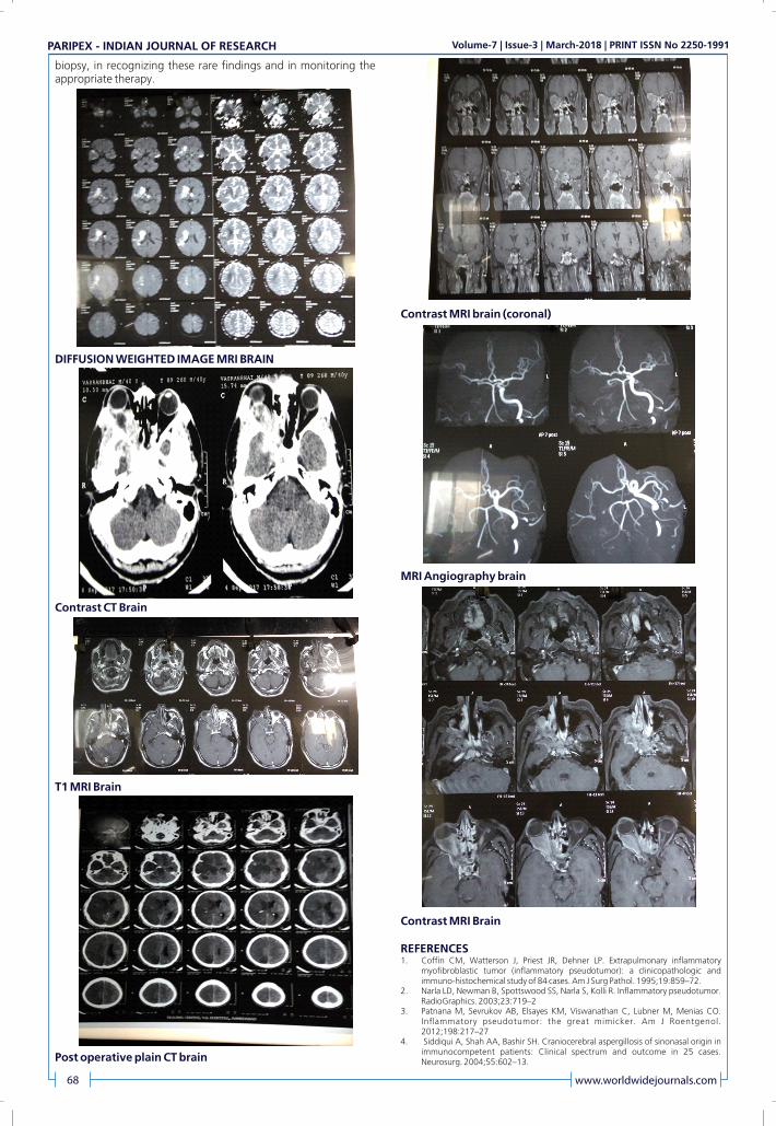
medial temporal lobe, right occipital lobe and right centrum semiovale. Ill defined heterogeneously enhancing altered signal intensity lesion involving intraconal as well as extraconal compartment of right retro orbital region causing compression over medial rectus and optic nerve medially with extension and involvement as described above. MRI angiography show complete occlusion in right internal carotid artery. No AV malformation or Aneurysm. Patient was operated by right orbito zygotomy with subtotal excision of right orbital mass in piecemeal. Intraoperatively mass was whitish in color,firm in consistency and encasing optic nerve. Mass was going intracranially through superior orbital fissure. Histopathology report showed idiopathic g ranu lomatous o rb i ta l inflammat ion ( inflammatory pseudotumor). Post operatively was uneventful. On 8 th day patient developed hemiplegia and was unconscious. Patient was intubated and was on ventilator. Intravenous dexamethasone and tablet ecosprin. Patient condition deterioted on POD 12 and developed right sided weakness. CT Brain was done which showed complete infarct in right cerebral hemisphere and brain stem infarct. Patient expired on pod 15.
Discussion-Previously reported cases of orbital pseudotumor with intracranial extension
Dr Abhishek Rajrd3 Year Resident, Department of Neurosurgery, Smt. N.H.L Medical College,
Ahmedabad
Dr Bhushan Kharche*
rd 3 Year Resident, Department of Neurosurgery, Smt. N.H.L Medical College, Ahmedabad *Corresponding Author
Dr Hemant Sablend 2 Year Resident, Department of Neurosurgery, Smt. N.H.L Medical College,
Ahmedabad
Dr Ankit Patnind 2 Year Resident, Department of Neurosurgery, Smt. N.H.L Medical College,
Ahmedabad
Dr Mukesh Patel H.O.D, Department of Neurosurgery, Smt. N.H.L Medical College, Ahmedabad
Dr Kalpesh Shah Professor, Department of Neurosurgery, Smt. N.H.L Medical College, Ahmedabad
66 www.worldwidejournals.com
Year Author Patient age (years) Gender Intracranial location/features
1984 Kaye et al. 71 M ACF (planum sphenoidale), dural-based
1986 Frohman et al. 48 M SOF, optic canal, bone erosion
48 F MCF/CS, bone erosion
72 M SOF, bone erosion
1986 Noble et al. 46 F ACF, dural thickening
Year Author Patient age (years) Gender Intracranial location/features
1992 Clifton et al. 49 M MCF/CS, pattern II
69 M SOF, pattern I
54 F MCF/CS, pattern II
36 M MCF, pattern I
86 M MCF/CS, dural thickening, bilateral involvement, pattern III
71 F MCF/CS, pattern II
30 M MCF/CS, dural thickening, pattern III
61 M MCF/CS, pattern II
1993 Bencherif et al. 23 M CS, SOF, left fronto-temporal dural thickening, sphenoid bone sclerosis
1993 Olmos et al. 64 F CS/Meckel cave, parasellar plaque, dural surface down to clivus and C2 body
1996 de Jesus et al. 16 F Optic canal, SOF, MCF; dural thickening of the left hemisphere and tentorium
Year Author Patient age (years) Gender Intracranial location/features
1998 Soares et al. - - Pituitary fossa/CS, ICA compression
2000 Ayala et al. 83 F ACF extra-axial mass without bone involvement, (possibly through the anterior etmoid foramen)
Volume-7 | Issue-3 | March-2018 | PRINT ISSN No 2250-1991PARIPEX - INDIAN JOURNAL OF RESEARCH
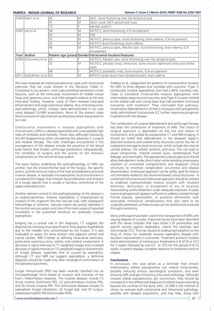
We have reviewed all orbital pseudotumor cases with intracranial extension that we could retrieve in the literature (Table 1). Compared to our patient, most cases exhibited somewhat similar features, such as the extra-axial involvement of middle cranial fossa and cavernous sinus, and mild vasogenic edema as the only intra-axial finding. However, none of them showed intra-axial enhancement and large subcortical edema, thus mimicking intra-axial pathology, which, instead, were demonstrated in our case using delayed CE-MRI acquisitions. None of the present cases show occlusion of right internal carotid artery which was present in our case.
Orbitocranial involvement in invasive aspergillosis with rhinosinusitis ( IARS) is a disease associated with unacceptably high rates of morbidity and mortality. These rates, although improving, are still disappointing when considering the advances in surgical and medical therapy. The main challenges encountered in the management of this disease include the presence of the blood brain barrier that hinders antifungal penetration intracerebrally, the morbidity of surgery and the gravity of the disease's complications on the central nervous system.
The exact factors underlying the pathophysiology of IARS are unclear, but the environmental load of the fungus, the specific strains, and the immune status of the host are believed to promote invasive disease. A favorable microaerophilic local environment is considered the trigger that initiates the proliferation and invasion of a fungal species that is usually a harmless commensal of the upper respiratory tract.
Another element central to the pathophysiology of this disease is its angioinvasiveness. Invasive aspergillosis is characterized by invasion of the organism into the vascular wall, with subsequent hemorrhage or ischemia. Vascular events are poorly tolerated in the central nervous system and one of the main causes of reported mortalities in the published literature on cerebrally invasive aspergillosis.
Imaging has a central role in the diagnosis; CT suggests the diagnosis by showing sinus opacification that appears hyperdense due to the metallic ions concentrated by the fungus. It is also invaluable to assess for bony erosion into adjacent orbital and cranial cavities. MRI is better at defining intracranial extension, particularly cavernous sinus, orbital, and cerebral involvement. A decrease in signal intensity on T1-weighted images and a marked decrease in signal intensity on T2-weighted images is characteristic of fungal disease, especially that of caused by aspergillus. Although CT and MRI can suggest aspergillosis, a definitive diagnosis should be made only after histological confirmation of the operative specimens.
Fungal rhinosinusitis (FRS) has been recently classified into six clinicopathologic forms based on invasion and character of the host's inflammatory response. The invasive diseases include (1) Acute invasive (fulminant) FRS, (2) granulomatous invasive FRS, and (3) chronic invasive FRS. The noninvasive diseases include (1) saprophytic fungal infestation, (2) fungal ball, and (3) fungus-related eosinophilic FRS that includes AFRS.
Siddiqui et al. categorized the patterns of craniocerebral invasion for IARS to three degrees that correlate with outcome: (Type 1) intradurally invasive aspergillosis that had a 66% mortality rate, (Type 2) extradural intracranially invasive aspergillosis with intermediate response and outcome, and (Type 3) invasion limited to the orbital wall and cranial base that had excellent functional outcomes with treatment. They concluded that judicious, conservative debridement is all that is necessary with preoperative orally administered itraconazole ICZ further improving prognosis in patients with this disease.
The combination of surgical debridement and antifungal therapy has been the cornerstone of treatment of IARS. The choice of surgical approach is dependent on the site and extent of involvement, and guided by preoperative CT and MRI imaging. It should be noted that debridement in the diseased region's distorted anatomy and eroded bony structures carries the risk of inadvertent damage to local structures, which include the internal carotid arteries, the orbital contents, and dura. This can lead to visual compromise, internal carotid injury, cerebrospinal fluid leakage, and meningitis. The appropriate surgical approach should allow debridement under direct vision while avoiding unnecessary violation of uninvolved compartments and displaced critical structures to minimize complication occurrence and disease dissemination. Endonasal approach can be safely used for lesions not intimately related to the aforementioned critical structures . A combined transcranial and endonasal approach should be utilized to eradicate craniocerebrally invasive sinusitis with resultant distortion, destruction, or encasement of any of structure necessitating careful dissection under adequate exposure. A solely transcranial approach appears sufficient to address isolated fungal frontal disease, sphenoiditis, or posterior ethmoiditis with associated intracranial complications that also need to be surgically addressed, as these sinuses can be satisfactorily accessed through craniotomy.
Many antifungals have been used in the management of IARS with varying degrees of success. Improved results have been described with the newer triazoles that have better CSF penetration and specific activity against Aspergillus, mainly the relatively new voriconazole VCZ. This has caused its widespread adoption as the drug of choice for cerebrally invasive aspergillus disease with resultant improvement in outcomes. Treatment protocol involves initial administration of intravenous Amphotericin B AT-B or VCZ for 2 weeks followed by oral ICZ or VCZ for the period of 8-12 weeks if patient displays complete response to treatment and no relapse.
ConclusionsIn conclusion, this case serves as a reminder that chronic inflammatory orbital pseudotumor can extend intracranially, possibly inducing serious neurological symptoms, and even showing MRI changes mimicking intra-axial pathology. Although invasive orbital pseudotumors are uncommon, they should be considered in the differential diagnosis of orbital masses extending beyond the confines of the bony orbit. CE-MRI is the method of choice to evaluate both extracranial and intracranial pathology, possibly with delayed acquisitions, and may help, along with
www.worldwidejournals.com 67
2004 Mahr et al. 40 M MCF, dural thickening over the temporal pole
41 M Optic canal, MCF paraclinoid mass
73 F Meckel cave/CS
2005 Lee et al. 58 M MCF/CS, dural thickening; ICA encasement
63 M SOF
55 M MCF/CS, petrous apex, dural thickening; brain edema; ICA encasement
32 M MCF/CS dural thickening; brain edema
46 M MCF/CS, petrous apex, Meckel cave, dural thickening; brain edema; ICA encasement
Year Author Patient age (years) Gender Intracranial location/features
2006 Zborowska et al. 45 F MCF/CS, Meckel cave, dural thickening over the temporal pole
32 M MCF/CS, pituitary fossa, tentorium; bone erosion (sphenoid wing and orbital roof)
48 F MCF/CS; parasellar mass, bone erosion, ICA encasement
2011 Saifudheen et al. 50 M MCF/CS large dural mass (temporal pole); brain edema
Volume-7 | Issue-3 | March-2018 | PRINT ISSN No 2250-1991PARIPEX - INDIAN JOURNAL OF RESEARCH
biopsy, in recognizing these rare findings and in monitoring the appropriate therapy.
DIFFUSION WEIGHTED IMAGE MRI BRAIN
Contrast CT Brain
T1 MRI Brain
Post operative plain CT brain
Contrast MRI brain (coronal)
MRI Angiography brain
Contrast MRI Brain
REFERENCES1. Coffin CM, Watterson J, Priest JR, Dehner LP. Extrapulmonary inflammatory
myofibroblastic tumor (inflammatory pseudotumor): a clinicopathologic and immuno-histochemical study of 84 cases. Am J Surg Pathol. 1995;19:859�72.
2. Narla LD, Newman B, Spottswood SS, Narla S, Kolli R. Inflammatory pseudotumor. RadioGraphics. 2003;23:719�2
3. Patnana M, Sevrukov AB, Elsayes KM, Viswanathan C, Lubner M, Menias CO. Inflammatory pseudotumor: the great mimicker. Am J Roentgenol. 2012;198:217�27
4. Siddiqui A, Shah AA, Bashir SH. Craniocerebral aspergillosis of sinonasal origin in immunocompetent patients: Clinical spectrum and outcome in 25 cases. Neurosurg. 2004;55:602�13.
68 www.worldwidejournals.com
Volume-7 | Issue-3 | March-2018 | PRINT ISSN No 2250-1991PARIPEX - INDIAN JOURNAL OF RESEARCH


![PARIPEX - worldwidejournals.com fileage of 50 to 62 years [8] Lipoma is a rare benign tumor that usually presents as an asymptomatic, slowly growing mass with a firm or soft consistency](https://static.fdocuments.in/doc/165x107/5d5e21c888c9932d118bc7da/paripex-of-50-to-62-years-8-lipoma-is-a-rare-benign-tumor-that-usually-presents.jpg)