Neonatal parenteral nutrition (PDF) | Neonatal parenteral ...

PARENTERAL FLUID
REPLACEMENT THERAPY Mgr. Martina Lepiešová, PhD.
Institute of Nursing JF MED CU in Martin
Basics of Nursing, May 2012
Purpose
to discuss factors influencing the administration of i.v. therapy
selection of proper venous access (device)
proper intravenous route of i.v. therapy
advantages / disadvantages of different routes / venipuncture
sites (peripheral/central)
risk of complications of i.v. therapy, etc.
to identify common types of vascular access devices and
describe their care and maintenance

Parenteral fluid replacement therapy
fluids infused directly into circulating blood volume to
supplement / replace body fluids
intravenous fluid and electrolyte therapy
blood therapy
partial parenteral nutrition (PPN) – combined with oral /
enteral nutrition (nutritional support <14 days), applied as
peripheral parenteral nutrition
total parenteral nutrition (TPN) – i.v. hyperalimentation – all
nutrients dilevered by parenteral routes (nutritional support
≥14 days), applied as central parenteral nutrition

Responsibilities – NURSE
preparation of medications to be administered i.v.
assistance to the physician within the application (legal norms
of the country)
insertion of peripheral venous catheters
assistance to the physician performing minor surgical
procedures (insertion of central venous or arterial lines)
maintenance of all the lines
prevention of complications – intravascular therapy
monitoring patient’s responses

Responsibilities – PHYSICIAN
order of the medications / infusion solutions to be
administered i.v.
insertion and removal of central lines – central venous
catheter (CVC), PICC, implantable venous port, arterial lines
application of all medications / infusion solutions intravenously
(in the conditions of the SR)

Indications
– i.v. administration of medications:
rapid effect of medication (onset)
if other parenteral route:
too risky for the patient
medication too irritating to tissues
discomfort

Indications
– parenteral fluid replacement / nutrition:
patient cannot absorb nutrients through GI tract / unable to
tolerate oral / enteral nutrition (diseases, trauma, surgery –
preOP bowel rest, postOP)
excessive nitrogen loss (wound infection, fistulas, abscesses)
increased energy needs (burns, sepsis, trauma)
nutritional deficits (nutrients - carbohydrates, fats, proteins,
vitamins, minerals – electrolytes and trace elements)
growth and development retardation

Contraindications
- TPN:
patients with normally functioning GI tract
well-nourished patients whose GI tract will normally function
within 10 days
patients with a poor prognosis (palliative care)

QUESTION/TASK
Try to describe 3 different routes of
intravenous application of medications...

1.) i.v. push method / i.v. bolus – “directly from hand”
concentrated dose of a drug directly into the systemic
circulation (vein / existing i.v. line)
medication usually diluted (20 ml), sterile normal saline as
diluents (0,9% NaCl; F 1/1)
advantages / (dis)advantages
2.) syringe pump / mini-infuser (intermittent application)
3.) i.v. infusion (intermittent / continual infusion by electronic
infusion pump) – i.e. adding medications into small-volume /
large-volume i.v. fluids/containers

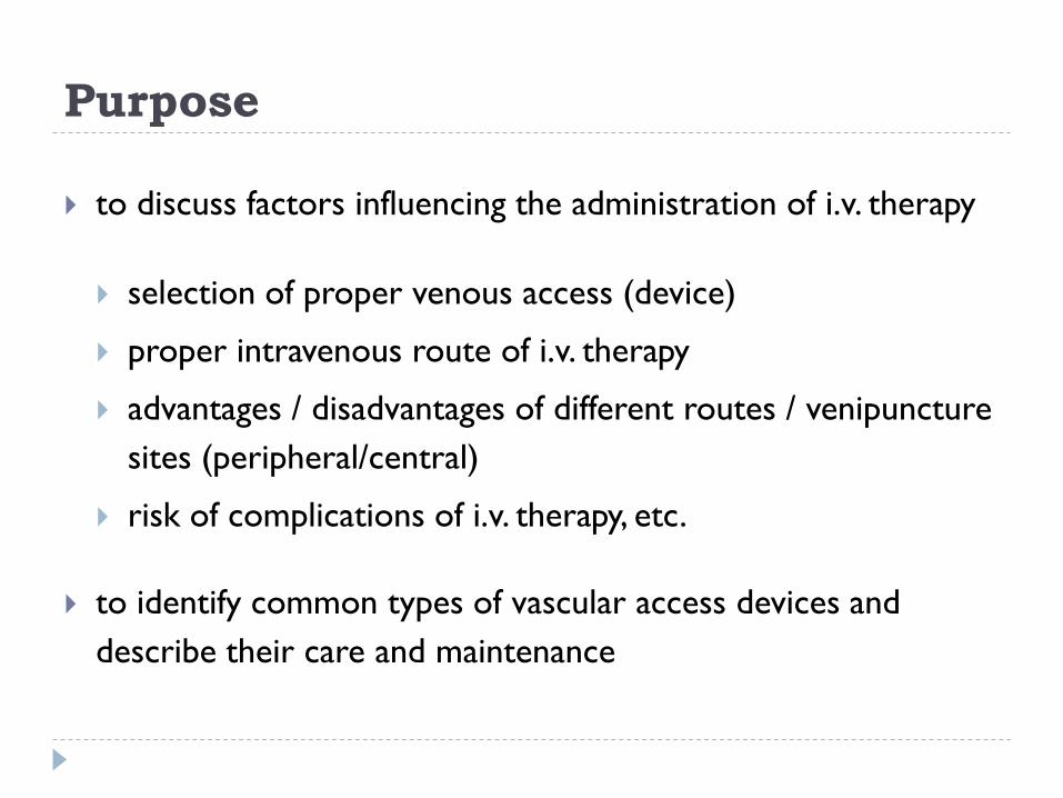
I.v. infusion solutions – 3 regimens
multi-bottle system (commonly used – glass/plastic bottles,
bags)
all-in-one system (prepared in institutions) – water,
electrolytes, glucose, lipids, amino acids, vitamins, trace
elements
multi-compartment system (commercially produced, 2-3
or more compartments) – hypertonic applied only by
central application and continually
advantages: reduction of mistakes (content / compatibility of
solutions), improvement of asepsis, time management
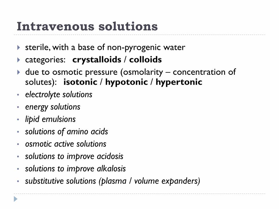
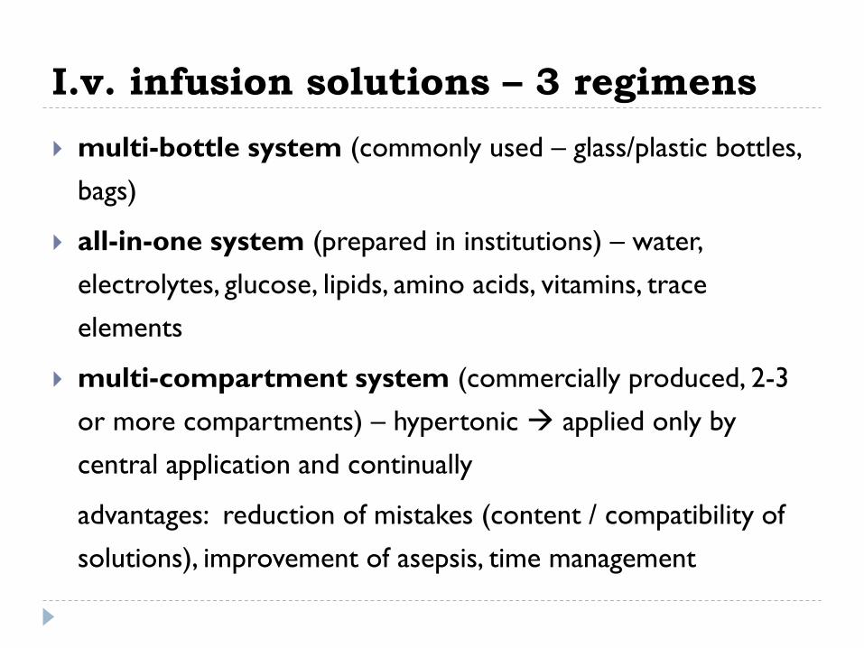
Intravenous solutions
sterile, with a base of non-pyrogenic water
categories: crystalloids / colloids
due to osmotic pressure (osmolarity – concentration of solutes): isotonic / hypotonic / hypertonic
• electrolyte solutions
• energy solutions
• lipid emulsions
• solutions of amino acids
• osmotic active solutions
• solutions to improve acidosis
• solutions to improve alkalosis
• substitutive solutions (plasma / volume expanders)
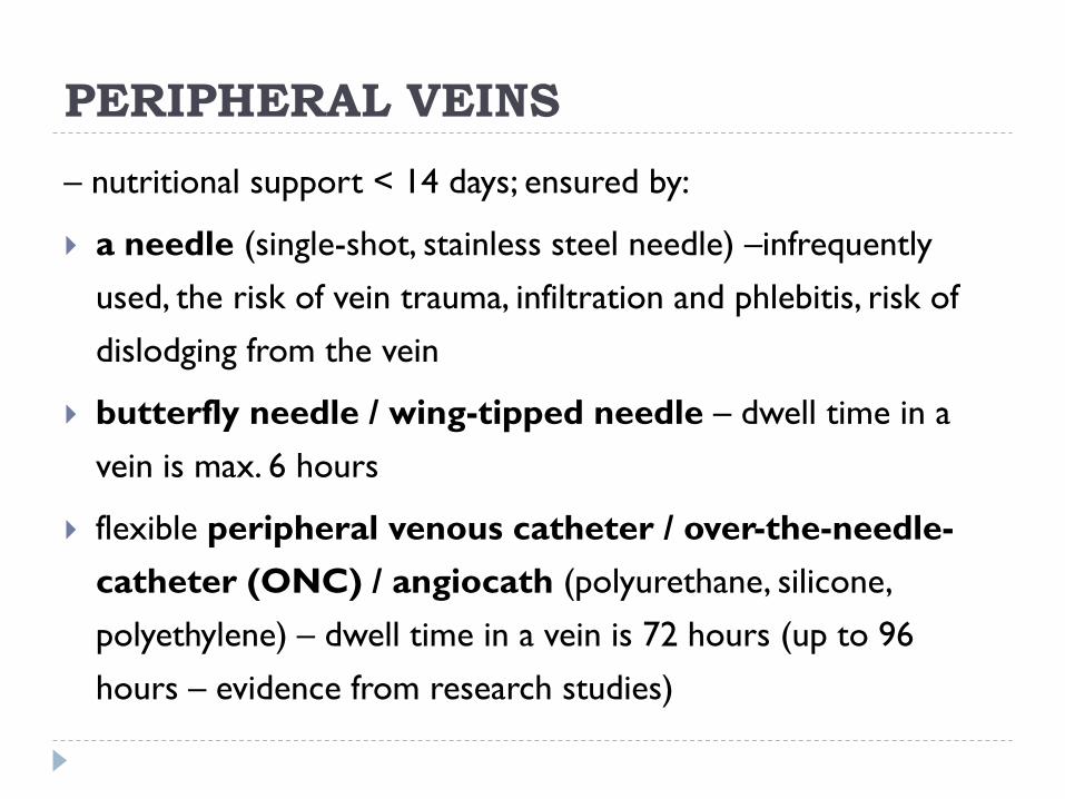
PERIPHERAL VEINS
– nutritional support < 14 days; ensured by:
a needle (single-shot, stainless steel needle) –infrequently
used, the risk of vein trauma, infiltration and phlebitis, risk of
dislodging from the vein
butterfly needle / wing-tipped needle – dwell time in a
vein is max. 6 hours
flexible peripheral venous catheter / over-the-needle-
catheter (ONC) / angiocath (polyurethane, silicone,
polyethylene) – dwell time in a vein is 72 hours (up to 96
hours – evidence from research studies)


Peripheral i.v. application
advantages: simple, quite safe method (peripheral
vascular devices insertion)
venipuncture sites (blood draw, bolus injection, insertion
sites for peripheral i.v. lines):
upper extremities – cubital space, antecubital fossa (basilic
vein, cephalic vein, median cubital vein, median antebrachial
vein, accessory cephalic vein), dorsal area of hand (dorsal
venous arch, superficial dorsal veins)
exceptionally lower extremities
frontal and temporal superficial veins in infants

Peripheral i.v. application
disadvantages:
short-time application (inflammation, phlebitis)
only low concentrated isotonic solutions – i.e. amino acids up
to 10%, amino acids mixed with dextrose up to 5%, dextrose
only up to 10% (hypertonic solutions too concentrated,
causing inflammation, phlebitis, thrombophlebitis, necrosis of
small-diameter peripheral veins)
risk of circulatory overload (large volume at once)
contraindicated in patients with site burns, sclerotic veins,
arteriovenous fistula, postmastectomy arm (the risk of
lymphoedema), oedematous or impaired arm or hand
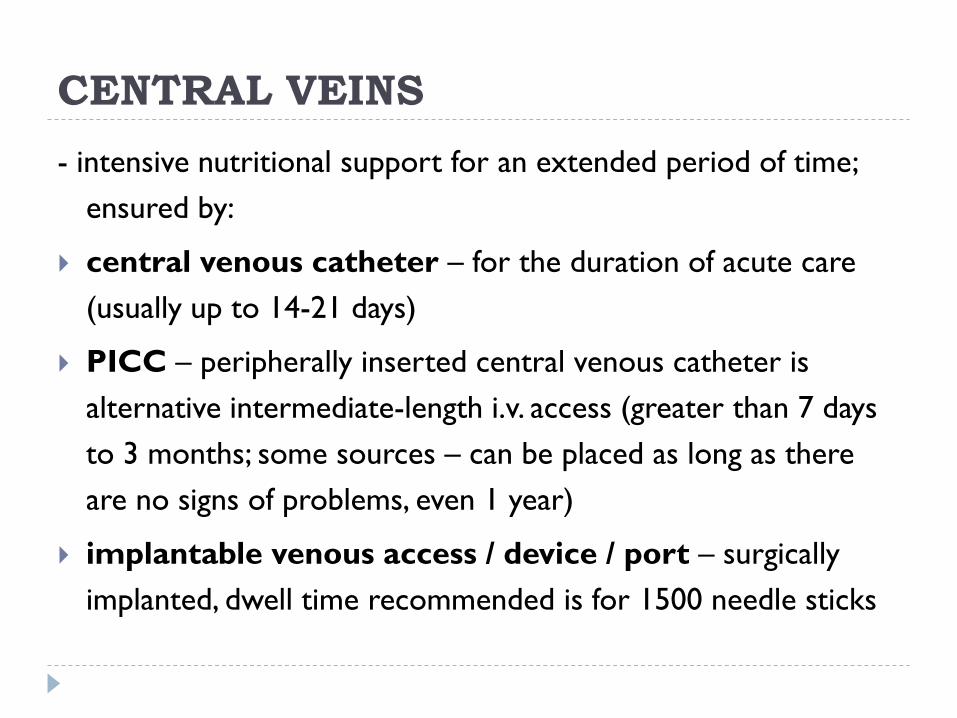
CENTRAL VEINS
- intensive nutritional support for an extended period of time;
ensured by:
central venous catheter – for the duration of acute care
(usually up to 14-21 days)
PICC – peripherally inserted central venous catheter is
alternative intermediate-length i.v. access (greater than 7 days
to 3 months; some sources – can be placed as long as there
are no signs of problems, even 1 year)
implantable venous access / device / port – surgically
implanted, dwell time recommended is for 1500 needle sticks


Central i.v. application
advantages:
long-time application (≥14 days)
no risk of circulatory overload (continual application)
possibility to apply concentrated and hyperosmolar solutions
no risk of phlebitis, thrombophlebitis, sclerosis of vein tissue
(high-flow and large-diameter central veins)
possibility to measure central venous pressure (CVP)
possibility to aspirate blood samples from it

Central i.v. application
venipuncture sites (puncture or surgically inserted)
subclavian or jugular veins (distal tip of catheter in superior
vena cava)
PICC – inserted into basilic or cephalic veins just above or
below the antecubital space of the right arm and threaded into
subclavian vein or superior vena cava
disadvantages:
complicated way of ensuring access
need to use full sterile-barrier precautions during central
venous device insertion (strict aseptic technique – surgical
sepsis, sterile field preparation)
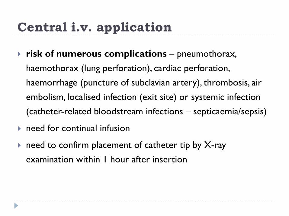
Central i.v. application
risk of numerous complications – pneumothorax,
haemothorax (lung perforation), cardiac perforation,
haemorrhage (puncture of subclavian artery), thrombosis, air
embolism, localised infection (exit site) or systemic infection
(catheter-related bloodstream infections – septicaemia/sepsis)
need for continual infusion
need to confirm placement of catheter tip by X-ray
examination within 1 hour after insertion
QUESTION/TASK
Describe everyday care of i.v. lines to decrease
the risk of intravascular infection...

Initiating i.v. therapy – assessment Review physician’s orders concerning type and amount of i.v.
injection/infusion (medication – dose, frequency, fluid/solution, rate of
administration).
Assess for clinical factors/conditions that will respond to or be
affected by i.v. fluid administration:
patient’s fluid volume or nutritional status – dry skin and mucous
membranes, inelastic skin turgor (failing to return to normal position
within 3 seconds after pinching), blood pressure changes, irregular pulse
rhythm, increased pulse rate, decreased capillary refill, distended neck
veins, peripheral oedema, body weight, behavioural changes – e.g.
confusion, restlessness, auscultation of crackles or rhonchi in lungs (as
for signs of deficient fluid volume / excess fluid volume); signs of
nutrient deficiency (e.g. thinning hair, hair loosing, poor muscle tone,
etc.), laboratory values
Cont. related factors of fluid volume or nutritional changes – e.g. thirst,
anorexia, nausea, vomiting, increased or decreased urine output,
excessive (profuse) sweating, excessive wound drainage (evaluate fluid
intake and output record), etc.
Assess for the following risk factors: child or older adult, presence of
heart failure or renal failure, skin lesions, infection, low platelet count,
anticoagulant therapy.
Determine if patient is to undergo any planned surgeries or blood
transfusion.
Assess the status of patient’s venous system – accessibility of
veins, the venipuncture sites (e.g. the presence of site burns,
oedematous or impaired arm or hand, sclerotic veins, presence of
casts, bandages or binders), contraindications of venipunctures (e.g.
arteriovenous fistula, postmastectomy arm), etc.
Cont.
Assess existing i.v. insertion sites for signs of infiltration or
phlebitis.
Assess patient’s history of allergies (to medication ordered,
antiseptic solutions, tapes or dressing materials), assess the risk
factors predisposing for latex allergy reaction – modify the selection
of supplies to use the correct material.
Assess patient’s previous or perceived experience with i.v.
therapy and arm placement preferences.
Assess patient’s comfort level or pain (on numeric scale of 0 to
10), symptoms of anxiety, elimination needs before preparing the
procedure.
Confirm the sterility of all packages and sterile supplies.

Procedures
preparing the medication from ampoule or vial
(assistance in) administering medications by i.v. bolus
performed by single-shot needle
preparing i.v. infusion (i.v. fluid container) – adding
medication/additive into i.v. fluid container
peripheral i.v. line insertion (venipuncture with butterfly
needle /over-the-needle-catheter ONC)
starting i.v. infusion
Complications
adverse drug reaction to medication
flow rate is incorrect – patient receives too little or too much
fluid
medication / infusion doesn’t infuse over desired period
sudden infusion of large volume of solution (fluid overload)
fluid volume excess (crackles in the lungs, shortness of breath,
oedema, jugular venous distension)
fluid volume deficit (decreased urine output, dry mucous
membranes, decreased capillary refill, a disparity in central and
peripheral pulses, tachycardia, hypotension, shock)

Cont.
electrolyte imbalance (abnormal serum electrolyte levels,
mental status changes, alterations in neuromuscular function,
cardiac arrhythmias, vital signs changes)
bleeding at venipuncture site (usually slow, continuous)
infiltration at site (swelling, possible pitting oedema, pallor,
coolness, pain, possible decrease in flow rate)
phlebitis at site (pain, increased skin temperature, erythema
along the path of vein)
loss of i.v. line patency (decreased or absent flow of i.v. fluid)
accidental removal of i.v. line
catheter tip is missing upon withdrawal

Expected patient outcomes
i.v. line is patent, intact, correctly placed
fluids and medications are infused without difficulties
no air bubbles are present in the syringe or in i.v. tube or
infusion set/tube
i.v. line site remains clear, free of any signs of inflammation, free
of infiltration
systemic signs of infection are absent (fever, malaise, increased
white blood cell count)
desired effect of medications is achieved without adverse
reactions occurring

Cont.
fluid and electrolyte balance returns to normal
patient will maintain stabilized fluid volume
patient will maintain or improve nutritional status (will meet
daily intake of required nutrients)
patient understands purpose and risks of i.v. therapy

BLOOD THERAPY

Special precautions
prepare documentation (transfusion book, patient’s record)
transfusion must always be warmed up (in the room temperature
/ warm bath) – don’t apply it cold / frozen
transfusion set (not infusion one) should be applied as closely to
transfusion as is possible and changed with every new unit
complete pre-transfusion tests must always be performed
transfusion has to be applied till 4 hours after being removed
from refrigerator (till 2 hours – erythrocyte concentrate), has to
be completed within 4 hours of initiation
empty bag with transfusion set should be stored in a refrigerator
for 24 hours
signs of adverse reactions to blood transfusion have to be
monitored

Pre-transfusion tests
blood pressure, pulse, temperature measurement (the same
after the transfusion)
chemical investigation of urine sample (the same after the
transfusion)
big cross-matching – imunnohaematological test in laboratory
(sending 5ml of native venous blood of the patient) – to
prepare compatible transfusion unit for the patient (as for
blood group and Rh system)
small cross-matching – “sangvitest” in the ward (the blood of
donor from transfusion bag, the blood of patient + diagnostic
serums)
biological test at the patient’s bedside

Adverse reactions to transfusion
haemolytic
febrile (pyretic)
allergic
circulatory overload
sepsis (bacterial – toxic)

References
BERMAN, A. - SNYDER, S. J. - KOZIER, B. - ERB, G. 2008. Kozier and Erb´s Fundamentals of
Nursing. Concepts, Process and Practice. 8th ed., New Jersey : Person Education, 2008. 1631 p.
ISBN 0-13-171468-6.
CRAVEN, R.F. - HIRNLE, C.J. 1992. Fundamentals of Nursing. Human Health and Function.
Philadelphia : J.B. Lippincott Company, 1992. 1522 p. ISBN 0-397-54669-6.
KOWALAK, J. P. 2008. Lippincott´s Nursing Procedures. 5th ed., Philadelphia : Lippincott Williams
and Wilkins, 2008. 949 pp. ISBN 13: 978-0-7817-8689-8, ISBN 10: 0-7817-8689-4.
KOZIER, B. - ERB, G. - BERMAN, A. - SNYDER, S. 2003. Kozier and Erb´s techniques in clinical
nursing. Basic to intermediate skills. 5th ed., Saddle River. N.J. : Prentice Hall, 2003. 752 p. ISBN 13:
978-0-13-114229-9.
KOZIER, B. - ERB, G. - BERMAN, A. - SNYDER, S. 2004. Fundamentals of Nursing. Concepts,
Process and Practice. 7th edition, New Jersey : Pearson Education, 2004. 1518 p. ISBN 0-13-
122878-1.
KUHN TIMBI, B. 2009. Fundamentals Nursing Skills and Concept. 9th ed., Philadelphia: Lippincott
Williams and Wilkins, 2009. ISBN 978-0-7817-7909-8.
McENTYRE, R.L. 1989. Practical Guide to the Care of the Surgical Patient. 3rd ed., St. Louis : The
C.V. Mosby Company, 1989. 348 p. ISBN 0-8016-3330-3.
Nursing procedures and protocols. 2003. 1st ed., Lippincott Williams & Wilkins. 2003, 672 p.
ISBN-13: 978-1582552378.
PERRY, A.G. - POTTER, P. A. 2004. Clinical Nursing Skills and Techniques. 6th ed., St. Louis : Mosby
Inc., 2006. 1611 p. ISBN-13: 978-0-323-02839-4, ISBN-10: 0-323-02839-X.
RICE, R. 1995. Handbook of Home Health Nursing Procedures. 1st ed., St. Louis : Mosby–Year
Book, Inc., 1995. 396 p. ISBN 0-8016-6946-4.
ZIMANOVÁ, Ľ. 2007. Infúzioterapia a transfúzioterapia. In Osacká, P. a kol. Techniky a postupy v
ošetrovateľstve. [CD-ROM]. 1.vyd. Martin : Ústav ošetrovateľstva, JLF UK, 2007. 505s. ISBN 978-
80-88866-48-0.