paravenous - British Journal of Ophthalmologybjo.bmj.com/content/bjophthalmol/76/4/250.full.pdf ·...
2
Bnitish Journal ofOphthalmology, 1992,76,250-251 Bilateral macular coloboma and pigmented paravenous retinochoroidal atrophy Muh-Shy Chen, Chang-Hao Yang, Jen-Shang Huang Department of Ophthalmology, College of Medicine, National Taiwan University, Taipei, Taiwan, ROC M-S Chen C-H Yang J-S Huang Correspondence to: Dr Muh-Shy Chen, Department of Ophthalmology, College of Medicine, National Taiwan University, Taipei, Taiwan, ROC. Accepted for publication 27 September 1991 Abstract A patient had bilateral macular coloboma with aggregations of pigment clumps located peri- vascularly, predominantly paravenously, and in other parts of the retina. The Toxoplasma IgG antibody was negative. The diagnosis of bilateral macular coloboma with pigmented paravenous retinochoroidal atrophy was made and seemed to be a developmental abnormality in origin. Figure I Retinal photograph of rightfundus. Figure 2 Retinal photograph of left fundus. Macular coloboma is thought to result from intrauterine inflammation or as an abnormality of development. Most cases are now recognised as being due to an intrauterine infection with Toxoplasma gondii. A developmental abnormality seems to be the cause in those patients with a hereditary or family origin,'2 and those with other ocular or systemic abnormalities.'-' We report a case of bilateral macular coloboma associated with pigmented paravenous retino- choroidal atrophy and negative Toxoplasma IgG antibody in which the macular lesions seem to be of a developmental origin. To our knowledge the appearance of these two congenital anomalies has not been previously reported in the English literature. Case report A 23-year-old woman presented with a history of progressive blurring of vision in both eyes for a duration of 8 years. There was no contributory medical or family history. Examination of the patient revealed the best corrected visual acuity as 20/400 in both eyes. The anterior segments were normal. Ophthal- moscopy showed a bilateral excavated, non-pig- mented macular coloboma about 4 x 4 disc diameter in size with ectatic sclera at the base. A few large choroidal vessels were visible at the base and the retinal vessels crossing the defect were attenuated. The optic discs were normal. Aggregations of pigment clumps were located perivascularly, mainly paravenously, and in some other parts of the retina. Zones of peripa- pillary and radial patches of chorioretinal atrophy were situated behind the pigmented muffs and mainly along the vessels (Figs 1 and 2). Fluore- scein angiography showed hypofluorescence with filling of the choroidal vasculature in both coloboma areas with a rim of hyperfluorescence surrounding the margin in the later phase of the examination. There were also blocked fluore- scence from the pigment clumps and the pig- ment epithelial window defect due to pigment epithelial atrophy (Figs 3 and 4). The Farns- worth-Munsell 100-hue test showed a mild colour vision defect. Goldmann perimetry examination revealed central scotoma with constricted peri- pheral fields. Electroretinography showed a reduction in b-wave amplitude. Dark adaptation curves showed monophasic and elevated final rod thresholds in both eyes. Ultrasonography showed a bilateral ectatic base with an axial length of 21 8 mm in the right eye and 22-0 mm in the left eye. A systemic evaluation, including skull and chest x rays, complete blood count, erythrocyte sedimentation rate, antinuclear antibody, 250 on 9 June 2018 by guest. Protected by copyright. http://bjo.bmj.com/ Br J Ophthalmol: first published as 10.1136/bjo.76.4.250 on 1 April 1992. Downloaded from
Transcript of paravenous - British Journal of Ophthalmologybjo.bmj.com/content/bjophthalmol/76/4/250.full.pdf ·...

BnitishJournal ofOphthalmology, 1992,76,250-251
Bilateral macular coloboma and pigmentedparavenous retinochoroidal atrophy
Muh-Shy Chen, Chang-Hao Yang, Jen-Shang Huang
Department ofOphthalmology, Collegeof Medicine, NationalTaiwan University,Taipei, Taiwan, ROCM-S ChenC-H YangJ-S HuangCorrespondence to:Dr Muh-Shy Chen,Department ofOphthalmology, College ofMedicine, National TaiwanUniversity, Taipei, Taiwan,ROC.Accepted for publication27 September 1991
AbstractA patient had bilateral macular coloboma withaggregations of pigment clumps located peri-vascularly, predominantly paravenously, andin other parts of the retina. The ToxoplasmaIgG antibody was negative. The diagnosis ofbilateral macular coloboma with pigmentedparavenous retinochoroidal atrophy was madeand seemed to be a developmental abnormalityin origin.
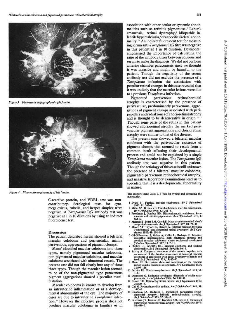
Figure I Retinal photograph ofrightfundus.
Figure 2 Retinal photograph ofleft fundus.
Macular coloboma is thought to result fromintrauterine inflammation or as an abnormalityof development. Most cases are now recognisedas being due to an intrauterine infection withToxoplasma gondii. A developmental abnormalityseems to be the cause in those patients with ahereditary or family origin,'2 and those withother ocular or systemic abnormalities.'-'We report a case of bilateral macular coloboma
associated with pigmented paravenous retino-choroidal atrophy and negative Toxoplasma IgGantibody in which the macular lesions seem to beof a developmental origin. To our knowledge theappearance of these two congenital anomalies hasnot been previously reported in the Englishliterature.
Case reportA 23-year-old woman presented with a history ofprogressive blurring of vision in both eyes for aduration of 8 years. There was no contributorymedical or family history.
Examination of the patient revealed the bestcorrected visual acuity as 20/400 in both eyes.The anterior segments were normal. Ophthal-moscopy showed a bilateral excavated, non-pig-mented macular coloboma about 4x 4 discdiameter in size with ectatic sclera at the base. Afew large choroidal vessels were visible at thebase and the retinal vessels crossing the defectwere attenuated. The optic discs were normal.Aggregations of pigment clumps were locatedperivascularly, mainly paravenously, and insome other parts of the retina. Zones of peripa-pillary and radial patches ofchorioretinal atrophywere situated behind the pigmented muffs andmainly along the vessels (Figs 1 and 2). Fluore-scein angiography showed hypofluorescencewith filling of the choroidal vasculature in bothcoloboma areas with a rim of hyperfluorescencesurrounding the margin in the later phase of theexamination. There were also blocked fluore-scence from the pigment clumps and the pig-ment epithelial window defect due to pigmentepithelial atrophy (Figs 3 and 4). The Farns-worth-Munsell 100-hue test showed a mild colourvision defect. Goldmann perimetry examinationrevealed central scotoma with constricted peri-pheral fields. Electroretinography showed areduction in b-wave amplitude. Dark adaptationcurves showed monophasic and elevated finalrod thresholds in both eyes. Ultrasonographyshowed a bilateral ectatic base with an axiallength of 21 8 mm in the right eye and 22-0 mmin the left eye.A systemic evaluation, including skull and
chest x rays, complete blood count, erythrocytesedimentation rate, antinuclear antibody,
250
on 9 June 2018 by guest. Protected by copyright.
http://bjo.bmj.com
/B
r J Ophthalm
ol: first published as 10.1136/bjo.76.4.250 on 1 April 1992. D
ownloaded from
Bilateral macular coloboma andpigmentedparavenous retinochoroidal atrophy
Figure 3 Fluorescein angiography ofrightfundus.
Figure 4 Fluorescein angiography ofleftfundus.
C-reactive protein, and VDRL test was non-
contributory. Serological tests for cyto-megalovirus, rubella, and herpes simplex werenegative. A Toxoplasma IgG antibody test was
negative at 1 in 10 dilutions by using an indirectfluorescence test.
DiscussionThe patient described herein showed a bilateralmacular coloboma and perivascular, mainlyparavenous, aggregations of pigment clumps.Mann9 classified macular coloboma into three
types, namely pigmented macular coloboma,non-pigmented macular coloboma, and macularcoloboma associated with abnormal vessels. Thepresent case did not fall clearly into any of thesethree types. Though the macular lesion seemedto be of the non-pigmented type paravenouspigment aggregations showed a peculiar mani-festation.Macular coloboma is known to develop from
an intrauterine inflammation or as a develop-mental abnormality of the eye. The majority ofcases are due to intrauterine Toxoplasma infec-tion.'0 However the infective process does notproduce macular coloboma in families or in
association with other ocular or systemic abnor-malities such as retinitis pigmentosa,3 Leber'samaurosis,4 retinal dystrophy,5 idiopathic in-fantile hypercalciuria,6or a specific skeletal abnor-mality.78 An indirect fluorescent test for measur-ing serum anti-Toxoplasma IgG titre was negativein this patient at 1 in 10 dilution. Desmonts"emphasised the importance of calculating theratio of the antibody titres between aqueous andserum to make the diagnosis. We did not performanterior chamber paracentesis since we thoughtit was invasive and might be harmful to thepatient. Though the negativity of the serumantibody test did not exclude the presence of aToxoplasma infection the association withpeculiar retinal changes in this case revealed thatit was unlikely that the macular lesions were dueto a previous Toxoplasma infection.Pigmented paravenous retinochoroidal
atrophy is characterised by the presence ofperivascular, predominantly paravenous, aggre-gations of pigment clumps associated with peri-papillary and radial zones ofchorioretinal atrophyand is thought to be degenerative in origin. 12-15Though some parts of the retina in this patientshowed chorioretinal atrophy the marked peri-vascular pigment aggregations and chorioretinalatrophy were similar to that of the disease.The present case showed a bilateral macular
coloboma with the perivascular existence ofpigment clumps that seemed to result from acommon insult affecting their developmentalprocess and could not be explained by a singleToxoplasma macular lesion. The Toxoplasma IgGantibody test was negative in this patient.Though the aetiology ofthis case is still unknownthe presence of a bilateral macular coloboma,pigmented paravenous retinochoroidal atrophy,and negative laboratory examinations lead us tospeculate that it is a developmental abnormalityin nature.
The authors thank Miss L S Yen for typing and preparing themanuscript.
1 Evans PJ. Familial macular colobomata. Br J Ophthalmol1937; 21: 503-6.
2 Miller SA, Bresnick G. Familial bilateral macular colobomata.BrJ Ophthalmol 1978; 62: 261-4.
3 Freedman J, Gombos GM. Bilateral macular coloboma, kera-toconus and retinitis pigmentosa. Ann Ophthalmol 1971; 3:664-6.
4 Margolis S, Scher BM, Carr RE. Macular colobomas in Leber'scongenital amaurosis. AmJ Ophthalmol 1977; 83: 27-31.
5 Moore AT, Taylor DS, Harden A. Bilateral macular dysplasia('colobomata') and congenital retinal dystrophy. BrJ Oph-thalmol 1985; 69: 691-9.
6 Gil-Gilberneau J, Galan A, Callis L, Rodrigo C. Infantileidiopathic hypercalciuria, high congenital myopia andatypical macular coloboma: a new oculorenal syndrome?J7 Pediatr Ophthalmol 1982; 19: 7-11.
7 Phillips CI, Griffiths DL. Macular coloboma and skeletalabnormality. BrJ Ophthalmol 1969; 53: 346-9.
8 Sorsby A. Congenital coloboma of the macula: together withan account of the familial occurrence of bilateral macularcoloboma in association with apical dystrophy of hands andfeed. BrJ Ophthalmol 1935; 19: 65-90.
9 Mann IC. On certain abnormal conditions of the macularregion usually classed as colobomata. BrJ Ophthalmol 1927;11:99-116.
10 Perkins ES. Ocular toxoplasmosis. BrJ Ophthalmol 1973; 57:1-17.
11 Desmonts G. Definitive serological diagnosis of ocular toxo-plasmosis. Arch Ophthalmol 1%6; 76: 839-51.
12 Brown TH. Retinochofoiditis radiata. BrJ Ophthalmol 1937;21: 645-8.
13 Chi H. Retinochoroiditis radiata. AmJ Ophthalmol 1948; 31:1485-7.
14 Chisholm IA, Dudgeon J. Pigmented paravenous retino-choroidal atrophy, helicoid retinochoroidal atrophy.BrJ Ophthalmol 1973; 57: 584-7.
15 Pearlman JT, Kamin DF, Kopelow SM, Saxton J. Pigmentedparavenous retinochoroidal atrophy. AmJ Ophthalmol 1975;80: 630-5.
251
on 9 June 2018 by guest. Protected by copyright.
http://bjo.bmj.com
/B
r J Ophthalm
ol: first published as 10.1136/bjo.76.4.250 on 1 April 1992. D
ownloaded from