Paraproteinaemia & Serum Free Light Chains
27
Paraproteinaemia & Serum Free Light Chains Dr. Alan Courtney Principal Clinical Scientist Clinical Biochemistry North West London Pathology Services
Transcript of Paraproteinaemia & Serum Free Light Chains

Paraproteinaemia & Serum Free Light
Chains
Dr. Alan Courtney
Principal Clinical Scientist
Clinical Biochemistry
North West London Pathology Services

Paraproteinaemia
• Increased production of a single monoclonal immunoglobulin/fragment
• Due to a diverse group of disorders: Myeloma, Waldenstrom’s, Lymphoma, CLL, Cryoglobulinaemia, AL Amyloidosis & MGUS
• MM: Presence of malignant plasma cells in the bone marrow, usually secrete a monoclonal immunoglobulin/fragment, diagnostic criteria includes detection/typing of paraprotein
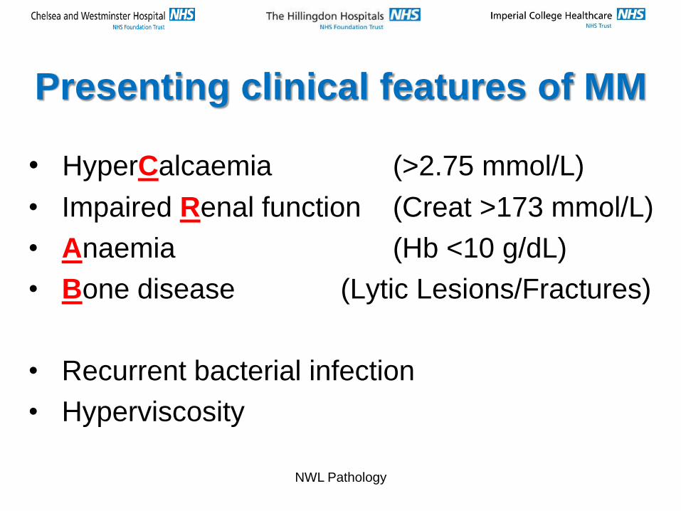
Presenting clinical features of MM
• HyperCalcaemia (>2.75 mmol/L)
• Impaired Renal function (Creat >173 mmol/L)
• Anaemia (Hb <10 g/dL)
• Bone disease (Lytic Lesions/Fractures)
• Recurrent bacterial infection
• Hyperviscosity
NWL Pathology

Paraprotein Types & Myeloma
Type Paraproteins Myeloma
IgG 53% 53%
IgA 22% 22%
IgM 11% 0.5%
IgD 1% 1.5%
IgE <0.001% 0.1%
BJP Only 12% 21%
Non-Secretory - 1%
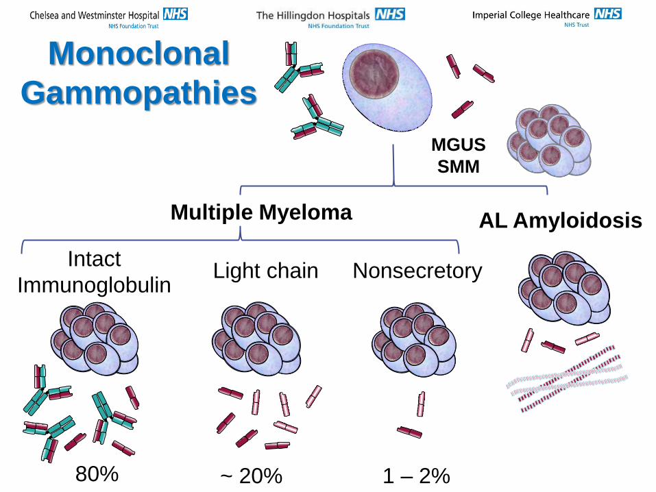
Intact
Immunoglobulin Light chain Nonsecretory
AL Amyloidosis
MGUS
SMM
Multiple Myeloma
Monoclonal
Gammopathies
80% ~ 20% 1 – 2%
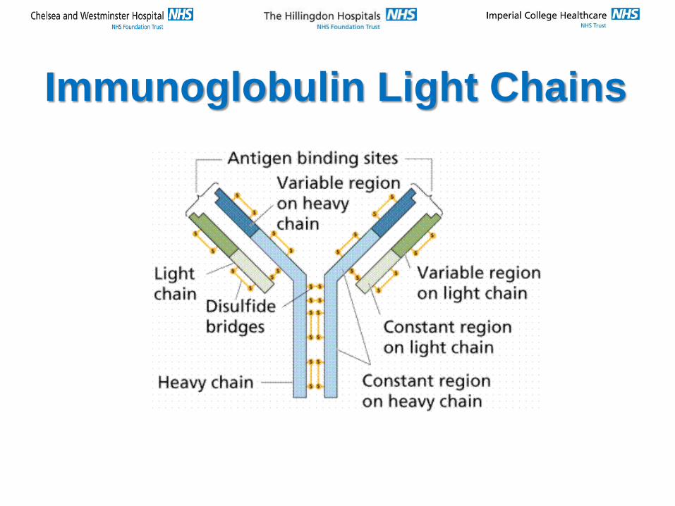
Immunoglobulin Light Chains

Immunoglobulin Light Chains
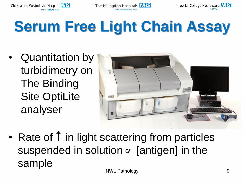
Serum Free Light Chain Assay
• Rate of in light scattering from particles
suspended in solution [antigen] in the
sample NWL Pathology 9
• Quantitation by
turbidimetry on
The Binding
Site OptiLite
analyser

3.3 – 19.4 mg/L 5.7 – 26.3 mg/L 0.26 – 1.65
10 mg/L 15 mg/L 0.67
Result Interpretation κ λ κ/λ
FLC levels and ratio NORMAL – No monoclonal FLCs
detected
N N N
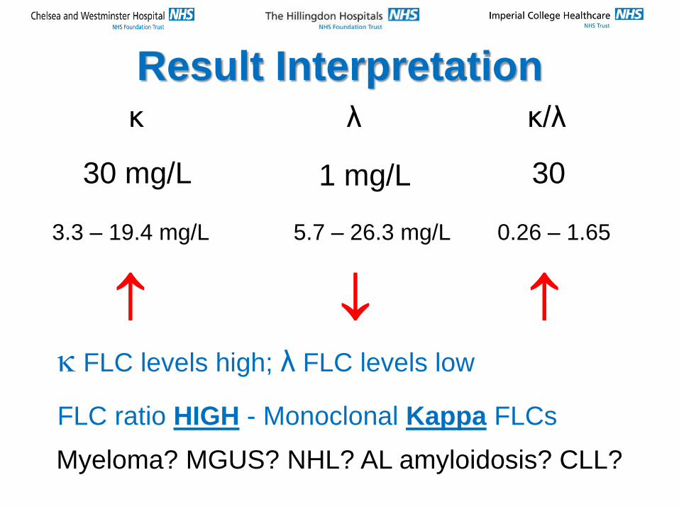
30 mg/L 1 mg/L 30
κ λ κ/λ
FLC levels high; λ FLC levels low
FLC ratio HIGH - Monoclonal Kappa FLCs
Myeloma? MGUS? NHL? AL amyloidosis? CLL?
3.3 – 19.4 mg/L 5.7 – 26.3 mg/L 0.26 – 1.65
Result Interpretation
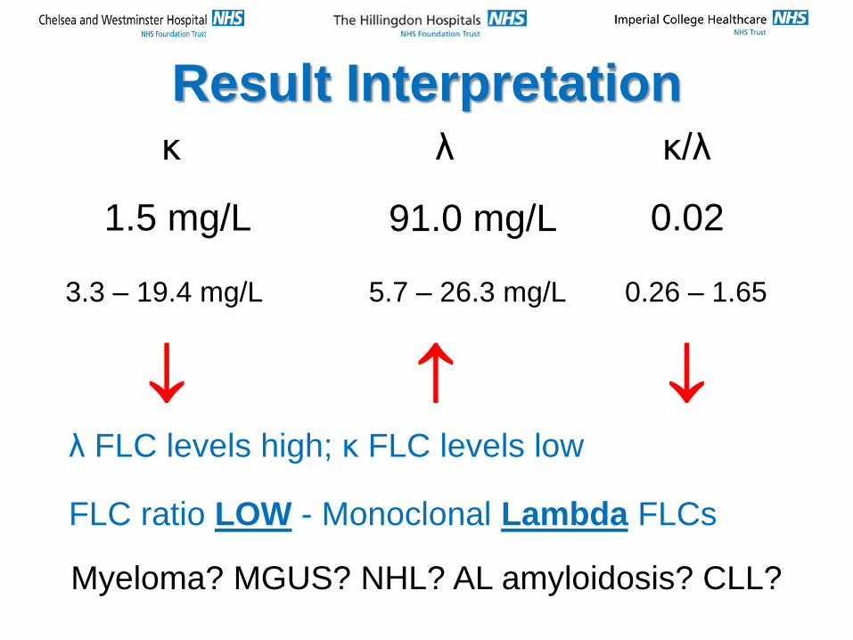
1.5 mg/L 91.0 mg/L 0.02
κ λ κ/λ
λ FLC levels high; κ FLC levels low
FLC ratio LOW - Monoclonal Lambda FLCs
Myeloma? MGUS? NHL? AL amyloidosis? CLL?
3.3 – 19.4 mg/L 5.7 – 26.3 mg/L 0.26 – 1.65
Result Interpretation
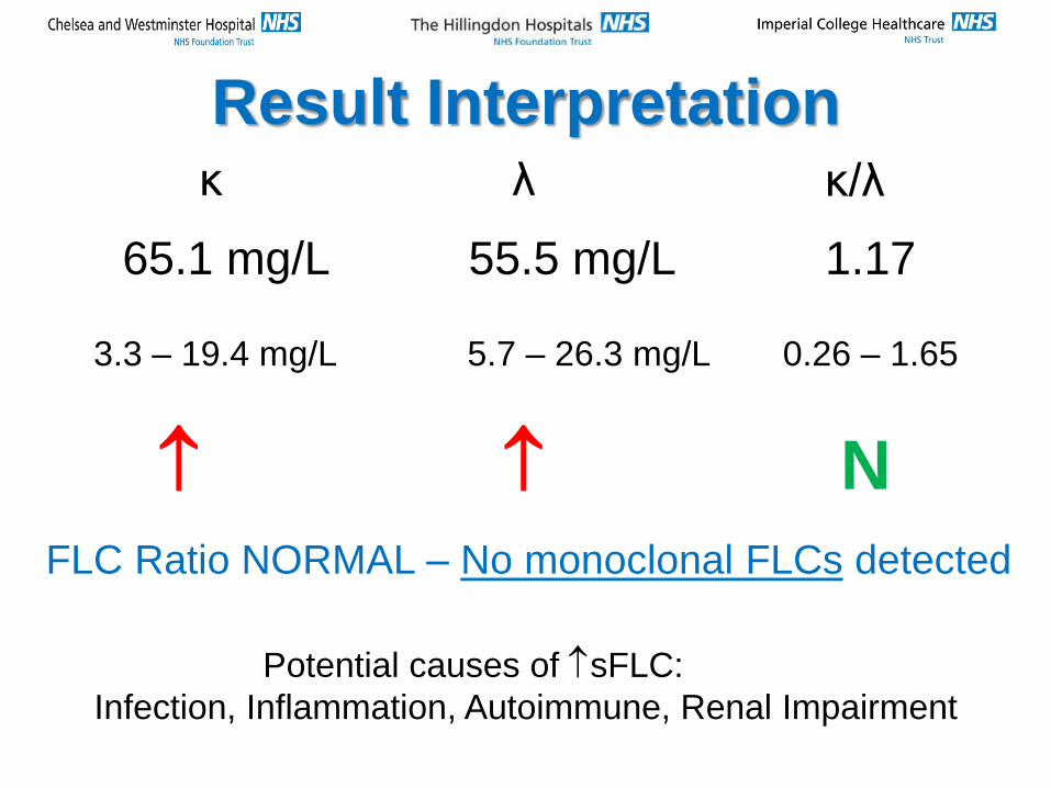
3.3 – 19.4 mg/L 5.7 – 26.3 mg/L 0.26 – 1.65
65.1 mg/L 55.5 mg/L 1.17
Result Interpretation κ λ κ/λ
FLC Ratio NORMAL – No monoclonal FLCs detected
N
Potential causes of sFLC:
Infection, Inflammation, Autoimmune, Renal Impairment
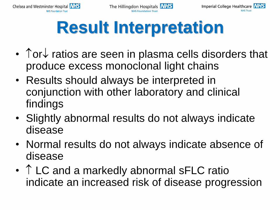
Result Interpretation
• or ratios are seen in plasma cells disorders that produce excess monoclonal light chains
• Results should always be interpreted in conjunction with other laboratory and clinical findings
• Slightly abnormal results do not always indicate disease
• Normal results do not always indicate absence of disease
• LC and a markedly abnormal sFLC ratio indicate an increased risk of disease progression
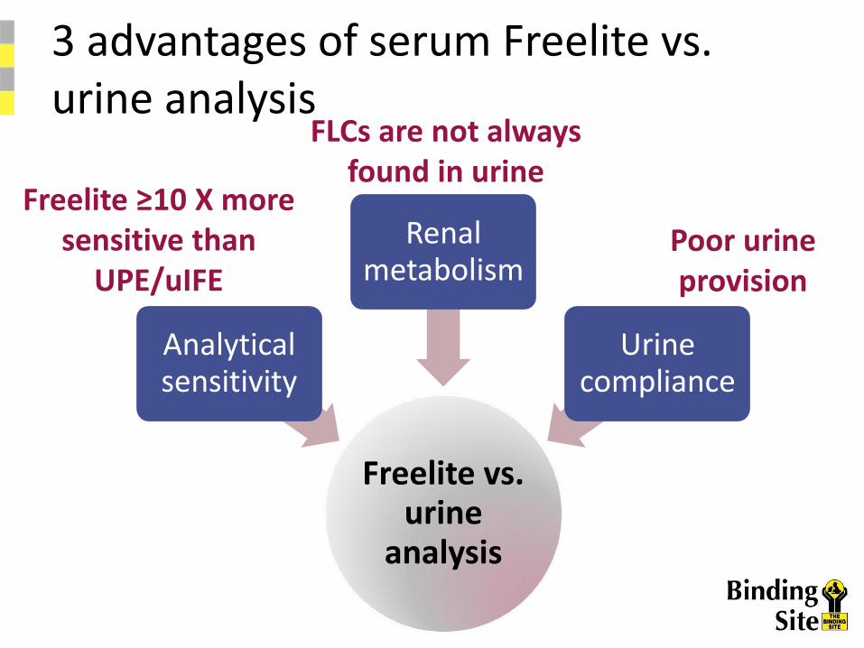
3 advantages of serum Freelite vs. urine analysis
Freelite vs. urine
analysis
Analytical sensitivity
Renal metabolism
Urine compliance
Freelite ≥10 X more sensitive than
UPE/uIFE
FLCs are not always found in urine
Poor urine provision

Analytical sensitivity
Lig
ht ch
ain
co
nce
ntr
atio
n (
mg
/L)
1
10
100
1000
SPE CZE
sIFE
Freelite
Normal range in serum
UPE
uIFE
Freelite is ~10-fold more sensitive than uIFE
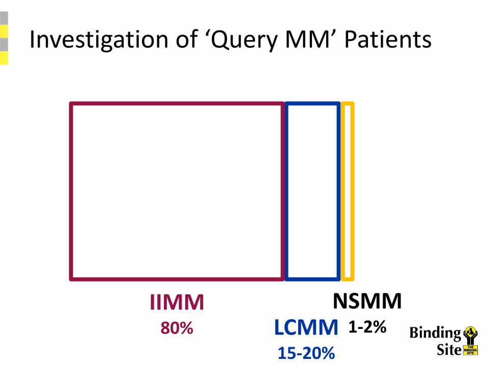
Investigation of ‘Query MM’ Patients
IIMM 80% LCMM
15-20%
NSMM 1-2%

SPE
IIMM LCMM
NSMM
Investigation of ‘Query MM’ Patients
Abraham Clin Chem 2002;48:655-7 Katzmann Am J Clin Pathol 1998;110:503-9
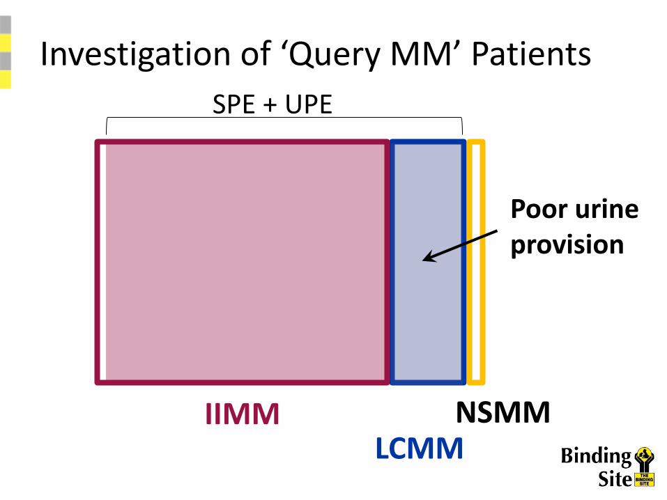
SPE + UPE
Poor urine provision
IIMM LCMM
NSMM
Investigation of ‘Query MM’ Patients
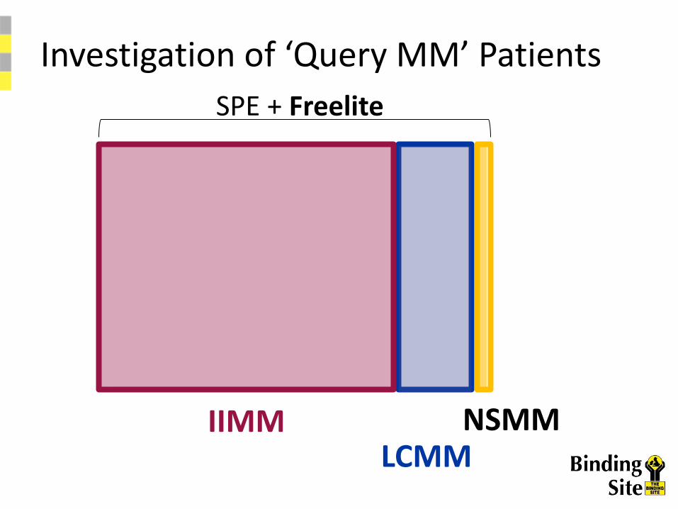
SPE + Freelite
IIMM LCMM
NSMM
Investigation of ‘Query MM’ Patients

SPE 25% 75%
98.7% MM detected
87.6% MM detected
90.4% diagnostic sensitivity
+ sFLCs
SPE
100%
100% diagnostic sensitivity
+ UPE, uIFE No urine
Sensitivity of ‘Query MM’ algorithms
Katzmann Clin Chem 2009;55:1517-22
SPE/CZE UPE sFLCs SPE/CZE vs. + +

Laboratory Investigation
• Screening Tests: FBC, ESR/Plasma Viscosity, Renal Function, Calcium, Albumin, Serum Immunoglobulins, Serum PEP (CZE or gel), Urine PEP (analysis by gel, 2nd Void/24hr Hr) &/or Serum Free Light Chains
• Establish Diagnosis: Immunofixation of serum/urine, Bone marrow aspirate + trephine biopsy with plasma cell phenotyping
• Estimation of Tumour Burden/Prognosis: FISH, Quantitation of Monoclonal (M) protein, albumin, 2M, Serum Free Light Chains
NWL Pathology

Sample Analysed for Immunoglobulins & Electrophoresis
M-Protein Detected Negative Equivocal
Immunofixation for Hv.
Chain G/A/M, + LCs
Hv. Chain not
detected, / LC
detected
IF for D/E Typed Paraprotein,
quantitation, referral
to haematologist,
send urine for
BJP/sFLC, Request:
FBC, Renal/Bone
profiles, LDH
IgD/E /
paraprotein or LC
only detected
IgG/A/M /
paraprotein
Analyse Urine for
BJP or measure
sFLC if clinically
suspected
Zone(s)/Multiple
Bands of Varying
Isotype
Repeat
in 3 – 6
months
Negative

BCSH/UKMF Guidelines:
Diagnosis & Management of MM 2014
• Serum and urine PEP/immunofixation
• sFLC: indicated when high suspicion of MM but
routine sPEP/immunofixation is negative
• sFLC: additional tool for assessment of LC
production/response to treatment, LC only
myeloma & oligosecretory/non-secretory disease
• Renal impairment sFLC ½-life, renal ref. range
• sFLC: Monitoring asymptomatic myeloma
NWL Pathology
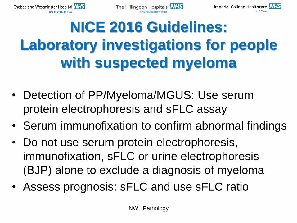
NICE 2016 Guidelines:
Laboratory investigations for people
with suspected myeloma
• Detection of PP/Myeloma/MGUS: Use serum
protein electrophoresis and sFLC assay
• Serum immunofixation to confirm abnormal findings
• Do not use serum protein electrophoresis,
immunofixation, sFLC or urine electrophoresis
(BJP) alone to exclude a diagnosis of myeloma
• Assess prognosis: sFLC and use sFLC ratio
NWL Pathology
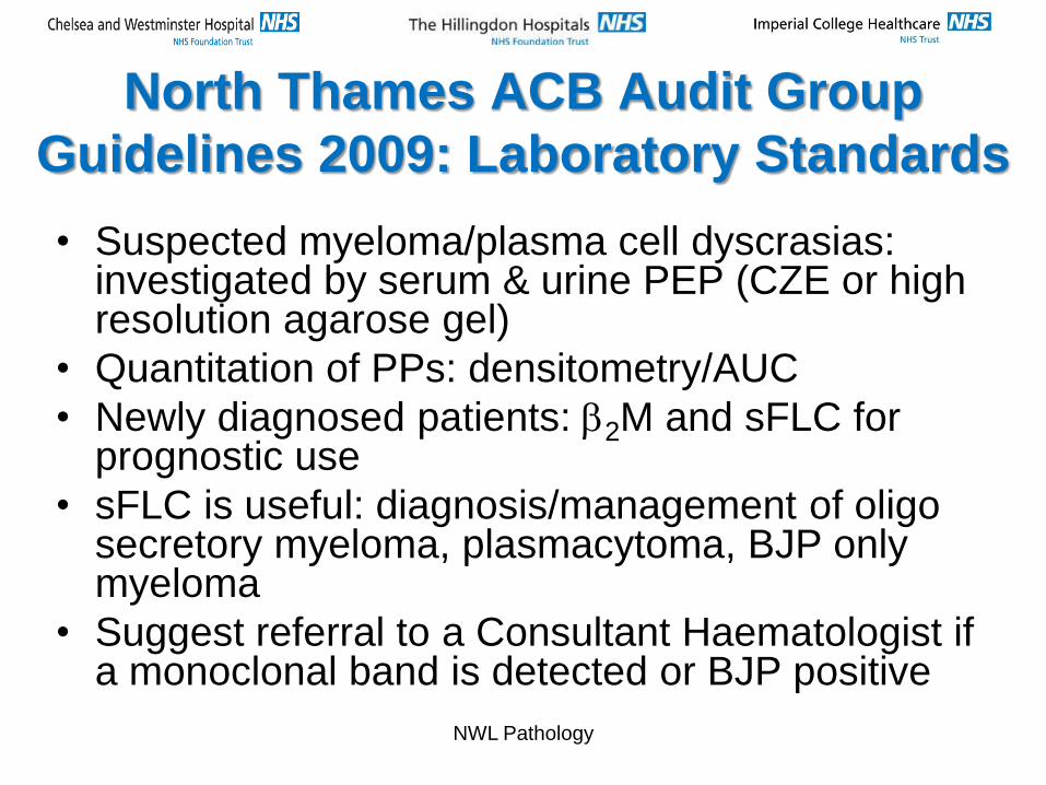
North Thames ACB Audit Group
Guidelines 2009: Laboratory Standards
• Suspected myeloma/plasma cell dyscrasias: investigated by serum & urine PEP (CZE or high resolution agarose gel)
• Quantitation of PPs: densitometry/AUC
• Newly diagnosed patients: 2M and sFLC for prognostic use
• sFLC is useful: diagnosis/management of oligo secretory myeloma, plasmacytoma, BJP only myeloma
• Suggest referral to a Consultant Haematologist if a monoclonal band is detected or BJP positive
NWL Pathology
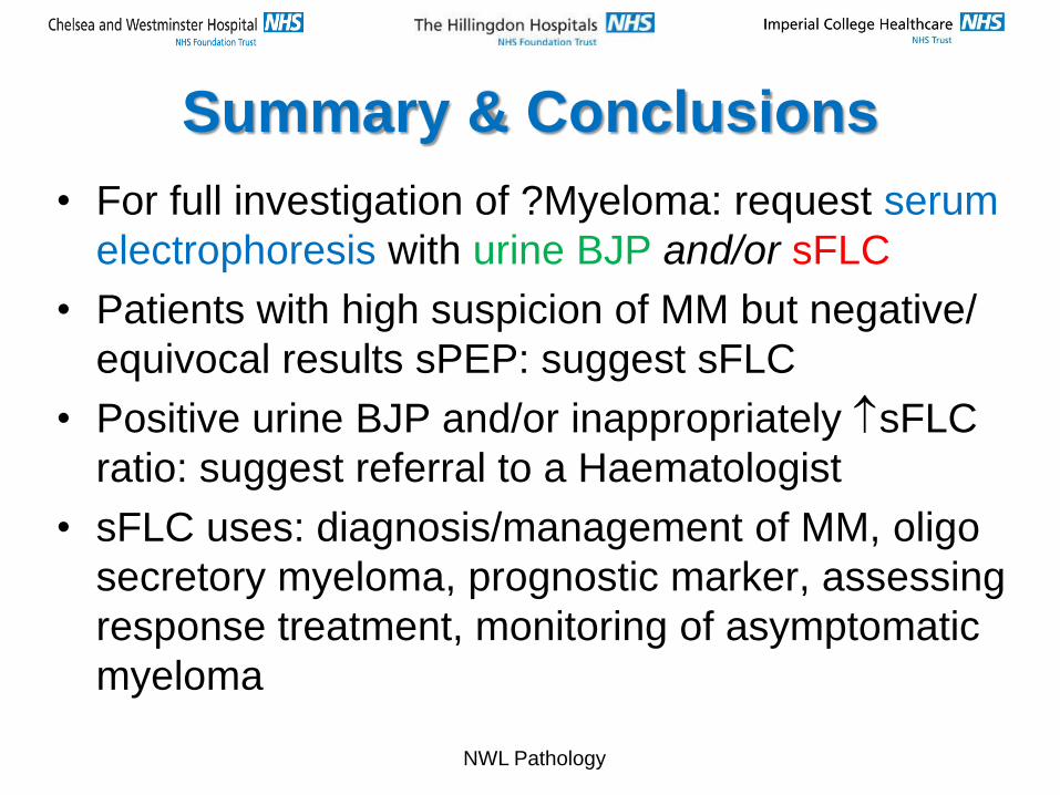
Summary & Conclusions
• For full investigation of ?Myeloma: request serum
electrophoresis with urine BJP and/or sFLC
• Patients with high suspicion of MM but negative/
equivocal results sPEP: suggest sFLC
• Positive urine BJP and/or inappropriately sFLC
ratio: suggest referral to a Haematologist
• sFLC uses: diagnosis/management of MM, oligo
secretory myeloma, prognostic marker, assessing
response treatment, monitoring of asymptomatic
myeloma
NWL Pathology

References
• BSCH and UKMF: Guidelines on the Management and Diagnosis of Multiple Myeloma, August 2014
• International Myeloma Working Group guidelines for serum-free light chain analysis in multiple myeloma and related disorders. Leukaemia 2008; 23: 2
• UK myeloma Forum and Nordic Myeloma Study Group: Guidelines for the investigation of newly detected M-proteins and the management of monoclonal gammopathy of undetermined significance (MGUS). British Journal of Haematology 2009; 147, 22-42
• ACB North Thames Audit Guidelines: 2009
• Capillary electrophoresis and its application in the clinical laboratory. 2003, Clinica Chimica Acta, 330: 1-2, 1-30