Paracoccidioidomycosis in southern Rio Grande do Sul: … · Paracoccidioidomycosis in southern Rio...
5
Paracoccidioidomycosis in southern Rio Grande do Sul: A retrospective study of histopathologically diagnosed cases Silvana Pereira de Souza 1 , Valéria Magalhães Jorge 1,2 , Melissa Orzechowski Xavier 3,4 1 Faculdade de Odontologia, Universidade Federal de Pelotas, Pelotas, RS, Brazil. 2 Santa Casa de Misericórdia de Pelotas, Pelotas, RS, Brazil. 3 Faculdade de Medicina, Universidade Federal do Rio Grande, Rio Grande, RS, Brazil. 4 Programa de Pós-Graduação em Ciências da Saúde, Universidade Federal do Rio Grande, Rio Grande, RS, Brazil Submitted: May 28, 2012; Approved: September 9, 2013. Abstract Paracoccidioidomycosis (PCM) is a systemic mycosis caused by the fungus Paracoccidioides brasiliensis and is endemic to Brazil. The aim of this study was to perform a retrospective analysis of the PCM cases in the countryside south of Rio Grande do Sul, Brazil. The files from four histo- pathology laboratories located in the city of Pelotas were obtained, and all of the epidemiological and clinical data from the PCM diagnosed cases were collected for analysis. A total of 123 PCM cases di- agnosed between 1966 and 2009 were selected. Of these patients, 104 (84.5%) were male, and 17 were female. The patients ranged from 02 to 92 years of age. Fifty-two cases (41.9%) were obtained from the oral pathology laboratory, and the remaining 71 cases (58.1%) were obtained from the three general pathology laboratories. Of all of the patients studied, 65.2% lived in rural zones and worked in agriculture or other related fields. Data on the evolution of this disease was available for 43 cases, and the time frame ranged from 20 to 2920 days (mean = 572.3 days). An accurate diagnosis per- formed in less than 30 days only occurred in 21% of the cases. PCM is endemic to the countryside of Rio Grande do Sul. Therefore, it is recommended that PCM be included as a differential diagnosis, mainly for individuals between 30 and 60 years of age, living in rural zones and who have respiratory signs and associated-oropharyngeal lesions. Key words: Paracoccidioides brasiliensis, epidemiology, systemic mycosis. Introduction Paracoccidioidomycosis (PCM) is a systemic mycosis that was first described in 1908 by Adolfo Lutz, who identified it as a South American blastomycosis. PCM is caused by Paracoccidioides brasiliensis, a dimorphic fungus that has a mycelial form at room temperature (25 °C) and a yeast form under conditions of parasitism (37 °C) (Shikanai-Yasuda et al., 2006; Ramos et al., 2008). PCM may appear as an acute / subacute case in chil- dren and adolescents, also known as the juvenile form, or as a chronic case, which is especially common in adults. Both types of cases can result in residual PCM. The slow pro- gressive infection primarily involves inhaling fungal pro- pagules into the lungs and tends to cause secondary lesions in the mucous membranes, lymph nodes and/or skin through hematogenous spread (Marques, 2003; Shikanai- Yasuda et al., 2006). The disease is endemic to Latin America and occurs in southern Mexico and northern Argentina. PCM cases found outside these areas are reported by patients who have visited or lived in a Latin American country. The majority of the PCM cases (»80%) are reported in Brazil, mainly in the states of São Paulo, Paraná, Rio Grande do Sul, Goiás, Rio de Janeiro and Rondônia (Palmeiro et al., 2005; Ramos et al., 2008; Colombo et al., 2011). The frequency of re- ported cases has also been increasing in the North and Cen- Brazilian Journal of Microbiology 45, 1, 243-247 (2014) Copyright © 2014, Sociedade Brasileira de Microbiologia ISSN 1678-4405 www.sbmicrobiologia.org.br Send correspondence to M.O. Xavier. Laboratório de Micologia, Faculdade de Medicina, Universidade Federal do Rio Grande, Campus Saúde, Visconde de Paranaguá 102, Centro, 96201-900 Rio Grande, RS, Brazil. E-mail: [email protected]. Research Paper
Transcript of Paracoccidioidomycosis in southern Rio Grande do Sul: … · Paracoccidioidomycosis in southern Rio...
Paracoccidioidomycosis in southern Rio Grande do Sul:
A retrospective study of histopathologically diagnosed cases
Silvana Pereira de Souza1, Valéria Magalhães Jorge1,2, Melissa Orzechowski Xavier3,4
1Faculdade de Odontologia, Universidade Federal de Pelotas, Pelotas, RS, Brazil.2Santa Casa de Misericórdia de Pelotas, Pelotas, RS, Brazil.
3Faculdade de Medicina, Universidade Federal do Rio Grande, Rio Grande, RS, Brazil.4Programa de Pós-Graduação em Ciências da Saúde, Universidade Federal do Rio Grande, Rio Grande,
RS, Brazil
Submitted: May 28, 2012; Approved: September 9, 2013.
Abstract
Paracoccidioidomycosis (PCM) is a systemic mycosis caused by the fungus Paracoccidioides
brasiliensis and is endemic to Brazil. The aim of this study was to perform a retrospective analysis of
the PCM cases in the countryside south of Rio Grande do Sul, Brazil. The files from four histo-
pathology laboratories located in the city of Pelotas were obtained, and all of the epidemiological and
clinical data from the PCM diagnosed cases were collected for analysis. A total of 123 PCM cases di-
agnosed between 1966 and 2009 were selected. Of these patients, 104 (84.5%) were male, and 17
were female. The patients ranged from 02 to 92 years of age. Fifty-two cases (41.9%) were obtained
from the oral pathology laboratory, and the remaining 71 cases (58.1%) were obtained from the three
general pathology laboratories. Of all of the patients studied, 65.2% lived in rural zones and worked
in agriculture or other related fields. Data on the evolution of this disease was available for 43 cases,
and the time frame ranged from 20 to 2920 days (mean = 572.3 days). An accurate diagnosis per-
formed in less than 30 days only occurred in 21% of the cases. PCM is endemic to the countryside of
Rio Grande do Sul. Therefore, it is recommended that PCM be included as a differential diagnosis,
mainly for individuals between 30 and 60 years of age, living in rural zones and who have respiratory
signs and associated-oropharyngeal lesions.
Key words: Paracoccidioides brasiliensis, epidemiology, systemic mycosis.
Introduction
Paracoccidioidomycosis (PCM) is a systemic
mycosis that was first described in 1908 by Adolfo Lutz,
who identified it as a South American blastomycosis. PCM
is caused by Paracoccidioides brasiliensis, a dimorphic
fungus that has a mycelial form at room temperature
(25 °C) and a yeast form under conditions of parasitism
(37 °C) (Shikanai-Yasuda et al., 2006; Ramos et al., 2008).
PCM may appear as an acute / subacute case in chil-
dren and adolescents, also known as the juvenile form, or as
a chronic case, which is especially common in adults. Both
types of cases can result in residual PCM. The slow pro-
gressive infection primarily involves inhaling fungal pro-
pagules into the lungs and tends to cause secondary lesions
in the mucous membranes, lymph nodes and/or skin
through hematogenous spread (Marques, 2003; Shikanai-
Yasuda et al., 2006).
The disease is endemic to Latin America and occurs
in southern Mexico and northern Argentina. PCM cases
found outside these areas are reported by patients who have
visited or lived in a Latin American country. The majority
of the PCM cases (�80%) are reported in Brazil, mainly in
the states of São Paulo, Paraná, Rio Grande do Sul, Goiás,
Rio de Janeiro and Rondônia (Palmeiro et al., 2005; Ramos
et al., 2008; Colombo et al., 2011). The frequency of re-
ported cases has also been increasing in the North and Cen-
Brazilian Journal of Microbiology 45, 1, 243-247 (2014) Copyright © 2014, Sociedade Brasileira de Microbiologia
ISSN 1678-4405 www.sbmicrobiologia.org.br
Send correspondence to M.O. Xavier. Laboratório de Micologia, Faculdade de Medicina, Universidade Federal do Rio Grande, Campus Saúde,
Visconde de Paranaguá 102, Centro, 96201-900 Rio Grande, RS, Brazil. E-mail: [email protected].
Research Paper
tral-West regions of the country (Paniago et al., 2003;
Shikanai-Yasuda et al., 2006).
Even though paracoccidioidomycosis is endemic to
Rio Grande do Sul, few studies have addressed the occur-
rence of this disease in the cities of the southern part of the
state. This study aimed to describe the clinical and epidemi-
ological data on the PCM diagnosed cases from the pathol-
ogy labs in the city of Pelotas, RS, Brazil.
Materials and Methods
The study was carried out retrospectively by evaluat-
ing the databases from the four major pathology labs, one
lab of which is odontology specific, in the city of Pelotas.
This region has altitudes between 100 and 429 m and a hu-
mid subtropical climate that consists of warm temperate
summers and cold winters with frequent frosts (an average
of 20 per year). Rainfall occurs regularly throughout the
year. The average annual rainfall is 1.379 mm, and the rela-
tive humidity is high, with an annual average of approxi-
mately 80%. The average temperature for the warmer
months is 23 °C, and the average temperature for the colder
months is 12 °C.
A survey of the total number of paracoccidioido-
mycosis cases diagnosed in each laboratory until the year
2010 was initially performed. These cases were confirmed
by the detection of multiple budding yeast cells typical of
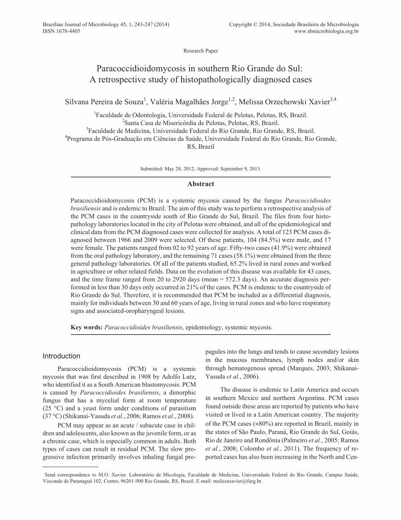
P. brasiliensis in tissue fragments (Figure 1). All of the
cases confirmed by histopathology (123 cases from 1966 to
2009) were included in this study. The following informa-
tion was collected for this study using the biopsy data
sheets and laboratory evaluations: age, sex, origin (rural or
urban), professional activity, course of the disease, location
of lesions and signs/symptoms. The data were compiled
and descriptively analyzed using the program Epi Info
3.5.1.
This study was approved by the ethics committee of
the institution (CEPAS-FURG 176/2011).
Results
From the four laboratories in Pelotas included in this
study, 123 patients diagnosed with PCM by histo-
pathological examination were identified. Four of the cases
have already been published as case reports (Jannke et al.,
1982, 1983, 1993). All of these cases occurred within a
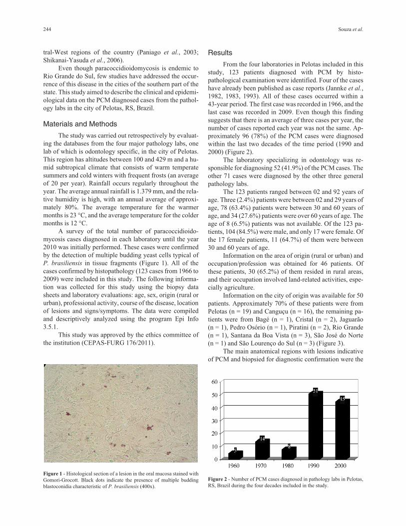
43-year period. The first case was recorded in 1966, and the
last case was recorded in 2009. Even though this finding
suggests that there is an average of three cases per year, the
number of cases reported each year was not the same. Ap-
proximately 96 (78%) of the PCM cases were diagnosed
within the last two decades of the time period (1990 and
2000) (Figure 2).
The laboratory specializing in odontology was re-
sponsible for diagnosing 52 (41.9%) of the PCM cases. The
other 71 cases were diagnosed by the other three general
pathology labs.
The 123 patients ranged between 02 and 92 years of
age. Three (2.4%) patients were between 02 and 29 years of
age, 78 (63.4%) patients were between 30 and 60 years of
age, and 34 (27.6%) patients were over 60 years of age. The
age of 8 (6.5%) patients was not available. Of the 123 pa-
tients, 104 (84.5%) were male, and only 17 were female. Of
the 17 female patients, 11 (64.7%) of them were between
30 and 60 years of age.
Information on the area of origin (rural or urban) and
occupation/profession was obtained for 46 patients. Of
these patients, 30 (65.2%) of them resided in rural areas,
and their occupation involved land-related activities, espe-
cially agriculture.
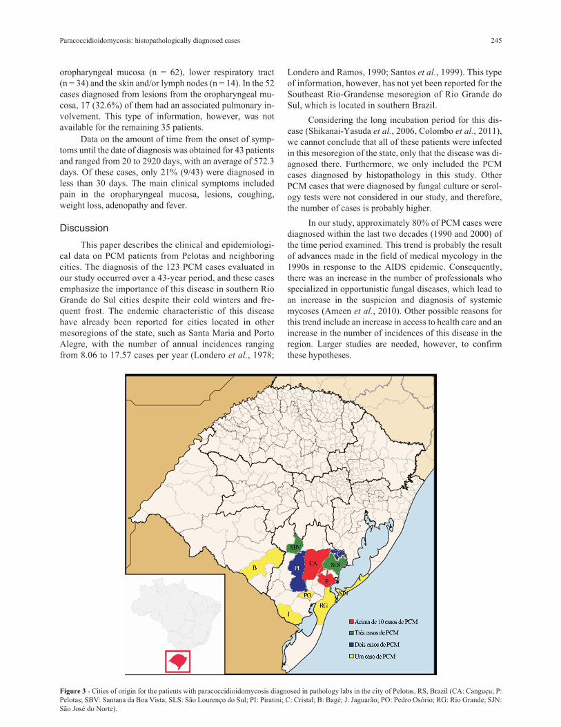
Information on the city of origin was available for 50
patients. Approximately 70% of these patients were from
Pelotas (n = 19) and Canguçu (n = 16), the remaining pa-
tients were from Bagé (n = 1), Cristal (n = 2), Jaguarão
(n = 1), Pedro Osório (n = 1), Piratini (n = 2), Rio Grande
(n = 1), Santana da Boa Vista (n = 3), São José do Norte
(n = 1) and São Lourenço do Sul (n = 3) (Figure 3).
The main anatomical regions with lesions indicative
of PCM and biopsied for diagnostic confirmation were the
244 Souza et al.
Figure 1 - Histological section of a lesion in the oral mucosa stained with
Gomori-Grocott. Black dots indicate the presence of multiple budding
blastoconidia characteristic of P. brasiliensis (400x).
Figure 2 - Number of PCM cases diagnosed in pathology labs in Pelotas,
RS, Brazil during the four decades included in the study.
oropharyngeal mucosa (n = 62), lower respiratory tract
(n = 34) and the skin and/or lymph nodes (n = 14). In the 52
cases diagnosed from lesions from the oropharyngeal mu-
cosa, 17 (32.6%) of them had an associated pulmonary in-
volvement. This type of information, however, was not
available for the remaining 35 patients.
Data on the amount of time from the onset of symp-
toms until the date of diagnosis was obtained for 43 patients
and ranged from 20 to 2920 days, with an average of 572.3
days. Of these cases, only 21% (9/43) were diagnosed in
less than 30 days. The main clinical symptoms included
pain in the oropharyngeal mucosa, lesions, coughing,
weight loss, adenopathy and fever.
Discussion
This paper describes the clinical and epidemiologi-
cal data on PCM patients from Pelotas and neighboring
cities. The diagnosis of the 123 PCM cases evaluated in
our study occurred over a 43-year period, and these cases
emphasize the importance of this disease in southern Rio
Grande do Sul cities despite their cold winters and fre-
quent frost. The endemic characteristic of this disease
have already been reported for cities located in other
mesoregions of the state, such as Santa Maria and Porto
Alegre, with the number of annual incidences ranging
from 8.06 to 17.57 cases per year (Londero et al., 1978;
Londero and Ramos, 1990; Santos et al., 1999). This type
of information, however, has not yet been reported for the
Southeast Rio-Grandense mesoregion of Rio Grande do
Sul, which is located in southern Brazil.
Considering the long incubation period for this dis-
ease (Shikanai-Yasuda et al., 2006, Colombo et al., 2011),
we cannot conclude that all of these patients were infected
in this mesoregion of the state, only that the disease was di-
agnosed there. Furthermore, we only included the PCM
cases diagnosed by histopathology in this study. Other
PCM cases that were diagnosed by fungal culture or serol-
ogy tests were not considered in our study, and therefore,
the number of cases is probably higher.
In our study, approximately 80% of PCM cases were
diagnosed within the last two decades (1990 and 2000) of
the time period examined. This trend is probably the result
of advances made in the field of medical mycology in the
1990s in response to the AIDS epidemic. Consequently,
there was an increase in the number of professionals who
specialized in opportunistic fungal diseases, which lead to
an increase in the suspicion and diagnosis of systemic
mycoses (Ameen et al., 2010). Other possible reasons for
this trend include an increase in access to health care and an
increase in the number of incidences of this disease in the
region. Larger studies are needed, however, to confirm
these hypotheses.
Paracoccidioidomycosis: histopathologically diagnosed cases 245
Figure 3 - Cities of origin for the patients with paracoccidioidomycosis diagnosed in pathology labs in the city of Pelotas, RS, Brazil (CA: Canguçu; P:
Pelotas; SBV: Santana da Boa Vista; SLS: São Lourenço do Sul; PI: Piratini; C: Cristal; B: Bagé; J: Jaguarão; PO: Pedro Osório; RG: Rio Grande; SJN:
São José do Norte).
Most of our PCM patients were adult men who had
the chronic form of this disease. The predominance of male
patients is consistent with findings from studies that found
male-to-female ratios between 5:1 to 16.3:1 in Mato Grosso
do Sul, Brasília, São Paulo and Rio Grande do Sul (Londero
and Ramos, 1990; Blotta et al., 1999; Paniago et al., 2003;
Shikanai-Yasuda et al., 2006; Campos et al., 2008). This
difference may be explained by a hormone protective factor
in women. The presence of estrogen receptors in P.
brasiliensis inhibits the transformation from the mycelial
phase to the yeast parasitic phase of the fungus (Borges-
Walmsley et al., 2002; Almeida et al., 2003; Vieira and
Borsatto-Galera, 2006; Bousquet et al., 2007). If there is a
hormone protective factor, then postmenopausal women
should become more susceptible to PCM. The majority of
the women (11/17) with diagnosed PCM in our study, how-
ever, were of fertile age, between 30 and 60 of age.
Consistent with previous findings (Verli et al., 2005;
Bousquet et al., 2007), a high portion of the PCM patients,
65.2%, evaluated in this study were involved in agricultural
activities. Agricultural activities predispose individuals to
mycosis because of their higher exposure to infectious fun-
gal propagules. For instance, the natural habitat of P.
brasiliensis includes forested areas with wet soils (Bous-
quet et al., 2007; Richini-Pereira et al., 2009). Furthermore,
agribusiness is the most common economic activity in Pe-
lotas and Canguçu, which are the cities with the highest
number of cases in our study.
The oropharyngeal mucosa and lungs were the most
common sites of lesions found in our study. According to
previous reports, these organs are the most common organs
involved in PCM (Almeida et al., 2003; Verli et al., 2005;
Vieira and Borsatto-Galera, 2006). We found that almost
half of our PCM cases (41.9%) were diagnosed in the
odontology pathology laboratory, which indicates that the
oropharyngeal mucosa is frequently affected by this dis-
ease. Moriform stomatitis is especially characteristic of this
disease. This finding indicates that the dental surgeon is an
important professional in the diagnosis of PCM because pa-
tients will frequently seek medical assistance for oral le-
sions and not respiratory symptoms, which are erroneously
associated with smoking (Araújo and Souza, 2000; Pal-
meiro et al., 2005; Verli et al., 2005; Vieira and Borsatto-
Galera, 2006). The clinical symptoms described by the pa-
tients included in our study are consistent with the symp-
toms described in the literature, such as pain in muco-
cutaneous lesions, coughing, weight loss, adenopathy and
fever (Ronquillo, 1983; Londero and Ramos, 1990; Shi-
kanai-Yasuda et al., 2006).
An early diagnosis of paracoccidioidomycosis and
the immediate patient referral for treatment are important
factors in reducing the number of complications caused by
this disease (Araújo and Souza, 2000; Palmeiro et al.,
2005). In our study, however, we found that a considerable
number of patients reported a long time period between the
onset of clinical symptoms and diagnosis. This delay may
be related to a difficulty for health professionals in making
an accurate diagnosis of PCM from early lesions as well as
rural patients waiting a longer time before seeking profes-
sional help. Furthermore, the long time period between the
onset of symptoms and diagnosis may also be attributed to a
lack of access to health services. This long time period be-
fore diagnosis can result in PCM progressing to the residual
form, which is often severe (Ronquillo, 1983; Shikanai-
Yasuda et al., 2006).
Conclusion
Paracoccidioidomycosis is a mycosis with an impor-
tant number of reported incidences in cities of the southeast
Rio-Grandense mesoregions of Rio Grande do Sul. This
study highlights the need to include PCM as a differential
diagnosis of respiratory infection, especially in patients
with oropharyngeal lesions and in rural males from Pelotas
or neighborhood cities who are between 30 and 60 years of
age.
ReferencesAlmeida OP, Jacks JR, Scully C (2003) Paracoccidioidomycosis
of the mouth: an emerging deep mycosis. Crit Rev Oral Biol
Med 14(5):377-383.
Ameen M, Talhari C, Talhari S (2010) Advances in paraco-
ccidioidomycosis. Clin Experimental Dermatol 35(6):576-
580.
Araújo MS, Souza SC (2000) Análise epidemiológica de pacien-
tes acometidos com paracoccidioidomicose em região endê-
mica do Estado de Minas Gerais. Rev Pós Grad 7:22-26.
Blotta MHSL, Mamoni RL, Oliveira SJ, Nouér SA, Papaiordanou
PMO, Goveia A, Camargo ZP (1999) Endemic regions of
paracoccidioidomycosis in Brazil: a clinical and epidemio-
logic study of 548 cases in the southeast region. Am J Trop
Med Hig 61:390-394.
Borges-Walmsley MI, Chen D, Shu X, Walmsley AR (2002) The
phatobiology of Paracoccidioides brasiliensis. Trends
Microbiol 10(2):80-87.
Bousquet A, Dussart C, Drouillard I, Charbel EC, Boiron P (2007)
Mycoses d’importation: le point sur la paracoccidioido-
mycose. Méd Mal Infect 37:210-214.
Campos MVS, Penna GO, Castro CN, Moraes MAP, Ferreira MS,
Santos JB (2008) Paracoccidioidomicose no Hospital Uni-
versitário de Brasília. Rev Soc Bras Med Trop 2:41.
Colombo AL, Tobón A, Restrepo A, Queiroz-Telles F, Nucci M
(2011) Epidemiology of endemic systemic fungal infections
in Latin America. Med Mycol 49:785-798.
Jannke HA, Isolan T, Pinto IO, Isaacsson JA (1982) Blastomicose
sul-americana com comprometimento genital. Rev Bras
Cirurgia 72(4):247-249.
Jannke HA, Lopez FS, Abrahao MC, Thofern P, Duarte AL,
Holthausen ET (1983) Blastomicose sul-americana pal-
pebral. Rev Bras Oftalmol 42(2):157-160.
Jannke HA, Deves ML, Roberti AG, Taddeu CAG, Bazzano MC,
Ferreira AD, Menezes FS, Oliveira Filho UL (1993) Para-
coccidioidomicose Associada a Neurofibromatose de Von
Recklinghausen e Fenda Palatina. Observação de dois casos.
246 Souza et al.
XXIX Congresso da Sociedade Brasileira de Medicina
Tropical 26:274.
Londero AT, Ramos CD, Lopes JO (1978) Progressive Pulmo-
nary Paracoccidioidomycosis a study of 34 cases observed
in Rio Grande do Sul (Brazil). Mycopathologia 63(1):53-56.
Londero AT, Ramos CD (1990) Paracoccidioidomicose. Estudo
clínico e micológico de 260 casos observados no interior do
Estado do Rio Grande do Sul. J Pneumol 1:129-132.
Marques SA (2003) Paracoccidiodomycosis: epidemiological,
clinical and treatment up-date. Arq Bras Dermatol
78(2):135-150.
Palmeiro M, Cherubini K, Yurgel LS (2005) Paracoccidioido-
micose-Revisão da Literatura. Scientia Medica 15(4):274-
278.
Paniago AMM, Aguiar JIA, Aguiar ES, Cunha RV, Pereira GRO,
Londero AT, Wanke B (2003) Paracoccidioidomicose: estu-
do clínico e epidemiológico de 422 casos observados no
Estado de Mato Grosso do Sul. Rev Soc Bras Med Trop
36(4):455-459.
Ramos E, Silva M, Saraiva, LES (2008) Paracoccidioidomycosis.
Dermatol Clinics 26(2):257-269.
Richini-Pereira VB, Bosco SM, Theodoro RC, Barrozo L, Pedrini
SC, Rosa PS, Bagagli E (2009) Importance of the xenar-
thrans in the ecoepidemiology of Paracoccidioides
brasiliensis. BMC Research Notes 2:228.
Ronquillo TEF (1983) Contribuição ao estudo da paracoccidioi-
domicose na República do Equador. Rev Patol Trop
12:345-419.
Santos JWA, Severo LC, Porto NS, Moreira JS, Silva LCC,
Camargo JJP (1999) Chronic Pulmonary Paracoccidioi-
domycosis in the state of Rio Grande do Sul, Brazil.
Mycopathologia 143:63-67.
Shikanai-Yasuda MA, Queiroz TL, Mendes R, Colombo A,
Moretti MA (2006) Consenso em paracoccidioidomicose.
Rev Soc Bras Med Trop 39:297-310.
Verli FD, Marinho SA, Souza SC, Figueiredo MAZ, Yurgel LS
(2005) Perfil clínico-epidemiológico dos pacientes porta-
dores de paracoccidioidomicose no Serviço de Estomato-
logia do Hospital São Lucas da Pontifícia Universidade
Católica do grande do Sul. Rev Soc Bras Med Trop 38:234-
237.
Vieira EMM, Borsatto-Galera B (2006) Manifestações clínicas
bucais da paracoccidioidomicose. Rev Patol Trop 35:23-30.
All the content of the journal, except where otherwise noted, is licensed under a
Creative Commons License CC BY-NC.
Paracoccidioidomycosis: histopathologically diagnosed cases 247