Papilledema - AJNR · cephal us 16 23 F Headache, de-Traumatic subdural Papilledema 00: 20/30,...
10
John R. Jinkins 1 This article appears in the July/August 1987 issue of AJNR and the October 1987 issue of AJR. Received May 6, 1986 ; accepted after revision October 28, 1986 . 'Department of Radiology, Neuroradiology Sec- tion, King Faisal Specialist Hospital and Research Centre, P.O. Box 3354, Ri yadh 11 211, Saudi Ara- bia. Address reprint requests to J. R. Jinkins. AJNR 8:681-690, July/ August 1987 0195-6108/87/0804-0681 © American Society of Neuroradiology "Papilledema": Neuroradiologic Evaluation of Optic Disk Protrusion with Dynamic Orbital CT 681 Current-generation CT scc·lners enable the visualization in vivo of structures and substructures that were previously unobservable. Certainly the orbit and optic nerve/ sheath complex have demonstrated a great number of pathologic and normal anatomic variations. It has been found in patients with elevated intracranial pressure that what was previously thought to be simple papilledema in fact masks a surprisingly large component of optic papilla protru<;ion, There may be a variable amount of increased intercellular/axonal fluid within tt .. - optic disk in patients with increased intracranial pressure; however, a significant factor in the " swollen disk" is the simple transmission of pressure along the optic nerve sheath to the papilla, causing it to bulge. Further investigations with dynamic CT reveal that there is decreased perfusion of the optic disk in the active phase of severe increased intracranial pressure in patients with papilledema and/or protrusion as compared with normal control subjects . This de- pressed flow pattern seems to originate subacutely and appears to resolve in certain patients after normalization of the elevated pressure. These findings apparently indicate that clinical intervention in cases of intracranial hypertension to restore the hemodynamic status of the optic disk would be timely, and thereby avert irreversible damage. This suggests and supports the theory that increased intracranial pressure may lead to rapid vision loss by the mechanical mechanism of pressure projected directly to the junction of the optic nerve and optic nerve head, leading to decreased perfusion , ischemia, axonal flow stasis , and resultant optic nerve atrophy . The medical definitions of papilledema range from the abbreviated "edema of the optic disc," [1] to the definitive, "edema of the optic disc . . . most co mmonly due to increased intracranial pressure, malignant hypertenSion, or thrombosis of the central retinal vein" [2]. Almost all definitions, however, stress a dominant or sole component of real edema in the optic nerve head accounting for the swelling without regard to differing pathologic processes [3, 4]. This study was undertaken to evaluate both the in vivo structure of the optic nerve/sheath co mpl ex at its junction at the optic nerve head in patients with clinical papilledema caused by intracranial hypertension as well as to study the perfusion characteristics of the optic papilla in patients with increased intracranial pressure (ICP) from various causes. Subjects and Methods The scann er used in this study was the GE 9800 with dynamic capability. Unenhanced static scans were obtained with 5-mm-thick axial sections through the orbit followed by 3- mm sections through the optic nerve head subsequent to IV administration of contrast material. The patient population encompassed a total of 20 subjects with increased ICP cau sed by various processes including trauma, premature craniosynostosis, hydrocephalus, true tumor, primary pseudotumor, and pseudotumor secondary to cerebral venous sinus thrombosis. In addition, a control group of eight patients scanned for reasons other th an increased ICP completed the study (Table 1).
Transcript of Papilledema - AJNR · cephal us 16 23 F Headache, de-Traumatic subdural Papilledema 00: 20/30,...
John R. Jinkins 1
This article appears in the July/August 1987 issue of AJNR and the October 1987 issue of AJR.
Received May 6, 1986; accepted after revision October 28 , 1986.
'Department of Radiology, Neuroradiology Section, King Faisal Specialist Hospital and Research Centre, P.O. Box 3354 , Riyadh 11 211, Saudi Arabia. Address reprint requests to J. R. Jinkins.
AJNR 8:681-690, July/ August 1987 0195-6108/87/0804-0681 © American Society of Neuroradiology
"Papilledema": Neuroradiologic Evaluation of Optic Disk Protrusion with Dynamic Orbital CT
681
Current-generation CT scc·lners enable the visualization in vivo of structures and substructures that were previously unobservable. Certainly the orbit and optic nerve/ sheath complex have demonstrated a great number of pathologic and normal anatomic variations. It has been found in patients with elevated intracranial pressure that what was previously thought to be simple papilledema in fact masks a surprisingly large component of optic papilla protru<;ion, There may be a variable amount of increased intercellular/axonal fluid within tt .. - optic disk in patients with increased intracranial pressure; however, a significant factor in the " swollen disk" is the simple transmission of pressure along the optic nerve sheath to the papilla, causing it to bulge. Further investigations with dynamic CT reveal that there is decreased perfusion of the optic disk in the active phase of severe increased intracranial pressure in patients with papilledema and/or protrusion as compared with normal control subjects. This depressed flow pattern seems to originate subacutely and appears to resolve in certain patients after normalization of the elevated pressure. These findings apparently indicate that clinical intervention in cases of intracranial hypertension to restore the hemodynamic status of the optic disk would be timely, and thereby avert irreversible damage. This suggests and supports the theory that increased intracranial pressure may lead to rapid vision loss by the mechanical mechanism of pressure projected directly to the junction of the optic nerve and optic nerve head, leading to decreased perfusion, ischemia, axonal flow stasis, and resultant optic nerve atrophy.
The medical definitions of papilledema range from the abbreviated "edema of the optic disc, " [1] to the definitive, "edema of the optic disc . . . most commonly due to increased intracranial pressure, malignant hypertenSion, or thrombosis of the central retinal vein" [2]. Almost all definitions, however, stress a dominant or sole component of real edema in the optic nerve head accounting for the swelling without regard to differing pathologic processes [3, 4]. This study was undertaken to evaluate both the in vivo structure of the optic nerve/sheath complex at its junction at the optic nerve head in patients with clinical papilledema caused by intracranial hypertension as well as to study the perfusion characteristics of the optic papilla in patients with increased intracranial pressure (ICP) from various causes.
Subjects and Methods
The scanner used in this study was the GE 9800 with dynamic capability . Unenhanced static scans were obtained with 5-mm-thick axial sections through the orbit followed by 3-mm sections through the optic nerve head subsequent to IV administration of contrast material.
The patient population encompassed a total of 20 subjects with increased ICP caused by various processes including trauma, premature craniosynostosis, hydrocephalus, true tumor, primary pseudotumor, and pseudotumor secondary to cerebral venous sinus thrombosis. In addition , a control group of eight patients scanned for reasons other than increased ICP completed the study (Table 1).

TA
BL
E
1:
Pa
pill
ed
em
a:
Su
mm
ary
of
Ca
ses
en
CD
t\
)
Ca
se
Ge
nde
r H
isto
ry
Dia
gno
sis
Clin
ica
l Fin
din
gs
CT
of
Op
tic
Dis
k
No
. A
ge
V
isua
l A
cuity
S
tatic
D
yna
mic
M
Orb
ital
sw
elli
ng
Co
ntra
late
ral r
etin
o-
Co
ntr
ala
tera
l o
rbita
l U
nkn
ow
n F
lat
dis
k N
ot
pe
rfo
rme
d
bla
sto
ma
m
ass
2
21
M
Hea
da
ches
N
on
org
an
ic h
ead
-N
orm
al
20
/20
Fla
t d
isks
N
ot
pe
rfo
rme
d
ache
s 3
30
F
R f
aci
al w
eak
ne
ss
Fa
cial
ne
rve
ne
uro
p-
Nor
mal
20
/20
Fla
t d
isks
N
ot
pe
rfo
rme
d
ath
y,
?e
tiolo
gy
4 2
2 F
D
iplo
pia
Te
mp
ora
l m
en
ing
i-R
III
& V
I cr
ania
l N
orm
al
Fla
t d
isks
N
orm
al d
isk
pe
rfu
-o
ma
n
erv
e p
als
y si
on
5 31
M
C
hro
nic
he
ada
che
N
on
org
an
ic h
ead
-?
Re
tiniti
s N
orm
al
Fla
t di
sk
Nor
mal
dis
k p
erf
u-
ach
es
sion
6
10
M
O
rbita
l sw
ellin
g C
on
tra
late
ral
Iym
-C
on
tra
late
ral
orb
ital
Un
kno
wn
F
lat
dis
k N
orm
al d
isk
pe
rfu
-p
ha
ng
iom
a
ma
ss w
ith h
em
or-
sion
rh
age
7 3
3 M
O
rbita
l sw
ellin
g C
on
tra
late
ral
he-
Co
ntr
ala
tera
l o
rbita
l 2
0/2
0
Fla
t d
isk
Nor
mal
dis
k p
erf
u-
ma
ng
iom
a
ma
ss
sion
8
18
F
H
ea
da
che
s N
on
org
an
ic h
ead-
No
rma
l 2
0/2
0 F
lat
dis
ks
Nor
mal
dis
k p
erf
u-
ach
es
sion
9
19
F
He
ad
ach
e,
naus
ea,
Pse
ud
otu
mo
r se
c-P
apill
edem
a: r
e-B
lind
Initi
al: o
pti
c sh
eath
dila
tatio
n;
Nor
mal
dis
k p
erf
u-
vom
itin
g, d
e-o
nd
ary
to
cer
e-so
lve
d t
o o
pti
c la
te:
flat
dis
ks
sian
aft
er
reso
lu-
cre
ase
d vi
sion
br
al v
en
ou
s si
nus
atr
op
hy
tion
of
ICP
th
rom
bo
sis
'-1
0
25
F
He
ad
ach
e,
nau
sea
, P
seu
do
tum
or
sec-
Pap
illed
ema
: re
-0
0:
20
/60,
In
itial
: o
pti
c sh
ea
th d
ilata
tion
; N
orm
al d
isk
pe
rfu
-z
vom
itin
g, d
e-
on
da
ry t
o c
ere
-so
lve
d t
o o
pti
c O
S: 2
0/5
0
late
: fla
t d
isks
si
on a
fte
r re
solu
-A
cre
ase
d v
isio
n b
ra I
ven
ou
s si
nus
atr
op
hy
tion
of
ICP
z
thro
mb
osi
s (f
)
11
4 F
H
ea
da
che
, vi
sion
P
rem
atu
re c
ran
io-
Op
tic
atr
op
hy
, pa
pil-
Blin
d O
pti
c sh
ea
th d
ilata
tion
, pa
-N
ot
pe
rfo
rme
d
loss
sy
no
sto
sis
led
em
a
pilla
pro
tru
sio
n
12
46
M
H
ea
da
che
, m
en
in-
Ba
cte
ria
l m
en
ing
itis
, P
apill
edem
a U
nkn
ow
n
Op
tic
she
ath
dila
tatio
n,
pa
-N
ot
pe
rfo
rme
d
gism
us
hyd
roce
ph
alu
s pi
lla p
rotr
usi
on
1
3
20
F
H
ea
da
che
, vo
miti
ng
P
rim
ary
pse
ud
otu
-P
apill
edem
a 0
0:
20
/40,
O
pti
c sh
ea
th d
ilata
tion
, pa
-N
ot
pe
rfo
rme
d
mo
r O
S:
20
/50
pi
lla p
rotr
usi
on
1
4
3 F
H
ea
da
che
T
B m
en
ing
itis
, hy-
Pap
illed
ema
Un
kno
wn
O
pti
c sh
ea
th d
ilata
tion
, pa
-N
ot
pe
rfo
rme
d
dro
cep
ha
lus
pilla
pro
tru
sio
n
15
9
F
Na
use
a, v
om
itin
g,
Th
ala
mic
ast
rocy
-P
ap
ille
de
ma
0
0:
20
/30
, O
pti
c sh
ea
th d
ilata
tion
, pa
-N
ot
pe
rfo
rme
d
de
cre
ase
d v
isio
n to
ma
and
hyd
ro-
OS
: 2
0/3
0
pilla
pro
tru
sio
n
cep
ha
l us
16
2
3
F
He
ad
ach
e,
de
-T
rau
ma
tic
sub
du
ral
Pap
illed
ema
00
: 2
0/3
0,
Op
tic
she
ath
dila
tatio
n,
pa
-N
ot
pe
rfo
rme
d
cre
ase
d v
isio
n,
R-
he
ma
tom
a
OS
: 2
0/3
0
pilla
pro
tru
sio
n
sid
ed
we
akn
ess
1
7
31
F
He
ad
ach
e,
na
use
a,
Pri
ma
ry p
seu
do
tu-
Pa
pill
ed
em
a
Un
kno
wn
O
pti
c sh
ea
th d
ilata
tion
, p
a-
No
t p
erf
orm
ed
vo
miti
ng
m
or
pilla
pro
tru
sio
n
18
1
4
M
He
ad
ach
e,
na
use
a,
Pin
eal
ge
rmin
om
a,
Otp
ic a
tro
ph
y 0
0:
20
/30
, O
pti
c sh
ea
th d
ilata
tion
, pa
-N
ot
pe
rfo
rme
d
l>
<-vo
miti
ng
h
ydro
cep
ha
lus
OS
: 2
0/7
0
pilla
pro
tru
sio
n
z :D
19
2
M
Pro
gre
ssiv
e w
ea
k-M
idb
rain
glio
ma
, h
y-P
apill
edem
a, r
etin
al
Un
kno
wn
O
pti
c sh
ea
th d
ilata
tion
, p
a-
No
t p
erf
orm
ed
~
ne
ss,
en
larg
ing
d
roce
ph
alu
s h
em
orr
ha
ge
s pi
lla p
rotr
usi
on
<- c
he
ad
-<
);
2
0
M
Pro
gre
ssiv
ely
en
-A
rte
rio
ven
ou
s fis
-P
ap
ille
de
ma
U
nkn
ow
n
Op
tic
she
ath
dila
tatio
n,
pa-
No
t p
erf
orm
ed
c <.0
larg
ing
he
ad
&
tula
, vei
n o
f G
alen
pi
lla p
rotr
usi
on
c ~
CH
F
an
eu
rysm
, h
ydro
-<D
ce
ph
alu
s <X
l .....
AJNR :8, July/August 1987
"0 <ll E
~ <ll a. o Z
c :;: o c ;£ c ::J
c :;: o c ;£ c ::J
C\J C\J
- 0 00 lll~ ----00 C\JC\J
oci) 00
(j) C\J
C') C\J
c :;: o c ;£ c ::J
c :;: o c ;£ c ::J
LL LL
o III
III C\J
;£
I .~ ::J"O 't:()
~E. "00 <ll_ en 0
~§ a. .<llen o
en E en ~ <ll <ll C ~"O
, C 0>= §.o -I
, '6. ro a. >.c a. Oro ~ E ro<ll
.~ "0 a.~ o
<D C\J
CT OF OPTIC DISK PROTRUSION 683
;£
1 .f!2 ::J"O 't:() ~ .E. "00 <ll_ en 0
~§ 0. ' <ll en o
- 0 00 ""C\J 00-C\JC\J
oci) 00
ro C
~~ - 0>
roro E.c <ll t: "00 .Q1E =<ll g..c
c...
cO <ll en ::J ro C
- 0> ~ .s (,) .'!::
.g~ al > I
C') C\J
'0 <ll en ro <ll U <ll o
ro E <ll "0
~ '6. ro c...
C') C\J
co C\J
c:' o
.~
E .2 ro E <J) ::J o c: OJ > o
.~
t ro II
:;;: > « Qi
.2 :§ t ro OJ r. OJ >
.~
OJ OJ c: o u II
LL I ()
iii 'u; o 'S 2 OJ D 3
o o Qi >OJ
.to $
The dynamic injection format was standardized at 4 ml/sec of Renografin-75 for a total of 50 ml introduced with a power injector in an antecubital vein through a short , 20-gauge venous catheter. Simultaneously, the scan sequence began with one initial scan followed by an 8-sec arm-to-head circulation delay , ending after 12 scans in rapid sequence (about 4.5 sec/scan); the entire sequence lasted 1 min. The dynamic scan thickness was 5 mm and the axial angle was through a plane bisecting the orbital apex. From the group of 28 patients , a complement of seven of the patients with clinical papilledema, two with resolved papilledema, and five "normal" subjects were scanned with this dynamic technique.
Results
Regardless of the cause of cranial hypertension, certain features were noted repeatedly in the study group of patients with papilledema as compared with normal subjects (Fig . 1). These features included (1) a dilated optic nerve sheath that was seen in all 20 patients on IV-contrast thin-section orbital CT (Fig. 2), as well as on metrizamide cisternography in one patient; (2) a "bulging" of the terminal optic sheath subarachnoid space into the posterior aspect of the globe at the optic nerve head in 18 of 20 patients with clinical papilledema (Figs. 2-4); and (3) depressed perfusion of the optic disk/nerve junction as measured by dynamic CT at the time of papilledema diagnosis in all si x patients with chronic symptomatology (greater than 4-weeks duration) (Figs. 5 and 6). This aberrant perfusion was seen in primary pseudotumor (two cases), pseudotumor secondary to cerebral venous sinus thrombosis (two cases) , and obstructive hydrocephalus caused by posterior fossa arachnoid cyst (one case) and metastases (one case).
With regard to this depressed perfusion , evaluations were based on reference to both normal subjects as well as to the choroid adjacent to the protruding optic disk . The latter seems a valid standard , as the choroid may receive its blood supply from a source other than the central retinal artery , namely , the ciliary arteries . Importantly , those ciliary arteries feeding the peripheral choroid surrounding the swollen disk are not subjected to the influences of the increased pressure within the optic nerve sheath . However, this pressure may directly affect both the ciliary vessels supplying the protruding optic papilla as well as the central retinal arterial and venous structures traversing the optic nerve itself (5-8].
In an attempt to follow this aberrant flow state, two other patients not scanned with dynamic techniques during the acute phase of cranial hypertension were later scanned after resolution of the papilledema (9] ; normal perfusion patterns were seen , despite blindness in one individual and moderate visual impairment in the other (Fig . 7).
(j) Discussion o £' OJ ·c
a: I
oj o z
Papilledema may result from many causes, including developmental changes, drug medication , metabol ic diseases , true tumors , primary or secondary psuedotumor cerebri , trauma, hemorrhage, vascular accidents, inflammatory processes, or anyone of many other acquired disease processes of the orbit or brain . The interest in papilledema lies not only
684 JINKINS
c D
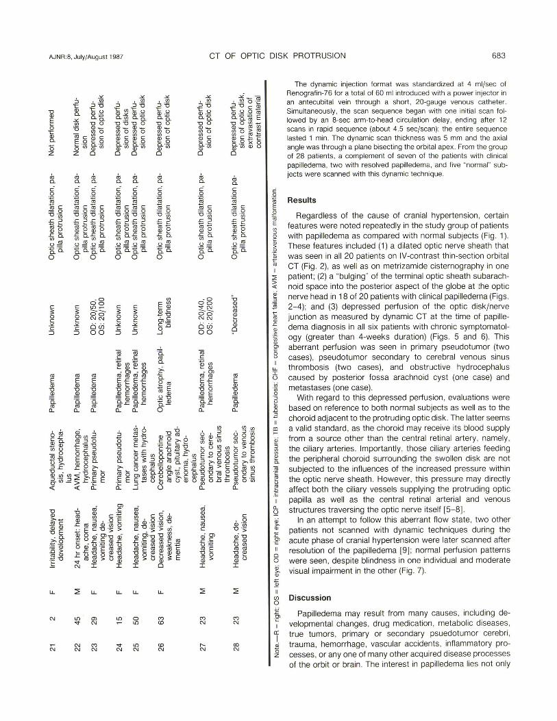
A B Fig, 2.- Case 16. A, Axia l section shows left hemispheric subdura l hematoma. B, Magnified view over orbits shows dilated opt ic nerve sheaths bilaterally.
AJNR:8, July/August 1987
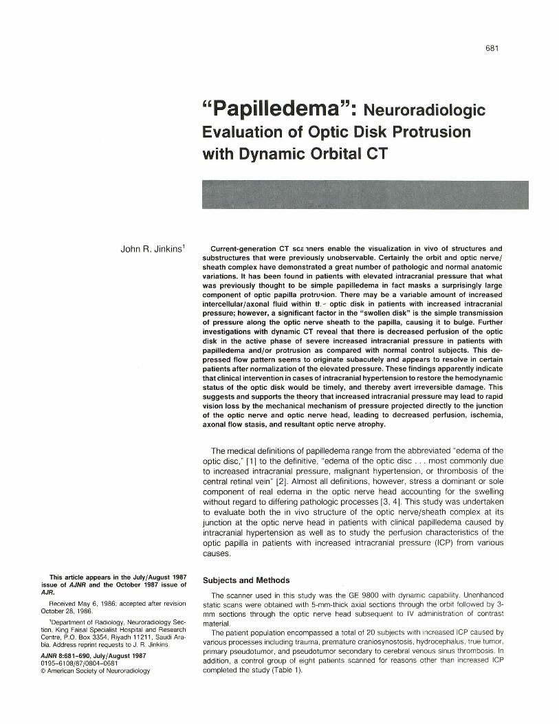
Fig, 1.-Case 8. A, Magnified view of right orbit shows normal
appearance of optic nerve/ sheath complex. B, Video-reverse magnified view of right
globe shows normal, flat appearance of optic disk (arrow).
C, Enlarged video-reverse view of globe during dynamic perfusion peak in normal subject shows complete rim of " choroidal " enhancement (large arrows) with equal enhancement of optic nerve head (small arrow).
D, Dynamic perfusion curves centered over orbits show almost identical perfusion patterns of left optic nerve head (L) as compared with adjacent choroid of left globe (S) ,
c
C, Video-reverse magnified image of right globe after IV enhancement shows optic papilla protruding into posterior aspect of globe (arrow).
AJNR :8, July/August 1987 CT OF OPTIC DISK PROTRUSION 685
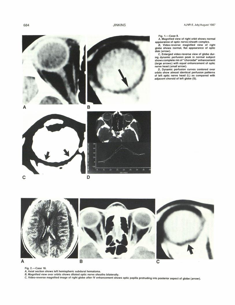
Fig. 3.-Case 26. A, Axial CT section at level of thalami shows
hydrocephalus and periventricular edema secondary to posterior fossa arachnoid cyst causing obstruction to CSF flow at fourth ventricular level.
B, Magnified contrast-enhanced view of right globe on video-reverse imaging shows optic papilla protruding into posterior aspect of globe (arrow).
Fig. 4.-Case 13: primary pseudotumor cerebri.
A B
A, Axial cranial CT after contrast administration shows compressed lateral ventricular system. B, Metrizamide cisternogram centered over orbits outlines markedly dilated optic nerve sheaths extending to globes, traversed by low-density optic
nerves. C, Magnified view of right orbit shows contrast-filled, ballooned terminal optic-sheath subarachnoid space protruding into posterior aspect of globe at
optic nerve head (arrows).
in its diagnostic potential as a measure of orbital or cerebral pathology, but also in the secondary effects of disk swelling leading to rapidly progressive blindness in certain cases [10-14].
To understand papilledema in the present context , it is perhaps best to isolate two major categories of swollen disks. The specific pathologic etiology of "optic disk swelling" covers a spectrum extending from true edema due to many acquired causes to the type of swelling observed in increased ICP [4]. It is the latter that is of concern here and that seems to involve in part the simple dilatation of the sheath surrounding the optic nerve terminating in a "ballooning" of the optic nerve head, causing it to protrude into the globe [4, 15-17]. In addition to this protrusion, there may be a variable amount of
associated intercellular fluid (true edema) accumulation within the optic papilla due to one or more factors : primary tissue ischemia [18 , 19], an increased pressure gradient between the subarachnoid space and the vitreous of the globe with resultant transarachnoid transudation of fluid , or quite likely an extravascular extravasation of fluid secondary to this pressure gradient (Fig . 8) [18 , 20 , 21] . In agreement with the latter theory was the evidence of extravascular accumulation of contrast material in the optic disk due to a breakdown in the "blood-nerve barrier" in a patient in the current study who had cranial hypertension secondary to extensive dural venous sinus thrombosis (Fig . 6C). Nevertheless, a significant factor in early disk swelling or papilledema is simple papillary protrusion (Fig . 9).
686 JINKINS AJNR:8, July/August 1987
A B
o
The more serious consideration previously alluded to is the progressive blindness that accompanies papilledema in some cases. Table 1 points out that this decrease of visual acuity is not strictly limited to patients with "benign" cranial hypertension , nor is it bilaterally symmetric. Many theories have been proposed as to the cause of this vision loss ; most of them hinge on abnormal pressure relationships and ischemia [16, 22- 24] . A most intriguing mystery is why the optic nerve is unusual in its vulnerability to damage from increased ICP. One line of investigation in this regard is to examine why the optic nerve is basically different from the other cranial nerves.
c
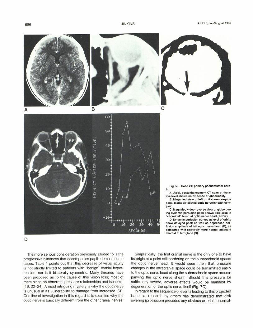
Fig. 5.-Case 24: primary pseudotumor cerebrio
A, Axial, postenhancement CT scan at thalamic level shows no evidence of abnormality.
S, Magnified view of left orbit shows serpiginous, markedly dilated optic nerve/sheath complex.
C, Magnified video-reverse view of globe during dynamic perfusion peak shows skip area in " choroidal " blush at optic nerve head (arrow).
D, Dynamic perfusion curves at level of orbits show delayed peak as well as depressed perfusion amplitude of left optic nerve head (P), as compared with relatively more normal adjacent choroid of left globe (S).
Simplistically, the first cranial nerve is the only one to have its origin at a point still bordering on the subarachnoid space: the optic nerve head. It would seem then that pressure changes in the intracranial space could be transmitted easily to the optic nerve head along the subarachnoid space accompanying the optic nerve sheath . Should this pressure be sufficiently severe, adverse effects would be manifest by degeneration of the optic nerve itself (Fig. 7C).
In regard to the sequence of events leading to this projected ischemia, research by others has demonstrated that disk swelling (protrusion) precedes any obvious arterial abnormal-
AJNR :8, July/August 1987 CT OF OPTIC DISK PROTRUSION 687
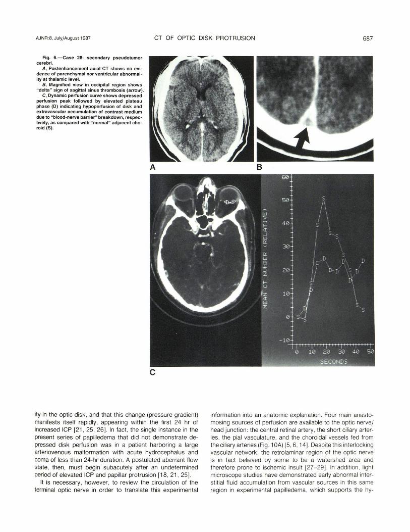
Fig. 5.-Case 28: secondary pseudotumor cerebri.
A, Postenhancement axial CT shows no evidence of parenchymal nor ventricular abnormality at thalamic level.
B, Magnified view in occipital region shows "delta" sign of sagittal sinus thrombosis (arrow) .
C, Dynamic perfusion curve shows depressed perfusion peak followed by elevated plateau phase (D) indicating hypoperfusion of disk and extravascular accumulation of contrast medium due to " blood-nerve barrier" breakdown, respectively, as compared with " normal" adjacent choroid (S).
A
c
ity in the optic disk , and that this change (pressure gradient) manifests itself rapidly , appearing within the first 24 hr of increased ICP [21 , 25, 26]. In fact , the single instance in the present series of papilledema that did not demonstrate depressed disk periusion was in a patient harboring a large arteriovenous malformation with acute hydrocephalus and coma of less than 24-hr duration. A postulated aberrant flow state, then , must begin subacutely after an undetermined period of elevated ICP and papillar protrusion [18 , 21 , 25] .
It is necessary , however, to review the circulation of the terminal optic nerve in order to translate this experimental
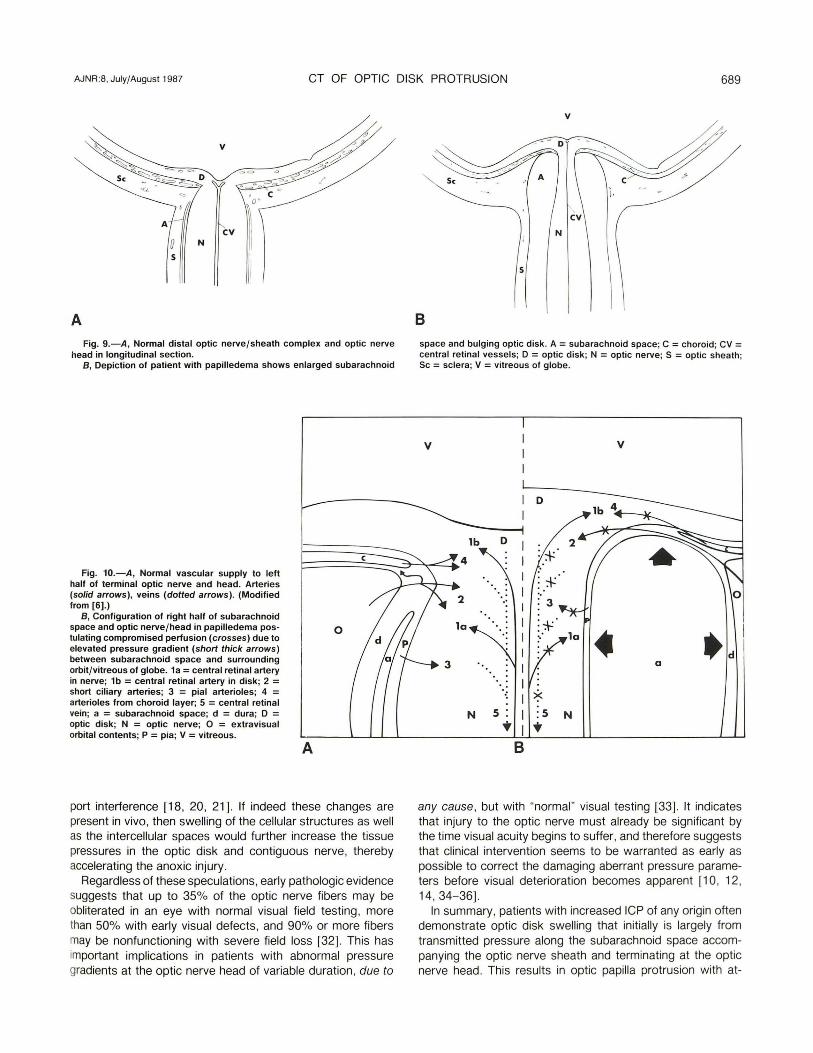
B
information into an anatomic explanation. Four main anastomosing sources of periusion are available to the optic nervej head junction: the central retinal artery, the short ciliary arteries, the pial vasculature, and the choroidal vessels fed from the ciliary arteries (Fig . 1 OA) [5 , 6, 14]. Despite this interlocking vascular network, the retrolaminar region of the optic nerve is in fact believed by some to be a watershed area and therefore prone to ischemic insult [27-29] . In addition , light microscope studies have demonstrated early abnormal interstitial flu id accumulation from vascular sources in th is same region in experimental papilledema, which supports the hy-
688 JINKINS AJNR :8, July/August 1987
A B
c
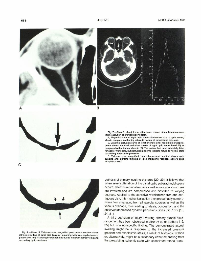
Fig. B.-Case 19. Video-reverse, magnified postcontrast section shows intrinsic swelling of optic disk (arrows) equating with true papi lledema in patient with long-standing hydrocephalus due to midbrain astrocytoma and secondary hydrocephalus.
Fig. 7.-Case 9: about 1 year after acute venous sinus thrombosis and after resolution of cranial hypertension.
A, Magnified view of right orbit shows diminutive size of optic nerve/ sheath complex, confirming return to normal of intracranial pressure.
B, Dynamic perfusion curve at level of orbits after resolution of papilledema shows identical perfusion curves of right optic nerve head (D) as compared with adjacent choroid (S). The patient had been subtotally blind for about 10 months, but perfusion patterns indicate return to normal state paralleling intracranial pressure.
C, Video-reverse, magnified, postenhancement section shows optic cupping and extreme thinning of disk indicating resultant severe optic atrophy (arrow) .
pothesjs of primary insult to this area [20, 30] . It follows that when severe dilatation of the distal optic subarachnoid space occurs, all of the regional neural as well as vascular structures are involved and are compressed and distorted to varying degrees. Applied to the sensitive retrolaminar area and contiguous disk, this mechanical action then presumably compromises flow emanating from all vascular sources as well as the venous drainage, thus leading to stasis, congestion , and the observed depressed dynamic perfusion curves (Fig . 1 OB) [16, 24 , 31 ].
A third postulate of injury involving primary axonal disarrangement has been observed in vitro by other authors [19 , 25] but is a nonspecific finding. The demonstrated axonal swelling might be a response to the increased pressure gradient and axoplasmic stasis , a result of histologic fixation or, alternatively, might be a secondary effect emanating from the preexisting ischemic state with associated axonal trans-
AJNR:8. July/August 1987 CT OF OPTIC DISK PROTRUSION 689
A Fig. 9.-A, Normal distal optic nerve/sheath complex and optic nerve
head in longitudinal section. B, Depiction of patient with papilledema shows enlarged subarachnoid
Fig. 10.-A, Normal vascular supply to left half of terminal optic nerve and head. Arteries (solid arrows), veins (dotted arrows). (Modified from [6].)
B, Configuration of right half of subarachnoid space and optic nerve/head in papilledema pos- 0 tulating compromised perfusion (crosses) due to elevated pressure gradient (short thick arrows) between subarachnoid space and surrounding orbit/vitreous of globe. 1a = central retinal artery in nerve; 1b = central retinal artery in disk; 2 = short ciliary arteries; 3 = pial arterioles; 4 = arterioles from choroid layer; 5 = central retinal vein; a = subarachnoid space; d = dura; D = optic disk; N = optic nerve; 0 = extravisual orbital contents; P = pia; V = vitreous.
A
port interference [18, 20, 21] . If indeed these changes are present in vivo, then swelling of the cellular structures as well as the intercellular spaces would further increase the tissue pressures in the optic disk and contiguous nerve, thereby accelerating the anoxic injury.
Regardless of these speculations, early pathologic evidence suggests that up to 35% of the optic nerve fibers may be obliterated in an eye with normal visual field testing , more than 50% with early visual defects , and 90% or more fibers may be nonfunctioning with severe field loss [32]. This has important implications in patients with abnormal pressure gradients at the optic nerve head of variable duration, due to
v
Sc
B space and bulging optic disk. A = subarachnoid space; C = choroid; CV = central retinal vessels; D = optic disk; N = optic nerve; S = optic sheath; Sc = sclera; V = vitreous of globe.
v v
D
1b D
10
~ td 0
','
~ N S :s N .. ..
B
any cause , but with "normal" visual testing [33] . It indicates that injury to the optic nerve must already be significant by the time visual acuity begins to suffer, and therefore suggests that clinical intervention seems to be warranted as early as possible to correct the damaging aberrant pressure parameters before visual deterioration becomes apparent [10 , 12, 14, 34-36].
In summary, patients with increased ICP of any origin often demonstrate optic disk swelling that initially is largely from transmitted pressure along the subarachnoid space accompanying the optic nerve sheath and terminating at the optic nerve head. This results in optic papilla protrusion with at-
I
690 JINKINS AJNR:8, July/August 1987
tendant depressed perfusion of the optic nerve head, which may be temporary, depending on the degree of injury. The multifactorial insult of direct pressure effects, decreased perfusion, an increase in the inter- and possibly intracellular fluid content, and a disruption in axoplasmic flow/exchange all combine to produce insidiously progressive blindness, leading to optic nerve atrophy. It would seem imperative , therefore, particularly in cases of visually labile cranial hypertension, that the focus clinically should be the immediate reduction of the increased ICP to phYSiologic levels so that both the perfusion of the optic disk as well as the axonal transport mechanisms can be restored, thereby averting permanent injury to the optic nerve.
ACKNOWLEDGMENTS
I thank A. Radford and C. Galban for manuscript preparation, C. Jinkins for manuscript research, and the King Faisal Specialist Hospital 's radiology and medical illustrations departments for technical assistance with the illustrations.
REFERENCES
1. Gould medical dictionary, 4th ed . New York: McGraw-Hili, 1979:984 2. Dorland 's illustrated medical dictionary , 26th ed. Philadelphia: Saunders ,
1981 :960 3. Stedman 's medical dictionary , 24th ed. Baltimore: Williams & Wilkins,
1982: 1022 4. Lepore FE. Toward a definition of papilledema: a historical review, 1851-
1911 . Surg Neuro/1982 ;17(3) :178-180 5. Hayreh SS. Pathogenesis of visual field defects. Br J Ophthalmol
1970;54 :289- 31 1 6. Lieberman MF, Maumenee E, Green WR . Histologic studies of the vascu
lature of the anterior optic nerve. Am J Ophthalmo/ 1976;82(3) :405-423 7. Anderson DR , Braverman S. Reevaluation of the optic disk vasculature.
Am J Ophthalmo/1976 ;(2): 165-174 8. Hayreh SS. Blood supply of the optic nerve head and its role in optic
atrophy, glaucoma, and edema of the optic disc. Br J Ophthalmol 1969;53 :721
9. O'Reilly GV, Hammerschlag SB, Bergland RM , Spark RE, Bienfang DC, Ronthal M. Pseudotumor cerebri : computed tomography of resolving papilledema. J Comput Assist Tomogr 1983;7(2) :364- 366
10. Boddie HG, Banna M, Bradley WG. "Benign- intracranial hypertenSion. A survey of the clinical and radiological features, and long-term prognosis. Brain 1974;97 ' 313- 326
11 . Katzman B, Lu LW, Tiwari RP, Bansal R. Pseudotumor cerebri: an observation and review. Ann Ophthalmo/1981 ;13(7) :887-892
12. Rush JA. Pseudotumor cerebri. Clinical profile and visual outcome in 63 patients. Mayo Clin Proc 1980;55 ;541 - 546
13. Barber SG, Garvan N. Is "benign intracranial hypertension" really benign? J Neurol Neurosurg Psychiatry 1980;43 : 136- 138
14. JeHerson A, Clark J. Treatment of benign intracranial hypertension by
dehydrating agents with particular reference to the measurement of the blind spot area as a means of recording improvement. J Neurol Neurosurg Psychiatry 1976;39: 627-639
15. Osborn AG, Thurman OJ, Van Dyk HJl. The angiographic ocular choroidal crescent: distortion with intraorbital and remote intracranial pathology. Neuroradiology 1978;15: 13-19
16. Green GJ, Lessell S, Loewenstein JI. Ischemic optiC neuropathy in chronic papilledema. Arch Ophthalmo/1980;98(3) : 502-504
17. Bird AC , Sanders MD. Choroidal folds in association with papilloedema. Br J Ophthalmo/1973 ;57:89-97
18. Hayreh SS . Optic disc edema in raised intracranial pressure. V. Pathogenesis. Arch Ophthalmol 1977;95: 1553-1565
19. Tso MOM, Fine BS. Electron microscopic study of human papilledema. Am J Ophthalmo/1976;82(3):424-434
20. Tso MOM, Hayreh SS. Optic disc edema in raised intracranial pressure. III. A pathologic study of experimental papilledema. Arch Ophthalmol 1977;95 : 1448-1457
21 . Hayreh SS, Hayreh MS. Optic disc edema in raised intracranial pressure. II. Early detection with fluorescein fundus angiography and stereoscopic color photography. Arch Ophthalmo/1977 ;95 : 1245-1254
22. Hayreh SS. Optic disc edema in raised intracranial pressure. Associated visual disturbances and their pathogenesis. Arch Ophthalmol 1977;95: 1566-1579
23. Ernest JT, Potts AM. Pathophysiology of the distal portion of the optic nerve. I. Tissue pressure relationships. Am J Ophthalmo/1968 ;66(3) :373-380
24. Ernest JT, Potts AM . Pathophysiology of the distal portion of the optic nerve. II . Vascular relationships . Am J Ophthalmo/1968;66(3) :380-387
25. Hayreh MS, Hayreh SS. Optic disc edema in raised intracranial pressure. I. Evolution and resolution . Arch Ophthalmo/1977;95: 1237-1244
26. Pagani LF. The rapid appearance of papilledema. J Neurosurg 1969;30:247-249
27. Rootman J, Butler D. Ischaemic optic neuropathy-a combined mechanism . Br J Ophthalmo/1980 ;64 : 826-831
28. Henkind P, Charles NC, Pearson J. Histopathology of ischemic optic neuropathy. Am J Ophthalmo/1970;69(1) :78-90
29. Lichter PR, Henderson JW. Optic nerve infarction. Am J Ophthalmol 1978;85:302-310
30. Tso MOM, Shih CY, McLean IW. Is there a blood-brain barrier at the optic nerve head? Arch Ophthalmo/1975;93 :815-825
31 . Rios-Montenegro EN , Anderson DR , David NJ. Intracranial pressure and ocular hemodynamics. Arch Ophthalmo/1973;89:52-58
32. Quigley HA. Glaucoma's optic nerve damage: changing clinical perspectives. Ann Ophthalmo/1982 ;14(7):611-612
33. Quigley HA, Addicks EM, Green WR . Optic nerve damage in human glaucoma. III. Quantitative correlation of nerve fiber loss and visual field defect in glaucoma, ischemic neuropathy, papilledema, and toxic neuropathy. Arch Ophthalmo/1982;100 :135- 146
34. Kaye AH, Galbraith JEK, King J. Intracranial pressure following optic nerve decompression for benign intracranial hypertenSion. J Neurosurg 1981;55:453-456
35. Smith JL, Hoyt WF, Newton TH . OptiC nerve sheath decompression for relief of chronic monocular chocked disc. Am J Ophthalmol 1969;68(4): 633-639
36. Burde RM, Karp JS, Miller RN. Reversal of visual deficii with optic nerve decompression in long-standing pseudotumor cerebri (correspondence). Am J Ophthalmo/1974;77(5) :770-772