Paper-Supported Three-Dimensional Cell Culture for...
21
S-1 Supporting Information for Paper-Supported Three-Dimensional Cell Culture for Tissue-Based Bioassays Ratmir Derda, Anna Laromaine, Akiko Mammoto, Sindy K. Y. Tang, Tadanori Mammoto, Donald E. Ingber and George M. Whitesides Methods. Cells and reagents We purchased MDA-MB-231 cells from ATCC and primary IMR- 90, HDF and primary HUVEC cells from Lonza. MDA-MB-231, IMR-90, HDF and LLC cells were cultured in Eagle’s minimal Essential medium (MEM) from Lonza supplemented with 10% fetal bovine serum (FBS) and 1% GlutaMax TM (both from Invitrogen). Primary HUVEC cells were cultured in endothelial growth medium (EGM-2, Lonza). Matrigel (growth factor-free) was purchased from BD-Bioscience. General protocol for plating and culture of cells on paper substrates To prepare paper substrates, we cut chromatography paper (Millipore) using scissors or a laser cutter (Versa Laser -Universal Laser VL-300, 50 Watt, using the settings for 200 μm deep-engraving plastic). The papers were rinsed them with deionized water, autoclaved, and dried them in a laminar flow hood. To plate the cells on paper substrates, we detached the cells from growth flasks using treatment with trypsin-EDTA (3-5 min); washed the cells with serum-containing media and pelleted them by centrifugation at 1,000 rpm for four minutes. The cells were re-
-
Upload
nguyenthien -
Category
Documents
-
view
215 -
download
0
Transcript of Paper-Supported Three-Dimensional Cell Culture for...

S-1
Supporting Information for Paper-Supported Three-Dimensional Cell Culture for
Tissue-Based Bioassays
Ratmir Derda, Anna Laromaine, Akiko Mammoto, Sindy K. Y. Tang, Tadanori
Mammoto, Donald E. Ingber and George M. Whitesides
Methods.
Cells and reagents We purchased MDA-MB-231 cells from ATCC and primary IMR-
90, HDF and primary HUVEC cells from Lonza. MDA-MB-231, IMR-90, HDF and LLC
cells were cultured in Eagle’s minimal Essential medium (MEM) from Lonza
supplemented with 10% fetal bovine serum (FBS) and 1% GlutaMaxTM (both from
Invitrogen). Primary HUVEC cells were cultured in endothelial growth medium (EGM-2,
Lonza). Matrigel (growth factor-free) was purchased from BD-Bioscience.
General protocol for plating and culture of cells on paper substrates
To prepare paper substrates, we cut chromatography paper (Millipore) using scissors or a
laser cutter (Versa Laser -Universal Laser VL-300, 50 Watt, using the settings for 200
μm deep-engraving plastic). The papers were rinsed them with deionized water,
autoclaved, and dried them in a laminar flow hood.
To plate the cells on paper substrates, we detached the cells from growth flasks using
treatment with trypsin-EDTA (3-5 min); washed the cells with serum-containing media
and pelleted them by centrifugation at 1,000 rpm for four minutes. The cells were re-

S-2
suspended in cold Matrigel (free of growth factor) to yield a concentration of 107
cells/mL. We spotted this suspension of cells on the specific areas in the paper with a
hand-held Gilson P10 pipette man or Eppendorf Repeater Plus pipette, placed the spotted
paper into a Petri dish filled with warm (36 ºC) growth media and cultured for 24 hours in
an incubator at 36 ºC and atmosphere of 5% CO2. Subsequently, we folded the paper
using sterile tweezers and we pressed them together using custom made plastic or
stainless steel holders. The stacks were cultured in 25 mm-deep Petri dish for the desired
time at 36 ºC, in a 5% CO2 incubator, on an orbital shaker set to 40-60 rpm. We
exchanged the media every two days. At a chosen time, each construct was processed
(pre-treatment, fixation, permeabilization, staining) according to the Table 1.
The paper containing labeled cells was scanned using a Typhoon gel scanner with the
proper setting for each stain. Table 1 lists scanner settings—the laser excitation, emission
wavelength, resolution, and value for the photo multiplier tube (PMT)—for each stain.
The images obtained after scanning were analyzed using ImageJ or MatLab.
S-3
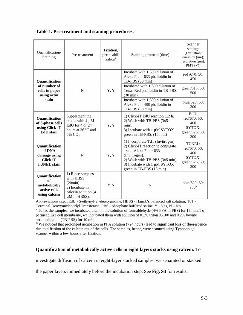
Table 1. Pre-treatment and staining procedures.
Quantification/ Staining Pre-treatment
Fixation, permeabili
zationa Staining protocol (time)
Scanner settings
(Excitation/ emission (nm);
resolution (µm); PMT (V))
Incubate with 1:500 dilution of Alexa Fluor 633 phalloidin in TB-PBS (30 min)
red /670; 50; 450
Incubated with 1:300 dilution of Texas Red phalloidin in TB-PBS (30 min)
green/610; 50; 500
Quantification of number of cells in paper
using actin stain
N Y, Y
Incubate with 1:300 dilution of Alexa Fluor 488 phalloidin in TB-PBS (30 min)
blue/520; 50; 300
Quantification of S-phase cells using Click-iT
EdU stain
Supplement the media with 4 µM EdU for 4 or 24 hours at 36 ºC and 5% CO2
Y, Y
1) Click-iT EdU reaction (12 h) 2) Wash with TB-PBS (3x5 min). 3) Incubate with 1 µM SYTOX green in TB-PBS. (15 min)
EdU: red/670; 50;
400 SYTOX:
green/526; 50; 300
Quantification of DNA
damage using Click-iT
TUNEL stain
N Y, Y
1) Incorporate TdT (Invitrogen) 2) Click-iT reaction to conjugate azido-Alexa Fluor 633 (Invitrogen). 2) Wash with TB-PBS (3x5 min) 3) Incubate with 1 µM SYTOX green in TB-PBS (15 min)
TUNEL: red/670; 50;
400 SYTOX:
green/526; 50; 300
Quantification of
metabolically active cells
using calcein
1) Rinse samples with HBSS (20min). 2) Incubate in calcein solution (4 µM in HBSS).
Y, N N blue/520; 50; 300b
Abbreviations used: EdU - 5-ethynyl-2´-deoxyuridine, HBSS - Hanck’s balanced salt solution, TdT - Terminal Deoxynucleotidyl Transferase, PBS - phosphate buffered saline, Y - Yes, N – No. a To fix the samples, we incubated them in the solution of formaldehyde (4% PFA in PBS) for 15 min. To permeabilize cell membrane, we incubated them with solution of 0.1% triton X-100 and 0.2% bovine serum albumin (TB-PBS) for 10 min. b We noticed that prolonged incubation in PFA solution (>24 hours) lead to significant loss of fluorescence due to diffusion of the calcein out of the cells. The samples, hence, were scanned using Typhoon gel scanner within a few hours after fixation.
Quantification of metabolically active cells in eight layers stacks using calcein. To
investigate diffusion of calcein in eight-layer stacked samples, we separated or stacked
the paper layers immediately before the incubation step. See Fig. S3 for results.
S-4
1. “Stack non-stack”: We cultured the stacked layers for nine days, then washed
the samples with warm HBSS, separated the layers using sterile tweezers and
placed them within one minute in a pre-warmed solution of calcein AM (4 µM in
HBSS). The separated layers were rocked for ten minutes at 36 ºC, and fixed with
cold PFA solution.
2. “Non-stack stack”: The non-stacked layers were cultured for nine days,
washed with warm HBSS, stacked, pressed together with a plastic holder and
placed them within one minute in the pre-warmed solution of calcein AM (4 µM
in HBSS). The separated layers were rocked for ten minutes at 36 ºC, and fixed
with cold PFA solution.
3. “Stack flip”: After nine days of culture, we washed the stacks with warm
HBSS media, removed the cellulose acetate layer from the bottom layer (L8), and
placed it atop (L1). The resulting stack was placed in a pre-warmed solution of
calcein AM (4 µM in HBSS) within one minute. The separated layers were
rocked for ten minutes at 36 ºC, and fixed with cold PFA solution.
Quantification of gene expression using real-time PRC. After nine days of culture, we
placed the cell-containing area in a 2-mL Eppendorf tube containing 500 µL of lysis
buffer (buffer RLT, RNAeasy Kit, Quiagen). Vortexing the tubes at 4 ºC for twenty min
turned the paper substrate to a homogenous pulp which was centrifuged at 14,000 rpm for
two minutes. Both supernatant and the pellet were transferred to QiaShredder tubes
(Quiagen) and centrifuged them at 14,000 rpm for two minutes. The flow-through
solution was processed using RNAeasy kit according to the manufacturer’s procedure

S-5
(Quiagen). Concentrations of RNA were determined using NanoDrop (Thermo
Scientific). The yield of RNA from the multi-layered samples correlated with the number
of cells in the layers, and ranged from 80 to 2000 ng/µL (A250/A280 ratio of >1.9). We
detected nearly no degradation in the samples as judged by 28S:18S rRNA bands on
agarose gels.
We synthesized cDNA from total RNA using the High Capacity cDNA Reverse
Transcription Kit (Applied Biosystems, #4368814) using 100 ng of total RNA in 20 µL
of the reaction volume. The resulting cDNA solution was diluted 10 fold and stored at -
80 ºC. For PCR, we mixed 5.1 µL of cDNA solution with 7.5 µL of Power SYBR®
Green PCR Master mix and 1.2 µL of 10 µM solution of forward and reverse primers for
human VEGF (forward: 5’-TGFAGTACATCTTCAAGCCATCCTGTGTGC-3’ and
reverse 5’-CCTATGTGCTGGCCTTGGTGAGGTTTGAT-3’), human IGFBP3
(forward: 5’-TCTGCGTCAACGCTAGTGC-3’, reverse 5’-
GCTCTGAGACTCGTAGTCAACT-3’), human beta-2 microglobulin (forward: 5’-
GAATGGAGAGAGAATTGAAAAAGTGGAGCA-3’, reverse 5’-
CAATCCAAATGCGGCATCTTCAAAC-3’), human actin (forward: 5-
CATGTACGTTGCTATCCAGGC-3’ and reverse 5’-
CTCCTTAATGTCACGCACGAT-3’). We performed the reaction on an Applied
Biosystems 7900HT Real-Time PRC system using 40 cycles of a two-step cycling
protocol (60 ºC for one min, 96 ºC, 15 seconds).a The concentration for VEGF, IFGPB3
transcripts was estimated using the ΔΔCt approach using Ct of beta-2 microglobulin
(human cells) from the same sample. We performed a pair-wise significance test for the
a The above conditions were tested with 100 fold serial dilution of total cDNA and yielded linear 2Ct vs. Log(concentration) plots and slopes corresponding to efficiency of DNA synthesis equal to 1.9-2.0.

S-6
samples calculating P-values for 2ΔCt values using the Wilcoxon rank sum test. To
simplify presentation, all 2ΔCt were divided by an average 2ΔCt of L1.
In vivo implantation assay. All animal studies were reviewed and approved by the
Animal Care and Use Committee of Children’s Hospital Boston. We mixed LLC cells
with 1:1 vol/vol mixture of Matrigel and growth medium (107 cells/mL), spotted 6 μL of
this suspension onto the patterned chromatography paper (size 8 mm x 8 mm, for SL
sample) or 8 x 6 μL onto the patterned strip of paper (64 mm x 8 mm, for L1L8 sample)
(Fig. S4D). The samples were incubated in the growth medium at 37 ºC overnight, folded
and implanted subcutaneously on the backs of C57BL/6 mice (left and right side). As
controls, we cultured the same samples in growth medium in vitro. After three days, we
injected rhodamine-conjugated ConA into the retro orbital sinus, excised the papers
surgically (Fig. S4E), fixed with 4% PFA for 24 hours, and de-stacked the layers for
analysis. For sectioning, excised and fixed samples were incubated in 30% sucrose in
PBS for 24 hours, embedded in optimal cutting temperature (OCT) compound; each
samples was cryosectioned from the edge to the middle of the sample (Fig. S4F, G). We
thawed the remaining halves of the samples, rinsed with PBS and de-stacked the layers
for further analysis. Sections and de-stacked layers were imaged using fluorescent gel
scanner (Fig. 6C-G).
We stained the sections (18-μm thick) with hematoxylin and eosin (H&E) (Fig. 6H-K) as
described previously(1, 2). For immunostaining, the sections were hydrated with PBS (10
min), permeabilized with PBS containing 0.1% Triton-X100 (30 min), blocked with PBS
containing 10% fetal calf serum (FCS) and 0.1% triton X-100 (30 min). We used 1:100
S-7
dilution of primary antibody (anti-CD31 or anti-CD45, overnight at 4 ºC), washed with
PBS (3 x 5 min), and incubated with 1:100 dilution of Alexa Fluor 633-conjugated
secondary antibody. We mounted the samples using Vectashield+DAPI and performed
four-color imaging (blue: DAPI; green: GFP; red Rhodamine ConA; far red:
immunostaining) using Carl Zeiss ApoTome or Leica laser scanning confocal microscope
(Fig. 6I-P, Fig. S4).

S-8
Supplementary Figure S1.

S-9
Supplementary Figure S1.
(A) To relate the fluorescent intensity of F-actin to the number of MDA-MB-231 cells
inside the paper, we spotted 4 μL of a suspension of MDA-MB-231 cells in Matrigel (106
– 3x10⁷ cells/mL) onto filter paper, incubated the samples for two hours in growth
medium, fixed them, and stained them with Texas Red- or Alexa Fluor 633-conjugated
phalloidin. Similarly to the previous results (Fig. 1I), we observed a non-linear relation
between F-actin fluorescence and the number of cells in paper. The nature of the
dependence (y=ax1/2) was similar for the two dyes we tested. Non-linearity is not the
result of the detector saturation, because we observed similar trends with different
photomultiplier (PMT) voltages applied to the detector. Both data sets on log-log plot fit
a line with slope equal to 0.5. (B) We also quantified the number of MDA-MB-231 cells
in paper using the rate of turnover of metabolic reagent (Alamar BlueTM). We incubated
papers presenting different number of cells with growth medium containing 10% of
Alamar Blue. Growth medium was sampled at different times (one, two, three and six
hours); fluorescence of the media at these times was determined, and the rate of change
of fluorescent intensity (units per hour) was plotted against the number of cells. The
observed linear correlation between Alamar Blue turnover, and the number of cells, was
used as a calibration curve in Fig. 3 and 5. (C) We cultured MDA-MB-231 cells in paper-
Matrigel matrix for one, three, five, seven or nine days and at those days, we removed
one sample from the medium and fixed it with 4% PFA solution. At the end of the
experiment, samples from different days were stained with fluorescently labeled
phalloidin and quantified using a fluorescent gel scanner and ImageJ. (D) We cultured
MDA-MB-231 cells in paper-Matrigel matrix for nine days; on days 1, 5 or 9, the media

S-10
was supplemented with Alamar Blue. After measuring the reagent turnover, the media
was changed back to normal growth media. Both F-actin stain intensity (C) and Alamar
Blue turnover rate (D) increased over time, but the results from F-actin stain did not
match that of Alamar Blue due to the non-linear relation between F-actin fluorescence
and the number of cells. The discrepancy can be resolved when F-actin fluorescence is
converted to the number of cells using y=x1/2 calibration. The resulting (2.5)2 ~ 6 fold
increase of F-actin stain over nine days resembles the result obtained by Alamar Blue
(five fold / nine days). Growth rate obtained from F-actin stain is higher, because this
staining method detects both metabolically active and inactive cells. (F) Long-term
proliferation of MDA-MB-231 cells monitored by Alamar Blue. (F-I) Confocal
microscopy images of MDA-MB-231 cells on day 1 (F-G) and day 9 (H-I) confirm the
results from Alamar Blue, and F-actin, and show that the number of cells in paper indeed
increase by 5-6 fold over nine days. We fixed the cells with PFA, stained with AF-633
phalloidin and Sytox to visualize F-actin (red) and nuclei (green), and imaged them using
a laser scanning confocal microscope (Leica) with 20x objective. Cellulose fibers were
detected via reflection (purple). 3D reconstruction was performed using Volocity. The
inside of the cell appear yellow due to overlap of red (F-actin) and green (nucleus)
channels in the 3D reconstructed images.

S-11
Supplementary Figure S2.

S-12
Supplementary Figure S2.
Investigation of the rate of proliferation of various cell types in a paper-supported
Matrigel matrix. (A-H) We spotted a suspension of cells in Matrigel (4 μL, 107 cells/mL)
onto filter paper, and allowed the cells to equilibrate for 24 hours in growth media. The
stacks of eight or six layers were cultured nine days. SL controls were cultured for one,
three, five, seven, or nine days and on those days, we removed one sample from the
growth medium and fixed it with 4% PFA. At the end of the experiment, L1-L8 samples
and SL samples from different days were stained with fluorescently labeled phalloidin,
and quantified using a fluorescent gel scanner and ImageJ. The blue line represents actin
stain intensity in SL samples on day one; the red line represents stain intensity in SL on
day nine. In B, D, F, H, an average from four to six measurements are presented and the
error bar is one standard deviation. In A, C, E, G, all data are presented, and the
overlaying grey bar is equal to 2x (standard deviation). We calculated p values (* p<0.05,
** p<0.01) using the two-tailed, two-sample unequal variance t-test. (I, L) Confocal
images and 3D-reconstruction of the confocal images of the endothelial lumens formed
on day seven by HUVEC cells in the top (L1) layer, and the small lumens in the bottom
(L8) layer of the eight-layered stack of HUVECs. (I-K) Cells were stained with AF488-
phalloidin or anti-VE-cadherin antibody (L) and imaged using laser scanning confocal
microscope. Color schemes are the following: purple: cellulose fibers (detected via
reflection), blue – nuclei, green – F-actin (I-K) or VE-cadherin (J). (L-M) The number of
nuclei per tube was counted in 11 images for “Layer 1” and six images for “Layer 8”.
Counting was performed manually by three independent participants. “Layer 1” has a

S-13
significantly higher fraction of large tubes, with more cells per tube (3-30 nuclei per
tube); whereas much smaller tubes (1-5 nuclei per tube) are observed in “Layer 8”.

S-14
Supplementary Figure S3.

S-15
Supplementary Figure S3.
Quantification of metabolically active cells using calcein dye. The outcome of the calcein
assay depended on the time and the concentration of cells, and could be described using
first order kinetics. The red line demonstrates the fit to the curve C=C0(1-e-kt). At a
defined time point (e.g. 20 minutes), the signal was proportional to the number of cells.
In 3D cultures, however, the outcome of calcein analysis was also determined by the
balance of diffusion rate and reaction rate. To address this issue, we investigated several
stacked and non-stacked geometries and determined that staining of thick 3D cultures led
to development of calcein signal only in layers exposed to medium (even though live
cells reside in every single layer). This discrepancy was resolved when the layers were
separated prior to the assay, to allow for unrestricted diffusion of calcein to cells in each
layer.

S-16
Supplementary Figure S4

S-17
Supplementary Figure S4
(A) LLC cells proliferate normally when cultured inside paper-Matrigel substrates in vitro
as demonstrated by Alamar Blue. In this experiment, we generated suspension of LLC
cells in 1:1 vol/vol mixture of Matrigel and growth medium (107 cells/mL) and spotted 4
μL of this suspension onto the 200 μL-thick chromatography paper. At designated time,
we supplemented the medium with 10% of Alamar Blue, measured rate of change of
fluorescence, and substituted back to the complete growth medium. (B-C) After nine days
of culture in eight layers of paper, distribution of cells (as judged by F-actin stain, B) and
distribution of S-phase cell (as determined by Click-iT EdU stain, C) resembles that of
MDA-MB-231 cells (Fig. 2B, D) and other cell lines (Fig. S2). (D) Patterned
chromatography paper before cell seeding and implantation. Black areas are patterned
with printer ink(3) and form hydrophobic, impermeable border; white areas are
unmodified paper. (E) Samples after three days implantation and excision. (F-G) Light
microscopy (2x) image of the OCT-embedded samples sectioned to the middle of the
sample. (H) 18 micron-thick sections of implanted SL sample stained with Hematoxylin
and Eosin (HE). (I-L) We stained the sections from SL samples (J, K) and L1L8 samples
(I, L) with anti-CD31 to detect endothelial cells or anti-CD45 antibody to detect
differentiated hematopoetic cells, and appropriate secondary antibody conjugated to
AlexaFluor633. Functional vasculature was labeled by intravenously injected
Rhodamine-ConA. Confocal imaging was performed using Carl Zeiss ApoTome
equipped with 10x objective, automated stage and mosaic imaging capability. An average
of 20 (SL) to 80 (L1L8) images were collected and stitched to obtain the overview

S-18
images. We used the following color scheme: green – GFP, red – Rhodamine ConA, blue
– DAPI, purple – CD31 or CD45 (as noted).

S-19
Supplementary Figure S5

S-20
Supplementary Figure S5
(A) To assess the effect of brief cold treatment on long-term proliferation of cells, we
plated the LLC cells atop paper permeated with Matrigel. We allowed the cells to adhere
for 24 hours and exposed one set of samples to ice-cold media (4 ºC) for 30 min, another
set of samples to ice cold-media for two hours, and the third, control set, we never
exposed to any cold media (N=8 in each set). We monitored the number of cells in all
samples over the next three days using Alamar Blue. (B) A plot of reagent turnover,
which is proportional to the number of metabolically active cells, before, and after the
treatment. All data from all samples is presented. (C) Increase in number of cells from
day two to day four in cold-treated samples is similar to that of control, untreated
samples. We calculated ratio of reagent turnover on day four to that on day two for each
sample and averaged the resulting values (N=8); error bad is one standard deviation.
Growth rates are statistically insignificant (p>0.05) as indicated by two-tailed unequal
variance t-test. We conclude that 24 hour incubation in 36 ºC-warm media is sufficient to
alleviate the effect of brief exposure (<2 h) of cells to cold medium.

S-21
References:
1. Yung CW, et al. (2007) Transglutaminase crosslinked gelatin as a tissue engineering scaffold. J Biomed Mater Res Part A 83A:1039-1046.
2. Mammoto A, Huang S, Moore K, Oh P, Ingber DE (2004) Role of RhoA, mDia, and ROCK in cell shape-dependent control of the Skp2-p27(kip1) pathway and the G(1)/S transition. J Biol Chem 279:26323-26330.
3. Carrilho E, Martinez AW, Whitesides GM (2009) Understanding Wax Printing: A Simple Micropatterning Process for Paper-Based Microfluidics. Anal Chem:DOI: 10.1021/ac901071p.