PAO Clinical Practice Guidelines: Management of Cataract …pao.org.ph/standard/CPG.pdf · Clinical...
31
Committee on Standards Philippine Academy of Ophthalmology 815 Medical Plaza Makati Makati City, MMLA, Philippines PAO Clinical Practice Guidelines: Management of Cataract among Adults
-
Upload
trinhnguyet -
Category
Documents
-
view
216 -
download
0
Transcript of PAO Clinical Practice Guidelines: Management of Cataract …pao.org.ph/standard/CPG.pdf · Clinical...

Committee on Standards Philippine Academy of Ophthalmology 815 Medical Plaza Makati Makati City, MMLA, Philippines
PAO Clinical Practice Guidelines: Management of Cataract among Adults

Clinical Practice Guidelines: Management of Cataract among Adults PAO
How to Use this Document Most readers would be immediately interested in the Recommendations in this CPG. A tabulation of
these recommendations appears in page 7. A detailed discussion and justification of each
recommendation follows sequentially from page 15. For readers and researchers more interested in a
in-depth critical analysis of the evidence, the sources used are in the References section and the
Evidence-Base and the proceedings of the CPG Panel session are on file at the PAO.
This CPG is an update of the original work in 2001. A brief historical perspective is given in the
Introduction. The research methodology and protocols used in the CPG formulation are found from
page 9 to 15. Consult the Table of Contents in the following page for an enumeration of the extensive
material contained herein.
The information can best be appreciated by reading this document from beginning to end, then keep
it on hand as a reference. These contents also appear in electronic media on the PAO website and in
the electronic copy of the Philippine Journal of Ophthalmology which can be accessed through any
internet portal.
While anyone is authorized to copy this CPG in its entirety or in part for personal use, the content is
the intellectual property of the PAO. Kindly provide the proper citations when utilizing the guidelines
and recommendations in lectures, research papers and other material used in public.
Queries, clarifications, suggestions, and other issues pertinent to this CPG may be directed to the
PAO office by email.
�2

Clinical Practice Guidelines: Management of Cataract among Adults PAO
�3

Clinical Practice Guidelines: Management of Cataract among Adults PAO
ACKNOWLEDGMENT This project would not have been possible without the initiative and financial support from the Philippine Academy of Ophthalmology (PAO) and the Fred Hollows Foundation. The latter provided an unrestricted grant to the PAO for the purpose of developing the CPG. To their credit, neither organization imposed any condition nor exerted any influence in the methodology, in the operations and in the formulation of the final output. The Asia Pacific Center for Evidence Based Healthcare (APCEBH) provided the extensive technical work in gathering and processing the evidence; in ensuring the adherence to correct procedures for greater objectivity, for rooting out conflicts of interest, and for eliminating bias; in facilitating the panel discussion and Delphi forum; and, in the documentation and writing of the final report. The PAO Secretariat was indispensable in carrying out the legwork and coordinating between the various individuals, groups, organizations and committees. The CPG Steering Committee of the PAO Committee on Standards was responsible for overall direction and management, and, is ultimately accountable for the quality of the output. Most importantly, this CPG is invaluable because of the contribution and enthusiastic participation of the panelists, who unselfishly committed much time and effort, sharing their knowledge, experience and expertise, in analyzing the evidence and synthesizing the recommendations into the nuggets of information that will serve as beacons for ophthalmologists in the treatment of adult cataracts for
years to come.
DISCLAIMER While the PAO encourages voluntary adherence to this guideline, it is cognizant of its limitations. The CPG is based on the best evidence available in scientific literature as of the time of its formulation, however, certain aspects of management not addressed by clinical studies are therefore not included. Hence, this is not a comprehensive guide. Like all CPGs, this is not meant to restrict the ophthalmologist, using sound clinical judgement and in concordance with the patient, from considering other modes of management especially when molded according to the patient's particular needs and preferences. Individual patients and their response to treatment cannot be always predicted nor guaranteed. The ophthalmologist must not use the CPG as a substitute for exercising good judgement in making treatment decisions and tailoring them according to each patient's peculiar circumstances. This CPG can serve to inform policy but it is not meant to serve as a basis for approving or denying financial coverage or insurance claims merely because of nonconformance with recommendations. Neither are the recommendations supposed to be considered as legal rules for dictating certain modes of action to the exclusion of others.
�4

Clinical Practice Guidelines: Management of Cataract among Adults PAO
ABBREVIATIONS
APCEBH Asia Pacific Center for Evidence Based Healthcare
COS Canadian Ophthalmological Society
CPG Clinical Practice Guidelines
CT Computerized Tomography
DSBCS Delayed Sequential Bilateral Cataract Surgery
EBM Evidence-Based Medicine
ECCE Extracapsular Cataract Extraction
FLACS Femtosecond Laser-Assisted Cataract Surgery
GRADE Grading of Recommendations Assessment, Development and Evaluation
ICD International Classification of Diseases
ISBCS Immediate Sequential Bilateral Cataract Surgery
MSICS Manual Small Incision Cataract Surgery
Nd:YAG Neodymium:Yttrium-Argon-Garnet Laser
NICE National Institute for Health and Care Excellence
NGO Non-governmental organizations
PAO Philippine Academy of Ophthalmology
PCO Posterior Capsular Opacification
PHIC Philippine Health Insurance Corporation
PI Povidone-Iodine
PSCRS Philippine Society of Cataract and Refractive Surgery
RCO Royal College of Ophthalmologists
�5

Clinical Practice Guidelines: Management of Cataract among Adults PAO
EXECUTIVE SUMMARY This Clinical Practice Guidelines (CPG) constitutes the 2016 update of the Philippine Academy of Ophthalmology (PAO) to the 2005 CPG on management of cataract among adults. The 2005 CPG in turn updated the 2001 CPG, which at that time was a collaboration between the PAO and the Department of Family & Community Medicine of the University of the Philippines - Philippine General Hospital.
The document provides selected practice recommendations on surgical techniques for cataract (phacoemulsification, extracapsular cataract extraction, manual small incision cataract surgery or laser assisted cataract surgery) and ancillary procedures (routine pre-operative ancillary testing or ‘clearance’), routine lacrimal duct irrigation, same sitting vs delayed bilateral cataract surgery, routine peri-operative antibiotic prophylaxis, povidone-iodine antisepsis and Nd:YAG laser capsulotomy for posterior capsular opacification after cataract surgery.
Recommendations are based on the best available evidence and are intended to be used by ophthalmologists and other eye care professionals, clinical staff, policy-makers, program managers, payors, NGOs and others. The guideline development process followed the widely accepted Grading of Recommendations Assessment, Development and Evaluation or the GRADE approach and included 1) identification of critical questions and critical outcomes, 2) retrieval of current evidence, 3) assessment and synthesis of the evidence base for these critical questions, 4) formulation of draft recommendations, 5) assembly of multi-sectoral stakeholder panel to assess the quality of the evidence and strength of the recommendations and 6) planning for dissemination, implementation, impact evaluation and updating.
The recommendations in this CPG shall hold until such time that technology, patient and provider preferences, or new evidence provides the motivation for revisiting and updating the guideline once
more.
�6

Clinical Practice Guidelines: Management of Cataract among Adults PAO
Table 1. Summary of Recommendations.
Recommendation Strength of Panel Recommendation
Quality of Evidence
1. Pre-operative medical ancillary testing prior to cataract surgery is recommended only if indicated by the patient's medical condition and the physician's assessment.
Strong Moderate to high
2. Lacrimal duct irrigation as a routine pre-operative procedure in cataract surgery does not reduce the incidence of endophthalmitis but may be performed when indicated.
Strong Very low
3. Instillation or irrigation of the conjunctiva with 5% povidone iodine solution pre-operatively is recommended to reduce the risk of postoperative endophthalmitis.
Strong Very low to low
4. The use of peri-operative antibiotic prophylaxis is recommended to reduce the risk of postoperative endophthalmitis in patients who undergo cataract surgery.
Strong Moderate
5. Delayed Sequential Bilateral Cataract Surgery is preferred over Immediate Sequential Bilateral Cataract Surgery (ISBCS) in the same sitting for patients with bilateral senile cataracts.
Strong Very low to low
6. MSICS is the preferred technique for cataract surgery over ECCE because of less surgically induced astigmatism despite the significantly higher risk for complications.
Strong Low to moderate
7. Phacoemulsification is the preferred technique for cataract surgery over MSICS because of faster visual improvement and lower risk of adverse events or complications.
Strong Very low to low
8. Phacoemulsification is the preferred technique for cataract surgery over ECCE because of significant benefits and lower risk of complications
Strong Low to moderate
9. The choice of FLACS or conventional phacoemulsification for routine cataract surgery will depend on accessibility, surgeon experience, and patient cost preferences.
Weak Very low
10. Regardless of time elapsed after cataract surgery, Laser (Nd:YAG) Capsulotomy is only recommended in patients who develop symptomatic posterior capsular opacification, because of the risk of macular edema, anterior chamber reaction, retinal detachment and other adverse events which may be associated with the procedure.
Strong Very low
�7

Clinical Practice Guidelines: Management of Cataract among Adults PAO
I. INTRODUCTION
Cataract is the leading cause of reversible blindness worldwide [1] and is associated with major economic, social, and health outcomes. The management of cataract is a growing field of interest facing a disproportionate number of challenges, including the inappropriate utilization of resources and ineffective and unsafe techniques. Cataract surgery is one of the top claimed procedures in the Philippines as reported by PhilHealth (PHIC), the country's leading health insurance provider. [2] As such, PHIC needs to ensure that the standards of care in cataract management are established throughout the nation.
Wide practice variations and the changing healthcare system engender the need for Clinical Practice Guidelines (CPG). By definition, CPG are "statements that include recommendations, intended to optimize patient care, that are informed by a systematic review of evidence and an assessment of the benefits and harms of alternative care options.” [3] They must be based on the best available research evidence and practice experience.
In response to need, the PAO, in collaboration with the Department of Family & Community Medicine of the University of the Philippines - Philippine General Hospital developed its first Evidence-based Clinical Practice Guidelines for the Management of Cataract among Adults in March 2001. The document was accepted for inclusion in the National Guideline Clearinghouse, an online database of evidence-based CPG put up by the Agency for Healthcare Research and Quality of the United States Department of Health and Human Services.
Rapid advances in technology and mounting scientific knowledge demand that we periodically review our clinical practice. In 2005, the PAO committee on Evidence-based Ophthalmology revisited the CPG and identified one particular recommendation that was deemed outdated, in reference to changes in the evidence, available resources and values placed on outcomes. At that time, a growing number of local ophthalmologists had made the shift from extracapsular cataract extraction (ECCE) to phacoemulsification. Thus, the goal of the update was to assess whether Recommendation #14 that stated that “both phacoemulsification and ECCE are acceptable techniques among patients undergoing cataract surgery,” still represented the best practice. The 2005 CPG Update was done in two stages. The first step involved a Technical Review team that identified new evidence by conducting a systematic review of the literature. A summary of the evidence was presented to the original panel of guideline developers (2001 CPG) who in turn assessed whether the new evidence warranted updating or withdrawal. Seventy-one percent (71%) of the panel voted to retain the original recommendation, while at the same time appending new and relevant information for clinicians. [4]
More than a decade has passed and considerable progress has taken place in cataract management and in the healthcare industry, as well as in the acceptable processes in CPG development. Thus, the PAO has once again embarked on updating the Cataract CPG. This time around, interest revolved
�8

Clinical Practice Guidelines: Management of Cataract among Adults PAO
around six specific clinical topics dictated by new scientific research, emerging ethical concerns and innovative trends in healthcare:
1. Which cataract surgical technique (phacoemulsification, extracapsular cataract extraction, manual small incision cataract surgery or femtosecond laser assisted cataract surgery) is most effective in improving visual outcomes
2. The role of routine pre-operative clearance in preventing mortality/morbidity 3. The effectivity of lacrimal apparatus irrigation in preventing endophthalmitis 4. The cost-effectiveness of bilateral delayed sequential cataract surgery compared to simultaneous
cataract surgery in improving visual outcomes 5. The use of prophylactic antibiotics in preventing endophthalmitis 6. The timing of Nd:YAG laser capsulotomy for patients who develop posterior capsular
opacification after cataract surgery
The impetus to update the CPG presented an opportunity to improve on the original CPG process. For this project, the PAO commissioned the APCEBH to serve as the Technical Review team, to assist in applying the now widely - accepted GRADE approach. Another important measure to ensure the integrity of the CPG was the strategic imperative in forming a balanced multidisciplinary CPG panel responsible for making the final recommendations. The Steering Committee gave deliberate attention to the issue of conflicts of interest, specifying group composition and exclusion criteria. To increase effective participation, briefing and orientation of entire panel in evidence appraisal was conducted. The working relationships between the Steering Committee, the Technical Review team
and the CPG panel were likewise defined to avoid bias.
This new set of guidelines is expected to reduce practice variation, discourage the use of procedures that are of minimal or questionable value, increase utilization of services that are effective but under utilized, and target populations most likely to benefit from those services. [5] It has the potential to impact cataract management nationwide for years to come and it is our hope that this shall be regarded as a trustworthy source of recommendations where the standards of patient care and safety are ensured. Target users include but are not limited to PAO members and referring physicians, training institutions, industry partners, regulatory agencies and payors, patients and the general public.
II. GUIDELINE DEVELOPMENT METHODS
Organization of the Process
Firstly, the PAO created an administrative group, herein referred to as the Steering Committee from the members of the PAO Committee on Standards to oversee the CPG formulation process. The Steering Committee engaged the APCEBH to provide technical support in the guideline development process.
�9
Clinical Practice Guidelines: Management of Cataract among Adults PAO
Creation of the Evidence Base
The APCEBH Technical Review team performed an appraisal of existing guidelines on the evaluation and management of senile cataracts. Five (5) primary clinical practice guidelines from other countries [7-11] and one locally developed CPG by the PAO with Family Medicine in 2001 (with an update in 2005) [12] were appraised using validity, applicability and equity lenses. This assessment was done to determine risk of bias, generalizability and whether disadvantaged populations were considered. Arising from the individual appraisal of the six guidelines, it was recommended that the guideline development focus on the research questions prioritized by the existing guidelines; that a systematic literature search be described in detail with evidence summaries written up for each research question; that values and preferences of local stakeholders be incorporated through a multi-sectoral stakeholder engagement and that equity and cost issues be addressed to cover for disadvantaged populations.
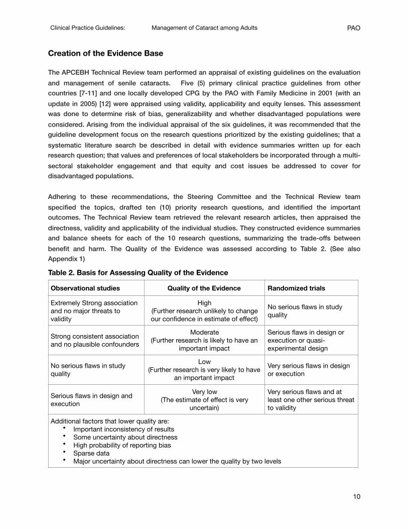
Adhering to these recommendations, the Steering Committee and the Technical Review team specified the topics, drafted ten (10) priority research questions, and identified the important outcomes. The Technical Review team retrieved the relevant research articles, then appraised the directness, validity and applicability of the individual studies. They constructed evidence summaries and balance sheets for each of the 10 research questions, summarizing the trade-offs between benefit and harm. The Quality of the Evidence was assessed according to Table 2. (See also Appendix 1)
Table 2. Basis for Assessing Quality of the Evidence
Observational studies Quality of the Evidence Randomized trials
Extremely Strong association and no major threats to validity
High(Further research unlikely to change our confidence in estimate of effect)
No serious flaws in study quality
Strong consistent association and no plausible confounders
Moderate(Further research is likely to have an
important impact
Serious flaws in design or execution or quasi-experimental design
No serious flaws in study quality
Low(Further research is very likely to have
an important impact
Very serious flaws in design or execution
Serious flaws in design and execution
Very low (The estimate of effect is very
uncertain)
Very serious flaws and at least one other serious threat to validity
Additional factors that lower quality are:• Important inconsistency of results• Some uncertainty about directness• High probability of reporting bias• Sparse data• Major uncertainty about directness can lower the quality by two levels
�10

Clinical Practice Guidelines: Management of Cataract among Adults PAO
The Technical Review team assessed the overall quality of evidence across the critical outcomes, basing this on the lowest quality of evidence for the outcomes that were critical to reaching a decision.
Balance between benefit and harm was weighed based on the critical and other important outcomes. Judgments about balance between benefits and harms did not take into account costs as shown in Table 3 (See also Appendix 1).
Composition of the CPG Panel
Pari passu with the Technical Review team’s preparation of the Evidence Base (EB), the Steering Committee undertook a systematic process of recruiting and selecting the CPG Panel considering all possible conflicts of interests of the potential panelists. To ensure fairness and transparency, this process was guided by principles and recommendations put forth by various guideline development groups. [13-17] The Steering Committee decided that the Panel would be composed of an equal number of ophthalmologists and non-ophthalmologists.
The ophthalmologists were to be chosen from nominees of the different ophthalmic groups with the understanding that they would be authorized to represent the views of their sponsoring organizations, namely: Chapter Societies of PAO, the PSCRS, the MSICS Interest Group and the PBO, and vote accordingly.
Non-ophthalmologists were either medical doctors well-versed in research methods, evidence-based medicine or CPG formulation, or, nonmedical personnel who had technical backgrounds in research,
Additional factors that may increase quality are:• All plausible residual confounding, if present, would reduce the observed effect• Evidence of a dose-response gradient
Factors that may lead to construction of separate evidence summaries and balance sheets for disadvantaged populations:
• Evidence of difference in effects in disadvantaged subgroups• Absence of direct evidence for disadvantaged subgroups
Table 3. Assessment of Benefits vs Harms
Category Benefits and Harms Balance
Net Benefits the intervention clearly doing more good than harm
Trade-offs important trade-offs between benefits and harms
Uncertain Net Benefits the intervention having unclear benefit over harm
No Net Benefits the intervention clearly not doing more harm than good
�11

Clinical Practice Guidelines: Management of Cataract among Adults PAO
statistics, ethics, administration or policy-making. Some had a dual role as patient advocates, having undergone cataract surgery themselves.
Formulation of the Recommendations
Recommendations were based on the trade-offs, the quality of evidence, the translation of evidence into practice in the specific situation (if specified) and uncertainty about the baseline risk. Recommendations were judged to be strong or weak based on the above, during the en banc Panel Review. (See Appendix 1)
For the purpose of determining the winning vote, the majority was considered to be 75% of all those who vote, i.e. 15 of 20 if all vote. Abstaining from voting was to be discouraged but not disallowed although unexpected if the evidence and the discussions were unambiguous. Two further rounds of voting on an issue would be conducted in case a majority decision was not obtained after the first round. After several rounds failing to reach a majority vote, a stalemate was to be declared and the issue would be decided again at a later date using a modified Delphi technique in order to reach a consensus. Evidence-based draft recommendations (as worded in the Evidence Base), were revised based on input arrived at by consensus in the en banc Panel Review.
Managing Conflicts of Interests
The Steering Committee facilitated the CPG formulation process but a priori decided to inhibit themselves from: assessing evidence, exerting influence on research methodology and the findings of the technical team, and voting as Panelists during the en banc review.
Planning for Dissemination and Implementation
Ultimately, this CPG will only be useful if it is becomes the reference for the standard of care for adult patients with cataract, and, if it positively influences the practice of ophthalmologists to the benefit of their patients. Wide dissemination and easy access will facilitate its utility, hence, it behooves the PAO to exert all efforts to reach all stakeholders. (See Dissemination and Implementation of the Guidelines).
The opportunity to react to the final recommendations was provided in the Public Forum with invited patient groups, healthcare providers, payors of healthcare, representatives from other specialty societies, the academe etc. Thereafter, planned implementation included a dissemination strategy, an information campaign specially targeting disadvantaged groups.
�12

Clinical Practice Guidelines: Management of Cataract among Adults PAO
III. RESULTS
Appraisal of Existing Guidelines
The Technical Review team evaluated five (5) international [7-11] and one (1) national CPG [12]. General findings in the scan of international guidelines were that 1) there was no detailed description of the prioritization of research questions and 2) there was no systematic searching and approach presented on how the evidence was summarized. Evidence summaries were not reported nor made available.
The National Institute for Health and Care Excellence (NICE) 2008 guidelines [9] and the PAO guidelines (2005) [12] in contrast, reported conduct of thorough literature searches and synthesis of evidence supporting the recommendations. Although the NICE 2008 guidelines [9] followed a search strategy, a number of non-English studies were removed from their yield that may have slightly affected some recommendations. The NICE 2008 guidelines [9] was also very focused on the interventional procedure (implantation of multifocal non-accommodative intraocular lenses during cataract surgery) and did not at all touch on non-surgical aspects.
A weakness common to all of the guidelines except the Royal College of Ophthalmology (RCO) 2010 [10] was the lack of incorporation of values and preferences. This could have been corrected by the incorporation of informed lay groups or a representative of such lay groups in the guideline process. This was also not evident even in the local guidelines. [12]
As for the use of the GRADE approach in developing the guidelines, only the Canadian Ophthalmological Society (COS) 2008 [8] was able to use the approach but it still has its limitations. None of the guidelines discussed issues on equity or targeted disadvantaged groups. In 2014, Wu and co-authors evaluated cataract surgery CPGs and arrived at similar conclusions, suggesting that stakeholder involvement must be improved to strengthen the area of values and preferences as well as applicability. [18]
The Research Questions
A total of ten (10) research questions were generated from the above guidelines scan and perceived gaps in knowledge: four (4) on comparisons of individual surgical procedures and six (6) on ancillary procedures in the management of senile cataracts. The research questions were worded as follows:
1. Among patients with senile cataracts scheduled for surgery, will routine pre-operative testing vs. no testing reduce mortality, morbidity, and adverse events?
2. Among patients with senile cataracts for cataract surgery, will routine lacrimal duct irrigation reduce postoperative endophthalmitis and adverse effects?
�13

Clinical Practice Guidelines: Management of Cataract among Adults PAO
3. Among patients with senile cataracts scheduled for surgery, will routine 5% povidone-iodine solution reduce postoperative endophthalmitis and adverse events?
4. Among patients with senile cataracts scheduled for surgery, will routine perioperative antibiotic prophylaxis reduce postoperative endophthalmitis and adverse events?
5. Among patients with bilateral senile cataracts about to undergo cataract surgery, how effective is same sitting or Immediate Sequential Bilateral Cataract Surgery (ISBCS) vs delayed operation or Delayed Sequential Bilateral Cataract Surgery (DSBCS) in preventing infection and reducing costs?
6. Among patients with senile cataracts, how effective is Manual Small Incision Cataract Surgery (MSICS) vs ECCE in improving vision and in terms of adverse outcomes/complications?
7. Among patients with senile cataracts, how effective is Manual Small Incision Cataract Surgery (MSICS) vs Phacoemulsification in improving vision and in terms of adverse outcomes/complications?
8. Among patients with senile cataracts, how effective is Phacoemulsification vs ECCE, in improving vision and in terms of adverse outcomes/complications?
9. Among patients with senile cataracts, how effective is Femtosecond Laser-assisted Cataract Surgery (FLACS) vs. conventional Phacoemulsification in improving vision and in terms of adverse outcomes/complications?
10. Among patients who develop posterior capsular opacification (PCO) after cataract surgery, how safe is less than 6 months versus 6 months and beyond laser (Nd:YAG) capsulotomy in preventing macular edema, retinal damage, anterior chamber reactions and other adverse events?
The Technical Review team then systematically searched the medical literature for the best available evidence for each of the research questions. Technical Review team members retrieved systematic reviews, randomized controlled trials and other research articles then appraised the directness, validity and applicability of each. For each of the questions and specific outcomes, evidence summaries and balance sheets were prepared. These summaries and balance sheets were then compiled, recommendations drafted and incorporated into the Evidence Base (EB) for the CPG on Management of Senile Cataracts (Appendix 2).
The CPG Panel
A total of 20 panelists were eventually selected from more than 30 nominees by the Steering Committee using weighted criteria. Nineteen (19) of the 20 confirmed participants made it to the en banc review on June 9, 2016; the absentee was called for an emergency meeting at the Department of Health. Among those who were not eye doctors, were 3 nurses, 2 economists, 2 biostatisticians, 1 internist/infectious disease specialist and 1 family medicine practitioner. There were 12 males and 7 females. The average age of the panelists was 54 years. At least 2 had experienced cataract surgery first hand. The CPG Panel in the en banc meeting developed judgments by consensus on each
�14

Clinical Practice Guidelines: Management of Cataract among Adults PAO
intervention, weighing the relative importance of their various outcomes and discussed acceptability, appropriateness and feasibility using the nominal group technique.
Final Recommendations
For each of the questions, the Panelists deliberated on the relative importance of the outcomes. They weighed the relative importance of outcomes scoring these from 1 to 9: with outcomes scoring from 1 to 3 as not so important; from 2 to 6 as important; and from 7 to 9 as critical (See Appendix 1). The Panelists considered all the reported outcomes critical.
Ten pre-determined specific questions were tackled by the Panelists to formulate recommendations. For each question, the session moderator explained the question to clarify the premise. Afterwards, a summary of the results of literature review responding to the question was presented, particularly the critical outcomes in relation to the procedure being discussed. This was followed correspondingly by a draft recommendation from the team based on the collected evidence.
Upon discussion of the available evidence and draft recommendation, the nominal group technique was employed to generate responses where each of the panelists were instructed to give concise 3 to 4 sentence inputs. Voting commenced afterwards, and consensus arrived at by majority rule and Delphi method when required (See Guideline Development Method). At the end of the session, the resulting recommendations and votes were presented to the plenary in summary for final validation.
Judgments on all but one of the 10 draft recommendations, as modified during the discussion, passed consensus by the Panelists in the en banc session. The only question that had to proceed to a Delphi survey was “Among patients with senile cataracts, how effective is Manual Small Incision Cataract Surgery (MSICS) vs ECCE in improving vision and in terms of adverse outcomes/complications?” (See Section Phacoemulsification vs ECCE).
IV. EVIDENCE AND RECOMMENDATIONS
1. Routine Medical Preoperative Testing
Based on moderate level of evidence, there was no significant difference in the rates of intra-operative or postoperative ocular and medical adverse events between routine medical preoperative testing and no routine medical preoperative testing.
A meta‐analysis of 3 randomized controlled trials that included 21,531 cataract surgeries done under local anesthesia showed that overall risk for adverse medical events from cataract surgery was low (3 out of 100 surgeries) [19]. The rate of adverse events was similar between the routine preoperative testing group (353 events out of 10,764 surgeries) and the no routine testing group (354 events out of 10,766 surgeries). Adverse events were mostly cardiovascular in nature (e.g. half were blood pressure
�15

Clinical Practice Guidelines: Management of Cataract among Adults PAO
elevations requiring treatment) and occurred intra-operatively. The rate of postponement or cancellation of surgery was also similar in the 2 groups (161 out of 10,287 surgeries in the routine testing group vs. 166 out of 10,295 surgeries in the no testing group) [20,21] (See Appendix 3: EB Table 1). Cost was evaluated in one study, which estimated that the cost was 2.5 times higher in those who underwent preoperative testing than those who did not. [20] Only one study reported rates of myocardial infarction, stroke, and hypoglycemia within 7 days of cataract surgery separately [21]. There was no difference between routine testing and no testing for these outcomes, however confidence intervals for the results were wide, because event rates were very low (less than 10 in 10,000 in both groups). None of these events occurred intra-operatively. Majority of the participants enrolled in the included studies were patients with mild to severe, non‐incapacitating systemic diseases (e.g. hypertension, diabetes, congestive heart failure, and bleeding disorders). Routine preoperative tests included ECG, CBC, blood sugar, serum electrolytes, BUN and creatinine. Outcomes evaluated were ocular complications, medical adverse events requiring treatment, and postponement or cancellation of surgery. The results of the meta-analysis were driven mainly by the largest study in the meta-analysis, which included a total of more than 19,000 surgeries [21]. However, this trial was methodologically sound, and the results were consistent across all the studies included in the meta-analysis. Given these findings, routine medical pre-operative testing is not recommended for patients with senile cataracts scheduled for surgery.
Recommendation 1
Pre-operative medical ancillary testing prior to cataract surgery is recommended only if indicated by the patient's medical condition and the physician's assessment.
(Strong recommendation. Moderate to high quality evidence.)Remarks
The physician must still determine a patient's health status through history taking and physical examination. It was emphasized that “preoperative testing” referred to the local procedure of “clearance” with ancillary testing totally separate from the routine history-taking and physical examination.
2. Routine Lacrimal Duct Irrigation
There is no randomized trial found in the literature search to answer the research question. Instead, a prospective cohort study is available where 282 (40%) patients who received no lacrimal system irrigation nor topical antibiotic prior to surgery was compared to 418 (60%) patients who received lacrimal system irrigation with or without neomycin eye drops [22]. No patient developed postoperative endophthalmitis in both groups. Although the overall bacterial contamination was
�16

Clinical Practice Guidelines: Management of Cataract among Adults PAO
reported at 14.1%, the actual number of patients per group was not mentioned. (See Appendix 3: EB Table 2).
Recommendation 2 Lacrimal duct irrigation as a routine pre-operative procedure in cataract surgery does not reduce the incidence of endophthalmitis but may be performed when indicated.
(Strong Recommendation. Very low quality evidence.) Remarks
Despite very low quality evidence, the panel decided NOT to recommend routine lacrimal duct irrigation because of low incidence of postoperative infections and even the risk of contamination with the procedure. In lieu of lacrimal duct irrigation, massaging the lacrimal sac to elicit purulent discharge from the puncta may be a better, less invasive way of determining if active dacryocystitis is present and prior treatment is indicated. Patients complaining of tearing and discharge or other symptoms of dacryocystitis, and, a prior history of lacrimal duct surgery are good indications to perform the procedure.
3. Routine Povidone-Iodine Solution
There is one randomized controlled trial (RCT) and one prospective cohort study found in the literature search to answer the research question. The RCT however does not specify the concentration of Povidone-Iodine (PI) solution used [23]. A total of 4111 eyes underwent cataract surgery and were included in this study. There was no significant difference between the PI group and the control (silver protein) group (RR 1.17, 95% CI 0.57 – 2.42). The prospective cohort study including 8,083 eyes showed significant difference between 5% PI and no 5% PI (p < 0.03) [24].
However, on rechecking, it turned out that this is not significant (RR 0.24, 95% CI 0.57 – 1.08). Pooled analysis of these two studies (PI vs control) showed a trend of benefit toward PI with an RR of 0.61 (95% CI 0.13 – 2.86, I2 72%). These two studies are both low quality (See Appendix 3: EB Table 3). In several case reports, conjunctival irritation is seen in 0.4% [25]. Contact dermatitis is less common (0.04%); however, the risk increases tenfold in the presence of allergy to shellfish or iodine [26]. Despite the increased risk for allergy, patients are still recommended to receive PI prior to surgery. [27] Keratoconjunctivitis sicca has also been reported [27]. Hyperemia of conjunctivae, superficial punctate epitheliopathy and epithelial defect, corneal symptoms (edema or Descemet’s membrane folds, and mild to moderate Tyndall were seen in randomized trial comparing different concentrations of PI solutions [26].
Recommendation 3
Instillation or irrigation of the conjunctiva with 5% povidone iodine solution pre-operatively is recommended to reduce the risk of postoperative endophthalmitis.
(Strong Recommendation. Very low to low quality evidence.)
�17

Clinical Practice Guidelines: Management of Cataract among Adults PAO
Remarks Despite the low to very low quality of evidence in published literature on its effectivity in reducing the
incidence of endophthalmitis but because of the relatively low incidence of adverse events in its use, the
instillation of 5% povidone-iodine solution was considered to be a prudent measure in ensuring asepsis
of the ocular surface during surgery.
4. Routine Perioperative Antibiotic Prophylaxis
There were three (3) randomized controlled trials (RCTs) found in the literature search to answer the research question but investigated different kinds of antibiotics. One RCT compared topical regimen (chloramphenicol-sulphadimidine ointment) plus periocular penicillin at the time of surgery with topical regimen alone [28] (See Appendix 3: EB Table 4). This study done in northern Pakistan found significant reduction of endophthalmitis with topical regimen plus periocular penicillin (RR 0.33, 95% CI 0.12–0.92) during surgery compared with topical regimen alone. In another RCT conducted in Turkey, balanced salt solution (BSS) irrigation with vancomycin and gentamycin was compared to BSS alone showed no benefit from vancomycin and gentamycin (RR 0.20, 95% CI 0.01–4.15) [29]. The last RCT of high quality evidence was based on a 2 x 2 factorial design, with intracameral cefuroxime and topical perioperative levofloxacin resulting in 4 treatment groups [30]. This was conducted by the European Society of Cataract & Refractive Surgeons (ESCRS) involving 16 603 eyes of 16 603 patients from multiple sites in Europe and Turkey. The results of this study showed that intracameral cefuroxime alone (RR 0.20, 95% CI 0.04–0.91) and combined intracameral cefuroxime with topical levofloxacin (RR 0.10, 95% CI 0.27–0.78) showed significant reduction in endophthalmitis as compared to no antibiotic prophylaxis. However, the reduction of endophthalmitis with topical levofloxacin alone as compared to no antibiotics was not statistically significant (RR 0.70, 95% CI 0.27–1.84). Other comparisons such as intracameral cefuroxime vs topical levofloxacin, combined intracameral cefuroxime and topical levofloxacin vs intracameral cefuroxime, intracameral cefuroxime and topical levofloxacin vs topical levofloxacin did not result in significant reduction in endophthalmitis. There was also no statistically significant reduction in the final visual acuity following endophthalmitis among patients given intracameral cefuroxime with/without topical levofloxacin as compared to no intracameral cefuroxime with/without topical levofloxacin.
Despite this reduction in endophthalmitis, a number of case reports and case series have been published on harm of intracameral and intravitreal cefuroxime. Anterior and posterior segment inflammation were reported in six patients with high doses of intracameral cefuroxime. [31] All six patients in this series had satisfactory final visual outcome even without surgical intervention. Inadvertent overdose of cefuroxime caused hemorrhagic retinal infarction in four patients [32] and macular infarction and associated cystoids macular edema. [33] Two patients developed anaphylactic reactions from intracameral [34] and intravitreal [35] cefuroxime (See Appendix 3: EB Table 4).
�18

Clinical Practice Guidelines: Management of Cataract among Adults PAO
Recommendation 4 The use of peri-operative antibiotic prophylaxis is recommended to reduce the risk of postoperative endophthalmitis in patients who undergo cataract surgery.
(Strong recommendation. Moderate quality evidence.)
Remarks
Since most of the antibiotics in the studies are not used locally, the panel could not recommend specific
antibiotics nor protocols for the perioperative prophylaxis.
5. Immediate versus Sequential Bilateral Cataract Surgery
Based on a low-quality cohort study, the risk of infection in delayed sequential bilateral cataract surgery (DSBCS) with intracameral prophylactic antibiotics is very small (1 in 29,582). A non-significant 2-fold increase in the risk of infection was observed among immediate sequential bilateral cataract surgery (ISBCS) patients who were given prophylactic intracameral antibiotics [36]. The subjective improvement in visual function is not significantly different in ISCBS and DSCBS patients based on a metaanalysis [37] of 2 randomized studies [38,39]. The two trials had conflicting results for this outcome and were significantly dissimilar. There is also no statistically significant difference in the risk of any intra‐ and post-operative complications (capsule tears, vitreous loss, iridectomy, sphincterotomy, sutures in wound, intraocular pressure > 30 mmHg, wound leak IOL decentration or deplacement, anterior chamber flare, capsular fibrosis, capsule opacification, foreign body sensation, dry eyes and more serious complications such as corneal edema, macular edema, wound leak, or iris prolapse) between patients who underwent ISCBS and DSCBS [38,39]. Likewise, there is no statistically significant difference in the risk of serious post‐operative complications (corneal edema, macular edema, wound leak, or iris prolapse) between the aforementioned groups. [38,39] (See Appendix 3: EB Table 5). Canadian estimates of crude cost savings from ISCBS over DSCBS is pegged at US$ 1,606 (valued in 2013) per patient with adjusted estimates pegged at US$ 1,431 per quality‐adjusted life-year gained [40]. Converting these based on the 2013 average peso‐dollar exchange rate, crude cost savings amount to Php 68,202 per patient while adjusted cost savings is Php 60,746 per QALY gained. Considering the number of patients needing cataract surgery, the cost savings can potentially add up even if we consider cost variation in different countries. In the Philippines, it is also important to consider the opportunity cost for the caregiver or watcher (bantay) of the patient. Often, patients will come for consultations and procedures with a handful of bantays. The collective loss of productivity and earnings of these bantays are substantial and may confer additional cost savings in favor of ISCBS.
Aside from benefits in terms of costs, ISCBS confers additional benefits to patients: rapid visual recovery and functional advantage in the short term. ISCBS patients tend to regain visual functions earlier than their DSCBS counterparts. Unfortunately, among patients who underwent ISCBS, the very
�19

Clinical Practice Guidelines: Management of Cataract among Adults PAO
short interval between the surgeries of the two eyes precludes adjustments in surgical plans based on the result of the first eye surgery [41].
Recommendation 5
Delayed Sequential Bilateral Cataract Surgery is preferred over Immediate Sequential Bilateral Cataract Surgery (ISBCS) in the same sitting for patients with bilateral senile cataracts.
(Strong recommendation. Very low to low quality evidence.) Remarks
The possibility of bilateral endophthalmitis, no matter how small its probability, and the devastating effect on the patient should such occur, outweighed the potential cost benefit of same sitting surgery which
came from very low-low quality evidence anyway.
6. MSICS versus ECCE for senile cataracts
Evidence from 2 randomized controlled trials (RCTs) showed that there was no significant difference in terms of improvement of visual acuity at 6-8 weeks between MSICS and ECCE with a relative risk (RR) of 1.57 (95% CI 0.88, 2.8) (See Appendix 3: EB Table 6) [42,43]. However, the data presented in these 2 RCTs reporting poor visual outcome were not combined due to difference on how the measurements were made. Both RCTs reported an insignificant difference between the 2 procedures with regards poor visual outcomes at 6-8 weeks. One study reported an RR of 1.58 (95% CI 0.45, 5.0) supporting the insignificant findings. [42] Surgically-induced astigmatism was significantly less for MSICS. One study reported a mean induced astigmatism in diopters of 1.77 ± 1.65 for ECCE vs 1.1 ± 0.95 for MSICS, p=0.012. [43] In another study, the incidence of astigmatism of ≥ 2D had a relative risk of 0.49, (95%CI 0.32-0.74) indicating less astigmatism in the MSICS technique. [43] Intraoperative and postoperative complications based on 1 RCT, were significantly higher with MSICS, with relative risks of 1.83 (95%CI 1.02-3.26), and 1.38 (95%CI 1.1-1.73), respectively. [42] Both intraoperative and postoperative complications reported in the RCT were graded and scored as described by the Oxford Cataract Treatment and Evaluation Team (OCTET). There was a limited number of studies with issues on allocation and concealment. It was unfortunate that most of the data could not be combined due to differences in reporting of outcomes.
Recommendation 6
MSICS is the preferred technique for cataract surgery over ECCE because of less surgically induced astigmatism despite the significantly higher risk for complications.
(Strong recommendation. Low to moderate quality evidence.)
�20

Clinical Practice Guidelines: Management of Cataract among Adults PAO
Remarks
Botched MSICS surgeries usually performed by novice surgeons not properly acquainted with the
technique has led many surgeons to prefer ECCE. This initial negative bias and higher risk of
complications in MSICS would be mitigated by improving access to training and thereby the surgical
expertise in MSICS.
7. MSICS versus phacoemulsification in senile cataracts
Evidence for MSICS versus phacoemulsification for the outcome of good functional vision at 3 months (uncorrected acuity 6/18 or better from 6-8 weeks of follow-up) showed significant benefit in favor of phacoemulsification with a relative risk (RR) of 0.90 (95% CI 0.84, 0.96) based on 3 randomized controlled trials (RCTs). [45-47] Other evidence summarized and combined from 13 RCTs [45-57] and 3 meta-analyses [58-60] (See Appendix 3: EB Table 7) failed to show significant difference in the following:
1. Good functional vision at 6 months (uncorrected acuity 6/18 or better) based on 1 RCT, with an RR of 1.07 (95% CI 0.91, 1.26). [54]
2. Good functional vision at 3 months (best-corrected acuity 6/18 to 6/12 or better) based on 6 RCTs, with an RR of 0.99 (95% CI 0.98, 1.01). [45-47, 59-61]
3. Good functional vision at 6 months (best-corrected acuity) based on 1 RCT, with an RR of 1.0 (95% CI 0.94, 1.06). [54]
4. Poor visual outcome at 3 months (best corrected acuity worse than 6/60), OR 2.48 (95% CI 0.74, 8.28). [45-47, 49-51]
5. Poor visual outcome at 6 months (best-corrected acuity worse than 6/18) based on 1 RCT is 1.9% in both groups with a computed RR of 1.0 (95% CI 0.06, 16). [54]
6. Uncorrected visual acuity in 1 week after surgery, RR 1.0 (95% CI 0.97, 1.03). [46,48,53,56,57] Furthermore, neither surgical techniques showed clear benefit in preventing any of the complications such as posterior capsular rupture, [45-47, 49-58] corneal edema postoperatively, [45-47, 50-53, 55-58] endothelial cell loss [49,51], and astigmatism. [46,47,49,51,56]
In light of the evidence, it seems that phacoemulsification has an edge over MSICS in terms of visual acuity improvement. Although astigmatism may be a problem, it has not been clearly established with the current evidence.
Recommendation 7
Phacoemulsification is the preferred technique for cataract surgery over MSICS because of faster visual improvement and lower risk of adverse events or complications.
(Strong recommendation. Very low to low quality evidence.)
�21

Clinical Practice Guidelines: Management of Cataract among Adults PAO
Remarks
The cost of the equipment and surgical consumables in phacoemulsification raises the issue of equity
and accessibility for disadvantaged groups. Hence, in areas where phacoemulsification is not available or
feasible, surgical expertise in MSICS must be developed as an alternative. Although faster visual
rehabilitation, due to less surgical trauma in phacoemulsification, is a patient-valued outcome, over time
MSICS and phacoemulsification achieve similar results.
8. Phacoemulsification versus ECCE for senile cataracts
These findings were derived from 6 randomized controlled trials (RCT) [61-66] and 1 meta-analysis [57] (See Appendix 3: EB Table 8). In the combined analysis, it was noted that good functional vision at 3 months (uncorrected visual acuity and best corrected visual acuity) significantly favored phacoemulsification. [61-64] Good functional vision at 12 months, in terms of uncorrected visual acuity, also significantly favored phacoemulsification. [62] However, at 12 months, measured by best corrected visual acuity, the difference between phacoemulsification and ECCE was insignificant. [62] Poor visual outcome at 3 months, in terms of best corrected acuity of 6/60 or worse, was significantly lower in phacoemulsification. [61-61] However, after 12 months, this outcome was insignificant between phacoemulsification and ECCE. [62] Adverse events or complications that did not show any significant difference between the 2 techniques were capsular rupture [62,63,65], retinal detachment [62] and endothelial cell loss [62,62,66]. However, posterior capsular opacification [62,65], cystoid macular edema [62,65] and iris prolapse [62] are complications that were significantly lower with phacoemulsification.
The studies generally had an unclear risk of bias due to poorly reported trial methods and although the quality per outcome ranged from low to moderate, overall quality of evidence was deemed low for this recommendation.
Recommendation 8
Phacoemulsification is the preferred technique for cataract surgery over ECCE because of significant benefits and lower risk of complications.
(Strong recommendation. Low to moderate quality of evidence.) Remarks
The benefits derived from the smaller incision, including faster visual rehabilitation and less risk of
devastating complications associated with larger incisions, make phacoemulsification favor over ECCE.
�22

Clinical Practice Guidelines: Management of Cataract among Adults PAO
9. Femtosecond laser-assisted cataract surgery (FLACS) versus conventional phacoemulsification in senile cataracts
There was no significant difference between FLACS and conventional phacoemulsification in the overall result, in terms of improvement in vision, measured by uncorrected distance visual acuity. [68,69] The difference was also insignificant when sub‐grouped by follow-up time (at 1 week, 1 month and 6 months). As for improvement in vision (measured by corrected distance visual acuity), there was a significant difference favoring FLACS when looking at the overall result, with a mean difference of -0.03 LogMAR units. This implies a significant improvement of visual acuity by 0.03 using the LogMAR chart favoring FLACS. The difference is also significant at 1 week and at 6 months postoperatively. [69-73] However, among the adverse outcomes, the differences were insignificant between FLACS and conventional phacoemulsification in the rates of anterior capsule tear [71,72], elevated intraocular pressure [71, 73], and macular edema [71,72].
One study showed that based on the simulated complication rates of phacoemulsification and FLACS and assuming resultant visual acuity outcome improvement of 5% in uncomplicated cases of FLACS, the cost‐effectiveness (dollars spent per QALY) gained from FLACS was not cost‐effective at $92 862 Australian Dollars. [74] There were 7 RCTs [68-74] and 1 meta-analysis [69] (See Appendix 3: EB Table 9) that showed these findings. Unfortunately, there was unclear to high risk for bias for the included studies. These were mainly from issues with randomization and allocation concealment. The studies were also at high risk for performance and detection bias. Consistency issues were seen in the primary outcomes, but not evident in the complications.
Recommendation 9
The choice of FLACS or conventional phacoemulsification for routine cataract surgery will depend on accessibility, surgeon experience, and patient cost preferences.
(Weak recommendation. Very low evidence.) Remarks
FLACS has not shown superior visual acuity results over conventional phacoemulsification and, in the light of cost concerns and limited access to the technology, FLACS was not recommended over phacoemulsification.
10.Laser (ND:YAG) Capsulotomy
Three studies were identified addressing the adverse effects of Nd:YAG laser capsulotomy in patients with PCO but none of these studies compared the less than 6 months versus 6 months and beyond
�23

Clinical Practice Guidelines: Management of Cataract among Adults PAO
in timing of the capsulotomy. One study however compared different time durations from 6 months up to greater or equal to 37 months [75] (See Appendix 3: EB Table 10). The sample sizes were 23 [76], 31 [77] and 314 [75] adult patients, with follow‐up observations ranging from as early as immediately after the procedure to three months post-capsulotomy.
From the very low quality evidence from the two studies with small sample sizes, the limited available data does not seem to show any statistically significant difference (p < 0.05) between the anterior chamber depth, intraocular pressure, macular foveal thickness and endothelial cell loss before and after the capsulotomy [76,77 ].
However, the cohort study by Shaikh et al. [75] in 2010 demonstrates that laser capsulotomy may induce potential complications, e.g. anterior chamber reactions, intraocular pressure, damage to intraocular lenses, retinal detachment, macular edema and vitreous hemorrhage. Recommendation 10
Regardless of time elapsed after cataract surgery, Laser (Nd:YAG) Capsulotomy is only recommended in patients who develop symptomatic posterior capsular opacification, because of the risk of macular edema, anterior chamber reaction, retinal detachment and other adverse events which may be associated with the procedure.
(Strong Recommendation. Very low quality evidence.) Remarks
There is no evidence that the timing of laser capsulotomy influences the probability of the occurrence of potential complications suggesting that other criteria should be used to determine appropriateness of treatment.
RESEARCH IMPLICATIONS The Technical Review team, the CPG Panel and the Steering Committee identified important knowledge gaps that need to be addressed through primary research.
These gaps were especially evident in the research questions for which evidence was deemed very low to low quality: Routine lacrimal duct irrigation; Routine 5% povidone-iodine solution; Immediate versus sequential bilateral cataract surgery; Phacoemulsification vs MSICS; (FLACS) versus conventional phacoemulsification; Laser (ND:YAG) capsulotomy.
Important research gaps were also pinpointed even for the research questions for which the evidence was deemed of moderate quality. Particular note is made of the lack of local data on the comparative effectiveness and safety of MSICS vs ECCE and MSICS vs phacoemulsification. This paucity of data has important equity issues, given the limited number of experts in MSICS and the resultant inaccessibility particularly of disadvantaged groups to this procedure.
�24

Clinical Practice Guidelines: Management of Cataract among Adults PAO
It must be noted that the evidence base for the various cataract surgery procedures consisted of comparisons between individual procedures. Panelists present at the en banc were able to formulate a “ranking” as an interim summary as follows: Phacoemulsification recommended over MSICS; Phacoemulsification recommended over ECCE; Neutral on phacoemulsification versus FLACS, and; undecided on MSICS versus ECCE (this latter was resolved after 2 rounds of Delphi surveys, concluding with MSICS recommended over ECCE). It was emphasized though that strictly seeking, such a “ranking” should be based on the results of a “network meta-analysis”, this being a priority research gap that merits attention.
Other gaps included: local cost-effectiveness data, e.g. expressed in QALYs; data on specific outcomes of visual improvement, e.g. immediate return to function as might be a more relevant outcome for disadvantaged groups like farmers, drivers, astigmatism as an outcome in the comparisons between phacoemulsification and MSICS, etc.; comparative safety between phacoemulsification and ECCE (arising from poorly reported trial methods and thus an unclear risk of bias in existing published reports); data on efficacy and safety of locally used antibiotics for routine perioperative antibiotic prophylaxis; publication of data on efficacy and safety of the commonly used 5% povidone-iodine for antisepsis.
Another important gap that was evident in the discussions, was that of a “mapping” of practitioners with expertise and facilities capacitated for particular cataract surgery procedures, e.g. for MSICS and phacoemulsification, the latter especially as it is a more costly procedure requiring special equipment. This has important implications to equity and targeting of disadvantaged groups.
A policy gap that needs to be addressed especially because of its implications to health financing was that of strengthening standardization of the practice of Laser (ND:YAG) Capsulotomy for symptomatic posterior capsular opacification.
DISSEMINATION AND IMPLEMENTATION OF THE GUIDELINES
Dissemination to the PAO Members
The CPG will be presented during the PAO Annual Convention. Before the meeting, copies would have been mailed to the members ahead of time for their perusal. Comments and questions will be elicited during the discussion period for the purpose of clarifying the recommendations. A full copy of the document will be published on-line in the PAO Website and as a supplement of the Philippine Journal of Ophthalmology (PJO).
�25

Clinical Practice Guidelines: Management of Cataract among Adults PAO
Dissemination to the Training Institutions
The Philippine Board of Ophthalmology (PBO) will be asked to endorse the CPG. Then, copies of the CPG with the PBO endorsement will be sent to the heads of hospital-based departments of ophthalmology, training institutions and ambulatory surgery centers, medical schools and libraries so as to incorporate the recommendations in their teaching and training curricula, with the support of the consultants and mentors.
Dissemination to Industry Partners, Regulatory Agencies, and Payors
The CPG will be transmitted to pharmaceutical industry partners; NGOs involved in eye care; health maintenance organizations (HMOs); the Department of Health and the Philippine Health Insurance Corporation through formal communications by the PAO Council.
Dissemination to the Patients and the Public in General
A simplified version of the CPG shall be formatted and made available to the PAO members in a format that will be ready for reproduction and dissemination to their patients in their clinics. The same will be available for interested parties who might browse the PAO website.
Implementation and Monitoring
A questionnaire will be distributed annually for the purpose of determining the preferred practices of PAO members with regard to cataract surgery. The results shall be compiled and tracked annually to monitor convergence of practice patterns with the CPG recommendations.
APPLICABILITY ISSUES
The PAO guideline development group using equity and applicability lenses flagged some caveats here re-emphasized:
The recommendation to not do routine pre-operative ancillary testing on healthy adults with cataracts are separate from the history taking and physical examination that are essential in their pre-operative evaluation. Should the history taking and physical examination uncover high risk conditions, this subgroup of patients may require direct testing under the individual discretion of their clinicians. Though the cost of phacoemulsification is prohibitive (largely because of the need for expensive equipment), it is the preferred procedure over ECCE and MSICS from the weighed tradeoff between benefits and harm.
�26

Clinical Practice Guidelines: Management of Cataract among Adults PAO
Though the surgical expertise in MSICS may not yet be widely distributed an increase in the capacity and improvement in accessibility to this procedure is being discussed, With more ophthalmologists trained in this procedure and deployed or committed to conduct periodic visits disadvantaged groups in remote areas.
UPDATING OF THE GUIDELINES
PAO plans to update this practice guideline within 2 years. The recommendations in the CPG shall hold until such time that technology, patient and provider preferences, or new evidence provides the impetus for revisiting and updating the guideline once more.
�27

Clinical Practice Guidelines: Management of Cataract among Adults PAO
REFERENCES I. INTRODUCTION
1. WHO. Prevention of Blindness and Visual Impairment: Priority Eye Diseases. http://www.who.int/blindness/causes/priority/en/index1.html
2. Philippine Health Insurance Corporation (PHIC). Stats and Charts. https://www.philhealth.gov.ph/about_us/statsncharts/snc2015_2nd.pdf
3. Institute of Medicine. Consensus report. Clinical practice guidelines we can trust. March 23, 2011. http://www.iom.edu/Reports/2011/Clinical-Practice-Guidelines-We-Can-Trust.aspx (Accessed on January 13, 2012).
4. Evidence-Based Ophthalmology Group. EBO technical review of the validity of Recommendation #14 of the Clinical Practice Guidelines for the Management of Cataract among Adults. Philippine Journal of Ophthalmology. 2005;Vol 30:No 2.
5. Institute of Medicine (2008) Knowing what works in health care: A roadmap for the nation. Washington, DC: The National Academies Press
II. GUIDELINE DEVELOPMENT METHODS6. Guyatt GH, Oxman AD, Vist GE, Kunz R, Falck-Ytter Y, Alonso-Coello P, Schunermann HJ for the GRADE
Working Group. GRADE: An emerging consensus on rating quality of evidence and strength of recommendations. British Medical Journal 2008; 336;924-926.
7. American Optometrists Association. Optometric Clinical Practice Guideline Care of the Adult Patient with Cataract. USA 2004.
8. Canadian Ophthalmological Society. Canadian Ophthalmological Society Evidence-based clinical practice guidelines for cataract surgery in the adult eye. Can J Ophthalmol 2008; 43: S7-57.
9. NICE. Implantation of multifocal (non-accommodative) intraocular lenses during cataract surgery. UK 2008.
10. The Royal College of Ophthalmologists. Cataract Surgery Guidelines. London, UK; September 2010.11. American Academy of Ophthalmology Cataract and Anterior Segment Panel. Preferred Practice Pattern
Guidelines. Cataract in the Adult Eye. San Francisco, CA: American Academy of Ophthalmology; 201112. Philippine Academy of Ophthalmology. Clinical Practice Guideline for the Management of Cataract Among
Adults. 2001. Updated 2005.13. Tricoci P, Allen JM, Kramer J, Califf RM, Smith SC. Scientific evidence underlying the ACC/AHA Clinical
Practice Guidelines. JAMA. 2009; 301(8):831-841.14. WHO. Handbook of Guideline Development, 2nd ed. World Health Organization.15. Lenzer J, Hoffman J, Furberg C, Ioannidis J. Ensuring the integrity of clinical practice guidelines: a tool for
protecting patient. BMJ 2013;347:15535.16. IOM (Institute of Medicine). 2011. Clinical Practice Guidelines We Can Trust. Washington, DC: The
National Academies Press.17. IOM (Institute of Medicine). 2009. Conflict of Interest in Medical Research, Education, and Practice.
Washington, DC: The National Academies Press.III. RESULTS
18. Wu CM, Wu AM, Young BK, Wu D, Chen A, Margo CE and Greenberg PB. An evaluation of cataract surgery clinical practice guidelines. Br J Ophthalmol 2015; 99: 401-404 originally published online September 24, 2014. doi: 10.1136/bjophthalmol-2014-305567
IV. EVIDENCE AND RECOMMENDATIONS
Routine Preoperative Testing 19. Keay L, Lindsley K, Tielsch J, Katz J and Shein O. Routine preoperative medical testing for cataract surgery.
Cochrane Database Syst Review 2012; 3: CD007293. doi:10.1002/14651858.CD007293.pub3. 20. Lira RP, Nascimento MA, Moreira-Filho DC, Kara-Jose N, Arieta CE. Are routine preoperative medical tests
needed with cataract surgery? Pan American Journal of Public Health 2001; 10(1):13-7 21. Shein OD, Katz J, Bass EB, Tielsch JM, Lubomski LH, Feldman MA, et al. The value of routine preoperative
medical testing before cataract surgery. Study of Medical Testing for Cataract Surgery. New England Journal of Medicine 2000; 342(3): 168-75.
Routine Lacrimal Duct Irrigation 22. Mistlberger A, Ruckhofer J, Raithel E, Müller M, Alzner E, Egger SF, Grabner G. Anterior chamber
contamination during cataract surgery with intraocular lens implantation. J Cataract Refract Surg. 1997 Sep;23(7):1064-9.
�28

Clinical Practice Guidelines: Management of Cataract among Adults PAO
Routine Povidone-Iodine Solution 23. Mørk P. Polyvinylpyrrolidone-iodine as a disinfectant in eye surgery for five years. Acta Ophthalmol
(Copenh). 1987 Oct;65(5):572-4. 24. Speaker MG, Menikoff JA. Prophylaxis of endophthalmitis with topical povidone-iodine. Ophthalmology.
1991 Dec;98(12):1769-75. 25. Zamora JL. Chemical and microbiologic characteristics and toxicity of povidone-iodine solutions. Am J
Surg. 1986 Mar;151(3):400-6. 26. Li B, Nentwich MM, Hoffmann LE, Haritoglou C, Kook D, Kampik A, Sheng M, Miño de Kaspar H.
Comparison of the efficacy of povidone-iodine 1.0%, 5.0%, and 10.0% irrigation combined with topical levofloxacin 0.3% as preoperative prophylaxis in cataract surgery. J Cataract Refract Surg. 2013 Jul;39(7):994-1001. doi:10.1016/j.jcrs.2013.02.039. Epub 2013 May 13.
27. Gills JP. Effective concentration of betadine. J Cataract Refract Surg. 1999 May;25(5):604.
Routine Perioperative Antibiotic Prophylaxis 28. Christy NE, Sommer A. Antibiotic prophylaxis of postoperative endophthalmitis. Ann Ophthalmol. 1979 Aug;
11(8):1261-5. 29. Sobaci G, Tuncer K, Taş A, Ozyurt M, Bayer A, Kutlu U. The effect of intraoperative antibiotics in irrigating
solutions on aqueous humor contamination and endophthalmitis after phacoemulsification surgery. Eur J Ophthalmol. 2003 Nov-Dec;13(9-10):773-8.
30. Endophthalmitis Study Group, European Society of Cataract & Refractive Surgeons. Prophylaxis of postoperative endophthalmitis following cataract surgery: results of the ESCRS multicenter study and identification of risk factors. J Cataract Refract Surg. 2007 Jun;33(6):978-88.
31. Delyfer MN, Rougier MB, Leoni S, Zhang Q, Dalbon F, Colin J, Korobelnik JF. Ocular toxicity after intracameral injection of very high doses of cefuroxime during cataract surgery. J Cataract Refract Surg. 2011 Feb;37(2):271-8.
32. Çiftçi S, Çiftçi L, Dağ U. Hemorrhagic retinal infarction due to inadvertent overdose of cefuroxime in cases of complicated cataract surgery: retrospective case series. Am J Ophthalmol. 2014 Feb;157(2):421-425.e2.
33. Qureshi F, Clark D. Macular infarction after inadvertent intracameral cefuroxime. J Cataract Refract Surg. 2011 Jun;37(6):1168-9.
34. Moisseiev E, Levinger E. Anaphylactic reaction following intracameral cefuroxime injection during cataract surgery. J Cataract Refract Surg. 2013 Sep;39(9):1432-4.
35. Villada JR, Vicente U, Javaloy J, Alió JL. Severe anaphylactic reaction after intracameral antibiotic administration during cataract surgery. J Cataract Refract Surg. 2005 Mar;31(3):620-1.
Immediate or Sequential Bilateral Cataract Surgery 36. Arshinoff SA, Bastianelli PA. Incidence of postoperative endophthalmitis after immediate sequential bilateral
cataract surgery. J Cataract Refract Surg 2011; 37:2105-2114.37. Kessel L, Andresen J, Tendal B et al. Immediate sequential bilateral cataract surgery: a systematic review
and metaanalysis. Journal of Ophthalmology 2015; 2015: 912481. 38. Serrano‐Aguilar P, Ramallo‐Fariña Y, Cabrera‐Hernández JM, et al. Immediately sequential versus delayed
sequential bilateral cataract surgery: safety and effectiveness. Journal of Cataract & Refractive Surgery 2012;38:10, pp. 1734–1742.
39. Sarikkola AU, Uusitalo RJ, Hellstedt T, Ess SL, Leivo T, and Kivelä T, Simultaneous bilateral versus sequential bilateral cataract surgery: Helsinki Simultaneous Bilateral Cataract Surgery Study Report 1. Journal of Cataract and Refractive Surgery. 2011; 37(6): 992–1002.
40. Malvankar-Mehta MS, Filek R, Iqbal M, Shakir A, Mao A, Si F, Malvankar MG, Mehta SS, Hodge WG. Immediate sequential bilateral cataract surgery: a cost-effective procedure. Canadian Journal of Ophthalmology. 2013; 48(6):482-488.
41. American Academy of Ophthalmology Cataract and Anterior Segment Panel. Preferred Practice Guidelines. Cataract in the Adult Eye. San Francisco CA: American Academy of Ophthalmology; 2011. Accessed at www.aao.org/ppp on June 20, 2016.
MSICS versus ECCE for senile cataracts 42. Gogate PM, Deshpande M, Wormald RP, Deshpande R, Kulkarni SR. Extracapsular cataract surgery
compared with manual small incision cataract surgery in community eye care setting in western India: a randomised controlled trial. British Journal of Ophthalmology 2003;87(6):667–72.
�29

Clinical Practice Guidelines: Management of Cataract among Adults PAO
43. Gurung A, Karki DB, Shrestha S, Rijal AP. Visual outcome of conventional extracapsular cataract extraction with posterior chamber intraocular lens implantation versus manual small‐incision cataract surgery. Nepalese Journal of Ophthalmology 2009;1(1):13–9.
44. Ang M, Evans JR, and Mehta JS. Manual small incision cataract surgery (MSICS) with posterior chamber intraocular lens versus Extracapsular cataract extraction (ECCE) with posterior chamber intraocular lens for age-related cataract. Cochrane Database of Systematic Reviews 2014, Issue 11. Art. No.: CD008811
MSICS versus phacoemulsification in senile cataracts 45. Cook C, Carrara H, Myer L. Phacoemulsification versus manual small incision cataract surgery in South
Africa. South African Medical Journal 2012;102(6):537–40. 46. Gogate PM, Kulkarni SR, Krishnaiah S, Deshpande RD, Joshi SA, Palimkar A, et al. Safety and efficacy of
phacoemulsification compared with manual small-incision cataract surgery by a randomized controlled clinical trial: six-week results. Ophthalmology 2005;112(5):869–74.
47. Venkatesh R, Tan CS, Sengupta S, Ravindran RD, Krishnan KT, Chang DF. Phacoemulsification versus manual small incision cataract surgery for white cataract. Journal of Cataract and Refractive Surgery 2010;36(11):1849–54.
48. Fu JM, Ying TR, Zheng HH. Clinical Study of senile cataract surgical treatment. Zhong Guo Xian Dai Yi Sheng 2012; 5036-38.
49. George R, Rupauliha P, Sripriya AV, Rajesh PS, Vahan PV, Praveen S. Comparison of endothelial cell loss and surgically induced astigmatism following conventional extracapsular cataract surgery, manual small-incision surgery and phacoemulsification. Ophthalmic Epidemiology 2005; 12(5):293–7.
50. Ghosh S, Roy I, Biswas PN, Maji D, Mondal LK, Mukhopadhyay S, et al. Prospective randomized comparative study of macular thickness following phacoemulsification and manual small incision cataract surgery. Acta Ophthalmologica 2010;88:e102–6.
51. Gogate P, Ambardekar P, Kulkarni S, Deshpande R, Joshi S, Deshpande M. Comparison of endothelial cell loss after cataract surgery: phacoemulsification versus manual small incision cataract surgery: six-week results of a randomized control trial. Journal of Cataract and Refractive Surgery 2010; 36(2):247–53.
52. Ji Z. Efficacy comparison between small incision ECCE and phacoemulsification surgery in cataract patients. Zhong Guo Yi Shi Jin Xiu Za Zhi 2011; 34: 26‐28.
53. Lin RJ, Li LJ. Study on Small incision sutureless cataract extraction and intraocular lens implantation surgery. Zhong Guo Yi Shi Jin Xiu Za Zhi 2007; 30: 21-23.
54. Ruit S, Tabin G, Chang D, Bajracharya L, Kline DC, Richheimer W, et al.A prospective randomized clinical trial of phacoemulsification vs manual sutureless small-incision extracapsular cataract surgery in Nepal. American Journal of Ophthalmology 2007;143(1):32–8.
55. Singh SK, Winter I, Surin L. Phacoemulsification versus small incision cataract surgery (SICS): which one is a better surgical option for immature cataract in developing countries?. Nepalese Journal of Ophthalmology 2009;1(2): 95–100.
56. Zhang L, Liu L. Small incision sutureless cataract surgery. Yan Wai Shang Zhi Ye Bing Za Zhi 2006; 28: 346-348.
57. Zhang SH, Liao RB, Cai SH. The clinical efficacy of nuclear techniques broken small incision cataract surgery. Guang Dong Yi Xue 2011; 32: 2305-2307.
58. Zi Y, Shou-Zhi H, and Zhao-Hui Li. Efficacy comparison between manual small incision cataract surgery and phacoemulsification in cataract patients: a meta-analysis. Int J Clin Exp Med. 2015. 8(6): 8848-8853.
59. Gogate P, Optom JJB, Deshpande S, Naidoo K. Meta-analysis to compare the safety and efficacy of manual small incision cataract surgery and phacoemulsification. Middle East African Journal of Ophthalmology 2015; 22(3): 362-369
60. Riaz Y, de Silva SR, Evans JR. Manual small incision cataract surgery (MSICS) with posterior chamber intraocular lens versus phacoemulsification with posterior chamber intraocular lens for age-related cataract. Cochrane Database of Systematic Reviews 2013, Issue 10. Art. No.: CD008813. DOI: 10.1002/14651858.CD008813.pub2.
Phacoemulsification versus ECCE for senile cataracts 61. Chee SP, Ti SE, Sivakumar M, Tan DT. Postoperative inflammation: extracapsular cataract extraction versus
phacoemulsification. Journal of Cataract and Refractive Surgery 1999;25(9):1280–5. 62. MEHOX. Bourne RR, Minassian DC, Dart JK, Rosen P, Kaushal S, Wingate N. Effect of cataract surgery on
the corneal endothelium: modern phacoemulsification compared with extracapsular cataract surgery. Ophthalmology 2004;111(4): 679–85.
�30

Clinical Practice Guidelines: Management of Cataract among Adults PAO
63. George R, Rupauliha P, Sripriya AV, Rajesh PS, Vahan PV, Praveen S. Comparison of endothelial cell loss and surgically induced astigmatism following conventional extracapsular cataract surgery, manual small-incision surgery and phacoemulsification. Ophthalmic Epidemiology 2005; 12(5):293–7.
64. Laurell CG, Zetterstrom C, Philipson B, Syren-Nordqvist S. Randomized study of the blood-aqueous barrier reaction after phacoemulsification and extracapsular cataract extraction. Acta Ophthalmologica Scandinavica 1998;76(5): 573–8.
65. Katsimpris JM, Petropoulos IK, Apostolakis K, Feretis D. Comparing phacoemulsification and extracapsular cataract extraction in eyes with pseudoexfoliation syndrome, small pupil, and phacodonesis. Klinische Monatsblätter für Augenheilkunde 2004;221(5):328 33.
66. Díaz-Valle D, Benítez del Castillo Sánchez JM, Castillo A, Sayagués O, Moriche M. Endothelial damage with cataract surgery techniques. Journal of Cataract and Refractive Surgery 1998;24(7):951–5.
67. de Silva SR, Riaz Y, Evans JR. Phacoemulsification with posterior chamber intraocular lens versus extracapsular cataract extraction (ECCE) with posterior chamber intraocular lens for age-related cataract. Cochrane Database of Systematic Reviews 2014, Issue 1. Art. No.: CD008812. DOI: 10.1002/14651858.CD008812.pub2
Femtosecond laser-assisted cataract surgery (FLACS) versus conventional phacoemulsification in senile cataracts 68. Kranitz, K. et al. Intraocular lens tilt and decentration measured by scheimpflug camera following manual or
femtosecond laser created continuous circular capsulotomy. J Refract Surg 2012; 28, 259–263. 69. Mastropasqua L, et al. Optical coherence tomography and 3-dimensional confocal structured imaging
system-guided femtosecond laser capsulotomy versus manual continuous curvilinear capsulorhesis. J Cataract Refract Surg. 2014; 40: 20135-2043.
70. Filkorn T. et al. Comparison of IOL power calculation and refractive outcome after laser refractive cataract surgery with a femtosecond laser versus conventional phacoemulsification. J Refract Surg 2012; 28, 540–544.
71. Conrad-Hengerer I, Al Juburi M, Schultz T, Hengerer FH, Dick HB. Corneal endothelial cell loss and corneal thickness in conventional compared with femtosecond laser-assisted cataract surgery: Three-month follow-up. J Cataract Refract Surg 2013; 39, 1307–1313.
72. Reddy KP, Kandulla J, Auffarth GU. Effectiveness and safety of femtosecond laser-assisted lens fragmentation and anterior capsulotomy versus the manual technique in cataract surgery. J Cataract Refract Surg 2013; 39, 1297–1306.
73. Conrad-Hengerer I, Hengerer FH, Al Juburi M, Schultz T, Dick HB. Femtosecond laser-induced macular changes and anterior segment inflammation in cataract surgery. J Refract Surg 2014; 30, 222–226.
74. Abell RG, Vote BJ. Cost-effectiveness of Femtosecond Laser-Assisted Cataract Surgery versus Phacoemulsification Cataract Surgery. Ophthalmology. 2014;121:10-16.
Laser (ND:YAG) Capsulotomy 75. Shaikh A, Shaikh F, Adwani JR & Shaikh ZA. Prevalence of different Nd:YAG laser induced complication in
patients with significant posterior capsule opacification and their correlation with time duration after standard cataract surgery. International Journal of Medicine & Medical Sciences January 2010; 2(1): 012-017.
76. Ozkurt YB, Sengor T, Evciman T et al. Refraction, intraocular pressure and anterior chamber depth changes after Nd:YAG laser treatment for posterior capsular opacification in pseudophakic eyes. Clin Exp Optom 2009; 92(5): 412-415.
77. Ruiz-Casas D, Barrancos C, Alio II JL, et al. Effect of posterior Neodymium:YAG capsulotomy. Safety evaluation of macular foveal thickness, intraocular pressure and endothelial cell loss in pseudophakic patients with posterior capsule opacification. Arch Soc Esp Oftalmol 2013; 88(11): 415-422.
�31