Pankreatitis Akut
32
ACUTE PANCREATITIS DR.RAHUL GARG M.D.MEDICINE(Std.) S.N.M.C.,AGRA DR. RAHUL GARG
-
Upload
ilmiah-bagus -
Category
Documents
-
view
27 -
download
2
description
Definisi, Etiologi, Patogenesis, Patofisiologi, Pemeriksaan, Diagnosis, Diagnosis Banding, Terapi, Penatalaksanaan, Prognosis Pankreatitis
Transcript of Pankreatitis Akut
DR. RAHUL GARG
ACUTE PANCREATITIS
DR.RAHUL GARGM.D.MEDICINE(Std.)
S.N.M.C.,AGRA
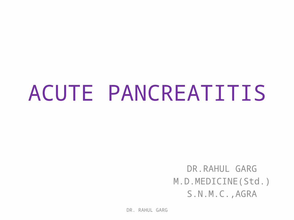
DEFINITIONS Atlanta Symposium definition of acute pancreatitis : An acute inflammatory process of the pancreas with variable involvement of other regional tissues or remote organ systems.
Acute pancreatitis is best defined clinically by a patient presenting with two of the following criteria:
(1)Symptoms, such as epigastric pain, consistent with the disease.
(2)A serum amylase or lipase greater than three times the upper limit of normal.
(3) Radiologic imaging consistent with the diagnosis, usually using CT or MRI.
Sleisenger and Fordtran's Gastrointestinal and Liver Disease ninth edition
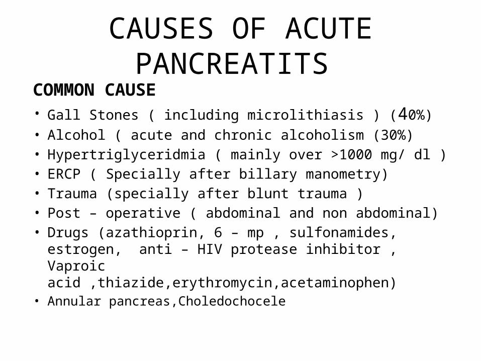
CAUSES OF ACUTE PANCREATITS COMMON CAUSE• Gall Stones ( including microlithiasis ) (40%)• Alcohol ( acute and chronic alcoholism (30%) • Hypertriglyceridmia ( mainly over >1000 mg/ dl )• ERCP ( Specially after billary manometry)• Trauma (specially after blunt trauma )• Post – operative ( abdominal and non abdominal) • Drugs (azathioprin, 6 – mp , sulfonamides, estrogen,
anti – HIV protease inhibitor , Vaproic acid ,thiazide,erythromycin,acetaminophen)
• Annular pancreas,Choledochocele
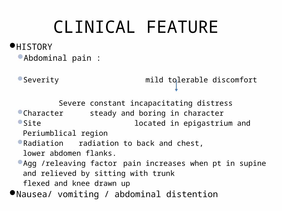
CLINICAL FEATURE HISTORY
Abdominal pain :
Severity mild tolerable discomfort
Severe constant incapacitating distressCharacter steady and boring in character Site located in epigastrium and
Periumblical region Radiation radiation to back and chest,
lower abdomen flanks.Agg /releaving factor pain increases when pt in supine
and relieved by sitting with trunk flexed and knee drawn up
Nausea/ vomiting / abdominal distention
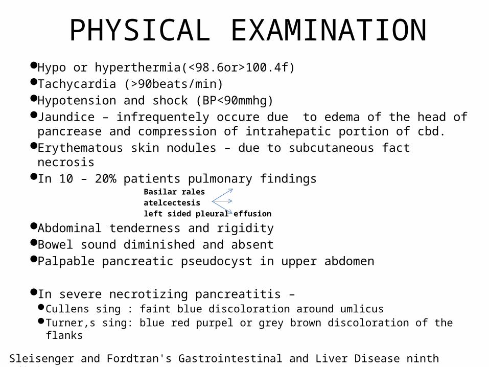
PHYSICAL EXAMINATION Hypo or hyperthermia(<98.6or>100.4f) Tachycardia (>90beats/min)Hypotension and shock (BP<90mmhg)Jaundice – infrequentely occure due to edema of the head of pancrease
and compression of intrahepatic portion of cbd.Erythematous skin nodules – due to subcutaneous fact necrosis In 10 – 20% patients pulmonary findings
Basilar rales atelcectesis left sided pleural effusion
Abdominal tenderness and rigidity Bowel sound diminished and absent Palpable pancreatic pseudocyst in upper abdomen
In severe necrotizing pancreatitis – Cullens sing : faint blue discoloration around umlicus Turner,s sing: blue red purpel or grey brown discoloration of the flanks Sleisenger and Fordtran's Gastrointestinal and Liver Disease ninth edition
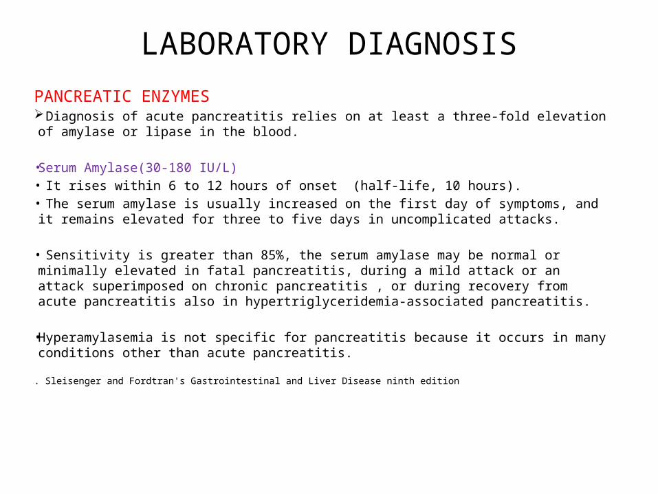
LABORATORY DIAGNOSISPANCREATIC ENZYMES Diagnosis of acute pancreatitis relies on at least a three-fold elevation of amylase or lipase in
the blood.
• Serum Amylase(30-180 IU/L)• It rises within 6 to 12 hours of onset (half-life, 10 hours). • The serum amylase is usually increased on the first day of symptoms, and it remains elevated
for three to five days in uncomplicated attacks.
• Sensitivity is greater than 85%, the serum amylase may be normal or minimally elevated in fatal pancreatitis, during a mild attack or an attack superimposed on chronic pancreatitis , or during recovery from acute pancreatitis also in hypertriglyceridemia-associated pancreatitis.
• Hyperamylasemia is not specific for pancreatitis because it occurs in many conditions other than acute pancreatitis.
. Sleisenger and Fordtran's Gastrointestinal and Liver Disease ninth edition
• Serum Lipase(0-160 IU/L)• The sensitivity of serum lipase is similar to that of serum
amylase and is between 85% and 100%.• Lipase may have greater specificity for pancreatitis than
amylase. • Serum lipase always is elevated on the first day of illness and
remains elevated longer than does the serum amylase.• Some believe that serum lipase is preferable to that of
serum amylase , whereas others find no clear advantage .
• Secretin-pancreozymin (CCK) test• Secretin leads to increased output of pancreatic juice and
HCO3; CCK leads to increased output of pancreatic enzymes.• Sensitive enough to detect occult disease; involves duodenal
intubation and fluoroscopy ,only role in acute on chronic pancreatitis.
Other Pancreatic Enzymes• They include PLA2, trypsin/typsinogen, carboxylester lipase,
carboxypeptidase A, colipase, elastase, and ribonuclease. • None, alone or in combination, are better than serum
amylase or lipase
Other blood test-
• White blood cell count -Increased• Haematocrit->44 show poor prognosis, moniter daily.• Serum Creatinine and serum electrolytes
• AST,ALT,alkaline phosphatase, and bilirubin also may increase .
• MCV- Alcoholic patients tend to have a higher MCV . • Serum triglyceride levels- increase in acute pancreatitis,
but also with alcohol use, uncontrolled diabetes mellitus, or defective triglyceride metabolism
RADIAOLIGICAL –
Chest X- ray / X – ray abdomen errectSee for pulmonary complication pleural effusionRule out other diagnosis,specially – a perforationUSG- Specially for diagnosis of gall stoneHelical CT Scan (Contrast Enhanced ,After 48 -72hrs of onset)Helpful to know severity and prognosis Allows estimation of presence and extend of pancreatic
necrosis.Conferm the cinical impression of acute pancreatitis in face of
normal s. Amylase. Severe allergy and renal impairment(s.crt>2mg/dl) are
contraindication for contrast use.EUS and/or MRCP are better than CT for choledocolithiasis.
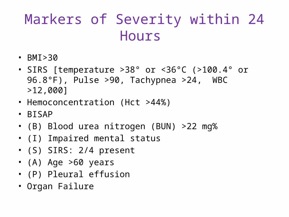
Markers of Severity within 24 Hours
• BMI>30• SIRS [temperature >38° or <36°C (>100.4° or 96.8°F), Pulse
>90, Tachypnea >24, WBC >12,000]• Hemoconcentration (Hct >44%)• BISAP • (B) Blood urea nitrogen (BUN) >22 mg%• (I) Impaired mental status• (S) SIRS: 2/4 present• (A) Age >60 years• (P) Pleural effusion• Organ Failure
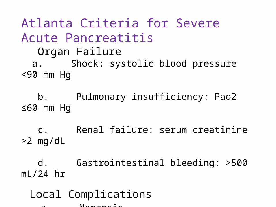
Atlanta Criteria for Severe Acute Pancreatitis Organ Failure a. Shock: systolic blood pressure <90 mm Hg b. Pulmonary insufficiency: Pao2 ≤60 mm Hg c. Renal failure: serum creatinine >2 mg/dL d. Gastrointestinal bleeding: >500 mL/24 hr
Local Complications a. Necrosis b. Abscess c. Pseudocyst
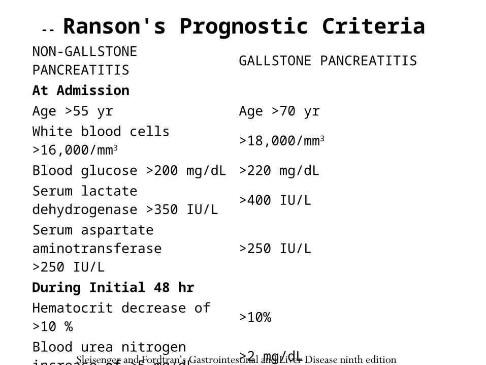
NON-GALLSTONE PANCREATITIS GALLSTONE PANCREATITIS At Admission Age >55 yr Age >70 yrWhite blood cells >16,000/mm3 >18,000/mm3
Blood glucose >200 mg/dL >220 mg/dLSerum lactate dehydrogenase >350 IU/L >400 IU/L
Serum aspartate aminotransferase >250 IU/L >250 IU/L
During Initial 48 hr Hematocrit decrease of >10 % >10%Blood urea nitrogen increase of >5 mg/dL >2 mg/dL
Serum calcium <8 mg/dL <8 mg/dLArterial po2 <60 mm Hg NASerum base deficit >4 mEq/L >5 mEq/LFluid sequestration >6 L >4 L
-- Ranson's Prognostic Criteria
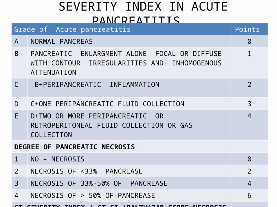
SEVERITY INDEX IN ACUTE PANCREATITIS Grade of Acute pancreatitis Points
A NORMAL PANCREAS 0
B PANCREATIC ENLARGMENT ALONE FOCAL OR DIFFUSE WITH CONTOUR IRREGULARITIES AND INHOMOGENOUS ATTENUATION
1
C B+PERIPANCREATIC INFLAMMATION 2
D C+ONE PERIPANCREATIC FLUID COLLECTION 3
E D+TWO OR MORE PERIPANCREATIC OR RETROPERITONEAL FLUID COLLECTION OR GAS COLLECTION
4
DEGREE OF PANCREATIC NECROSIS
1 NO – NECROSIS 0
2 NECROSIS OF <33% PANCREASE 2
3 NECROSIS OF 33%-50% OF PANCREASE 4
4 NECROSIS OF > 50% OF PANCREASE 6
CT SEVERITY INDEX ( CT SI )BALTHAZAR SCORE+NECROSIS SCORE
CT GRADE + NECROSIS GRADE (0 - 4) + ( 0 – 6 ) ( 0 – 10 )
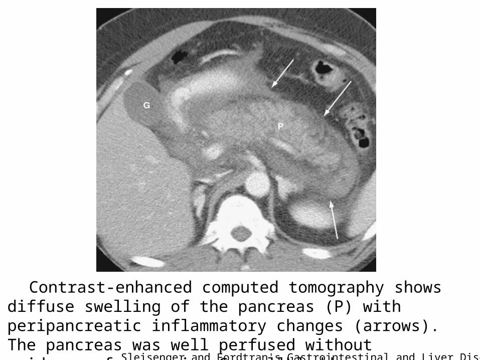
Contrast-enhanced computed tomography shows diffuse swelling of the pancreas (P) with peripancreatic inflammatory changes (arrows). The pancreas was well perfused without evidence of necrosis. G, gallbladder. Sleisenger and Fordtran's Gastrointestinal and Liver Disease ninth edition
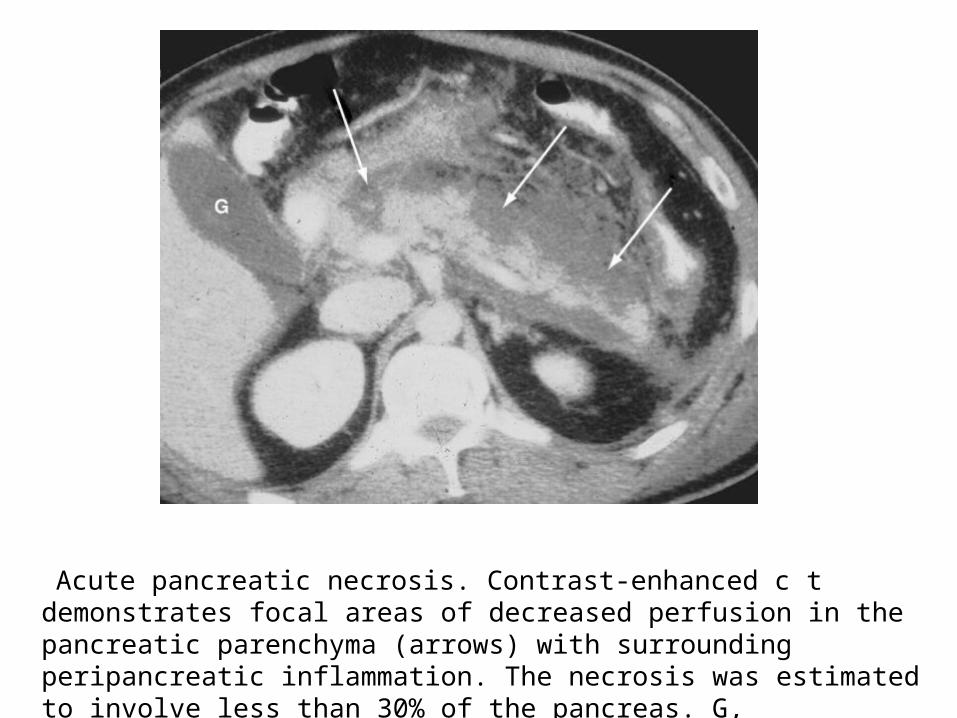
Acute pancreatic necrosis. Contrast-enhanced c t demonstrates focal areas of decreased perfusion in the pancreatic parenchyma (arrows) with surrounding peripancreatic inflammation. The necrosis was estimated to involve less than 30% of the pancreas. G, gallbladder
MANAGEMENT;• Patients with acute pancreatitis require adequate intravenous
hydration and adequate analgesia to eliminate or markedly reduce pain.
• The patient is usually on npo until any nausea and vomiting have subsided.
• Opiate dosing is monitored carefully and adjusted on a daily basis according to ongoing needs.
• Nasogastric intubation is not used routinely because it is not beneficial in mild pancreatitis.
• It is used only to treat gastric or intestinal ileus or intractable nausea and vomiting.
• Similarly, proton pump inhibitors or H2-receptor blocking agents are not beneficial and not used.
Sleisenger and Fordtran's Gastrointestinal and Liver Disease ninth edition

• The patient should be carefully monitored for any signs of early organ failure such as hypotension, vital signs and urinary output.
• Tachypnea should not be assumed to be due to abdominal
pain; monitoring oxygen saturation and, if needed, blood gases is advised and oxygen supplementation is mandatory if there is hypoxemia.
• Any patient who exhibits signs of early organ dysfunction should be immediately transferred to intensive care monitoring.
FLUID RESUSCITATION;• The goal is to decrease the hematocrit. • Laboratory and clinical studies with intravenous dextran to
promote hemodilution have suggested efficacy in preventing severe disease.
• One of the markers of severity of pancreatitis defined by Ranson and colleagues is intravascular losses (“fluid sequestration”).
• Requirements of a 70-kg person during the first 48 hours, is should be at least 250 to 300 mL/hour for 48 hours.
• The rate of volume replacement is more important during the first
24 hours, when a rising hematocrit has been shown to correlate closely with severe disease.
• Maintaining adequate intravascular volume in patients with severe disease may require 5 to 10 L of fluid such as isotonic saline daily for the first several days (200 to 400 mL/hour).
• In a patient with unclear cardiac output, a Swan-Ganz catheter can be useful .
Sleisenger and Fordtran's Gastrointestinal and Liver Disease ninth edition
RESPIRATORY CARE;
• ARDS is the most serious respiratory complication of acute pancreatitis.
• It generally occurs between the second and seventh day of illness (but can be present on admission) .
• Chest radiography may show multilobar alveolar infiltrates. • Treatment is endotracheal intubation with positive end-
expiratory pressure ventilation, often with low tidal volumes to protect the lungs from volutrauma.
• No specific treatment will prevent or resolve ARDS.
• Hypoxemia (oxygen saturation <90%) requires supplemental oxygen.
• Current guidelines recommend the initial routine use of nasal cannula oxygen to all patients with acute pancreatitis.
• Cardiac complications of severe acute pancreatitis include congestive heart failure, myocardial infarction, cardiac dysrhythmia, and cardiogenic shock.
• If hypotension persists even with appropriate fluid
resuscitation, intravenous dopamine may help maintain the systemic blood pressure.
• Dopamine does not impair the microcirculation of the pancreas as do other vasoconstrictors.
METABOLIC COMPLICATION;
• Hyperglycemia may present but usually normalizes as the inflammatory process subsides.
• Blood sugars fluctuate, and insulin should be administered cautiously.
• Reduced serum ionized calcium may occur and cause neuromuscular irritability.
• If hypomagnesemia coexists, magnesium replacement should restore serum calcium to normal.
• Once the serum magnesium is normal, signs or symptoms of neuromuscular irritability may require administering intravenous calcium gluconate as long as the serum potassium is normal and digitalis is not being given.
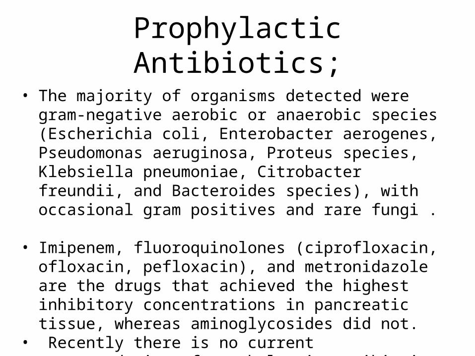
Prophylactic Antibiotics;
• The majority of organisms detected were gram-negative aerobic or anaerobic species (Escherichia coli, Enterobacter aerogenes, Pseudomonas aeruginosa, Proteus species, Klebsiella pneumoniae, Citrobacter freundii, and Bacteroides species), with occasional gram positives and rare fungi .
• Imipenem, fluoroquinolones (ciprofloxacin, ofloxacin, pefloxacin), and metronidazole are the drugs that achieved the highest inhibitory concentrations in pancreatic tissue, whereas aminoglycosides did not.
• Recently there is no current recommendation of prophylactic antibiotic.
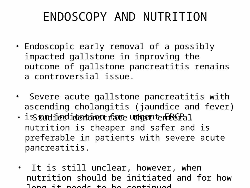
ENDOSCOPY AND NUTRITION
• Endoscopic early removal of a possibly impacted gallstone in improving the outcome of gallstone pancreatitis remains a controversial issue.
• Severe acute gallstone pancreatitis with ascending cholangitis (jaundice and fever) is an indication for urgent ERCP .
• Studies demonstrate that enteral nutrition is cheaper and safer and is preferable in patients with severe acute pancreatitis.
• It is still unclear, however, when nutrition should be initiated and for how long it needs to be continued.
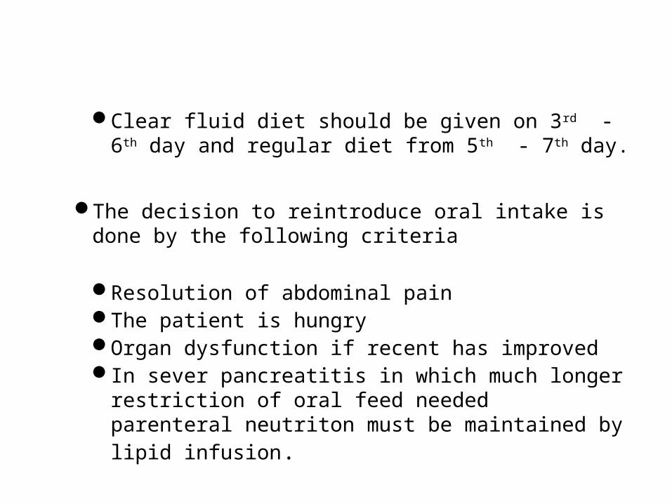
Clear fluid diet should be given on 3rd - 6th day and regular diet from 5th - 7th day.
The decision to reintroduce oral intake is done by the following criteria
Resolution of abdominal pain The patient is hungry Organ dysfunction if recent has improved In sever pancreatitis in which much longer restriction of
oral feed needed parenteral neutriton must be maintained by lipid infusion.
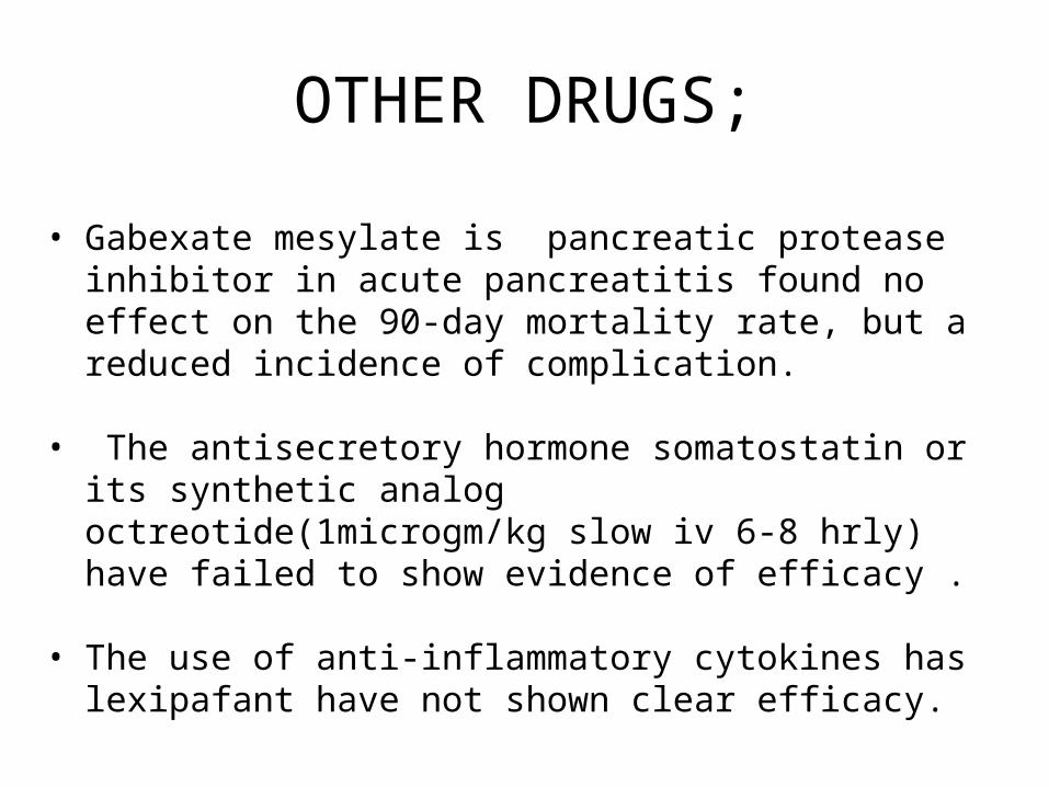
OTHER DRUGS;
• Gabexate mesylate is pancreatic protease inhibitor in acute pancreatitis found no effect on the 90-day mortality rate, but a reduced incidence of complication.
• The antisecretory hormone somatostatin or its synthetic
analog octreotide(1microgm/kg slow iv 6-8 hrly) have failed to show evidence of efficacy .
• The use of anti-inflammatory cytokines has lexipafant have not shown clear efficacy.
ROLE OF SURGERY;• Cholecystectomy should be performed as soon as the
patient has recovered and the acute inflammatory process has subsided.
• A second potential role for surgery in pancreatitis is to debride pancreatic necrosis (necrosectomy) .
COMPLICATION;LOCAL
• Necrosis– Sterile / infected
• Pancreatic fluid collection– Pancreatic abscess – Pancreatic pseudocyst – Pancreatic ascitis
• Involvement of contiguous organ by necrotising pancreatitis – Massive intraperitioneal haemorrhage – Thrombosis of blood vessels
• Obstructive jaundice

SYSTEMIC Pulmonary
Pleural effusion Atelectasis Pneumonitis ARDS
Cardiovascular Hypotension / hypervolemia Sudden deathPericardial effusion
Hematological DIC
GI haemorrhagePeptic ulcer disease Erosive gastritis Hemorrhagic pancreatitis erosion in blood vessel Portal vein thrombosis
SYSTEMIC Renal
Oliguria Azotemia Renal artery / vein thrombosis
Metabolic HyperglycemiaHypertriglyceridemia Hypocalcemia Purtscher’s retinopathy ( due to occulsion of post retinal artery
by aggregated granulocyte)CNS
Psychosis Fat emboli
Fat necrosisSubcutaneous tissue - erythematous nodule

THANK YOU…