Allogeneic Hematopoietic Stem Cell Transplant for the Medical Oncologist John Kuruvilla MD FRCPC.

Pandemic (H1N1) 2009 influenza in Canadian pediatriccancer and hematopoietic stem cell transplant patients
Dat Trana,b,c, Michelle Sciencea, David Dixd, Carol Portwinee, Shayna Zelcerf, Donna L. Johnstong,
Rochelle Yanofskyh, Adam Gassasi,c, Marie-Chantal Ethier j, Lillian Sung ,c,i,j
aDivision of Infectious Diseases, The Hospital for Sick Children, Toronto, ON, Canada. bProgram in Genetics & Genome Biology, Research
Institute, The Hospital for Sick Children, Toronto, ON, Canada. cDepartment of Paediatrics, University of Toronto, Toronto, ON, Canada.dBC Children’s Hospital, Vancouver, BC, Canada. eHamilton Health Sciences, Hamilton, ON, Canada. fLondon Health Sciences, London, ON,
Canada. gChildren’s Hospital of Eastern Ontario, Ottawa, ON, Canada. hCancer Care Manitoba, Winnipeg, MB, Canada. iDivision of
Hematology ⁄ Oncology, The Hospital for Sick Children, Toronto, ON, Canada. jProgram in Child Health Evaluative Sciences, Research Institute,
The Hospital for Sick Children, Toronto, ON, Canada.
Correspondence: Lillian Sung, Division of Hematology ⁄ Oncology, The Hospital for Sick Children, 555 University Ave., Toronto, Ontario, Canada
M5G 1X8. E-mail: [email protected]
Accepted 6 February 2012. Published Online 14 March 2012.
Background The impact of pandemic H1N1 influenza (pH1N1)
virus in pediatric cancer is uncertain. The objectives of this study
were to characterize the clinical course of pH1N1 and identify
factors associated with severe outcomes.
Methods We conducted a Canadian multicenter retrospective
review of children with cancer and stem cell transplant (SCT)
recipients who were diagnosed with laboratory-confirmed pH1N1
infection between May 1, 2009 and January 31, 2010.
Results We identified 100 (19 in wave 1 and 81 in wave 2) cases
of pH1N1 infection. Median age was 8Æ7 years. 71% had a
hematologic malignancy, and 20% received SCT. Median duration
of fever and illness was 2 and 12Æ5 days, respectively. 51 (51Æ5%)
were hospitalized for a median of 5 days, with no deaths and only
1 requiring admission to the intensive care unit. Radiologically
confirmed pneumonia was diagnosed in 10 (10%). Interruption of
chemotherapy or conditioning occurred in 43 patients. In
multivariable analyses, age <5 years (relative to ‡10 years) and
neutropenia were associated with hospitalization while
neutropenia was associated with pneumonia. Despite oseltamivir
use in 89%, viral shedding was prolonged (median, 46 days) and
often persisted after symptom resolution. However, an extended
treatment course (>5 days) correlated with shortened duration of
viral shedding (P = 0Æ041).
Conclusions pH1N1 infection in pediatric cancer and SCT
patients infrequently caused complications but commonly
interrupted cancer treatment. Persistent shedding of virus after
illness resolution was common. Further research is needed to
verify this finding as it could have implications for treatment
guidelines and infection control practices.
Keywords cancer, child, pandemic H1N1 influenza, stem cell
transplant.
Please cite this paper as: Tran et al. (2012) Pandemic (H1N1) 2009 influenza in Canadian pediatric cancer and hematopoietic stem cell transplant patients.
Influenza and Other Respiratory Viruses DOI: 10.1111/j.1750-2659.2012.00352.x.
Introduction
First detected in April of 2009,1 the pandemic H1N1 influ-
enza (pH1N1) virus rapidly spreads globally, triggering the
first pandemic of the 21st century. By August 1, 2010,
more than 214 countries reported laboratory-confirmed
cases to the World Health Organization, including over
18 449 deaths.2
Children with cancer and pediatric hematopoietic stem
cell transplant (SCT) recipients comprise an important
group of immunocompromised patients in whom pro-
longed viral shedding and serious influenza-associated mor-
bidity are common.3–10 If pH1N1 substantially contributes
to future influenza seasons as may be expected via anti-
genic drift, an understanding of its clinical course and
severity in pediatric cancer patients in multiple geographic
regions would help inform healthcare needs, provide infor-
mation to facilitate decision-making regarding cancer treat-
ment interruption, and identify early interventions to
improve outcomes.
An analysis of pooled surveillance data on laboratory-
confirmed pH1N1 cases from 19 countries or administra-
tive regions demonstrated that the odds ratio (OR) for
death given hospitalization was >5 for immunocompro-
mised patients, but did not determine ORs for subgroups
of these patients.11 Several studies of pH1N1 infection in
pediatric cancer patients have yielded conflicting informa-
tion, with some demonstrating a mild course,12–15 while a
DOI:10.1111/j.1750-2659.2012.00352.x
www.influenzajournal.comOriginal Article
ª 2012 Blackwell Publishing Ltd 1

Brazilian study documented a high mortality rate among
hospitalized cases.16 Almost all of these studies were con-
ducted at single centers, thus raising concerns about the
generalizability of findings.
Consequently, we conducted a Canadian multicenter
retrospective analysis of pH1N1 in pediatric cancer. Our
objectives were to describe the clinical course and identify
factors associated with severe outcomes of children with
cancer or pediatric SCT recipients diagnosed with labora-
tory-confirmed pH1N1.
Methods
Patients and settingWe identified eligible cases through retrospective review of
virology reports and health records from a base population
of patients cared for by the Divisions of Haematology ⁄Oncology at six Canadian pediatric referral centers (BC
Children’s Hospital, Vancouver; The Children’s Hospital of
Winnipeg, Winnipeg; Children’s Hospital London Health
Sciences Centre, London; McMaster Children’s Hospital,
Hamilton; The Hospital for Sick Children, Toronto; and
Children’s Hospital of Eastern Ontario, Ottawa).
Subjects eligible for study inclusion were (i) £18 years of
age; (ii) receiving or about to receive chemotherapy for can-
cer or conditioning for SCT, or received treatment within
the preceding 3 months (for chemotherapy patients),
6 months (for autologous SCT recipients), 2 years (for allo-
geneic SCT recipients), or receiving immunosuppressive
therapy for graft-versus-host disease for those ‡2 years post-
allogeneic SCT; and (iii) diagnosed with pH1N1 infection by
RT-PCR [using primers developed by the Centers for Disease
Control and Prevention (CDC; Atlanta, GA)17 or the
National Microbiology Laboratory (NML; Public Health
Agency of Canada, Winnipeg, MB),18 or the Astra influenza
Screen & Type (Astra Diagnostics, Hamburg, Germany)19]
of nasopharyngeal or flocked nasal mid-turbinate swabs
between May 1, 2009 and January 6, 2010.
Case details abstracted from health records were reported
on a standardized case report form. We collected patient
demographics, underlying conditions, treatment informa-
tion, clinical manifestations, pandemic H1N1 vaccination
history, antiviral and antibiotic use, level of care required
and influenza-related complications. All centers obtained
research ethics board approval for the study.
Outcomes and definitionsNorth America experienced two waves of pH1N1 activity,
peaking in most locations around June and October
2009.20 Based on reporting of laboratory-confirmed pH1N1
hospitalized cases and deaths to the Public Health Agency
of Canada, we defined wave 1 as the period prior to August
30, 2009.21
Severe outcomes examined were hospitalization, radio-
logically confirmed pneumonia (defined as new pulmonary
infiltrate on chest imaging associated with compatible clini-
cal symptoms), intensive care unit (ICU) admission, need
for mechanical ventilation, and mortality. Potential predic-
tors of severe outcomes were identified from the literature
or based on clinical experience and were defined a priori.
Other complications of interest that developed between
symptom onset and resolution were classified as respiratory
complications, extra-respiratory complications or microbio-
logically confirmed infections. Extra-respiratory complica-
tions were further subcategorized into hematologic or non-
hematologic complications. Hematologic complications of
interest were neutropenia (absolute neutrophil count
<500 ⁄ mm3), lymphopenia (<500 ⁄ mm3), and thrombocyto-
penia (<50 000 ⁄ mm3). Non-hematologic complications
specifically sought for included clinical diagnoses of sepsis
(hypotension requiring fluid bolus or inotrope administra-
tion), pericarditis ⁄ myocarditis, myositis (elevated creatine
phosphokinase), encephalitis, and hepatitis (aspartate ami-
notransferase or alanine aminotransferase > twice the
upper limit of normal for age).
Obesity and underweight were covariates of interest and
were examined for children at least 2 years of age. Obesity
was defined as a body mass index (BMI) ‡ 95th percentile,
and underweight was defined as BMI < 5th percentile
based on the gender- and age-specific BMI reference from
Centers for Disease Control and Prevention.22 Children
<2 years were not included in this analysis because of
uncertainty in how to categorize children in this age-group.
Reflecting the recommendations of the Advisory Com-
mittee on Immunization Practices,23 full vaccination with
pH1N1 vaccine for children 6 months to <9 years was
defined as receipt of two vaccine doses separated by at least
4 weeks with dose 2 received ‡14 days prior to illness
onset. Those who received only one dose ‡14 days before
illness onset were classified as partially vaccinated. Children
aged 9–18 years were considered fully vaccinated with
receipt of one vaccine dose ‡14 days prior to illness onset.
Statistical considerationsStandard descriptive statistics were used to summarize data
on the study population. For all statistical testing, only the
first pH1N1 episode was included in cases with multiple
episodes within the same child because these episodes can-
not be considered independent. Comparisons of data cate-
gorized by hospitalization versus no hospitalization,
presence or absence of radiologically confirmed pneumo-
nia, and pandemic wave (wave 2 versus 1) were made using
the Mann–Whitney test for continuous variables and the v2
or Fisher’s exact test as appropriate for categorical vari-
ables. We performed univariate and multivariable linear or
logistic regression, as appropriate, to adjust for potential
Tran et al.
2 ª 2012 Blackwell Publishing Ltd
confounding variables and to identify independent predic-
tors of severe outcomes. We considered for the multivariable
models all variables with P values <0Æ1 in univariate analy-
sis and retained in the final model all variables with an
adjusted P-value <0Æ05. All tests were two-sided, and a P
value <0Æ05 was considered statistically significant. Data
were analyzed by spss statistical software (version 16Æ0,
SPSS Inc, Chicago, IL, USA).
Results
We identified a total of 102 episodes of laboratory-confirmed
pH1N1 infection in 100 children with 100 unique first epi-
sodes. Unique episodes were distributed in two distinct
waves: 19 in wave 1 prior to August 29, 2009, and 81 in wave
2 from August 30, 2009 onward. In total, 51 ⁄ 99 were hospi-
talized (information missing in one child) and 10 ⁄ 100 were
diagnosed with radiologically confirmed pneumonia.
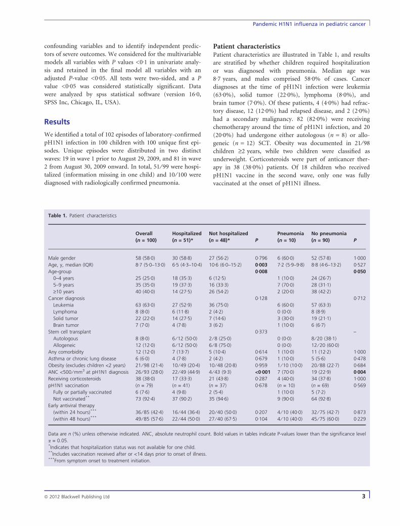
Patient characteristicsPatient characteristics are illustrated in Table 1, and results
are stratified by whether children required hospitalization
or was diagnosed with pneumonia. Median age was
8Æ7 years, and males comprised 58Æ0% of cases. Cancer
diagnoses at the time of pH1N1 infection were leukemia
(63Æ0%), solid tumor (22Æ0%), lymphoma (8Æ0%), and
brain tumor (7Æ0%). Of these patients, 4 (4Æ0%) had refrac-
tory disease, 12 (12Æ0%) had relapsed disease, and 2 (2Æ0%)
had a secondary malignancy. 82 (82Æ0%) were receiving
chemotherapy around the time of pH1N1 infection, and 20
(20Æ0%) had undergone either autologous (n = 8) or allo-
geneic (n = 12) SCT. Obesity was documented in 21 ⁄ 98
children ‡2 years, while two children were classified as
underweight. Corticosteroids were part of anticancer ther-
apy in 38 (38Æ0%) patients. Of 18 children who received
pH1N1 vaccine in the second wave, only one was fully
vaccinated at the onset of pH1N1 illness.
Table 1. Patient characteristics
Overall
(n = 100)
Hospitalized
(n = 51)*
Not hospitalized
(n = 48)* P
Pneumonia
(n = 10)
No pneumonia
(n = 90) P
Male gender 58 (58Æ0) 30 (58Æ8) 27 (56Æ2) 0Æ796 6 (60Æ0) 52 (57Æ8) 1Æ000
Age, y, median (IQR) 8Æ7 (5Æ0–13Æ0) 6Æ5 (4Æ3–10Æ4) 10Æ6 (6Æ0–15Æ2) 0Æ003 7Æ2 (5Æ9–9Æ8) 8Æ8 (4Æ6–13Æ2) 0Æ527
Age-group 0Æ008 0Æ050
0–4 years 25 (25Æ0) 18 (35Æ3) 6 (12Æ5) 1 (10Æ0) 24 (26Æ7)
5–9 years 35 (35Æ0) 19 (37Æ3) 16 (33Æ3) 7 (70Æ0) 28 (31Æ1)
‡10 years 40 (40Æ0) 14 (27Æ5) 26 (54Æ2) 2 (20Æ0) 38 (42Æ2)
Cancer diagnosis 0Æ128 0Æ712
Leukemia 63 (63Æ0) 27 (52Æ9) 36 (75Æ0) 6 (60Æ0) 57 (63Æ3)
Lymphoma 8 (8Æ0) 6 (11Æ8) 2 (4Æ2) 0 (0Æ0) 8 (8Æ9)
Solid tumor 22 (22Æ0) 14 (27Æ5) 7 (14Æ6) 3 (30Æ0) 19 (21Æ1)
Brain tumor 7 (7Æ0) 4 (7Æ8) 3 (6Æ2) 1 (10Æ0) 6 (6Æ7)
Stem cell transplant 0Æ373 –
Autologous 8 (8Æ0) 6 ⁄ 12 (50Æ0) 2 ⁄ 8 (25Æ0) 0 (0Æ0) 8 ⁄ 20 (38Æ1)
Allogeneic 12 (12Æ0) 6 ⁄ 12 (50Æ0) 6 ⁄ 8 (75Æ0) 0 (0Æ0) 12 ⁄ 20 (60Æ0)
Any comorbidity 12 (12Æ0) 7 (13Æ7) 5 (10Æ4) 0Æ614 1 (10Æ0) 11 (12Æ2) 1Æ000
Asthma or chronic lung disease 6 (6Æ0) 4 (7Æ8) 2 (4Æ2) 0Æ679 1 (10Æ0) 5 (5Æ6) 0Æ478
Obesity (excludes children <2 years) 21 ⁄ 98 (21Æ4) 10 ⁄ 49 (20Æ4) 10 ⁄ 48 (20Æ8) 0Æ959 1 ⁄ 10 (10Æ0) 20 ⁄ 88 (22Æ7) 0Æ684
ANC <500 ⁄ mm3 at pH1N1 diagnosis 26 ⁄ 93 (28Æ0) 22 ⁄ 49 (44Æ9) 4 ⁄ 43 (9Æ3) <0Æ001 7 (70Æ0) 19 (22Æ9) 0Æ004
Receiving corticosteroids 38 (38Æ0) 17 (33Æ3) 21 (43Æ8) 0Æ287 4 (40Æ0) 34 (37Æ8) 1Æ000
pH1N1 vaccination (n = 79) (n = 41) (n = 37) 0Æ678 (n = 10) (n = 69) 0Æ569
Fully or partially vaccinated 6 (7Æ6) 4 (9Æ8) 2 (5Æ4) 1 (10Æ0) 5 (7Æ2)
Not vaccinated** 73 (92Æ4) 37 (90Æ2) 35 (94Æ6) 9 (90Æ0) 64 (92Æ8)
Early antiviral therapy
(within 24 hours)*** 36 ⁄ 85 (42Æ4) 16 ⁄ 44 (36Æ4) 20 ⁄ 40 (50Æ0) 0Æ207 4 ⁄ 10 (40Æ0) 32 ⁄ 75 (42Æ7) 0Æ873
(within 48 hours)*** 49 ⁄ 85 (57Æ6) 22 ⁄ 44 (50Æ0) 27 ⁄ 40 (67Æ5) 0Æ104 4 ⁄ 10 (40Æ0) 45 ⁄ 75 (60Æ0) 0Æ229
Data are n (%) unless otherwise indicated. ANC, absolute neutrophil count. Bold values in tables indicate P-values lower than the significance level
a = 0.05.*Indicates that hospitalization status was not available for one child.**Includes vaccination received after or <14 days prior to onset of illness.***From symptom onset to treatment initiation.
Pandemic H1N1 influenza in pediatric cancer
ª 2012 Blackwell Publishing Ltd 3

Clinical presentation, course, and managementTable 2 illustrates symptoms at presentation of pH1N1 and
during the illness course. The most commonly reported pre-
senting symptoms were fever (84Æ0%) and cough (80Æ0%);
24Æ0% had abdominal pain, nausea, vomiting, and ⁄ or
diarrhea. Median interval between symptom onset and clin-
ical encounter was 1 day [interquartile range (IQR), 0–2].
The treatment and clinical course of pH1N1 infection
are presented in Tables 1 and 3. Antiviral therapy was pre-
scribed to most patients during the pandemic, with more
frequent antiviral use in wave 2 than 1 (P = 0Æ001). Over-
all, antiviral therapy was extended beyond the standard
5-day course for 17 (19Æ5%) patients, 12 of whom received
a 10-day course. Antibiotics were given in 56 (56Æ0%) cases.
Median duration of symptoms (defined as the interval
between onset of any symptom and resolution of all symp-
toms) and that of fever was 12Æ5 and 2 days, respectively.
We were able to determine the duration of shedding of
pH1N1 virus for 35 patients, the median of which was
46 days. In the 22 patients for whom both duration of
symptoms and viral shedding were available, 13 continued
to shed virus after symptom resolution. The median dura-
tion of viral shedding did not differ significantly based on
whether or not the patient was hospitalized (63 days [IQR,
14–88Æ5] versus 44 days [IQR, 19–78Æ5]; P = 0Æ986) or diag-
nosed with pneumonia (65Æ5 days [IQR, 20Æ25–95Æ75]
versus 44 days [IQR, 14–85]; P = 0Æ836). The median dura-
tion of viral shedding was, however, significantly shorter in
wave 2 of the pandemic (38 days [IQR, 13–70] versus 98.5
days [IQR, 28.25–157.25]; P = 0Æ034). As antiviral use dif-
fered between wave 1 and 2, we explored the possible con-
founding effects of different aspects of antiviral
prescription (antiviral treatment versus no treatment, antiv-
iral treatment within 24 hours versus after 24 hours of
symptom onset, extended versus standard antiviral course)
and other factors on the logarithm of length of viral shed-
ding in linear regression analyses (Table 4). After multiple
regression analysis, we found that an extended antiviral
treatment course, but not wave 2, was significantly associ-
ated with shortened duration of viral shedding.
In terms of other complications, 2 experienced asthma
exacerbation. Upper respiratory tract disease associated
with pH1N1 infection were otitis media (n = 4), sinusitis
(n = 2), and croup (n = 1). Hematologic abnormalities
were neutropenia (n = 32), lymphopenia (n = 29), and
thrombocytopenia (n = 17). Of the 11 patients classified as
having a non-hematologic, extra-respiratory complication,
nine had hepatitis. Microbiologically confirmed bacterial
infection was documented in two cases (one coagulase-neg-
ative staphylococcus bacteremia and one Staphylococcus
aureus cellulitis). Infection with a herpes group virus dur-
ing the pH1N1 illness was reported in two patients (one
herpes simplex virus and one Epstein–Barr virus), both
managed as outpatients. Of the 82 patients scheduled for
chemotherapy or conditioning for SCT, 43 had their che-
motherapy or conditioning delayed (n = 23), modified
(n = 5), or stopped ⁄ canceled (n = 11).
Severe outcomes of pH1N1As previously described, 51 children were hospitalized and
10 had radiologically confirmed pneumonia (Table 3).
The median duration of hospitalization was 5 days (IQR,
3-13 days); for those with absolute neutrophil count
(ANC) < 500 cells ⁄ mm3 at pH1N1 diagnosis, it was 6 days
(IQR, 4–11Æ5 days). Overall, few patients developed critical
illness. There was no mortality; one child was admitted to
ICU for pneumonia requiring ‡60% supplemental oxygen
and continuous positive airway pressure by face mask but
not intubation or mechanical ventilation; and one child
had pneumonia resulting in ‡60% supplemental oxygen
therapy but did not require ICU admission.
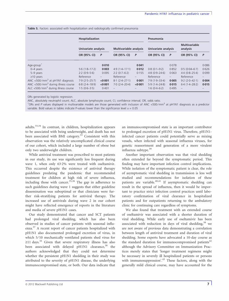
Table 5 displays univariate and multivariable analyses for
predictors of pH1N1-associated hospitalization and pneu-
monia. In univariate analyses, children <5 years were more
likely to be hospitalized (P = 0Æ003) and those aged
5–9 years trended toward being more likely to have
pneumonia (P = 0Æ063), compared to children ‡10 years.
Neutropenia at pH1N1 diagnosis was associated with
Table 2. Clinical manifestations at presentation and during the
course of the influenza illness
At
presentation
(n = 100)
During
illness
(n = 100)
n % n %
Fever 84 84Æ0 88 88Æ0Maximum temperature,
�C, mean (SD)
38Æ8 (0Æ6) 38Æ9 (0Æ7)
Rhinorrhea 46 46Æ0 46 46Æ0Sore throat 22 22Æ0 26 26Æ0Cough 80 80Æ0 85 85Æ0Hemoptysis 0 0Æ0 1 1Æ0Wheezing 3 3Æ0 6 6Æ0Respiratory distress 2 2Æ0 4 4Æ0Rash 6 6Æ0 7 7Æ0Headache 13 13Æ0 15 15Æ0Myalgia 12 12Æ0 13 13Æ0Abdominal symptoms* 24 24Æ0 28 28Æ0Fatigue 20 20Æ0 26 26Æ0Irritability 3 3Æ0 3 3Æ0Altered level of
consciousness
1 1Æ0 1 1Æ0
Seizure 0 0Æ0 0 0Æ0
*Includes nausea and ⁄ or vomiting and ⁄ or diarrhea.
Tran et al.
4 ª 2012 Blackwell Publishing Ltd

hospitalization (P < 0Æ001) and pneumonia (P = 0Æ005), as
was neutropenia detected at any time during the illness
(P < 0Æ001 and P = 0Æ015, respectively). We did not collect
data on lymphopenia at pH1N1 diagnosis, but lymphopenia
detected at any time during the illness was not associated
with hospitalization or pneumonia. Multiple logistic
regression revealed that age category and neutropenia (both
at diagnosis and during the illness) were significant
independent predictors of hospitalization, with children
<5 years at highest risk. Neutropenia, but not age, was a
significant predictor of pneumonia in multiple logistic
regression analysis. The models that included age and
neutropenia at diagnosis fit the observed data well
(Hosmer–Lemeshow P = 0Æ955 and 0Æ778 for hospitaliza-
tion and pneumonia, respectively) and discriminated
well between those who were and were not hospitalized
[c-index, 0Æ767 (95% CI, 0Æ670–0Æ864)] as well as between
children with and without pneumonia [c-index, 0Æ839
(95% CI, 0Æ735–0Æ942)].
Discussion
This study resulted in several important observations. First,
pH1N1 infection was uncomplicated in most children with
cancer although commonly interrupted anticancer therapy.
Second, neutropenia was an important predictor of hospi-
talization as well as pneumonia. Third, antiviral prescrip-
tion increased significantly during wave 2 of the pandemic.
Finally, prolonged viral shedding often persisted after
symptom resolution, and an extended antiviral treatment
Table 3. Clinical course, treatment, and complications
Overall (n = 100) Wave 1 (n = 19) Wave 2 (n = 81) P
Clinical course and management
Days of symptoms, median (IQR) 12Æ5 (6–18) 18 (7–19Æ25) 10 (6–16Æ75) 0Æ149
Days of fever, median (IQR) 2 (1–4) 2 (1–3) 2 (1–4) 0Æ443
Days of viral shedding, median (IQR) 46 (14–85) 98Æ5 (28Æ25–157Æ25) 38 (13–70) 0Æ034
Antiviral therapy 89 (89Æ0) 12 (63Æ2) 77 (95Æ1) 0Æ001
Early antiviral therapy
Within 24 hours of symptom onset 36 ⁄ 85 (42Æ4) 2 ⁄ 11 (18Æ2) 34 ⁄ 74 (45Æ9) 0Æ108
Within 48 hours of symptom onset 49 ⁄ 85 (57Æ6) 6 ⁄ 11 (54Æ5) 43 ⁄ 74 (58Æ1) 1Æ000
Antiviral duration
Standard (5 days) 70 ⁄ 87 (80Æ5) 8 ⁄ 11 (72Æ7) 62 ⁄ 76 (81Æ6) 0Æ443
Extended (>5 days) 17 ⁄ 87 (19Æ5) 3 ⁄ 11 (27Æ3) 14 ⁄ 76 (18Æ4)
Antibiotic treatment 56 (56Æ0) 9 (47Æ4) 47 (58Æ0) 0Æ400
Oxygen supplementation 4 (4Æ0) 2 (10Æ5) 2 (2Æ5) 0Æ162
Respiratory complications* 9 (9Æ0) 1 (5Æ3) 8 (9Æ9) 1Æ000
Upper respiratory tract 7 (7Æ0) 1 6
Asthma exacerbation 2 (2Æ0) 0 2
Extra-respiratory complications 39 (39Æ0) 8 (42Æ1) 31 (38Æ3) 0Æ758
Hematologic 50 (50Æ0) 13 37
Non-hematologic 11 (11Æ0) 5 6
Laboratory-confirmed infections 5 (5Æ0) 1 (5Æ3) 4 (4Æ9) 1Æ000
Bacterial 2 (2Æ0) 0 2
Fungal 1 (1Æ0) 1 0
Viral 2 (2Æ0) 0 2
Alteration of chemotherapy** 43 ⁄ 82 (52Æ42) 9 ⁄ 17 (52Æ9) 34 ⁄ 65 (52Æ3) 0Æ963
Severe outcomes
Hospitalization 51 ⁄ 99 (51Æ5) 10 (52Æ6) 41 ⁄ 80 (51Æ2) 0Æ914
Duration, d, median (IQR) 5 (3–13) 8 (4–68Æ5) 5 (3–10Æ75) 0Æ143
Pneumonia*** 10 (10Æ0) 2 (10Æ5) 8 (9Æ9) 1Æ000
ICU admission� 1 (1Æ0) 0 (0Æ0) 1 (1Æ2) 1Æ000
Need for mechanical ventilation 0 (0Æ0) – – –
Death 0 (0Æ0) – – –
Data are n (%) unless otherwise indicated. Bold values in tables indicate P-values lower than the significance level a = 0.05.*Excludes radiologically confirmed pneumonia.**Denominators exclude patients not scheduled for chemotherapy.***Radiologically confirmed.�None required ventilation, inotropes, dialysis, or extracorporeal membrane oxygenation.
Pandemic H1N1 influenza in pediatric cancer
ª 2012 Blackwell Publishing Ltd 5

course >5 days was associated with shortened length of
viral shedding.
In our cohort, 51Æ5% of patients were hospitalized and
10% developed lower respiratory tract infection. Although
only one patient was admitted to the ICU and none died,
52Æ4% of those scheduled for chemotherapy or condition-
ing had treatment modified or held. These results are com-
parable to those of modest-sized case series of pediatric
cancer patients from Italy,13 Texas,12 Jordan,14 and Tur-
key.15 In contrast, a Brazilian study found that 8 ⁄ 14 pediat-
ric cancer patients hospitalized with pH1N1 required ICU
admission and ventilator support. This is consistent with
the high rate of severe complications from pH1N1 infec-
tion in the general Brazilian population.16 Potential expla-
nations for the discrepant findings of this study include
increased predisposition to severe pH1N1 infection in the
Brazilian population owing to host genetic and ⁄ or socio-
economic factors.
Several factors may contribute to the generally favorable
outcome of pH1N1 infection in cancer patients. First, par-
ents of children with cancer are likely to seek prompt med-
ical attention in the presence of fever, resulting in early
diagnosis and antiviral treatment. A meta-analysis of 11
randomized controlled trials (RCTs) showed that oseltami-
vir treatment was associated with a reduced risk of lower
respiratory tract complications in adolescents and adults
with flu symptoms.24 A recent RCT demonstrated that
early antiviral treatment in children aged 1–3 years reduced
illness duration and incidence of otitis media.25 Second,
increased cytokine and chemokine plasma levels have been
shown to be markers of critical illness with pH1N1 infec-
tion, suggesting that immunopathology may contribute to
disease severity.26,27 It is possible that the relatively benign
course of illness in children with cancer is partly related to
decreased cytokine and chemokine production related to
immune suppression. Third, high levels of cross-reacting
but non-neutralizing antibodies that result from previous
seasonal influenza exposures, leading to immune complex-
mediated pulmonary disease, have been advanced as a
novel biological mechanism of severe cases of pH1N1 in
middle-aged individuals.28 Pediatric cancer and SCT
patients have impaired serum antibody responses to both
seasonal influenza vaccination and natural infection and
would, therefore, likely be protected from this mechanism
of severe pH1N1 disease. Finally, concurrent bacterial infec-
tion has been documented in 29% of lung tissue specimens
from fatal human cases of pH1N1.29 Neutropenia was an
independent predictor of pneumonia in our study, and
neutropenia, in association with fever, would trigger hospi-
talization and broad-spectrum antibiotic therapy resulting
in early treatment of established or subclinical bacterial
lower respiratory tract infection along with more intensive
supportive therapy.
It is possible that neutropenia was merely a surrogate for
profound lymphopenia that has been shown to correlate
with the risk of influenza-associated lower respiratory tract
infection 30,31 and that the lack of association of concurrent
lymphopenia with pneumonia in our study reflected a higher
cut-off point compared to previous studies demonstrating
association (<500 ⁄ mm3 versus <100–200 ⁄ mm3).30,31 More
detailed analysis of ANC and absolute lymphocyte count
(ALC) in relation to the pattern of radiographic changes
(viral versus bacterial) in future studies could provide
further insight. In addition to neutropenia, our study
indicated that younger age (particularly those <5 years)
was associated with hospitalization for pH1N1 infection.
This is consistent with reported rates of pH1N1-associated
hospitalization in the general pediatric population, being
highest for children <5 years and especially for those
<12 months.32 Morbid obesity has been associated with
hospitalization and death from pH1N1 infection in
Table 4. Duration of viral shedding and potential predictive factors
Log10 (days of viral shedding)
Univariate
analysis
Multivariable
analysis
Coefficient b
(SE) P
Coefficient b
(SE) P
Male gender )0Æ07 (0Æ16) 0Æ685 – –
Age (years) )0Æ01 (0Æ02) 0Æ578 – –
Receiving
corticosteroids
0Æ13 (0Æ17) 0Æ441 – –
ANC < 500 ⁄ mm3 at
diagnosis of pH1N1
0Æ13 (0Æ21) 0Æ529 – –
ALC < 500 ⁄ mm3
during illness course
)0Æ03 (0Æ18) 0Æ893 – –
Fully or partially
vaccinated
)0Æ19 (0Æ36) 0Æ600 – –
Hospitalized versus
not hospitalized
)0Æ03 (0Æ16) 0Æ857 – –
Pneumonia versus no
pneumonia
0Æ05 (0Æ25) 0Æ837 – –
Pandemic wave
(wave 2 versus 1)
)0Æ35 (0Æ18) 0Æ059 )0Æ25 (0Æ20) 0Æ219
Antiviral therapy )0Æ26 (0Æ25) 0Æ304 – –
Early antiviral therapy
Within 24 hours of
symptom onset
0Æ04 (0Æ18) 0Æ809 – –
Within 48 hours of
symptom onset
0Æ05 (0Æ17) 0Æ754 – –
Antiviral duration
(extended versus
standard)
)0Æ36 (.017) 0Æ048 )0Æ37 (0Æ17) 0Æ041
ALC, absolute lymphocyte count. Bold values in tables indicate
P-values lower than the significance level a = 0.05.
Tran et al.
6 ª 2012 Blackwell Publishing Ltd
adults.33,34 In contrast, in children, hospitalization appears
to be associated with being underweight, and death has not
been associated with BMI category.33 Consistent with this
observation was the relatively uncomplicated clinical course
of our cohort, which included a large number of obese but
only two underweight children.
While antiviral treatment was prescribed to most patients
in our study, its use was significantly less frequent during
wave 1, when only 63Æ2% were treated with oseltamivir.
This occurred despite the existence of antiviral therapy
guidelines predating the pandemic that recommended
treatment for children at high risk of severe influenza,
including those with cancer.35,36 The gap in adherence to
such guidelines during wave 1 suggests that either guideline
dissemination was suboptimal or that clinicians were fur-
ther risk-stratifying patients for antiviral therapy. The
increased use of antivirals during wave 2 in our cohort
might have reflected emergence of reports in the literature
and media of severe pH1N1 cases.
Our study demonstrated that cancer and SCT patients
had prolonged viral shedding, which has also been
observed in studies of cancer patients with seasonal influ-
enza.37 A recent report of cancer patients hospitalized with
pH1N1 also documented prolonged excretion of virus, in
which 5 ⁄ 10 mechanically ventilated patients shed virus for
‡11 days.16 Given that severe respiratory illness has also
been associated with delayed pH1N1 clearance,38 the
authors acknowledged that they could not determine
whether the persistent pH1N1 shedding in their study was
attributed to the severity of pH1N1 disease, the underlying
immunocompromised state, or both. Our data indicate that
an immunocompromised state is an important contributor
to prolonged excretion of pH1N1 virus. Therefore, pH1N1-
infected cancer patients could potentially serve as mixing
vessels, when infected with seasonal influenza viruses, for
genetic reassortment and generation of a more virulent
influenza subtype.39
Another important observation was that viral shedding
often extended far beyond the symptomatic period. This
finding may have important infection control implications.
While isolation of the symptomatic patient is clear, the role
of asymptomatic viral shedding in transmission is less well
studied and recommendations for isolation of these
patients are variable.40,41 If asymptomatic shedding can
result in the spread of influenza, then it would be impor-
tant to practice strict infection control practices until labo-
ratory confirmation of viral clearance in hospitalized
patients and for outpatients returning to the ambulatory
clinic for continuing care regardless of symptoms.
We also found that treatment with an extended course
of oseltamivir was associated with a shorter duration of
viral shedding. While early use of oseltamivir has been
associated with reduction in days of viral shedding,38 we
are not aware of previous data demonstrating a correlation
between length of antiviral treatment and duration of viral
shedding. Some experts have advocated a 10-day course as
the standard duration for immunocompromised patients42
although the Advisory Committee on Immunization Prac-
tices merely states that ‘longer treatment regimens might
be necessary in severely ill hospitalized patients or persons
with immunosuppression’.43 These factors, along with the
generally mild clinical course, may have accounted for the
Table 5. Factors associated with hospitalization and radiologically confirmed pneumonia
Hospitalization Pneumonia
Univariate analysis Multivariable analysis Univariate analysis
Multivariable
analysis
OR (95% CI) P OR (95% CI) P OR (95% CI) P OR (95% CI) P
Age-group* 0Æ010 0Æ041 0Æ078 0Æ086
0–4 years 5Æ6 (1Æ8–17Æ2) 0Æ003 4Æ9 (1Æ4–17Æ1) 0Æ012 0Æ8 (0Æ1–9Æ2) 0Æ852 0Æ5 (0Æ04–6Æ7) 0Æ626
5–9 years 2Æ2 (0Æ9–5Æ6) 0Æ095 2Æ2 (0Æ7–6Æ2) 0Æ155 4Æ8 (0Æ9–24Æ6) 0Æ063 4Æ4 (0Æ8–25Æ4) 0Æ099
‡10 years Reference – Reference – Reference – Reference –
ANC <500 ⁄ mm3 at pH1N1 diagnosis 7Æ9 (2Æ5–25Æ7) <0Æ001 8Æ1 (2Æ4–27Æ1) 0Æ001 7Æ9 (1Æ9–33Æ4) 0Æ005 9Æ2 (2Æ0–42Æ1) 0Æ004
ANC <500 ⁄ mm3 during illness course 6Æ8 (2Æ4–18Æ9) <0Æ001 7Æ0 (2Æ4–20Æ4) <0Æ001 5Æ9 (1Æ4–24Æ8) 0Æ015 6Æ4 (1Æ4–28Æ2) 0Æ015
ALC <500 ⁄ mm3 during illness course 1Æ5 (0Æ6–3Æ5) 0Æ401 – – 1Æ6 (0Æ4–6Æ2) 0Æ495 – –
ORs generated by logistic regression.
ANC, absolutely neutrophil count; ALC, absolute lymphocyte count; CI, confidence interval; OR, odds ratio.*ORs and P values displayed in multivariable models are those generated with inclusion of ANC <500 ⁄ mm3 at pH1N1 diagnosis as a predictor
variable. Bold values in tables indicate P-values lower than the significance level a = 0.05
Pandemic H1N1 influenza in pediatric cancer
ª 2012 Blackwell Publishing Ltd 7

infrequent use of extended antiviral treatment in the study.
Our findings, if confirmed in future studies, would lend
further support to a recommendation for longer treatment
regimens and may have beneficial infection control impli-
cations. However, any potential benefits from prolonged
antiviral therapy would have to be balanced against the risk
of selection for resistance mutations.44
Limitations of the study include its retrospective design
and inability to calculate population-based rates. Based on
its retrospective nature, our ability to determine whether the
hematologic complications were attributable to influenza
infection or to chemotherapy was limited, and our results
may be affected by unobserved confounders or insufficient
adjustment for observed confounders. The precision in our
estimate of the duration of viral shedding may have been
limited given that follow-up influenza testing was performed
at the clinicians’ discretion. The extent of prolonged viral
shedding may have been overestimated by overrepresenta-
tion of serial sampling in patients with longer courses. We
did not capture data on the duration of individual symp-
toms and therefore were not able to identify those which
could potentially be used to guide optimal retesting and
treatment. Although larger than previous studies of pediatric
cancer and SCT patients, our sample size was still relatively
small. Some of our metrics of influenza severity such as hos-
pitalization, length of hospital stay, and ICU admission may
be influenced by variation in clinical practice across centers
as well as non-medical factors such as publicity surrounding
the pandemic and bed availability.
In conclusion, pH1N1 infection was associated with an
uncomplicated course in most pediatric cancer and SCT
patients but commonly altered scheduling of chemotherapy
or transplant conditioning. Although neutropenia was an
independent predictor of radiologically confirmed pneumo-
nia, it is unclear whether it can be used as a criterion sepa-
rate from lymphopenia for decision-making regarding
cancer treatment modification given the limitations in our
analysis of lymphopenia as a risk factor. Despite the
uncomplicated course in most, viral shedding was pro-
longed and often continued after symptom resolution. Pro-
spective studies evaluating the effects of extended antiviral
treatment on viral shedding in cancer and SCT patients,
while addressing the limitations in this study, could have
an important bearing on infection control practices and
treatment guidelines.
Acknowledgements
We would like to thank Valla Sahraei (Vancouver), Teresa
Benton (Vancouver), Martha Rolland (London), Deanna
Lawson (Ottawa), Krista Mueller (Winnipeg), and Rebekah
Hiebert (Winnipeg) for their assistance in data abstraction
and the participating sites for supporting this study.
Conflict of interest
The authors have no commercial or other association that
might pose a conflict of interest.
Funding
LS is supported by a New Investigator Award from the
Canadian Institutes of Health Research (Grant no. 87719).
References
1 Perez-Padilla R, de la Rosa-Zamboni D, Ponce de Leon S et al.
Pneumonia and respiratory failure from swine-origin influenza A
(H1N1) in Mexico. N Engl J Med 2009; 361:680–689.
2 World Health Organization. Pandemic (H1N1) 2009 – update 104.
Available at: http://www.who.int/csr/don/2010_06_11/en/index.html
(Accessed 21 June 2010 [cited 2010 21 June]: Available from:
http://www.who.int/csr/don/2010_06_11/en/index.html).
3 Maltezou HC, Kafetzis DA, Abisaid D, Mantzouranis EC, Chan
KW, Rolston KV. Viral infections in children undergoing hemato-
poietic stem cell transplant. Pediatr Infect Dis J 2000; 19:307–
312.
4 Kempe A, Hall CB, MacDonald NE et al. Influenza in children with
cancer. J Pediatr 1989; 115:33–39.
5 Christensen MS, Nielsen LP, Hasle H. Few but severe viral infections
in children with cancer: a prospective RT-PCR and PCR-based 12-
month study. Pediatr Blood Cancer 2005; 45:945–951.
6 Lujan-Zilbermann J, Benaim E, Tong X, Srivastava DK, Patrick CC,
DeVincenzo JP. Respiratory virus infections in pediatric hemato-
poietic stem cell transplantation. Clin Infect Dis 2001; 33:962–
968.
7 Feldman S, Webster RG, Sugg M. Influenza in children and young
adults with cancer: 20 cases. Cancer 1977; 39:350–353.
8 Moore DL, Vaudry W, Scheifele DW et al. Surveillance for influenza
admissions among children hospitalized in Canadian immunization
monitoring program active centers, 2003–2004. Pediatrics 2006;
118:e610–e619.
9 Roberts A, Bitnun A, McGeer A et al. Laboratory-confirmed influ-
enza-associated hospitalizations among children in the metropolitan
Toronto and Peel region by active surveillance, 2004–2005. Can
Commun Dis Rep 2006; 32:203–207.
10 Quach C, Piche-Walker L, Platt R, Moore D. Risk factors associated
with severe influenza infections in childhood: implication for vaccine
strategy. Pediatrics 2003; 112:e197–e201.
11 Van Kerkhove MD, Vandemaele KA, Shinde V et al. Risk factors for
severe outcomes following 2009 influenza A (H1N1) infection:
a global pooled analysis. PLoS Med 2011; 8:e1001053.
12 Cost C, Brock E, Adams-Huet B, Siegel JD, Ardura MI. 2009 pan-
demic influenza A (H1N1) virus infection in pediatric oncology and
hematopoietic stem cell transplantation patients. Pediatr Blood Can-
cer 2011; 56:127–133.
13 Caselli D, Carraro F, Castagnola E et al. Morbidity of pandemic
H1N1 influenza in children with cancer. Pediatr Blood Cancer 2010;
55:226–228.
14 Amayiri N, Madanat F. Retrospective analysis of pediatric cancer
patients diagnosed with the pandemic H1N1 influenza infection.
Pediatr Blood Cancer 2011; 56:86–89.
15 Ozdemir N, Celkan T, Midilli K et al. Novel influenza a (H1N1) infec-
tion in a pediatric hematology oncology clinic during the 2009-
2010 pandemia. Pediatr Hematol Oncol 2011; 28:288–293.
Tran et al.
8 ª 2012 Blackwell Publishing Ltd

16 Souza TM, Salluh JI, Bozza FA et al. H1N1pdm influenza infection
in hospitalized cancer patients: clinical evolution and viral analysis.
PLoS ONE 2010; 5:e14158.
17 Dawood FS, Jain S, Finelli L et al. Emergence of a novel swine-origin
influenza A (H1N1) virus in humans. N Engl J Med 2009;
360:2605–2615.
18 Cutler J, Schleihauf E, Hatchette TF et al. Investigation of the first
cases of human-to-human infection with the new swine-origin
influenza A (H1N1) virus in Canada. CMAJ 2009; 4:159–163.
19 Petrich A, Luinstra K, Chong S, Mahony JB, Smieja M. Perfor-
mance comparison of commercial molecular assays to detect and
identify pandemic influenza A in a sensitivity panel and in clinical
specimens (abstract). 26th Clinical Virology Symposium. 2010 April
25–28:S35.
20 Zimmer SM, Crevar CJ, Carter DM, et al. Seroprevalence following
the second wave of pandemic 2009 H1N1 influenza in Pittsburgh,
PA, USA. PLoS One 2010; 5:e11601.
21 Helferty M, Vachon J, Tarasuk J, Rodin R, Spika J, Pelletier L. Incidence
of hospital admissions and severe outcomes during the first and sec-
ond waves of pandemic (H1N1) 2009. CMAJ 2010; 182:1981–1987.
22 Kuczmarski RJ, Ogden CL, Grummer-Strawn LM et al. CDC growth
charts: United States. Adv Data 2000; 8:1–27.
23 Litchfield SM. Summary recommendations by the advisory commit-
tee on immunization practices (ACIP) for the use of H1N1 influenza
vaccine for the 2009–2010 vaccination season. AAOHN J 2009;
57:354.
24 Hernan MA, Lipsitch M. Oseltamivir and risk of lower respiratory tract
complications in patients with flu symptoms: a meta-analysis of ele-
ven randomized clinical trials. Clin Infect Dis 2011; 53:277–279.
25 Heinonen S, Silvennoinen H, Lehtinen P et al. Early oseltamivir treat-
ment of influenza in children 1–3 years of age: a randomized con-
trolled trial. Clin Infect Dis 2010; 51:887–894.
26 To KK, Hung IF, Li IW et al. Delayed clearance of viral load and
marked cytokine activation in severe cases of pandemic H1N1 2009
influenza virus infection. Clin Infect Dis 2010; 50:850–859.
27 Hagau N, Slavcovici A, Gonganau DN et al. Clinical aspects and
cytokine response in severe H1N1 influenza A virus infection. Crit
Care 2010; 14:R203.
28 Monsalvo AC, Batalle JP, Lopez MF et al. Severe pandemic 2009
H1N1 influenza disease due to pathogenic immune complexes. Nat
Med 2010; 17:195–199.
29 Centers for Disease Control and Prevention. Bacterial coinfections in
lung tissue specimens from fatal cases of 2009 pandemic influenza
A (H1N1) – United States, May–August 2009. MMWR Morb Mortal
Wkly Rep 2009; 58:1071–1074.
30 Nichols WG, Guthrie KA, Corey L, Boeckh M. Influenza infections
after hematopoietic stem cell transplantation: risk factors, mortality,
and the effect of antiviral therapy. Clin Infect Dis [Research Sup-
port, N.I.H., Extramural Research Support, Non-U.S. Gov’t Research
Support, U.S. Gov’t, P.H.S.]. 2004; 39:1300–1306.
31 Chemaly RF, Ghosh S, Bodey GP et al. Respiratory viral infections in
adults with hematologic malignancies and human stem cell trans-
plantation recipients: a retrospective study at a major cancer center.
Medicine 2006; 85:278–287.
32 Louie JK, Acosta M, Winter K et al. Factors associated with death
or hospitalization due to pandemic 2009 influenza A(H1N1) infec-
tion in California. JAMA 2009; 302:1896–1902.
33 Morgan OW, Bramley A, Fowlkes A et al. Morbid obesity as a risk
factor for hospitalization and death due to 2009 pandemic influ-
enza A(H1N1) disease. PLoS ONE 2010; 5:e9694.
34 Louie JK, Acosta M, Samuel MC et al. A novel risk factor for a
novel virus: obesity and 2009 pandemic influenza A (H1N1). Clin
Infect Dis 2011; 52:301–312.
35 American Academy of Pediatrics Committee on Infectious Diseases.
Antiviral therapy and prophylaxis for influenza in children. Pediatrics
2007; 119:852–860.
36 Harper SA, Bradley JS, Englund JA et al. Seasonal influenza in adults
and children – diagnosis, treatment, chemoprophylaxis, and institu-
tional outbreak management: clinical practice guidelines of the
infectious diseases society of America. Clin Infect Dis 2009;
48:1003–1032.
37 McMinn P, Carrello A, Cole C, Baker D, Hampson A. Antigenic drift
of influenza A (H3N2) virus in a persistently infected immunocom-
promised host is similar to that occurring in the community. Clin
Infect Dis 1999; 29:456–458.
38 Meschi S, Selleri M, Lalle E et al. Duration of viral shedding in hos-
pitalized patients infected with pandemic H1N1. BMC Infect Dis
2011; 11:140.
39 Schrauwen EJ, Herfst S, Chutinimitkul S et al. Possible increased
pathogenicity of pandemic (H1N1) 2009 influenza virus upon reas-
sortment. Emerg Infect Dis 2011; 17:200–208.
40 Siegel JD, Rhinehart E, Jackson M, Chiarello L. 2007 guideline for
isolation precautions: preventing transmission of infectious agents
in health care settings. Am J Infect Control 2007; 35(10 Suppl
2):S65–S164.
41 Ontario Agency for Health Protection and Promotion, Provincial
Infectious Diseases Advisory Committee. Routine Practices and
Additional Precautions in All Health Care Settings. Toronto, ON:
Queen’s Primer for Ontario, 2011.
42 Casper C, Englund J, Boeckh M. How I treat influenza in patients
with hematologic malignancies. Blood 2010; 115:1331–1342.
43 Fiore AE, Fry A, Shay D, Gubareva L, Bresee JS, Uyeki TM. Antiviral
agents for the treatment and chemoprophylaxis of influenza –
recommendations of the advisory committee on immunization
practices (ACIP). MMWR Recomm Rep [Practice Guideline]. 2011;
60:1–24.
44 Memoli MJ, Hrabal RJ, Hassantoufighi A, Eichelberger MC,
Taubenberger JK. Rapid selection of oseltamivir- and peramivir-
resistant pandemic H1N1 virus during therapy in 2 immunocompro-
mised hosts. Clin Infect Dis 2010; 50:1252–1255.
Pandemic H1N1 influenza in pediatric cancer
ª 2012 Blackwell Publishing Ltd 9


![Bone marrow transplants for cancer (other than …...An autologous or allogeneic (ablative and non-myeloablative [mini-transplant]) hematopoietic stem cell transplantation, single](https://static.fdocuments.in/doc/165x107/5f0ea6807e708231d440431f/bone-marrow-transplants-for-cancer-other-than-an-autologous-or-allogeneic-ablative.jpg)