
Palladium Nanoparticles and Bimetal Palladium...
14
Palladium Nanoparticles and Bimetal Palladium Layers for Enhanced Hydrogenation Properties by Manika Khanuja Depa 比ment of Physics Submitted in fulfillment of the requirement of the Doctor of Philosophy to the INDIAN INSTITUTE OF TECHNOLOGY DELHI NEW DELHI-110016, INDIA February 2009
Transcript of Palladium Nanoparticles and Bimetal Palladium...
Palladium Nanoparticles and Bimetal Palladium Layers for Enhanced
Hydrogenation Properties
by
Manika Khanuja Depa比ment of Physics
Submitted
in fulfillment of the requirement of the Doctor of Philosophy to the
INDIAN INSTITUTE OF TECHNOLOGY DELHI NEW DELHI-110016, INDIA
February 2009

I would like to dedicate my thesis to
Sameer, myfamily and my in-laws
Certifficate
We approve that the thesis of M面ka 助如可a entitled "Palladium Nanoparticles
and B血etal Palladium L習ers for Enhanced Hydrogenation Prope市es" is
worthy of consideration for the award of the degree of Doctor of Philosop畑加d is a
record of original and bonaffide research work carried out by her under our
supervision. OEe results contained in it have not been submitted in part or full to any
other university or institute for awards of any degree/diploma.
Date: c ・え・ユ。。へ
Prof. B. R Mehta
OEmn Film Laboratory
Department of P取sics
Indian Institute of Technology Delhi
New Delhi-i 10016
India
Prof. S. M. Shivaprasad
Chemistry&Physics of Materials Unit (CPMU),
Jawaharlal Nehru Centre for Advance Scientiffic
Research (JNCASR), Jakkur-P.O.
Bangalore-560064
India
Acknowledgement This research started in November 2003 and this thesis is the result of ffive years of
work whereby I have been accompanied and supported by many people. It is a
pleasant aspect that I have now the opportunity to express my gratitude for all of
them.
Prof B. R. Mehta, my supervisor. I would like to express my gratitude for
his continuous support. His enthusiastic and integral view on research and his
mission for undertaking 'only high-quality work and not less', has made a deep
impression on me. I owe him lots of gratitude for initiating me into this fleld of
化se町ch.
Prof. S. M. Shiv叩rasad, my supervisor for being supportive and encouraging in
my research work. His valuable inputs always helped me, both in XPS analysis
and paper publication. His wisdom, knowledge and commitment always inspired
and motivated me.
Dr F. E. Kruis, Institute for Nano Structures Technique 但ST), University of
Duisburg-Essen, Duisburg Campus, Germany, for inviting me at Duisburg
University as a Guest Scientist and supporting me to cany out part of my research
work at Germany.
Dr. D. K. Awasthi, Senior Scientist, Inter-University Accelerator Center (IUAC),
New Delhi, India and Mr. P. K. Kulriya for fruitful collaboration in carrying out
experiments related to in-situ X-ray diffraction measurements
CSIR (Council of Scientiffic and Industrial Research) India for their ffinancial
support in the form of "Junior Research Fellowship" and "Senior Research
Fellowship".
'''
. Dr. Deepak Varandani and Dr. I. Aruna for their continuous guidance, support and
motivation during my experimental and thesis work.
. Dr. V. N. Singh for providing support by carrying out TEM&HRTEM
measurements.
. Special thanks to Shubhra Kala for her support in Synthesis of Size selected
nanoparticles.
. Pragya Agar, Dr. Vandana, Mandeep Singh, Himani Shanna, Rupali Nagar and
Mukesh for being supportive d面ng my research work and thesis writing.
・ K. Gopinathan, Kanwal Preet Bhatti, Mandeep Singh, Sangeeta Handuja, and
Cham Dube for lending their support during XRD analysis. Mr. Vinod Khanna for
TEM measurements.
National Physical Laboratory 国PL) for providing me inifiastructure where I have
done XPS studies and Dr. Govind, Dr. Amish Joshi, Dr. Mahesh Kum叫 Dr.
Him面 Sharma, Praveen, Manoj and Jithesh for backing me up in numerous
ways.
Mr. Balraj Khatri, Mr. Nagendra Chaudhary and members ofIDDC, lIT Delhi, for
providing technical help during modiffication ofthe experimental set up.
. My school and college teachers Ms. Poonam Tandon, Ms. U. Chugh and Dr.斑ta
Chandra for providing me their valuable guidance and motivation to pursue higher
studies.
Dr. Sujeet chaudhary for allowing me to use conductivity measurement set up.
Suchitra, Sudesh and K. Gopinathan for helping me carrying out these
measuremenis.
I would like to appreciate support provided 句 my lab-mates Suneet Arora,
Mandeep Singh, Mukesh Kumar, Ajay Kumar Mann, Sanju Rani, Jaswinder Kaur,
Iv

Sudesh, Sangeeta Handuja, Priyanka Gupta, Bhawna Pandey, Ranga Rao and
Sandeep Chokker.
Finally, I need to thanks my partner, Sameer, my family and my in-laws for so
much patience over the last ffive years and making sure that I could focus on my
research.
Manika Khanuja
v
Abstract
In the present study, two approaches (i) nanoparticle route and (ii) bimetal
layer route have been employed to enhance the Pd-H interaction. Pd nanoparticles are
synthesized by two different techniques of inert gas evaporation and gas phase
synthesis. Pd thin flims have been deposited using vacuum evaporation technique. Pd
nanoparticle layers have been prepared by inert gas evaporation technique. Size-
selected, monodisperse Pd nanoparticles having well-deffined size distributions have
been synthesized by using an integrated set up based on gas phase synthesis. OEe
hydrogen sensing properties of Pd nanoparticle layers have been compared with that
of the Pd thin flim. In Pd nanoparticles, a unique pulsed like hydrogen sensing
response has been obtained in which electrical resistance increases due to electronic
effect (EE) sharply followed by a sudden decrease due to geometric eLect (GE), when
the hydrogen is switched on. Electronic effect 面ses due to the hydrogen acting as
scattering centres in Pd lattice that is responsible for increase in resistance on Pd-H
interaction. Geometric effect arises due to the filing of interparticle gaps during
lattice expansion on hydride formation due to which electrical resistance decreases.
Due to well-connected crystallite formation in thin flim samples, decrease in electrical
resistance due to filing of the interparticle gaps is not signifficant. In comparison, Pd
thin flims exhibit a slow and subdued sensing response because of the overlap of the
above two opposing effects and hydrogen induced lattice strain.
OEe hydrogen sensing response is studied at different H2 concentrations and
measurement temperatures, in size-selected and monodisperse Pd nanoparticles. Two
types of sensing behaviours are observed as a function of H2 concentration (He): (i) a
normally observed 'saturated' response and (ii) a 'pulsed' response. At 20OC, pulsed
VI
response is observed at H2 concentration と 2.5% and at H2 concentration 5 2.0%, the
saturated response is observed. OEe geometric effect is absent at H2 concentration 5
2.0% as at such a low H2 concentration negligible or the small change in the
nanoparticle dimensions occurred that is insuffficient for causing signifficant changes
in the interparticle gaps. Thus, occurrence of both the EE and GE is observed to be a
pre-requisite for observing pulsed like sensing response. At 20OC, pulsed response is
observed at 取drogen concentration 但C)と2.5% and at H。5 2.0%, the saturated
response is observed. This shows that at 20OC, HC 5 2%is insuffficient to cause
signifficant change in nanoparticle dimensions required for observing GE. At 80OC,
saturated response is observed at all Hc fflom 0.25 to 5%. This is due to a smaller
amount of hydrogen getting incorporated into Pd due to reduced p珂sical-adsorption.
It was observed that the threshold H2 concentration is a strong function of
measurement temperature. OEus, measurement of the temperature, at which the
saturated response changes to pulsed response, can give important information about
the H2 concentration level. This type of concentration-speciffic sensor can be quite
useful in a variety of applications, like fuel cell, nuclear reactors, and Ni-H batteries
for detecting the H2 concentration level. The present stu町 shows that the hydrogen
sensing response of Pd nanoparticle due to EE is much faster (about 7-8 times) than
that of GE, whereas, the sensitivity of GE is larger (about 5 times for HC=4%) in
comparison to EE. Thus, the concentration-speciffic Pd nanoparticle based H sensor
has the useful features of faster response of initial EE and higher sensitivity of the GE.
In-situ XRD measurements have been carried out on Pd nanoparticle layers
and thin flims to directly observe the changes in lattice structure during hydrogen
loading and reversibility of Pd-H interaction at structural level d面ng hydrogen
VII
deloading. It is observed that change in lattice constant is a strong function of
measurement temperature and hydrogen concentration.
In the second approach of using bimetal layer, properties of 加o elements are
combined in a synergic manner to yield a surface which is more reactive than either of
the two. Cu and Ag thin flims have been deposited onto Pd layers and 取drogenation
properties of Cu (thin ffilm)/Pd (thin ffilm) and Ag (thin ffilm)/Pd (thin ffilm) bimetal
layers have been studied. Stable and reversible hydrogen sensing response is observed
over a number of hydrogen loading and deloading cycles. OEe si師fficant sensing
signal is observed in CulPd bimetal layer at 取drogen concentration up to 0.5%.
Percentage change in resistance on 取drogen loading was observed to decrease till on
annealing up to 300OC. On annealing at 350OC, 400OC and 450OC, percentage change
in resistance was found to increase. To explain the 取drogen sensing response in
terms of changes that occur during annealing, surface sensitive X-ray photoelectron
spectroscopy (XPS) and glancing angle X-ray di缶action (GAXRD) studies have been
done. OEe variation of the Pd to Cu core line intensity m面fests the compositional
changes and its temperature dependence. XPS results show that with increasing
annealing temperature, intermixing occurs between Cu and Pd, and a surface alloy is
formed at the intermediate temperature region of 267OC to 534OC. Pd (3d5 and 3d3n)
core level peaks exhibit two components, one at higher binding energy of 337. 1 eV
corresponding to alloyed component and other at lower binding energy of 335.6 eV
corresponding to metallic component. Formation of new states in the valence band,
that are different fflom the pure Cu and Pd features, conffirms the surface alloy
formation. GAXRD studies also show that d spacing values lie in between those of
v川
pure palladium and copper in case of annealed samples co面rming the surface alloy
formation.
In as-deposited Cu/Pd bimetal layer, two factors are important. First is 'ligand'
effect that arises due to the presence of different kinds of the Pd atoms. Pd-H
interaction decreases in as-deposited Cu/Pd bimetal layer system due to electron flow
from Pd to Cu. Second is ' lattice-mismatch' effect, due to 7. 1 % lattice-mismatch
between Cu and Pd, compressive stress occurs that suppress the catalytic activity of
Pd towards 比. Due to surface alloy formation on annealing, the influence of both the
effects on catalytic interaction of Pd with H2 decreases. On annealing, Cu and Pd
forms common valence band whose catalytic activities are completely different from
those of the pure Pd and Cu. Thus, catalytic interaction of Pd towards hydrogen
increases on annealed Cu/Pd bimetal layer samples.
In Ag/Pd b加eta! layer system, the eLect of converting the metal over layer
fflom thin fllm (TF) to metal nanoparticle 伽P) layers has been investigated. Two
加es of samples have been studied (i) Ag(TF)/Pd(TF) and (ii) Ag(NP)/Pd(TF).OEe
observed hydrogen sensing response is stable and reversible over a number of
取drogen loading and deloading cycles in both bimetallic systems. OEe GAXRD and
xPS studies carried on AgPd bimetal layer sample shows that alloying between Ag
and Pd is suppressed in Ag国P)/Pd(TF) bimetal layers as compared to
Ag(TF)/Pd(TF). On 取drogen loading, resistance change of about 2% and 5% is
observed in 300OC annealed Ag(TF)/Pd(TF) and Ag(NP)/Pd(TF) samples. Along with
low sensitivity, large response time has been observed in Ag(TF)/Pd(TF) bimetal
layers as compared to Ag倒P)/Pd(TF). Hence alloying, suppresses the 珂drogen
sensing response in Ag(TF)/Pd(TF) bimetal layer system. This is due to the d-hand
Ix
centroid positions of Pd and Ag. The d-band centroids of these metals are quite far
apart. On annealing, there is no common valence band formation as occurs in case of
Cu/Pd bimetal layer system. Density of states near the Fermi level also reduces on
alloy formation between Ag and Pd. As a result, interaction of Pd 面th 取drogen gets
reduced on annealing due to alloy formation between Ag and Pd in Ag(TF)/Pd(TF)
bimetal layers. This effect can be reduced by using Ag nanoparticles in place of Ag
thin ffilms.
X
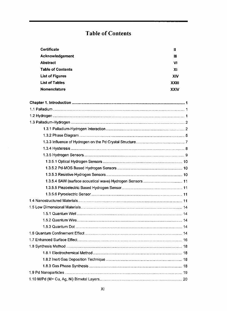
Table of Contents
Certificate II
Acknowledgement Ill Abstract VI Table of Contents XI List of Figures XIV List of Tables XXIII Nomenclature XXIV
Chapter 1.Introduction...............■................................................-..........................................I
1.1 Palladium .............................................................................................................................. i
i.2 Hydrogen .............................................................................................................................. i i .3 Palladium-Hydrogen ..................。. ......................................................................................... 2
1.3.1 Palladium-Hydrogen Interaction ........................................................................... 2
1.3.2 Phase Diagram ...............................,..................................................................... 5
1.3.3 Influence of Hydrogen on the Pd Crystal Structure.............,................................ 7
1.3.4 Hysteresis ............................................................................................................. 8 1.3.5 Hydrogen Sensors .................,.............................................................................. 9
i .3. 5. 1 Optical Hyd rogen Sensors ......................................................,..................... lo
1.3.5.2 Pd-MOS Based Hydrogen Sensors .....................................,........................ lo i.3.5.3 Resistive Hydrogen Sensors.........................................................................10
I .3.5.4 SAW (surface acoustical wave) Hydrogen Sensors ..................................... li
1 .3.5.5 Piezoelectric Based Hydrogen Sensor..........................................................i i 1.3.5.6 Pyroelectric Sensor....................................................................................... 11
I .4 Nanostructured Materials....................................................................................................11 1.5 Low Dimensional Materials.................................................................................................i4
i.5.i Quantum Well................................................................................................... 14 1.5.2 Quantum Wire.....................................................................................................14
1.5.3 Quantum Dot ................................................,..................................................... 14 1.6 Quantum Confinement Effect ............................................................................................. 14
1.7 Enhanced Surface Effect....................................................................................................16 1.8 Synthesis Method ............................................................................................................... 18
1.8.1 Electrochemical Method . .. . . .. . . . .. . . . ... .. . .. ... ... . .. .. ... .. .. . .. .. .. ... .. .. .. .. . ... . . .. .. .. . . .. .. . .. .. .. . 18
1 .8.2 Inert Gas Deposition Technique ......................................................................... 18 1 .8.3 Gas Phase Synthesis ......................................................................................... 18
I .9 Pd Nanoparticles ................................................................................................................ 19
1 . 10 M/Pd (M= Cu, Ag, Nり BimetalLayers...............................................................................20
xl
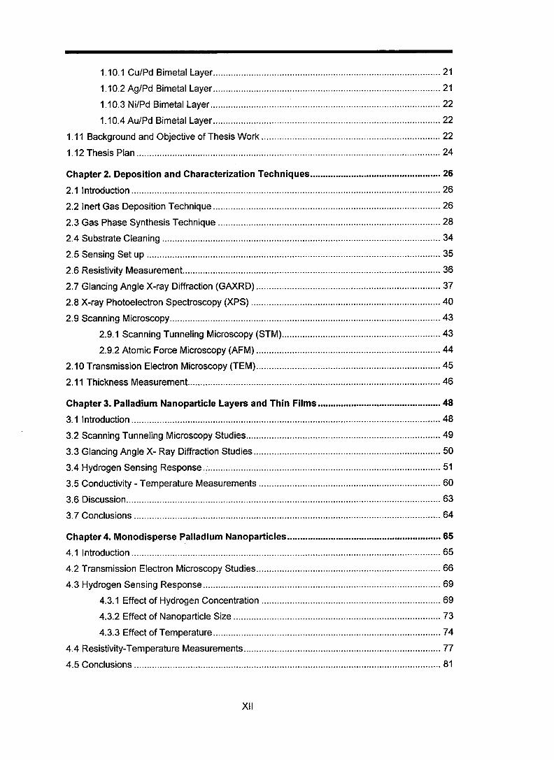
1.10.1 Cu/Pd Bimetal Layer ..........................。. ............................................................. 21
1.10.2 Ag/Pd Bimetal Layer ......................................................................................... 21 1.10.3 Ni/Pd Bimetal Layer .......................................................................................... 22
1.10.4 Au/Pd Bimetal Layer ................................................................'........................ 22
i . 1 1 Background and Objective of Thesis Work .........................。. ........................................... 22
1.12 Thesis Plan ....................................................................................................................... 24
Chapter 2. Deposition and Characterization Techniques...・・・・. ..............・..・・ー・・・・・・・. .............. 26
2.1 Introduction.........................................................................................................................26
2.2 Inert Gas Deposition Technique ......................................................................................... 26
2.3 Gas Phase Synthesis Technique ....................................................................................... 28 2.4 Substrate Cleaning ............................................................................................................. 34 2.5 Sensing Set up .............................................................,..................................................... 35
2.6 Resistivity Measurement.....................................................................................................36 2.7 Glancing Angle X-ray Diffraction (GAXRD)........................................................................ 37
2.8 X-ray Photoelectron Spectroscopy (XPS)........................'................................................. 40
2.9 Scanning Microscopy..........................................................................................................43
2.9.1 Scanning Tunneling Microscopy (STM)..............................................................43 2.9.2 Atomic Force Microscopy (AFM)........................................................................ 44
2.10 Transmission Electron Microscopy (TEM)........................................................................45 2. 1 1 Thickness Measurement...................................................................................................46
Chapter 3. Palladium Nanoparticle Layers and Thin Films...............・.・・・ー・..ー・ーー・. ....、. ...…48
3.1 Introduction ..................'...................................................................................................... 48
3.2 Scanning Tunneling Microscopy Studies............................................................................49 3.3 Glancing Angle X- Ray Diffraction Studies ......................................................................... 50
3.4 Hydrogen Sensing Response.............................................................................................51 3.5 Conductivity-Temperature Measurements .. ...... ... .. .... .. .. ..... ..'.'.. .... .................... ....... ....... 60
3.6 Discussion...........................................................................................................................63 3.7 Conclusions . .... ... . .. .. .. .. .. ................. ... .. ......... .... .. .. .. .. ............... .. ....... ...... ..... .. .. ... .. ....... .... ... 64
Chapter4. Monodisperse Palladium Nanoparticles............................................................65
4.1 Introduction ......................................................................................................................... 65
4.2 Transmission Electron Microscopy Studies........................................................................66 4.3 Hydrogen Sensing Response.............................................................................................69
4.3.1 Effect of Hydrogen Concentration ...................................................................... 69
4.3.2 Effect of Nanoparticle Size ................................................................................. 73 4.3.3 Effect of Temperature.........................................................................................74
4.4 Resistivity-Temperature Measurements.............................................................................77
4.5 Conclusions ........................................................................................................................ 81
XII
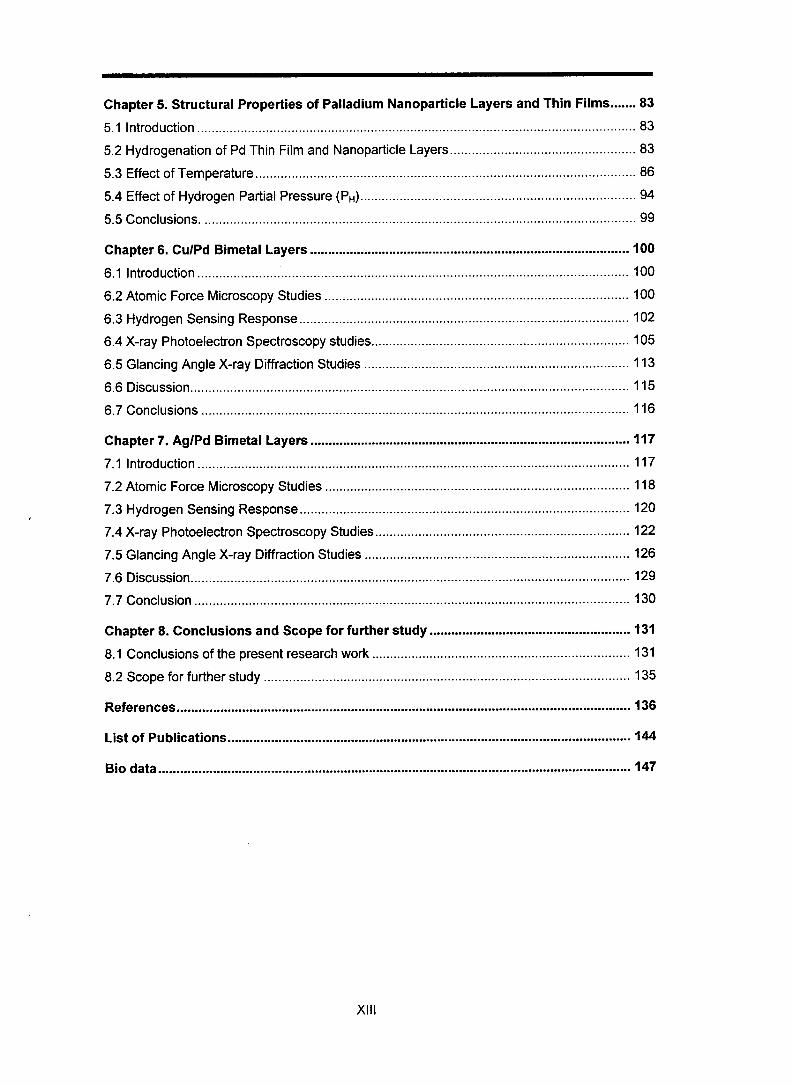
Chapter 5. StructuraI Properties of Palladium Nanoparticle Layers and Thin Films ....... 83 5.1 Introduction ......................................................................................................................... 83 5.2 Hydrogenation of Pd Thin Film and Nanoparticle Layers...................................................83 5.3 Effect of Temperature ...........,............................................................................................. 86 5.4 Effect of Hydrogen Partial Pressure (PH)............................................................................94 5.5 Conclusions. ....................................................................................................................... 99
Chapter 6. Cu!Pd Bimetal Layers ..............■ー. .............-................................................,...... I 00 6.1 Introduction ................,...................................................................................................... ico 6.2 Atomic Force Microscopy Studies .................................................................................... ico 6.3 Hydrogen Sensing Response........................................,.................................................. I 02 6.4 X-ray Photoelectron Spectroscopy studies.......................................................................I 05 6.5 Glancing Angle X-ray Diffraction Studies ......................................................................... 113 6.6 Discussion.........................................................................................................................115 6.7 Conclusions ...................................................................................................................... 116
Chapter 7. Ag/Pd Bimetal Layers .........■. .....■ー. ..-..-.-..-..............-.....■. -..........、. ................ 117 7.1 Introduction ....................................................................................................................... 117 7.2 Atomic Force Microscopy Studies .................'.................................................................. 118 7.3 Hydrogen Sensing Response...........................................................................................120 7.4 X-ray Photoelectron Spectroscopy Studies......................................................................i 22 7.5 Glancing Angle X-ray Diifiraction Studies ....................................................................'.... I 26 7.6 Discussion.........................................................................................................................129 7.7 Conclusion ...........,............................................................................................................ 130
Chapter 8. Conclusions and Scope for further study ......・.・・……■. ..........・、’ '.・・・・・・ー・・・..・・・…… 1 31 8.1 Conclusions of the present research work ....................................................................... I 31 8.2 Scope for further study .................................................................,................................... 135
References.............................................................................................................................136
Listof Publications.....................................................................ーー. ..............ーー. .................. 144
Biodata..................................................................................................................................147
XIII


![Palladium nanoparticles and nanowires deposited ... - Pd-JSSE.pdf · processes such as catalysis, fuel cells, hydrogen storage and metal embrittlement [22]. Research on palladium](https://static.fdocuments.in/doc/165x107/5f0236a27e708231d403233a/palladium-nanoparticles-and-nanowires-deposited-pd-jssepdf-processes-such.jpg)


