Pain & Palliative Care in Homeopathy
50
www.similima.com Page 1 Pain & Palliative Care in Homeopathy A study on the Effectiveness of Homeopathic Medicines in the Management of Pain in Cancer Dr Rithesh B ACKNOWLEDGEMENT I would like to express my sincere thanks to my respected teacher and guide Dr.T.AbduRahiman, Principal, Govt. Homoeopathic Medical College, Kozhikode for his valuable guidance and constant encouragement given to me throughout my post-graduation course and necessary directions in the preparation of this thesis. I am very grateful to Dr.Suresh, (Director, Pain & Palliative clinic Medical college, Kozhikode) for his favorable suggestions in the conduction of this study particularly regarding assessment criterias.. I am deeply indebted to Dr.K.L.Babu,, Reader, Dept. of Organon of medicine, Govt. Homoeopathic Medical College, Kozhikode, who admitted sufficient number of cases for the study and for his informative suggestions and guidance which helped me to complete this work.. I sincerely thank Mr.P.I.Narayanan, MSc.D.H.S, Retired Associate Professor, Department of Biostatistics, Medical College, Kozhikode for conducting the analysis of this work.. I extend my gratitude to my colleagues and to the staff of various departments of this college for their sincere co-operation. The gratitude I owe to my wife Dr.Narda cannot be expressed in words, without her constant encouragement and co-operation this work couldn’t have been possible. Finally I owe my unlimited indebtedness to all my patients involved in this study for their co-operation, without which this study would not have been possible. Last but not the least I thank almighty God for giving me the strength and perseverance to fulfill the project. - Dr. Rithesh.B Introduction Cancer is a common and widely publicized disease, and in spite of ever increasing effort to understand it as a process, its incidence in the population is rising. The main reason for this is the close correlation of the number of cancer cases with the increasing age of the patients and the number of more aged people in the western society (and also in Kerala) at least is rising .It used to be suggested that same aspect of the ageing process increased the susceptibility to cancer, perhaps by impairing immune surveillance .However it is now generally accepted that the relationship of many cancer cases to increasing age is rather a reflection of time required to accumulate a critical number of genetic abnormalities for cancer to arise .Cancer may affect any organ or tissue ,but while some cancer are common, Eg; lung ,breast, gut, prostate, others are very rare ,those affecting the young people often being amongst the rarest .In particular ,cancer affect epithelial tissue and over a 99% of tumor are derived from this tissue .This is not surprising since many of the known cancer causing agents (carcinogens) are from natural radiation. In the air we breathe and from the food stuff we ingest, and epithelial cells are the first line of defense to the outside world, in the skin, lung, and gastrointestinal tract 1 The prevalence of acute and chronic cancer pain and the profound psychological and physical burdens engendered by this symptom oblige all treating physician to be skilled in pain management. 2 Since homoeopathic treatment has found to be effective in managing cancer pain, and since a scientific study on this subject is not known to be conducted, an attempt is made to evaluate the effectiveness of homoeopathic medicines with appropriate statistical analysis. A prospective study is conducted by studying the cancer patient with pain attending the out patient and in patient department of Govt. Homoeopathic medical college, Calicut. The patients are assessed at the time of consultation using, Pain rating scale, Degree of distress score and performance status score to rate the pain and quality of life. Even though the aim of study is palliation of pain, cases are taken according to homoeopathic philosophy giving importance to the general symptoms .The selection of medicine are also based on the homoeopathic philosophy giving more importance to the general symptoms and if case demands taking sectoral totality of pain giving importance to its modifying factors and the characteristics of pain. Miasmatic aspect of the cases are studied and given due importance in the selection of medicine.
Transcript of Pain & Palliative Care in Homeopathy
www.similima.com Page 1
Pain & Palliative Care in Homeopathy
A study on the Effectiveness of Homeopathic Medicines in the Management of Pain
in Cancer Dr Rithesh B
ACKNOWLEDGEMENT I would like to express my sincere thanks to my respected teacher and guide Dr.T.AbduRahiman, Principal, Govt. Homoeopathic Medical College, Kozhikode for his valuable guidance and constant encouragement
given to me throughout my post-graduation course and necessary directions in the preparation of this thesis. I am very grateful to Dr.Suresh, (Director, Pain & Palliative clinic Medical college, Kozhikode) for his favorable suggestions in the conduction of this study particularly regarding assessment criterias.. I am deeply indebted to Dr.K.L.Babu,, Reader, Dept. of Organon of medicine, Govt. Homoeopathic Medical College, Kozhikode, who admitted sufficient number of cases for the study and for his informative suggestions and guidance which helped me to complete this work.. I sincerely thank Mr.P.I.Narayanan, MSc.D.H.S, Retired Associate Professor, Department of Biostatistics, Medical College, Kozhikode for conducting the analysis of this work.. I extend my gratitude to my colleagues and to the staff of various departments of this college for their sincere co-operation. The gratitude I owe to my wife Dr.Narda cannot be expressed in words, without her constant
encouragement and co-operation this work couldn’t have been possible. Finally I owe my unlimited indebtedness to all my patients involved in this study for their co-operation, without which this study would not have been possible. Last but not the least I thank almighty God for giving me the strength and perseverance to fulfill the project. - Dr. Rithesh.B Introduction
Cancer is a common and widely publicized disease, and in spite of ever increasing effort to understand it as a process, its incidence in the population is rising. The main reason for this is the close correlation of the number of cancer cases with the increasing age of the patients and the number of more aged people in the western society (and also in Kerala) at least is rising .It used to be suggested that same
aspect of the ageing process increased the susceptibility to cancer, perhaps by impairing immune surveillance .However it is now generally accepted that the relationship of many cancer cases to increasing age is rather a reflection of time required to accumulate a critical number of genetic abnormalities for cancer to arise .Cancer may affect any organ or tissue ,but while some cancer are common, Eg; lung ,breast, gut, prostate, others are very rare ,those affecting the young people often being amongst the rarest .In particular ,cancer affect epithelial tissue and over a 99% of tumor are derived from this tissue .This is not surprising since many of the known cancer causing agents (carcinogens) are from natural radiation. In the air we breathe and from the food stuff we ingest, and epithelial cells are the first line of defense to the outside world, in the skin, lung, and gastrointestinal tract1 The prevalence of acute and chronic cancer pain and the profound psychological and physical burdens engendered by this symptom oblige all treating physician to be skilled in pain management.2
Since homoeopathic treatment has found to be effective in managing cancer pain, and since a
scientific study on this subject is not known to be conducted, an attempt is made to evaluate the effectiveness of homoeopathic medicines with appropriate statistical analysis. A prospective study is conducted by studying the cancer patient with pain attending the out patient and in patient department of Govt. Homoeopathic medical college, Calicut. The patients are assessed at the time of consultation using, Pain rating scale, Degree of distress score and performance status score to rate the pain and quality of life. Even though the aim of study is palliation of pain, cases are taken according to homoeopathic philosophy giving importance to the general symptoms .The selection of medicine are also based on the homoeopathic philosophy giving more importance to the general symptoms and if case demands taking sectoral totality of
pain giving importance to its modifying factors and the characteristics of pain. Miasmatic aspect of the cases are studied and given due importance in the selection of medicine.

www.similima.com Page 2
AIM AND OBJECTIVE OF THE STUDY To assess the efficacy of homoeopathic treatment in the management of pain in cancer. Review of Literature Neoplasia literally means ‘the process of new growth’ and a new growth is called “neoplasm”. However all new growth are not neoplasms, since examples of new growth of tissues and cells also exist in the process
of embryogenesis, regeneration and repair, hyperplasia and hormonal stimulation. There fore a satisfactory definition of neoplasm or tumor is “a mass of tissue formed as a result of abnormal, excessive, uncoordinated, autonomous and purposeless proliferation of cells”. 3 The branch of science dealing with the study of neoplasm is called “Oncology” (oncos –tumor, logos –study). Neoplasm may be “benign” when they are slow growing and localized with out causing much difficulty to the host .or “Malignant” when they proliferate rapidly, spread through out the body and may eventually cause death of the host .The common term used for all malignant tumor is cancer. Hippocrates (460-377 BC) coined the term “Karkinos”for the cancer of the breast .The word cancer means “Crab” thus reflecting the prime character of cancer since it sticks to the part stubbornly like a crab4. International union against cancer (IUAC) has defined cancer as a “disturbance of growth characterized primarily by excessive proliferation of cells with out apparent relation to the physiological demand of the organ involved”5.
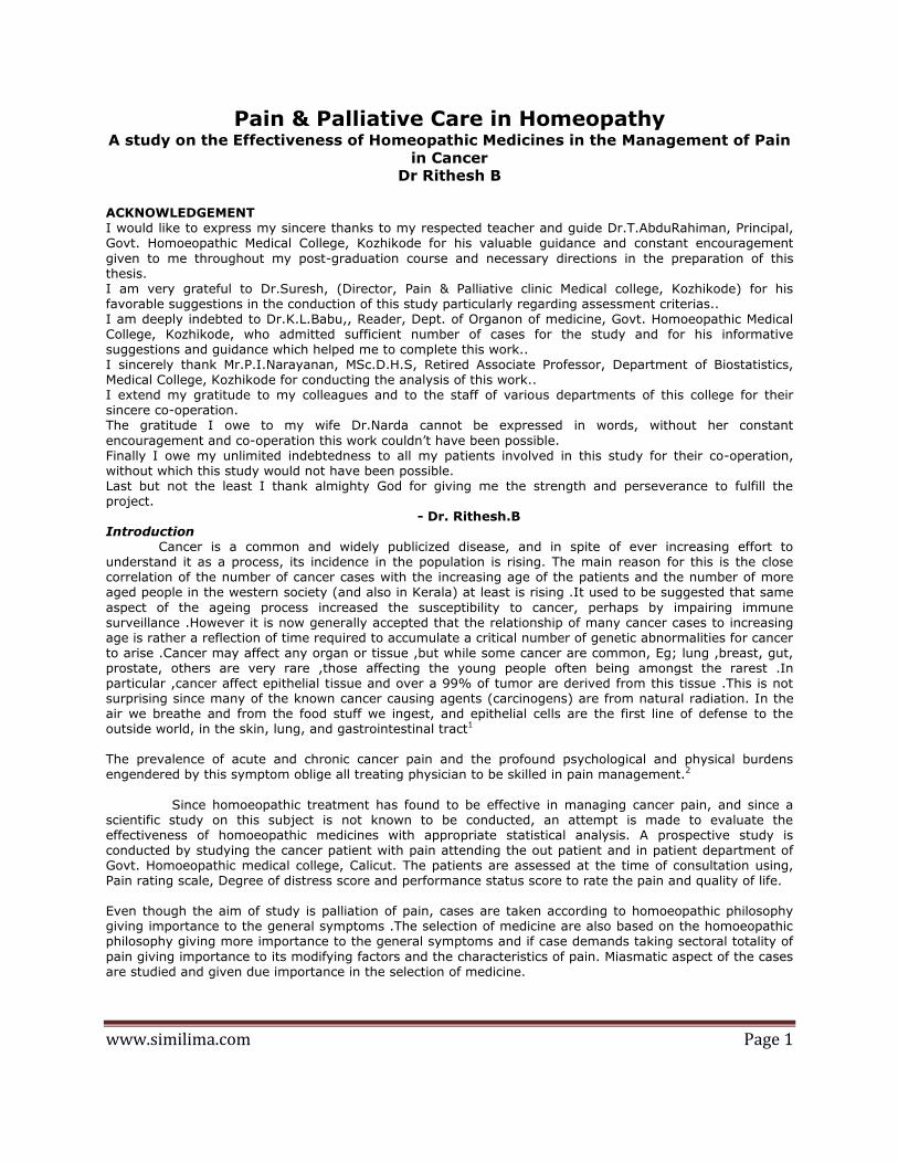
All tumors benign and malignant have the basic component,
1) Parenchyma composed of proliferating tumor cell –parenchyma determines the nature and evolution of the tumor.
2) Supportive stroma – comprised of fibrous connective tissue and blood vessels, it provides the framework on which the parenchyma tumor cell grow .3
3) The tumor derives their nomenclature on the basis of the parenchymal component comprising them. The suffix “oma”is added to denote benign tumors, malignant tumors of epithelial origin are called “carcinomas” while malignant mesenchymal tumors are named “sarcomas” (sarcos-fleshy) however some cancers are composed of highly undifferentiated cells and are referred to as undifferentiated malignant tumors. Although the broad generalization regarding nomenclature of tumors usually holds true in majority of instances, some examples contrary to
the concept are –melanoma for carcinoma of melanocytes, hepatoma for carcinoma of hepatocytes, lymphoma for malignant tumor of lymphoid tissue and seminoma for malignant tumor of testis .3
Tumors composed of a single type of parenchyma cells that differentiate towards more than one cell line are called mixed tumor. Teratomas on the other hand are made up of a number of parenchymal cell types arising from totipotent cells derived from more than one germ cell layer. Choristoma refers to ectopic rests of normal tissues. Hamartoma is a mass of disorganized but mature cells of tissue indigenous to the particular site .3
Classification of tumor 3
Tumor of origin Benign Malignant
I. Tumors of one parenchymal cell type A) Epithelial tumors
: 1.Squamous epithelium
2. Transitional epithelium 3. Glandular epithelium 4.Basal cell layer skin
5. Neuroectoderm 6. Hepatocytes
Squamous cell papilloma Transitional cell papilloma Adenoma - Nevus Liver cell adenoma Hyaditidiform mole
Squamous cell carcinoma
Transitional cell carcinoma Adenocarcinoma Basal cell carcinoma Melanoma Hepatoma Choriocarcinoma
Liposarcoma
www.similima.com Page 3
7. Placenta B. Non epithelial (mesenchymal) tumors 1.Adipose tissue
2.Adult fibrous tissue 3.Embryonic fibrous tissue 4. Cartilage 5. Bone 6. Synovium 7. Smooth muscle 8.Skeletal muscle 9. Mesothelium 10. Blood vessels 11. Lymph vessels
12. Glomus 13. Meninges 14. Hematopoetic cells 15. Lymphoid tissue 16. Nerve sheath 17. Nerve cells II) Mixed tumors Salivary glands III) Tumors of more
than one germ cell layer Totipotent cells in gonads or in embryonal nests
Lipoma Fibroma Myxoma Chondroma
Osteoma Benign synovioma Leiomyoma Rhabdomyoma - Hemangioma Lymphangioma Glomus tumor Meningiomas -
Pseudolymphomas Neurilemmoma, neurofibroma Ganglioneuroma Pleomorphic adenoma
Mature teratoma
Fibrosarcoma Myxosarcoma Chondrosarcoma Osteosarcoma
Synovial sarcoma Leiomyosarcoma Rhabdomyosarcoma Mesothelioma Angiosarcoma Lymphangiosarcoma - Invasive meningoma Leukemias Malignant lymphomas Neurogenic sarcoma
Neuroblastoma Malignant mixed salivary tumor Immature teratoma
Table: 1
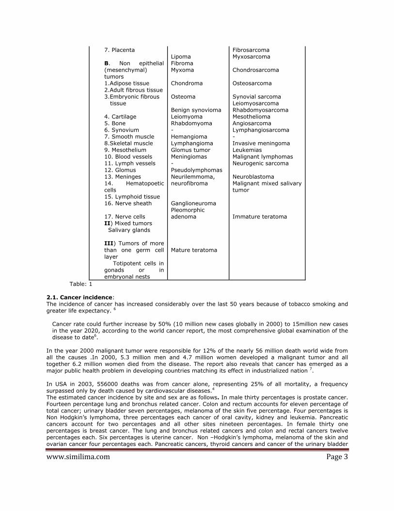
2.1. Cancer incidence: The incidence of cancer has increased considerably over the last 50 years because of tobacco smoking and greater life expectancy. 6
Cancer rate could further increase by 50% (10 million new cases globally in 2000) to 15million new cases in the year 2020, according to the world cancer report, the most comprehensive global examination of the disease to date6.
In the year 2000 malignant tumor were responsible for 12% of the nearly 56 million death world wide from all the causes .In 2000, 5.3 million men and 4.7 million women developed a malignant tumor and all together 6.2 million women died from the disease. The report also reveals that cancer has emerged as a major public health problem in developing countries matching its effect in industrialized nation 7.
In USA in 2003, 556000 deaths was from cancer alone, representing 25% of all mortality, a frequency surpassed only by death caused by cardiovascular diseases.4 The estimated cancer incidence by site and sex are as follows. In male thirty percentages is prostate cancer. Fourteen percentage lung and bronchus related cancer. Colon and rectum accounts for eleven percentage of total cancer; urinary bladder seven percentages, melanoma of the skin five percentage. Four percentages is Non Hodgkin’s lymphoma, three percentages each cancer of oral cavity, kidney and leukemia. Pancreatic cancers account for two percentages and all other sites nineteen percentages. In female thirty one percentages is breast cancer. The lung and bronchus related cancers and colon and rectal cancers twelve percentages each. Six percentages is uterine cancer. Non –Hodgkin’s lymphoma, melanoma of the skin and ovarian cancer four percentages each. Pancreatic cancers, thyroid cancers and cancer of the urinary bladder

www.similima.com Page 4
two percentages each. All other sites accounts for the rest twenty percentages.4The estimated frequency of cancer death by site and sex are as follows, in male thirty one percentages death are due to cancer related to lung and bronchus. Prostate cancer accounts for eleven percentages, ten percentage death due to colon and rectal cancer. Pancreatic cancer and Non-Hodgkin’s lymphoma accounts for five percentages of death each. Four percentages due to leukemia, three percentages each due to carcinoma of the esophagus, liver, urinary bladder and kidney. Two percentages due to all other sites. In females twenty-five percentage
deaths is due to cancer related to lung and bronchus. Breast cancer accounts for fifteen percentages of death. Eleven percentages due to cancer related to colon and rectum, six-percentages death due to cancer of pancreas and five percentages due to cancer of the ovary. Four percentages of death each due to leukemia and Non-Hodgkin’s lymphoma. Two percentages each due to cancer of the uterus, brain and multiple myeloma. Twenty-three percentages of the remaining death are due to cancer of all other sites.4 Over the past 50 years, the overall age adjusted cancer death rate has significantly increased in men, where as it has fallen significantly in women .The increase in men can be largely attributed to lung cancer .The improvement in women is mainly attributed to a significant decline in death rate from the cancer uterus, stomach, liver, and most notably from cancer of the cervix, one of the most common malignant neoplasia in women. Striking is the alarming increase in the death rate form the carcinoma of the lung in both sexes .In women carcinoma of the breast occur 2.5 times more frequently than those of the lung Because of the large difference in the cure rates of these two cancers, however lung cancer has become the leading cause of
cancer death in woman. 4 2.2. Cancer incidence in India and Kerala The rate of cancer occurrence in Kerala and in India is much lower compared to western countries .It is now estimated that 25000 new cancer cases occur in Kerala in one year. Among the males 50% of cancers in the mouth, throat and lung are caused by tobacco and alcohol habits. Among women the incidence of tobacco related cancer is 15%.8
Most prevalent cancer in India 9
Rank In men In women Overall
1 Lung Breast Lung
2 Stomach Cervix Stomach
3 Colon /rectum Liver Liver
4 Prostate Stomach Colon/rectom
5 Oral Lung Esophagus
6 Liver Oral Breast
7 Esophagus Ovary Oral
8 Bladder Body of the uterus Cervix
Table: 2
Cancer incidence in Kerala-Trivandrum Male, age [0-85+] 10
Cancer Cases Crude rate
Lip 11 0.4
Tongue 124 4.5
Salivary glands 13 0.5
Mouth 207 7.6
Pharynx 128 4.7
Esophagus 70 2.6
Stomach 81 3.0
Small intestine 5 0.2
Colon 42 1.5
Rectum and anus 52 1.9
www.similima.com Page 5
Liver 57 2.1
Gallbladder etc. 5 0.2
Pancreas 40 1.5
Nose sinuses etc. 9 0.3
Larynx 93 3.4
Lung (incl. trachea and bronchus) 197 7.2
Bone 17 0.6
Melanoma of skin 16 0.6
Other skin 37 1.4
Connective tissue 22 0.8
Breast 3 0.1
Penis 19 0.7
Prostate 88 3.2
Testis 13 0.5
Kidney etc. 28 1.0
Bladder 44 1.6
Eye 2 0.1
Brain, central nervous system 60 2.2
Thyroid 46 1.7
Other endocrine 5 0.2
Hodgkin lymphoma 20 0.7
Non-Hodgkin lymphoma 64 2.3
Multiple myeloma 25 0.9
Leukemia 94 3.4
All sites 2017 73.9
All sites but skin 1980 72.6
Table: 3
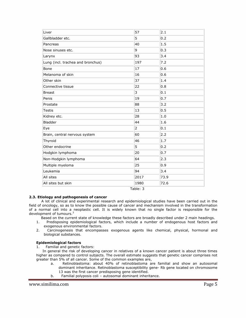
2.3. Etiology and pathogenesis of cancer A lot of clinical and experimental research and epidemiological studies have been carried out in the
field of oncology, so as to know the possible cause of cancer and mechanism involved in the transformation of a normal cell into a neoplastic cell. It is widely known that no single factor is responsible for the development of tumours.3 Based on the current state of knowledge these factors are broadly described under 2 main headings.
1. Predisposing epidemiological factors, which include a number of endogenous host factors and exogenous environmental factors.
2. Carcinogenesis that encompasses exogenous agents like chemical, physical, hormonal and biological substances.
Epidemiological factors 1. Familial and genetic factors:
In general the risk of developing cancer in relatives of a known cancer patient is about three times higher as compared to control subjects. The overall estimate suggests that genetic cancer comprises not greater than 5% of all cancer. Some of the common examples are,
a. Retinoblastoma: about 40% of retinoblastoma are familial and show an autosomal dominant inheritance. Retinoblastoma susceptibility gene- Rb gene located on chromosome 13 was the first cancer predisposing gene identified.
b. Familial polyposis coli – autosomal dominant inheritance.
www.similima.com Page 6
c. Multiple endocrine Neoplasia. d. Von Recklinghausen’s disease. e. Cancer of the breast- female relatives of breast cancer patients have 2 to 3 times higher
risk of developing breast cancer
2. Racial and geographical factors
Differences in racial incidence of some cancer are largely attributed to the influence of environmental and geographic differences affecting the whole population such as climate, water, soil, diet habits, customs, etc. some of the examples of racial and geographical variations in various cancer are as under,
a. White Europeans and Americans develop most commonly malignancies of lung, breast and colon. Liver cancer is uncommon in these races.
b. Black Africans on the other hand have more commonly cancer of the glans penis, cervix and liver. c. Japanese have five times higher incidence of carcinoma of the stomach than the Africans.
3. Environmental and cultural factors: Surprising, as it may seem, we are surrounded by an environment of carcinogens we eat, drink, inhale and touch. Some examples are, a. Cigarette smoking is the single most important environmental factor implicated in the etiology of cancer
of the oral cavity, pharynx, larynx, esophagus, lung, pancreas and urinary bladder. b. Alcohol abuse predisposes to the development of cancer of the oropharynx, larynx, esophagus and liver. c. Cancer of the cervix is linked to a number of factors such as age of the first coitus, frequency of coitus, multiplicity of partners, parity etc,. d. Penile cancer is rare in Jews and Muslims as they are customarily circumcised . e .Betel nut cancer of the cheek and tongue is quite common in some parts of India due to the habitual practice of keeping the bolus of pan in a particular place in the mouth for a long time. f. A large number of industrial and environmental substances are carcinogens and are occupational hazards for some population-arsenic, benzene, vinyl chloride. g. Certain constituent of diet have been implicated in the causation of cancer. Over weight individuals, deficiency of Vit.A and people consuming diet rich in animal fat and low in fiber content are more at risk of developing certain cancer of the colon.
4. Age: Generally cancer occurs in older individuals past 5th decade of their life. Some tumors have two peaks of incidence, example acute leukemia occurs in children and older age group.
f one plots on a semi log scale, the age distribution of some of the most important cancers, then most carcinoma are rare under the age of 30, but then the incidence rate increases dramatically (103 -104 times) with the age .The most attractive explanation for this exponential relationship is that, three to seven hits (mutations) are required for a cancer to form. Of course not all cancers show a sharp rise in incidence rate with age, testicular cancer show a peak incidence between the second and fifth decades and then declines, while the peak incidence of leukemia and nervous system cancers occur not only at greater age but also in early childhood, suggesting the influence of prenatal factors1.
5. Sex: - Most tumors are generally more common in men than in women except cancer of the breast, gall bladder, thyroid and hypopharynx. Cancer of the breast is the commonest cancer in women through out the world, while lung cancer is the commonest cancer in men. 6. Pre-malignant lesions: Pre-malignant lesions are a group of conditions, which predisposes to the subsequent development of cancer. Some examples of pre-malignant lesions are,
1) Carcinoma in situ (intraepithelial neoplasia) –when the malignant cells are confined to the epithelium without invasion across the basement membrane it is called as carcinoma in situ or intra epitheleal neoplasia. e.g.: uterine cervix at the junction of the ecto and endo cervix.
Bowen’s disease of the skin. 2) Some benign tumors
Eg: multiple adenoma of the large intestine have high incidence of developing adenocarcinoma. Neurofibromatosis may develop in to sarcoma.
3) Miscellaneous conditions – certain inflammatory and hyperplasic conditions are prone to the development of cancer. Eg: patients of long standing ulcerative colitis
www.similima.com Page 7
Cirrhosis of the liver Chronic bronchitis
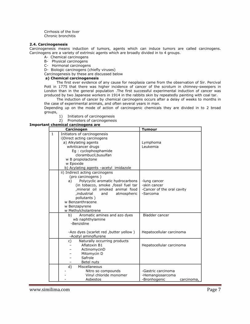
2.4. Carcinogenesis Carcinogenesis means induction of tumors, agents which can induce tumors are called carcinogens. Carcinogens are a variety of extrinsic agents which are broadly divided in to 4 groups.
A- Chemical carcinogens B- Physical carcinogens C- Hormonal carcinogens D- Biologic carcinogens (chiefly viruses) Carcinogenesis by these are discussed below a) Chemical carcinogenesis The first ever evidence of any cause for neoplasia came from the observation of Sir. Percival Pott in 1775 that there was higher incidence of cancer of the scrotum in chimney-sweepers in London than in the general population .The first successful experimental induction of cancer was produced by two Japanese workers in 1914 in the rabbits skin by repeatedly painting with coal tar. The induction of cancer by chemical carcinogens occurs after a delay of weeks to months in the case of experimental animals, and often several years in man.
Depending up on the mode of action of carcinogenic chemicals they are divided in to 2 broad groups,
1) Initiators of carcinogenesis 2) Promoters of carcinogenesis
Important chemical carcinogens are
Carcinogen Tumour
1 Initiators of carcinogenesis i)Direct acting carcinogens a) Alkylating agents wAnticancer drugs Eg : cyclophosphamide clorambucil,busulfan
w B propiolactone w Epoxide b) Acylating agents –acetyl imidazole
Lymphoma Leukemia
ii) Indirect acting carcinogens (pro carcinogens )
a) Polycyclic aromatic hydrocarbons (in tobacco, smoke ,fossil fuel tar ,mineral oil smoked animal food ,industrial and atmospheric pollutants )
w Benzanthracene
w Benzapyrene w Methylcholantrene
-lung cancer -skin cancer -Cancer of the oral cavity -Sarcoma
b) Aromatic amines and azo dyes wb naphthylamine -Benzidine -Azo dyes (scarlet red ,butter yellow )
-Acetyl aminoflurene
Bladder cancer Hepatocellular carcinoma
c) Naturally occurring products – Aflatoxin B1 – ActinomycinD – Mitomycin D – Safrole – Betel nuts
Hepatocellular carcinoma
d) Miscellaneous - Nitro so compounds - Vinyl chloride monomer - Asbestos
-Gastric carcinoma -Hemangiosarcoma -Bronhogenic carcinoma,
www.similima.com Page 8
- Arsenical compounds - Metals (nickel, lead, cobalt ) - Insecticides, fungicides (eldrin,
dieldrin, chlordane)
mesothelioma Epithelial hyperplasia, basal cell carcinoma Lung cancer Experimental cancer
2 Promoters of carcinogenesis a) Phorbol esters b) Phenols c) Hormones (estrogen) d) Drugs (eg-phenobarbitol) e) Artificial sweeteners (Eg- saccharin. cyclamates)
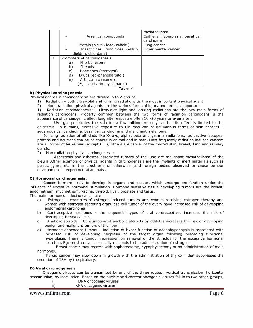
Table: 4 b) Physical carcinogenesis Physical agents in carcinogenesis are divided in to 2 groups
1) Radiation – both ultraviolet and ionizing radiations ,is the most important physical agent 2) Non –radiation physical agents are the various forms of injury and are less important 1) Radiation carcinogenesis: - ultraviolet light and ionizing radiations are the two main forms of radiation carcinogens. Property common between the two forms of radiation carcinogens is the appearance of carcinogenic effect long after exposure often 10 -20 years or even after. UV light penetrates the skin for a few millimeters only so that its effect is limited to the
epidermis .In humans, excessive exposure to UV rays can cause various forms of skin cancers –squamous cell carcinoma, basal cell carcinoma and malignant melanoma. Ionizing radiation of all kinds like X-rays, alpha, beta and gamma radiations, radioactive isotopes, protons and neutrons can cause cancer in animal and in man. Most frequently radiation induced cancers are all forms of leukemias (except CLL); others are cancer of the thyroid skin, breast, lung and salivary glands. 2) Non radiation physical carcinogenesis: Asbestosis and asbestos associated tumors of the lung are malignant mesothelioma of the pleura .Other example of physical agents in carcinogenesis are the implants of inert materials such as plastic ,glass etc in the prosthesis or otherwise ,and foreign bodies observed to cause tumour development in experimental animals .
C) Hormonal carcinogenesis: Cancer is more likely to develop in organs and tissues, which undergo proliferation under the influence of excessive hormonal stimulation. Hormone sensitive tissue developing tumors are the breast, endometrium, myometrium, vagina, thyroid, liver, prostate and testis. The main hormones inducing cancer are
a) Estrogen – examples of estrogen induced tumors are, women receiving estrogen therapy and women with estrogen secreting granulosa cell tumor of the ovary have increased risk of developing endometrial carcinoma.
b) Contraceptive hormones – the sequential types of oral contraceptives increases the risk of developing breast cancer.
c) Anabolic steroids – Consumption of anabolic steroids by athletes increases the risk of developing
benign and malignant tumors of the liver. d) Hormone dependant tumors – induction of hyper function of adenohypophysis is associated with
increased risk of developing neoplasia of the target organ following preceding functional hyperplasia. There is tumour regression on removal of the stimulus for the excessive hormonal secretion, Eg: prostate cancer usually responds to the administration of estrogens.
Breast cancer may regress with oopherectomy, hypophysectomy or on administration of male hormones. Thyroid cancer may slow down in growth with the administration of thyroxin that suppresses the secretion of TSH by the pituitary.
D) Viral carcinogenesis Oncogenic viruses can be transmitted by one of the three routes –vertical transmission, horizontal
transmission, by inoculation. Based on the nucleic acid content oncogenic viruses fall in to two broad groups, i) DNA oncogenic viruses ii) RNA oncogenic viruses
www.similima.com Page 9
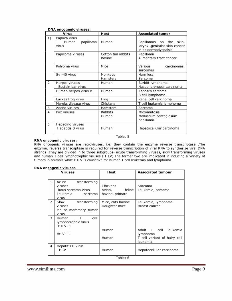
DNA oncogenic viruses:
Virus Host Associated tumor
1) Papova virus Human papilloma virus
Human
Papillomas on the skin, larynx ,genitals: skin cancer in epidermodyspalsia
Papilloma viruses Cotton tail rabbits Bovine
Papilloma Alimentary tract cancer
Polyoma virus Mice Various carcinomas, sarcomas
Sv -40 virus Monkeys Hamsters
Harmless Sarcoma
2 Herpes viruses Epstein bar virus
Human Burkitt lymphoma Nasopharyngeal carcinoma
Human herpes virus B Human Kaposi’s sarcoma B cell lymphoma
Luckes frog virus Frog Renal cell carcinoma
Mareks disease virus Chickens T cell leukemia lymphoma
3 Adeno viruses Hamsters Sarcoma
4 Pox viruses Rabbits Human
Myxomatosis Molluscum contagiosum
papilloma
5 Hepadino viruses Hepatitis B virus
Human
Hepatocellular carcinoma
Table: 5 RNA oncogenic viruses: RNA oncogenic viruses are retroviruses, i.e. they contain the enzyme reverse transcriptase .The enzyme, reverse transcriptase is required for reverse transcription of viral RNA to synthesize viral DNA strands .They are divided in to three subgroups- acute transforming viruses, slow transforming viruses and human T cell lymphotrophic viruses (HTLV).The former two are implicated in inducing a variety of tumors in animals while HTLV is causative for human T cell leukemia and lymphoma. RNA oncogenic viruses
Viruses Host Associated tumour
1 Acute transforming viruses Rous sarcoma virus
Leukemia –sarcoma virus
Chickens Avian, feline
bovine, primate
Sarcoma Leukemia, sarcoma
2 Slow transforming viruses Mouse mammary tumor virus
Mice, cats bovine Daughter mice
Leukemia, lymphoma Breast cancer
3 Human T cell lymphotrophic virus HTLV- 1 HtLV-11
Human Human
Adult T cell leukemia lymphoma T cell variant of hairy cell leukemia
4 Hepatitis C virus HCV
Human
Hepatocellular carcinoma
Table: 6

www.similima.com Page 10
Molecular genetics of cancer Broadly speaking genes and molecular factors involved in pathogenesis of cancer can be grouped in to 4 categories
1) Oncogenes (i.e. cancer causing genes) 2) Anti- oncogenes (i.e., cancer suppressing genes)
3) Mutator genes (i.e., genes that regulate DNA repair) 4) Telomerase in cancer (i.e., telomere shortening as cancer suppressor mechanism)
Oncogenes: - Oncogenes are cancer causing genes. They are derived from proto-oncogenes or cellular oncogenes (c-oncs) which are detected on normal animal and human cell and promote normal growth and differentiation of cells. Human oncogenes are identified by DNA transfection and by non-random chromosomal abnormalities.(e.g.: translocations) Mechanism of activation of cellular oncogenes: 3 There is similarity between normal genes coding for proteins for growth and differentiation on one hand and
oncogenes of viral and tumor origin on the other .How these normal genes are activated to become oncogenes is explained on the basis of two type of mechanisms i) Change in the structure of the gene & ii) Change in the regulation of gene expression Based on this, examples of activation of human oncogenes in human tumors are under,
a) Point mutations and deletion, Eg: ras oncogene b) Chromosomal translocations, Eg: Philadelphia chromosome seen in 95 % of the case of chronic
myeloid leukemia .In 75% case of Burkit’s lymphoma, translocations of c- myc –proto-oncogene from its site on chromosome 8 to a portion on chromosome 14.
c) Gene amplification - chromosomal alterations that results in increase in the number of a gene, eg: Neuroblastoma having n-myc –HSR region Erb-B in breast and ovarian cancer.
Mechanism of action of oncogenes:
Oncogenes have oncoproteins, which are altered form of their normal counter parts, proto-oncogenes,
regulating growth and differentiation. Thus proliferation of cells by oncogenes induces the steps shown
below3,
The important oncogenes and their mode of activation and associated human tumor :
Type Oncogenes Associated tumor
1) Extra cellular growth
factor
β chain of PDGF
FGF
sis
hst,k53
Sarcoma, glioblastoma
Stomach cancer, kaposi’s
sarcoma

www.similima.com Page 11
2) Trans membrane growth
factor receptors .EGF –
receptors
CSF receptor
erb B1
her -2 /neu
Fms
Carcinoma lung
Ca breast, ovary
Leukemia
3) Intracellular signal
transduction proteins GTP
binding
Non receptor tyrosine
kinase
4) Transcription proteins
(nuclear regulatory
proteins)
Transcription
activators
Ras
Abl
Myc
N -myc
Ca lung ,colon ,pancreas
CML,ALL
Burkit's lymphoma
Neuroblastoma
5) Cell cycle regulatory
proteins
Cyclins
Cyclin dependent
kinase
Cyclin D
CDK 4
Mantle cell lymphoma
Glioblastoma,
melanoma
6) Apoptotic inhibitors
Anti apoptotic genes
bcl -2
Follicular B cell lymphoma
Table: 7
Anti –oncogenes (tumor suppressor genes):
Tumor suppresser gene or anti –oncogenes, just as proto- oncogenes are also a pair of normal genes
which perform the physiologic function of regulation of cell growth. Mechanism of stimulation of
carcinogenesis by tumor suppresser gene is mutation in both alleles producing deficiency of normal gene
product, which normally suppresses tumor formation. Homozygous deletion or mutation at specific genetic
loci are seen in such tumor cells Eg: in retinoblastoma, Wilm’s tumor, familial adenomatous polyposis coli,
breast cancer etc.

www.similima.com Page 12
Important tumor suppresser gene and associated human tumors:
Gene Location Associated human tumor
1) Rb Nucleus Retinoblastoma
Osteosarcoma
2) p53 Nucleus Colorectal cancer
50% of all human cancers
3) APC Cytosol Familial APC
4) WT1 Nucleus Wilm’s tumor
5) NF 1 Plasma
membrane
Neurofibromatosis type 1
6)BRCA 1,BRCA 2 Nucleus Ca breast ,Ca Ovary
Table: 8
Mutator oncogenes :
Normal cells have caretaker genes to take care of the integrity of genetic information in response to DNA
damage. The mutated version of mutator gene is characterized by loss of normal surveillance function and
render the DNA susceptible to accumulation of mutations and therefore progression to cancer.
Telomerase in cancer:
Telomeres are the terminal tips of chromosome which progressively shorten due to repetitive cell division
.Telomerase is the enzyme required for continued recognition of telomere in successive cell divisions. Cancer
cells express telomerase with consequent telomere lengthening and further immortalization of cancer cells.
High risk factors for cancer
a) Excess alcohol drinking
b) Excess tobacco chewing, smoking
c) Lack of green and yellow vegetables
d) Excess consumption of high fat diet
e) Early age at marriage, multiple sexual partners, poor genital hygiene.
2.5. Clinical effects of Neoplasia:
Two major aspects of clinical; significance in assessing the course and management of neoplasia are: tumor
host inter-relationship and laboratory diagnosis of cancer
www.similima.com Page 13
Effects of tumor on host:
Malignant tumors produce more ill effects than the benign tumors
1) Local effects:
Both benign and malignant tumors cause local effects on the host due to their size or location. Some of
the local effects of tumors are
i) Compression
ii) Mechanical obstruction
iii) Tissue destruction
iv) Infarction, ulceration, hemorrhage
2) Cancer cachexia:
Patients with advanced and disseminated cancers terminally have asthenia, emaciation and anorexia,
together referred to as cancer cachexia .Cachectin or tumor necrosis factor alpha derived from
macrophages play a contributory role in cachexia.
3) Fever:
Fever of unexplained origin may be presenting feature in some malignancies such as in Hodgkin’s
disease, adenocarcinoma of the kidney, osteogenic sarcoma and many other tumors. The exact
mechanism of tumor-associated fever is not known but probably the tumor cells themselves elaborate
pyrogens.
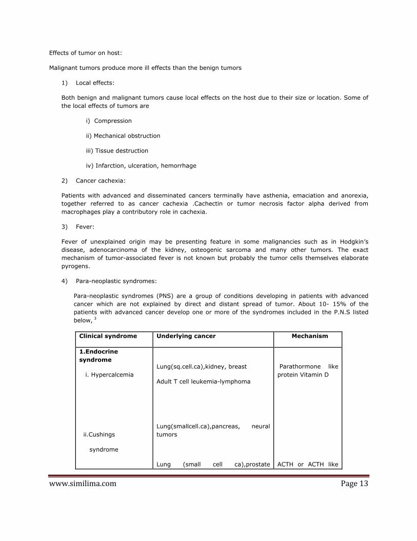
4) Para-neoplastic syndromes:
Para-neoplastic syndromes (PNS) are a group of conditions developing in patients with advanced
cancer which are not explained by direct and distant spread of tumor. About 10- 15% of the
patients with advanced cancer develop one or more of the syndromes included in the P.N.S listed
below, 3
Clinical syndrome Underlying cancer Mechanism
1.Endocrine
syndrome
i. Hypercalcemia
ii.Cushings
syndrome
Lung(sq.cell.ca),kidney, breast
Adult T cell leukemia-lymphoma
Lung(smallcell.ca),pancreas, neural
tumors
Lung (small cell ca),prostate
Parathormone like
protein Vitamin D
ACTH or ACTH like

www.similima.com Page 14
iii. Inappropriate
anti diuresis
iv. Hypoglycemia
v. Carcinoid
syndrome
vi. Polycythemia
2.Neuromuscular
syndrome
i. Myasthenia gravis
ii. Neuromuscular
disorders
3.Osseous,joint&soft
tissues
i. Hypertrophic
osteoarthropathy
ii.Clubbing of
fingers
4.Hematologic
syndromes
i.Thrombophlebitis
ii. Non bacterial
intracranial tumor
Pancreas(islet cell tumor)
mesothelioma, Fibrosarcoma
Bronchial carcinoid tumor , Ca
pancreas, stomach
Kidney,liver,cerebellar hemangioma
Thymoma
Lung (small cell ca), breast
Lung
Lung
Pancreas ,lung ,GIT
Advanced cancers
AML, adenocarcinoma
Thymoma
substances
ADH or atrial
natriuretic factor
Insulin or insulin like
substance
Serotonin
Bradykinin
Erythropoietin
Immunologic
Immunologic
Not known

www.similima.com Page 15
thrombotic
endocarditis
ii. DIC
iii. Anemia
5.Gastrointestinal
syndromes
i. Malabsorption
6.Renal syndromes
I. Nephritic
syndrome
7.Cutaneus
syndromes
i. Acanthosis
nigricans
ii.Seborrheic
keratosis
iii.Exfoliative
dermatitis
8.Amyloidosis
Lymphoma of small bowel
Advanced cancers
Stomach, large bowel
Bowel
Lymphoma
Multiple myeloma
Kidney, lymphoma, solid tumors.
Not known
Hypercoagulabilty
Hypercoagulability
Chronic thrombotic
phenomina
Unknown
Hypoalbuminemia
Renal vein
thrombosis,Systemic
amyloidosis
Immunologic
Immunologic
Immunologic

www.similima.com Page 16
i. Primary
ii.Secondary
Immunologic
AA protein
Table: 9
2.6. Warning signals of cancer:
? A change in character of pain
? Unexplained and persistent or increasing pain in the body
? Convulsion originating in adult life
? Unremitting continued fever not responding to treatment
? Haemoptysis with out any apparent cause
? A persistent cough
Warning signals of oral cancer and precancerous lesions:
Cancer of the oral cavity is predominantly seen among those who chew tobacco. The red and white coloured
patches, non healing ulcers of long duration, ulcers caused by sharp tooth. Inability to tolerate spices along
with glistening appearance of the tongue and lining of cheeks especially in tobacco habituates.
Warning signals of breast cancer:
Lump in the breast which is increasing in size and or causing pain and changes in the overlying skin .Red or
brown coloured discharge from the nipple or any swelling in the axilla.
Warning signals of uterine cervical cancer:
Bleeding per vagina after intercourse, excess discharge from the vagina, bleeding after attaining menopause
and bleeding between menstrual periods.
Head and Neck cancer:
Painless enlargement of lymph nodes in the neck, appearance of tumors or nodules in the neck especially in
tobacco habituates and alcoholics.
Gastro intestinal cancer or colorectal cancer:
Bleeding per rectum with altered bowel habits like constipation alternating with diarrhea and passing
mucous with abdominal discomfort.
Skin cancer:
Changes in moles or pigmented skin lesions like appearance of fissuring, ridges, furrows and ulceration in the moles, appearance of satellite lesions, itching with bleeding and sudden enlargement of pigmented moles. Cancer of larynx (Vocal cord):
Progressive hoarseness of voice ultimately resulting in absence of sound especially in a smoker or alcoholic. Cancer of the Esophagus:
Progressive dysphagia i.e. difficulty in swallowing solid food and later on leading to difficulty in drinking. Tumors:
Swelling or enlargement of lymph nodes in the neck, bony swellings or tumors anywhere in the body. Leukemia:
Combined symptoms of lymph node enlargement, pallor, loss of weight, fatigue, bleeding gums, weakness, recurrent fever and bone pain. Non healing ulcers:
www.similima.com Page 17
Non-healing ulcers of long duration anywhere in the body which is not responding to antibiotic treatment may transform to malignancy. Others: Bleeding from any orifice like nose, ear, anus, vagina, mouth or from the conjunctiva may be warning signal
of cancer in rare cases.
Any individual having any of the above-mentioned warning signals need not panic, as it does not mean that they have cancer. But they should consult a doctor so that they can have the necessary investigations done to rule out cancer.
2.7. Host response against tumor: 3
It has long been thought that host defense mechanism in the form of immunological response exist so as to
counter the growth and spread of cancer, albeit more often partially.
1) Certain cancers evoke significant lymphocytic infiltrates. E.g.: medullary carcinoma breast,
seminoma testis.
2) Rarely a cancer may spontaneously regress partially or completely, E.g: malignant melanoma.
3) It is highly unusual to have primary and secondary tumor in the spleen due to its ability to destroy
the growth and proliferation of tumor cells .
4) There is an increased frequency of cancer in immuno deficient hosts, Eg: AIDS.
A) Tumor antigens:
There are two types of antigens,
i) Tumor specific antigens – (TSA) these are located on tumor cells but are not present on normal
cells Examples of TSA are ,
* Mutant form of ras and p53 proteins and
* bcr –abl gene
ii) Tumor associated antigens (TAA)
These are present on the tumor cells as well as on some normal cells, Examples of TAA are,
· CD -10 antigen on the early B lymphocytes expressed in B –cell leukemia, lymphomas.
· Prostate specific antigen (PSA) expressed by normal as well as malignant prostatic
epithelium.
B) Immune responses
The nature of host immune response to tumors can be categorized as under,
i)Cell mediated mechanism:
a) Specifically sensitized cytotoxic T lymphocytes (CTL) which are directly cytotoxic,
b) Natural killer cells (NK) destroy tumor cells with out sensitization, either directly or by
antibody dependent cellular cytotoxicity (ADCC),
www.similima.com Page 18
c) Macrophage mediated cytotoxicity by ADCC or by cytotoxic products,
ii) Humoral mechanism:
Humoral antibodies are capable of killing free tumor cells in the blood and in the serosal cavities.
iii) Inhibitory (regulatory) mechanisms:
a) CD8+T suppresser cells may play a regulatory role in humoral and cell mediated tumor
immunity.
b) Humoral blocking factors, possibly antigen –antibody complexes, may either block the antigen
sites on the tumor cells or block the receptors on the immune competent cell.
2.8. Diagnosis of cancer 3
The most certain and reliable method which has stood the test of time is the histological examination of
biopsy.
Histological methods:
These methods are based on the microscopic examination of properly fixed tissue (excised tumor mass or
open needle biopsy from the mass), supported with complete clinical and investigative data. These methods
are most valuable in arriving at the accurate diagnosis.
Cytological methods:
Cytological methods for diagnosis consists of study of cells shed in to the body cavities (exfoliative cytology)
and study of the cells by putting a fine needle introduced under vacuum in to the lesion (fine needle
aspiration cytology -FNAC).
Histochemistry and cytochemistry:
Histochemistry and cytochemistry are additional diagnostic tools, which help in identifying the chemical
composition of cells and their products by special staining methods.
Immnohistochemistry:
This is an immunological method of recognizing a cell by one or more of its specific component in the
cytoplasm, cell membrane or nucleus .These cell component (called antigens) combine with specific
antibodies on the formalin fixed paraffin sections or cytological smears .The complex of antigen –antibody
on the slide is made visible for light microscope identification by either fluorescent dyes or by enzyme
system. The specific antibody against a particular cellular antigen is now a days obtained by hybridoma
technique for monoclonal antibody production. These monoclonal antibodies impart objectivity to the
subjective tumor diagnosis made by the surgical pathologist. 3
One important group of such antibody stains is directed against various classes of intermediate filaments
which is useful in classification of poorly differentiated tumors of epithelial or mesenchymal origin.
Tumor markers (Biochemical assays):
Tumor markers are biochemical assays of products elaborated by tumor cells in blood or other body fluids, it
is there for, a pertinent to keep in mind that many of these products are produced by normal cells too, and
thus the biochemical estimation of the product in the blood reflects the total substance and not by the tumor
cells alone.

www.similima.com Page 19
Tumor markers include -cell surface antigens (or oncofetal antigens) cytoplasmic proteins, enzymes,
hormones and cancer antigens. However two of the best known examples of oncofetal antigens are secreted
by fetal tissues as well as by tumors are alpha fetoproteins (AFP) and carcino embryonic antigens (CEA). 3.
Modern aids in tumor diagnosis:
In addition to the methods described above some more modern techniques have emerged for pathologic
diagnosis but their availability as well as applicability is limited, Eg:,
i) Flow cytometry
ii) In situ hybridization
iii) Molecular diagnostic techniques.3
2.9. Pain
Pain, it has been said is one of natures earliest signs of morbidity, and it stands pre eminent among all the
sensory experiences by which humans judge the existence of disease within themselves.11
Pain is mainly a protective mechanism for the body, it occurs whenever any tissue is being damaged, and it
causes the individual to react to remove the pain stimulus. 12
The painful experiences of the sick pose manifold problems for the physicians. They must be prepared to
diagnose the disease in patients who have felt only the first rumbling of discomfort before other symptoms
and signs have appeared. To deal intelligently with pain problems, the physician requires familiarity with the
anatomy of sensory pathways and the sensory supply of the body segments, insight into the psychological
factors that influence behavior and a knowledge of medical and psychiatric diseases.11
The dual nature of pain is responsible for some of our difficulty in understanding it. Easier to comprehend is
its evocation by particular stimuli and the transmission of pain impulse along certain pathways i.e, the
sensation of pain. Far more abstruse is its quality as a mental state, i.e. the quality of anguish or suffering –
“a passion of the soul” –in the words of Aristotle- which defies definition and quantiification.11
Unlike most sensory modalities, which are aroused by a specific (adequate) symptom, such as pressure,
heat or cold, pain may be evoked by each of these stimuli, if it is intense enough.
The theory of specificity of sensations proposed at the end of last century maintains that each type of
sensation is conveyed by a separate anatomical pathway. This theory has repeatedly been questioned.
Sir.Henry Head postulated the existence of two sets of sensory inputs to the central nervous system, the
epicritic and protopathic inputs. Epicritic sensation allowed discrimination of touch, temperature and pain,
where as protopathic sensation was a poorly localized, unpleasant and long-lasting sensation. The
protopathic sensation is physiologically inhibited in the central nervous system by the epicritic system. Later
Trotter and Davies dismissed this theory after performing careful examinations on themselves.13
Pain has been classified into two major types. Fast pain and slow pain.12
Fast pain is felt within 0.1 second after a pain stimuli is applied. Where as slow pain begins only after 1
second or more and then increase slowly over many seconds and sometimes even minutes.
When pain is the result of physiologic activity in the normal pain receptors and there is no primary
dysfunction of the nervous system, it is called nociceptive pain. Nociceptive pain may indicate a disorder in
any other system or organ, whereas pain resulting from the dysfunction of the central or peripheral nervous
system is called neuropathic pain.13

www.similima.com Page 20
Pain and temperature are sensations mediated at a primary afferent level by fibers of smaller diameter than
the fibers mediating touch, vibration and position sense. Cold sensation is mediated by small myelinated
fibers (A delta fibers); warm sensation mediated by unmyelinated warm specific C-fibers and pain is
mediated by small myelinated A-delta nociceptor and unmyelinated C- nociceptors.13
The anatomical and functional aspect of cutaneous pain afferents have been studied in detail. The skin,
subcutaneous tissue, muscles and joints are sensitive to a variety of potentially harmful mechanical, thermal
and chemical stimuli. Numerous free nerve endings are responsible for conveying sensory information that is
decoded as pain in the central nervous system. Although the ultra structural characteristics of pain receptors
(nociceptor) are not well known, classically two types of nociceptors have been characterized in the human
skin according to their receptor-response features,
They are 1. Those associated with unmyelinated C-fibers and
2. Those associated with small myelinated A-delta fibres2
The glabrous and hairy skin is richly innervated by nociceptors with unmyelinated C-fiber. These are known
as C-polymodal nociceptors (CPN) because they respond to a variety of noxious stimuli i.e, mechanical,
thermal and chemical stimuli. A simple C-polymodal nociceptor innervates an area of skin approximately
1cm2 usually in a single continuous receptive field. These nociceptors have threshold that are well below the
level at which tissue damage occurs. These nociceptors also respond to temperature below 15oc.13
Another receptor is the A-delta nociceptor. They mainly respond to mechanical stimulation. These nociceptor
display a smaller, usually punctiform receptive field and have higher mechanical and heat threshold than C-
polymodal nociceptors.13
A third type of cutaneous nociceptor composed of unmyelinated C-fibers has been recently described. These
nociceptors are activated only during inflammation. In the absence of inflammation they do not respond
even to high noxious stimulation.13
The sensations evoked by activation of A-delta and C- nociceptors are different. Excitations of cutaneous C-
polymodal nociceptors evoke a burning sensation. Where as A-delta nociceptors evoke a sharp pain that is
projected to a punctiform area. 13
In addition to their role in pain sensation, C- polymodal nociceptors are involved in neurogenic inflammation.
Excitation of C-polymodal nociceptors determines the release of algogenic substances from nociceptive
terminals in the skin, causing local vasodilatation and thus reddening of skin. This flare reaction spreads
some centimeters around the site of stimulation through an axonal reflux that depends on a network of fine
dermal afferent fibers, originally described as a nocifensor system. Denervation of skin impairs the flare
reaction.13
The peripheral afferent fibers have their cell bodies in the dorsal root ganglia. Central extension of these
nerve cells project via the dorsal root to the dorsal horn of the spinal cord or in the case of cranial pain
afferents to the nucleus of the trigeminal nerve, i.e. the medullary dorsal horn. The fine myelinated and
unmyelinated fibers occupy mainly the lateral part of the root entry zone and with in the spinal cord many of
the thinnest fibers form a discrete bundle the tract of Lissauer.11
The afferent pain fibers after traversing Lissauer’s tract terminate in the posterior grey matter of dorsal
horn, predominantly in the marginal zone. Most of the fibers terminate with in the segment of their entry in
to the cord, but some extend rostrally and caudally to one or two adjacent segment ipsilaterally and some
via the anterior commissure to the contra lateral dorsal horn. The neurons in the dorsal horn are arranged in
a series of six layers or laminae.

www.similima.com Page 21
Transverse section through the sixth cervical segment of the spinal cord of the cat, illustrating the subdivision of the gray matter into laminae according to Rexed. LM and VM, later-omedial and ventromedial groups of motor neurons.
Fine myelinated A-delta fibers terminates principally in lamina 1 of Ryxed and also in outermost part of
lamina 2 , some A-delta pain fibers penetrate the dorsal gray matter and terminate in the lateral part of
lamina 5.Unmyelinated C-fibers terminate in lamina 2(substantia gelatinosa).From these cells of
termination,secondary neurons connect with central and lateral horn of cells in the same and adjacent spinal
segments and sub serve both somatic and autonomic reflexes .Other secondary neurons sub serving pain
sensation decussate in the anterior spinal commissure and ascent in the antero lateral fasciculus to the brain
stem and thalamic structures. The axons from each dermatome decussate one to three segments higher
than the level of root of entry. In this way the dorsal horn and anterior spinal commissure form a continuous
pain pathway, the full length of the spinal cord. Crossing fibers are added to the inner side of the
spinothalamic tract, so that the longest fibres from the sacral segments come to the most superficial and
fibers from successively more rostra level occupy a progressively deeper position.11
In addition to the lateral spinothalamic tract the anterolateral fasciculus of the spinal cord contain
a more slowly conducting medially placed system of fibers, which project via short interneuron chain to the
reticular core of the medulla and midbrain and then to the medial and intra laminar nuclei of the thalamus.
This group of fibers is known as the spinoreticulo thalamic or paleo spinothalamic pathway. This tract
conducts diffuse poorly localized pain arising from deep structures like gut peritoneum etc.
The direct spinothalamic fibers as they approach the thalamus segregate in to two bundles. The lateral
division terminates in the ventro basal and posterior group of nuclei. The medial division terminates mainly
in the intralaminar complex of nuclei and in the nucleus submedius. Spinoreticulo thalamic fibers project in
to the medial intralaminar thalami nuclei. Projections from the dorsal column nuclei, which have a
modulating influence on pain transmission, are mainly to the ventrobasal and posterior group of nuclei. Each
of the four thalamic nuclear groups that receive nociceptive projections from the spinal cord has a distinct
cortical projection and is thought to play a different role in pain sensation.11
The ventrobasal complex and probably the posterior group of nuclei send their axons to two mass cortical
areas, the post central cortex (a small number terminate in the perceptual cortex) and the upper bank of
sylvian fissure. The areas are mainly concerned with the reception of tactile and proprioceptive stimuli and
with discriminative sensory function including pain.11
Stimuli that activate pain receptors vary from one tissue to another. An adequate stimulus for skin is one
that injures tissue, i.e. pricking, cutting, crushing, burning and freezing. These stimuli are ineffective when
applied to the stomach and intestine where pain is produced by local effects of an engorged or inflamed
mucosa, distension or spasm of the smooth muscle, and traction on the mesenteric attachment. In skeletal
muscle pain is caused by ischemia as well as by injuries of connective tissue sheath, necrosis and
heamorrhage.11
In the painful lesions due to tissue damage, proteolytic enzymes are released which act on gamma globulins
to liberate substances that excite peripheral nociceptors. Bradykinins, histamines, prostaglandins, serotonins
and similar polypeptides as well as potassium ion elicit pain. Vascular permeability may also be increased by
these substances. In addition, direct stimulation of nociceptor release substance that enhances pain
perception. The best studied of these is substance- P, which is released from C-fiber terminals in the skin
during peripheral nerve stimulation. It causes erythema by dilating cutaneous vessels and edema by
releasing histamine from mast cells. This reaction is called neurogenic inflammation and this is mediated by
antichromic action potential from the small nerve cells in the spinal ganglia. 11
www.similima.com Page 22
In 1965, Melzac and Wall propounded a new theory to explain the mechanism of pain. They observed in
decerebrate and spinal cats, that peripheral stimulation of ‘large myelinated fibers’ produced a negative
dorsal root potential and that stimulation of small C fibers caused a positive root potential. They postulated
that these potentials which were a reflection of pre synaptic inhibition or excitation modulated the activity of
secondary transmitting neuron (T cells) in the dorsal horn, and that this modulation mediated through an
inhibitory interneuron (I cell). The essence of this theory is that the larger diameter fiber excites the
inhibitory cells; conversely the small pain afferents inhibit the inhibitor cells, leaving the transmitter cells in
an excitatory state. Melzac and Wall emphasized that the transmission of pain impulses from the dorsal horn
must also be under the control of a descending system of fibers from the brainstem, thalamus and limbic
lobes.11
The gate control hypothesis of Melzak &Wall: A stimulus presented to the skin activates both large and small diameter fibers. If the stimulus is light, large fiber input predominates, the inhibitory inter neuron (I) is excited and the transmission cell does not fire. If the stimulus is intense, small fiber input predominates, the
inhibitory interneuron is shut off and the transmission cell (T) is activated resulting in pain
The most important contribution in recent years to our understanding of pain has been the discovery of an
endogenous neural system for analgesia, which can be activated by the administration of opiates or by
naturally occurring brain substances with the pharmacological properties of opiates.11
The endogenous analgesic system was first demonstrated by Reynolds in 1969.This analgesic system consists of three major components,11 1. The peri acquiductal gray and peri-ventricular areas of the mesencephalous and upper pons surrounding
the aqueduct of sylvius and portions of the third and fourth ventricles. Neurons from these areas send their
signals to 2. The raphe magnus nucleus, a thin midline nucleus located in the lower pons and upper
medulla, and the nucleus reticularis para giganto cellularis located laterally in the medulla. From these
nuclei, the signals are transmitted down the dorsolateral column in the spinal cord to 3. Pain inhibitory
complex located in the dorsal horn of the spinal cord. At this point the analgesia can block the pain before it
is relayed to the brain. 12
Further investigation disclosed that stimulation produced analgesia (SPA) produces its effects by inhibiting
the neurons of laminae 1 and V of the dorsal horn, i.e. the neurons that are stimulated by nervous stimuli.11
The opiates also act on the neurons of laminae 1 and V of the dorsal horn, suppressing the inputs from A-
delta and C- fibers. There are several opiate receptors in the CNS, they have been found in the spinal cord
in the terminals of the A- delta and C fibers in the dorsal horn neurons, as well as in medullary reticular
nuclei, medial thalamus and amygdaloid nuclei. The analgesic effects of the opiates are both presynaptic and
post synaptic after the discovery of specific opiate receptor in the CNS. Several naturally occurring peptides,
which proved to have a potent analgesic effect and bind specifically to opiate receptors, were identified.
These endogenous morphine like compounds are known as ‘endorphins’ meaning morphine within. The most
important endorphins in the body are beta endorphin (a fragment of pituitary hormone – beta lipoprotein)
and enkephalin, they are found in greatest concentration in relation to opiate receptors in the mid brain.11
Theoretical mechanism of action of enkephalin (endorphin and morphine) on the transmission of pain
impulses from the periphery to the CNS is as follows. Spinal inter neurons containing enkephalin synapse
with the terminals of pain fibers and inhibit the release of the presumptive transmitter, substance P. As a
result, the receptor neurons in the dorsal horn receive less excitatory (pain) impulses and transmit fewer
pain impulses to the brain. Morphine binds to the unoccupied enkephalin receptors, mimicking the pain
suppressing effects of the endogenous opiate enkephalin.

www.similima.com Page 23
There are two varieties of enkephalin met-enkephalin and leucoenkephalin. These are found most
importantly in the brain stem and spinal cord. Beta endorphins is present in both the hypothalamus and
pituitary gland.12
Although all the finer details of brain opiate system are not understood, activation of analgesia system by
nervous signals entering the peri acquiductal gray and periventricular areas or inactivation of pain pathways
by morphine like drugs can totally or almost totally suppress many pain signals entering through the
peripheral nerves.
2.10. Cancer pain:
Relief of pain in cancer patients is an ethical imperative and it is incumbent up on clinician to maximize the
knowledge, skill and diligence needed to attend this task. Unfortunately under treatment continues to be
common. Under treatment have many causes, among the most important of which is inadequate
assessment. Assessment is an ongoing and dynamic process that includes evaluation of presenting
problems, elucidation of pain syndromes and pathophysiology and formulation of a comprehensive plan for
continuing care. In this process pain treatment must be incorporated with in a broader therapeutic agenda,
so that needs for tumour control and symptom palliation are concurrently addressed. 2
Pain assessment in cancer population begins with an appreciation of the relationship among pain,
nociception and suffering.
Nociceptors as we already discussed is the activity produced in the nervous system by potentially tissue
damaging stimuli. Nociceptor is not equivalent to pain. There may be no report of pain despite overt tissue
injury and should pain occur it may or may not be perceived by the clinician to be commensurate with the
degree of injury. 2
Pain can be conceptualized as the perception of nociceptors. Like other perceptions pain is determined by an
interaction between activity and sensori-neural pathway and other factors- the neuropathic process and the
psychological disturbances. Although psychological process can strongly influence the expression and impact
of pain, nociceptive and neuropathic factors predominate in the cancer population. Elucidation of the lesion
that induce these process is an essential element in the assessment and may alter prognosis, provide an
opportunity for selecting the method of treatment.2
Suffering can be defined on the global perception of distress engendered by adverse factors that together
undermine quality of life. Pain may contribute to suffering, but numerous other factors such as the
experience of other physical symptoms, progressive physical impairment and psychological disturbances
may be equally important.2
Factors contributing to pain and suffering and relationship between pain and suffering: 2
Nociception Neuropathic psychological Other physiological process
mechanism process physical impairment
Social isolation
Family distress
Sense of financial loss
www.similima.com Page 24
Analgesia alone may not lessen the suffering and consequently pain therapy is not the sole objective in the supportive care of the cancer patients. Rather pain therapy must be the critical component of a more comprehensive therapeutic plan designed to address the diverse factors that impair quality of life. Frequency with which pain occurs varies with the stage of the disease and with the primary site of the tumour.6 ; Moderate or severe pain occurs in
· 1/3rd (30% - 40%) of the patients at the time of diagnosis · More than 2/3rd (60% - 100%) 0f patients with advanced cancer 15
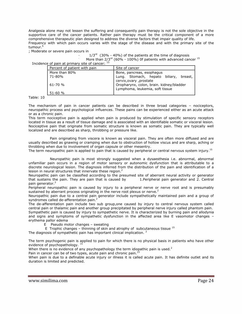
Incidence of pain at primary site of cancer: 15
Percent of patient with pain Site of cancer
More than 80% 71-80% 61-70 % 51-60 %
Bone, pancreas, esophagus Lung. Stomach, hepato biliary, breast, cervix,ovary ,prostate Oropharynx, colon, brain. kidney/bladder Lymphoma, leukemia, soft tissue
Table: 10
The mechanism of pain in cancer patients can be described in three broad categories – nociceptors, neuropathic process and psychological influences. These pains can be experienced either as an acute attack or as a chronic pain. This term nociceptive pain is applied when pain is produced by stimulation of specific sensory receptors located in tissue as a result of tissue damage and is associated with an identifiable somatic or visceral lesion. Nociceptive pain that originate from somatic structure is known as somatic pain. They are typically well localized and are described as sharp, throbbing or pressure like.
Pain originating from viscera is known as visceral pain. They are often more diffused and are
usually described as gnawing or cramping when due to obstruction of hollow viscus and are sharp, aching or throbbing when due to involvement of organ capsule or other mesentry. The term neuropathic pain is applied to pain that is caused by peripheral or central nervous system injury.15
Neuropathic pain is most strongly suggested when a dysaesthesia i.e. abnormal, abnormal
unfamiliar pain occurs in a region of motor sensory or autonomic dysfunction that is attributable to a discrete neurological lesion. The diagnosis inferred from the distribution of the pain and identification of a lesion in neural structures that innervate these region.2 Neuropathic pain can be classified according to the presumed site of aberrant neural activity or generator that sustains the pain. They are pain that is caused by 1.Peripheral pain generator and 2. Central pain generator.2 Peripheral neuropathic pain is caused by injury to a peripheral nerve or nerve root and is presumably sustained by aberrant process originating in the nerve root plexus or nerve.2 Neuropathic pain due to a central pain generator include sympathetically maintained pain and a group of
syndromes called de-afferentation pain.2 The de-afferentation pain include two sub group,one caused by injury to central nervous system called central pain or thalamic pain and another group precipitated by peripheral nerve injury called phantom pain. Sympathetic pain is caused by injury to sympathetic nerve. It is characterized by burning pain and allodynia and signs and symptoms of sympathetic dysfunction in the affected area like E vasomotor changes – erythema pallor edema E Pseudo motor changes – sweating E Trophic changes – thinning of skin and atrophy of subcutaneous tissue 15 The diagnosis of sympathetic pain has important clinical implication. 2 The term psychogenic pain is applied to pain for which there is no physical basis in patients who have other evidence of psychopathology. 15
When there is no evidence of any psychopathology the term idiopathic pain is used.2 Pain in cancer can be of two types, acute pain and chronic pain.15 When pain is due to a definable acute injury or illness it is called acute pain. It has definite outlet and its duration is limited and predicted.

www.similima.com Page 25
This pain is accompanied by anxiety and clinical signs of sympathetic over activity like tachycardia, tachypenea, hypertension, sweating, papillary dilatation and pallor. These are characteristics of a patient obviously in pain.15 When pain is the result of a chronic pathological process it is called chronic pain. It has gradual or ill-defined onset, continuos unabated and may become progressively more severe. Patients
with chronic pain appear depressed and withdrawn and as there are usually no signs and symptoms of sympathetic over activity. They are frequently labeled as not looking like somebody in pain. These patients have symptoms of depression with lethargy, apathy, anorexia and insomnia. Personality changes may occur due to progressive alternation in lifestyle and functional ability.15 For patients with chronic pain of non-malignant origin, the pain is to said to lack positive meaning, where as for patients with chronic pain related to cancer the pain not only lacks positive meaning but it may have definite negative implication with regard to progress and life expectancy. 15 Pain lasting more than two weeks should be considered as chronic and should be treated accordingliy.15 Evaluation of pain in cancer is based on the following factors, 1, Intensity of pain: In cancer population evaluation of pain intensity is pivoted to therapeutic decision making. The selection of
the medicine, potency and dosage may all be influenced by reported pain intensity. Furthermore intensity may also help characterize the pain mechanism and underlying syndrome. For example clinical observation strongly suggests that the pain associated with radiation induced nerve injury is rarely severe; the occurrence of severe pain in previously irradiated region therefore suggests the existence of occult neoplasm. 2 There are various factors that will affect the pain intensity, 6 Pain will be increased if there is discomfort insomnia, fatigue, anxiety, fear, anger, sadness, depression and isolation . Pain will be decreased if there is relief of other symptoms good sleep, understanding , companionship, creative activity relaxation ,reduction in anxiety and elevation of mood.
2. Quality of pain: The quality of pain often suggests its pathophysiology. As said earlier, somatic nociceptive pain is usually well localized and described as sharp, aching, throbbing or pressure like. Visceral nociceptive pains are generally diffuse and may be crampy or gnawing when due to obstruction of a hollow viscus or aching sharp or throbbing when due to involvement of organ capsule or mesentry. Neuropathic pains are often described as burning tingling or shock like. 2
1. Distribution of pain: Patients with cancer pain commonly experience pain at more than one site. The topographic distribution of a specific pain may also have implication for diagnosis and treatment. The distribution of pain often clarifies its relationship to the underlying organic lesion. The term focal pain is used to denote one site of pain or pain
that is experienced in the region of the underlying lesion. The cause of pain in cancer can be grouped into 4 categories6. 1. Cancer itself Eg: soft tissue, visceral, bone neuropathic. 2. Anti cancer treatment Eg: chemotherapy causes mucositis, neuropathy and abdominal cramp 3. Debility Eg: constipation muscle tension and sprain. 4. Concurrent disorders. Eg: spondylosis, OA
Cancer related pain could present as an acute pain syndrome. Cancer related acute pain syndrome is most commonly due to diagnostic or therapeutic intervention, where as chronic pains are mainly tumor related.2 Acute pain associated with diagnostic interventions are mainly due to, lumbar puncture headache, arterial or venous blood sampling, bone marrow biopsy, lumbar puncture, colonoscopy, myelography, per cutaneous biopsy and thoracocentesis.

www.similima.com Page 26
Acute pain associated with therapeutic interventions is mainly, acute post-operative pain. Acute pain caused by other therapeutic interventions mainly pleurodesis, tumor embolisation, suprapubic catheterization, intercostal catheter and nephrostomy insertion. Acute pains associated with analgesic technique are injection pain, opioid headache, spinal opioid hyperalgesia syndrome and epidural injection pain. Acute pain associated with anti cancer therapies like, acute pain associated with chemotherapy infusion
techniques that include intravenous infusion pain due to venous spasm, chemical phlebitis, vesicant extravasations, anthracycline associated flair reaction. Hepatic artery infusion pain and intraperitonial chemotherapy causing abdominal pain. Acute pain associated with chemotherapies toxicity like mucositosis, corticosteroid induced perineal discomfort, steroid pseudo rheumatism, painful peripheral neuropathy, headache -due to intrathecal methotrexate meningitis syndrome, L-asparaginase associated dural sinus thrombosis and trans retinoic acid headache-, diffuse bone pain-due to trans retinoic acid and colony stimulating factors-, 5 –fluorouracil induced anginal chest pain, post chemotherapy gyenecomastia. Acute pain associated with hormonal therapy like leutinising hormone releasing factor tumor flares in prostate cancer and hormone induced pain flare in breast cancer. Acute pain associated with immuno therapy like interferon induced acute pain. Acute pain associated with radiotherapy like incident pain associated with positioning, oropharyngeal
mucositis, acute radiation enteritis and proctocolitis, early onset brachial plexopathy and sub acute radiation myelopathy. Acute pain associated with infection like acute herpetic neuralgia. 2.11. Psychological aspects of Cancer Pain The cancer patient faces a wide range of psychological and physical stresses throughout the course of illness. These stresses include fear of a painful death, physical disability, disfigurement and growing dependency on others. Although such fears exist in most, if not all cancer patients, the degree of psychological distress experienced varies greatly between individuals and depends on part on the patient’s personality style, coping abilities, available social supports and medical factors. One of the most feared consequences of cancer is the pain. Pain has a profound impact on the patient’s level of emotional distress
and psychological factors such as mood, anxiety and the meaning attributed to pain can intensify a patients experience on cancer pains. 2 Patient diagnosed with cancer often demonstrates a consistent pattern of emotional response. These responses usually consists of an initial period of shock, denial and disbelief, followed by a period of anxiety and depression. Disturbed sleep, diminished appetite and concentration, irritability, pervasive thoughts about cancer and fears about the future often interfere with normal daily activities. These stress responses usually occur at specific points in the course of cancer and its treatment - after diagnosis, with relapse , prior to diagnostic tests, surgery, radiation and chemotherapy, as well as after treatment has concluded and patient enters the phase of survival ship. Distress usually resolves slowly over a period of several weeks and patient gradually returns to their prior level of homoeostasis once a treatment plan has been agreed upon and emotional supports arrive.
Not only does pain have a profound impact on psychological distress in cancer patients, but also psychological factors appear to influence the experience and intensity of cancer pain. Psychological variables such as perceived control, meaning attributed to the pain experience, fear of death, hopelessness and anxious or depressed mood all appear to contribute to the experience of cancer pain and suffering.2 Pain is no longer considered simply as a nociceptive event, but is widely recognized and accepted as a psychological process involving nociceptors, perception and expression. Because of the important role played by psychological variables in the experience and intensity of cancer pains, appropriate and effective management of cancer pain requires detailed care taking that covers the mental, emotional and physical characteristics of the patient.2 Uncontrolled pain is a major factor in cancer suicides. Cancer is perceived by the public as an extremely painful disease compared to other medical conditions. The majority of suicides observed among patients
with cancer had severe pain, which was often inadequately controlled or tolerated poorly. Factors associated with increased risk of suicide in cancer patients are ,
? Pain -suffering aspects ? Multiple physical symptoms ? Advanced illness, poor prognosis ? Depression, hopelessness
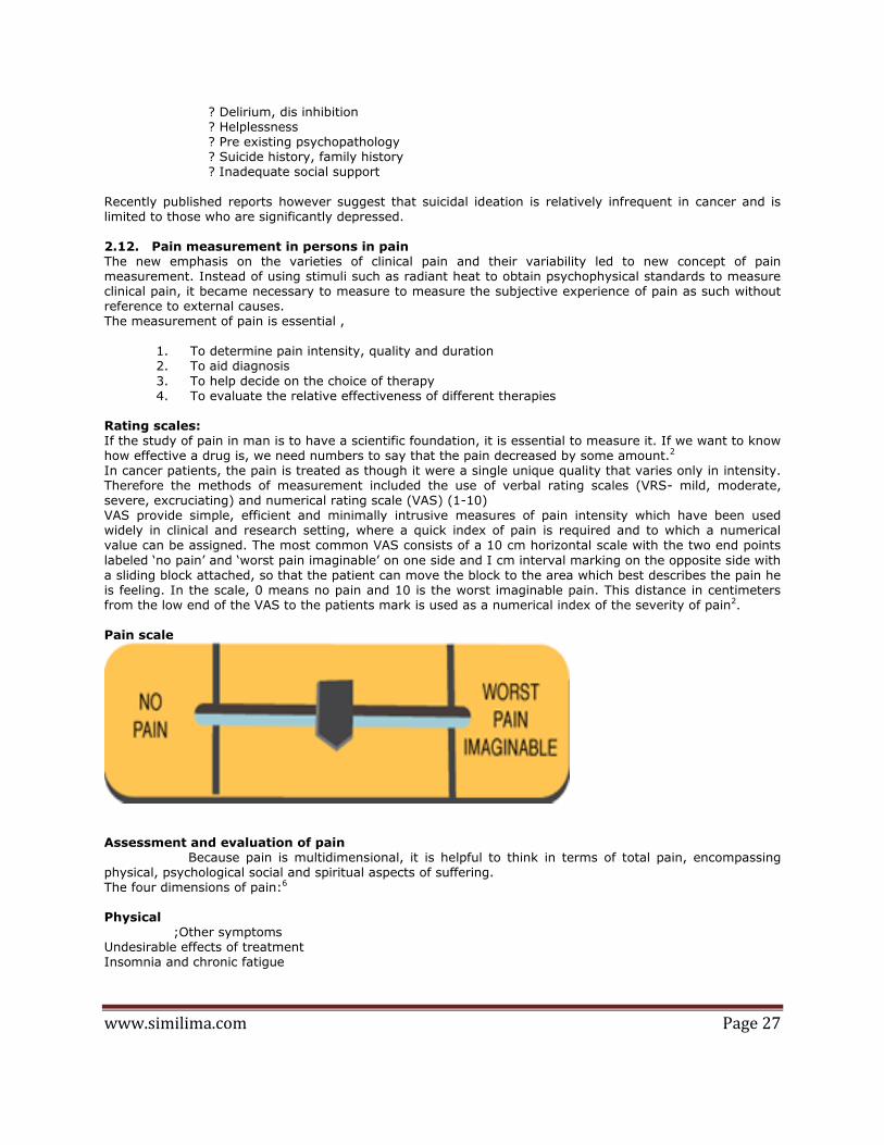
www.similima.com Page 27
? Delirium, dis inhibition ? Helplessness ? Pre existing psychopathology ? Suicide history, family history ? Inadequate social support
Recently published reports however suggest that suicidal ideation is relatively infrequent in cancer and is limited to those who are significantly depressed. 2.12. Pain measurement in persons in pain The new emphasis on the varieties of clinical pain and their variability led to new concept of pain measurement. Instead of using stimuli such as radiant heat to obtain psychophysical standards to measure clinical pain, it became necessary to measure to measure the subjective experience of pain as such without reference to external causes. The measurement of pain is essential ,
1. To determine pain intensity, quality and duration 2. To aid diagnosis
3. To help decide on the choice of therapy 4. To evaluate the relative effectiveness of different therapies
Rating scales: If the study of pain in man is to have a scientific foundation, it is essential to measure it. If we want to know how effective a drug is, we need numbers to say that the pain decreased by some amount.2 In cancer patients, the pain is treated as though it were a single unique quality that varies only in intensity. Therefore the methods of measurement included the use of verbal rating scales (VRS- mild, moderate, severe, excruciating) and numerical rating scale (VAS) (1-10) VAS provide simple, efficient and minimally intrusive measures of pain intensity which have been used widely in clinical and research setting, where a quick index of pain is required and to which a numerical value can be assigned. The most common VAS consists of a 10 cm horizontal scale with the two end points
labeled ‘no pain’ and ‘worst pain imaginable’ on one side and I cm interval marking on the opposite side with a sliding block attached, so that the patient can move the block to the area which best describes the pain he is feeling. In the scale, 0 means no pain and 10 is the worst imaginable pain. This distance in centimeters from the low end of the VAS to the patients mark is used as a numerical index of the severity of pain2. Pain scale
Assessment and evaluation of pain Because pain is multidimensional, it is helpful to think in terms of total pain, encompassing physical, psychological social and spiritual aspects of suffering. The four dimensions of pain:6 Physical ;Other symptoms Undesirable effects of treatment Insomnia and chronic fatigue
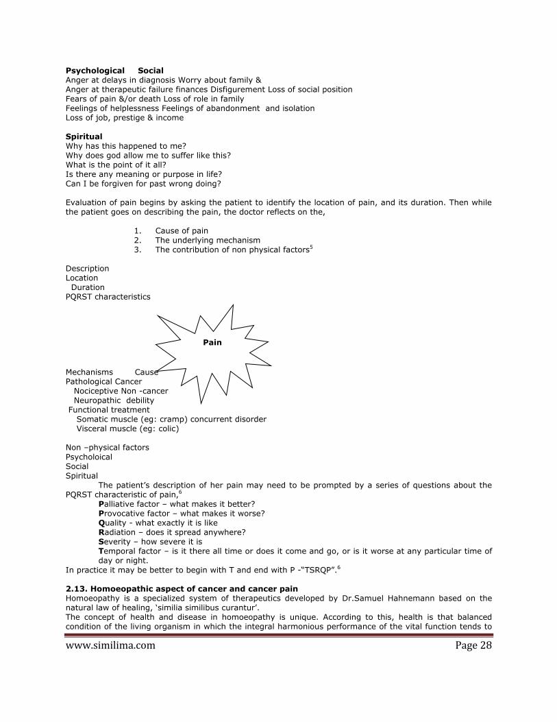
www.similima.com Page 28
Psychological Social Anger at delays in diagnosis Worry about family & Anger at therapeutic failure finances Disfigurement Loss of social position Fears of pain &/or death Loss of role in family Feelings of helplessness Feelings of abandonment and isolation Loss of job, prestige & income
Spiritual Why has this happened to me? Why does god allow me to suffer like this? What is the point of it all? Is there any meaning or purpose in life? Can I be forgiven for past wrong doing? Evaluation of pain begins by asking the patient to identify the location of pain, and its duration. Then while the patient goes on describing the pain, the doctor reflects on the,
1. Cause of pain
2. The underlying mechanism 3. The contribution of non physical factors5
Description Location Duration PQRST characteristics
Mechanisms Cause Pathological Cancer Nociceptive Non -cancer Neuropathic debility Functional treatment Somatic muscle (eg: cramp) concurrent disorder Visceral muscle (eg: colic) Non –physical factors
Psycholoical Social Spiritual
The patient’s description of her pain may need to be prompted by a series of questions about the PQRST characteristic of pain,6
Palliative factor – what makes it better? Provocative factor – what makes it worse? Quality - what exactly it is like Radiation – does it spread anywhere? Severity – how severe it is Temporal factor – is it there all time or does it come and go, or is it worse at any particular time of day or night.
In practice it may be better to begin with T and end with P -“TSRQP”.6 2.13. Homoeopathic aspect of cancer and cancer pain Homoeopathy is a specialized system of therapeutics developed by Dr.Samuel Hahnemann based on the natural law of healing, ‘similia similibus curantur’. The concept of health and disease in homoeopathy is unique. According to this, health is that balanced condition of the living organism in which the integral harmonious performance of the vital function tends to
Pain
www.similima.com Page 29
the preservation of the organism and the normal development of the system. Disease is an abnormal vital process, a changed condition of life, which is inimical to the true development of the individual and tends to organic dissolution. Vital phenomena in health and disease are caused by the reaction of the vital substantial power or principle of the organism to various external stimuli. So long as healthy man lives normally in a favorable environment, he moves, feels, thinks, acts and reacts in an orderly manner. If he violates this law of life or become the victim of an unfavorable environment, disorder takes the place of order, disease
destroy ease, he suffers and his body deteriorates.16 Dr.Hahnemann in §3 explains the things physician needs to know in order to become a true practitioner of the healing art, and the first thing he says is the physician should clearly perceive what is to be cured in every individual case of disease. In §72 he further says to perceive what is to be cured in disease, the classification of disease will serve as a general preliminary view, and he defines acute and chronic disease as follows, “they are diseases of such a character that, with small, often imperceptible beginnings, dynamically derange the living organism, each in its own peculiar manner, and cause it gradually to deviate from the healthy condition, in such a way that the automatic life energy, called vital force, whose office is to preserve the health, only opposes to them at the commencement and during their progress imperfect, unsuitable, useless resistance, but is unable of itself to extinguish them, but must helplessly suffer (them to spread and) itself to be ever more and more abnormally deranged, until at length the organism is destroyed; these
are termed chronic diseases. They are caused by infection with a chronic miasm”.17 In homoeopathy disease are classified in to indispositions, surgical disease and dynamic diseases. Hahnemann in §150 explains indispositions are nothing but those, manifested by one or more trivial symptoms, that have been only observed a short time previously .A slight alteration in diet and regimen will usually suffice to dispel. The dynamic diseases are divided in to acute and chronic as already described. The acute diseases are further classified in to individual, sporadic, endemic and epidemic according to the nature of onset, causation and number of person affected. The chronic disease are also further classified in to artificial chronic disease, inappropriately named chronic disease and true natural chronic disease(miasmatic disease).Artificial chronic disease are those caused by allopathic or antipathic drug overuse and these cases are the most difficult type of cases to cure. Inappropriately named chronic disease are those caused by improper way of living, occupation or habits of long standing duration. These diseases disappear of
themselves without any treatment if the maintaining cause is removed. The true natural chronic disease are those arise from dynamic infection of chronic miasm.This true natural chronic disease can present with fully developed symptoms or with few symptoms only. Those with few symptom can be those with only mental symptoms or with physical symptoms, either presenting with few internal symptoms or with external symptoms ,the later variety is called local disease due to causa interna. The chronic disease with fully developed symptoms can be either due to a single miasm (psora, sycosis or syphilis) or due to a combination of miasms (psora+syphilis, psora+sycosis, syphilis+sycosis, psora +syphilis+sycosis)17 . On the basis of this classification we can say that cancer fall under the category of true natural chronic disease. Hahnemann in §80, while describing the nature of psoric miasm says, the monstrous internal chronic miasm - the psora, the only real fundamental cause and producer of all the other numerous, I may say
innumerable, forms of disease, which, under the names of nervous debility, hysteria, hypochondriasis, mania, melancholia, imbecility, madness, epilepsy and convulsions of all sorts, softening of the bones (rachitis), scoliosis and hypnosis, caries, cancer, fungus haematodes, neoplasms, gout, hemorrhoids, jaundice, cyanosis, dropsy, amenorrhea, hemorrhage from the stomach, nose, lungs, bladder and womb, of asthma and ulceration of the lungs, of impotence and barrenness, of megrim, deafness, cataract, amaurosis, urinary calculus, paralysis, defects of the senses and pains of thousands of kinds, &c., figure in systematic works on pathology as peculiar, independent diseases17. From this it is clear that Hahnemann considered neoplasm to be of psoric origin. This may be because, during Hahnemann’s period, not much was known about the etiology, pathogenesis and different types of tumors. At that time this type of long standing cases which rapidly proliferates and which were incurable, eventually causing the death of the patient was considered as something stubbornly sticks to the system like a crab and they named it as ‘cancer’ means crab. So the concept of cancer as something deep rooted in the system might have made Hahnemann to
include cancer under psoric miasm. But considering the clinical presentation, the dominant miasm can be any of the three miasm depending up on the pathological process, their pace and their various expressions. As already discussed in the aetiopathogenesis of cancer, the cause of cancer can be endogenous factors or environmental factors. The initiating cause of malignant change is a mutation in the gene. So that a normal gene become an abnormal gene. The genetic change may be a deletion or translocation of genes. So there is a deviation from normal functioning of the gene. The cells that undergone these changes deny the rules of
www.similima.com Page 30
normal growth regulation of the body and starts multiplying on its own deriving nutrition from the body. So the abnormal cell with the tendency for excess growth corresponds with the nature of sycotic miasm. As Dr.H.A.Roberts said, “the sycotic patient is too susceptible to the available constructive elements, he seizes up on and assimilate to the point of overgrowth of tissues. It explain the reason of pathology in all parts of the body that manifest overgrowth of natural tissues and where we find malignancies with overgrowth of tissues and infiltration we are almost always able to trace the sycotic trait”18. If there is a syphilitic trait the
manifestations of the tumor will be different. Dr.H.A.Roberts says “one of the cardinal manifestation of syphilitic trait is destruction of tissues”18 .If there is a syphilitic trait, it will present as rapidly progressing destructive change involving the bones with sever night pain, ulcer with punched out edges and glandular involvement. If the psoric miasm is the predominant one, the patient will present with more functional symptom like weakness, anorexia, cachexia etc., than due to structural change. Even though we have good idea about the aetiopathogenesis of cancer and its miasmatic nature, most of the cancer cases remain incurable. Dr.Kent in his lesser writings discuss why is cancer incurable. He tells that in most cases there is paucity of symptoms ,and there is nothing discoverable but the malignant growth and its associated features of hardness ,stinging pain ulceration ,enlarged glands and the tendency to involve the surrounding parts in its own development .If the child’s mental symptoms could be fully ascertained and the symptom from child
hood to the adult age ,some thing might be done .Cancer generally comes on in later life ,when childhood action have forgotten .If the symptoms that have appeared from birth to the present date are undiscovered ,it is no wonder that cancer is incurable. 19 Hahnemann says all curable disease make themselves known to the intelligent physician in signs and symptoms Pathological conditions as also the patient are incurable when there are no signs and symptoms .In proportion as the pathology progress signs and symptoms decreases. This is true in cancer cases. In terminal cancer cases there are paucity of symptoms and even if there is any that are mainly the common symptoms or pathological symptoms. 19 As Dr. Stuart close says, the gross tissue changes, organic lesions, morphological disproportion and the physical effect of mechanical causes are not primarily with in the domain of similia and there for are not the
object of homoeopathic treatment, the morbid process from which they arise, or to which they lead are amenable to homoeopathic medications. Homoeopathic remedies by virtue of their power to control vital function and increase resistance often exercise a favorable influence upon the physical development as well as up on the tangible products of disease or accident .Thus the growth of the tumor may be retarded or arrested .So the conditions like cancer with gross tissue changes we can’t expect a cure and the only thing a homoeopath can do is to palliate the suffering.16 2.14. Miasmatic aspect of pain: Hahnemann included most of the pain in to psoric miasm. In § 80 under Psora he says “the monstrous internal chronic miasm - the psora, the only real fundamental cause and producer of all the other numerous, I may say innumerable, forms of disease, which, under the names of nervous debility, hysteria,
hypochondriasis, mania, melancholia, imbecility, madness, epilepsy and convulsions of all sorts, softening of the bones (rachitis), scoliosis and kyphosis, caries, cancer, fungus haematodes, neoplasms, gout, haemorrhoids, jaundice, cyanosis, dropsy, amenorrhea, hemorrhage from the stomach, nose, lungs, bladder and womb, of asthma and ulceration of the lungs, of impotence and barrenness, of megrim, deafness, cataract, amaurosis, urinary calculus, paralysis, defects of the senses and pains of thousands of Kinds, &c., figure in systematic works on pathology as peculiar, independent diseases”17. The symptom pain cannot be included in to one miasm alone. The miasm of pain can only be identified on the basis of its causation, its modifying factors, and the nature of pain. So the pain of Psora is usually of a neuralgic type, and the pain is better by rest and warmth and worse by motion. The pain of Sycosis are usually joint pains .The rheumatic pains are worse during cold, damp, better moving or stretching. Stitching, pulsating, wandering pains are sycotic.
The Syphilitic miasm has usually bone pains. Burning, bursting and tearing pain are syphilitic .The pain is aggravated at night20. 2.15. Palliative Care: Palliative care is the active total care of patients with life limiting diseases, and their families, by a multi professional team, when the disease is no longer responsive to curative or life prolonging treatments.

www.similima.com Page 31
The word ’Palliative’ is derived from the Latin word “pallium”- means a ‘cloak’. In palliative care symptoms are ‘cloaked’ with treatments whose primary action is to promote comfort. However palliative care extends far beyond physical symptom relief. It seeks to integrate physical, psychological, social and spiritual aspects of care, so that the patient may come to terms with their impending death as fully and constructively as they can6. The three essential components of palliative care bound together by the cement of skilled
communication. Symptom relief Psychological support Teamwork and partnership Palliative care is,
E Patient centered than disease focused E Death accepting but also life enhancing E A partnership between the patient and the carers E Concerned with healing rather than curing
Palliative care neither intentionally hastens nor intentionally postpones death. Palliative care is often said to be “low tech and high touch”. It seeks to ensure that compassion and sciences are both governing forces in patient care. Accordingly high tech investigations and treatments are used only when their benefits clearly outweigh any potential burdens. Palliative care is about quality of life. It seeks to help patients achieve and maintain their maximum potential physically, psychologically, socially and spiritually; however limited they have become as a result of disease progression. Quality of life is what a person says it is. Quality of life relates to an individual’s subjective satisfaction with life and is influenced by all aspects of personhood, physical, psychological, social and spiritual6.
Quality of life issues: 21 To people living with cancer life is precious, when pain become part of each day of ones life these days are diminished and quality of life is eroded . The list of damage that pain does to quality of life include ,
i) Sleep is disturbed. ii) Ability to work is impaired. iii) Exhaustion can become a constant companion. iv) Sadness, depression and worry are commonly felt emotions. v) Appetite diminished. vi) Simple pleasures such as enjoying once family are impaired or given up.
vii) Trips and vacations are uncomfortable or impossible. viii) Reluctance to move or exercise is experienced. ix) Feeling of isolation from the worlds increase. x) Family and friends who are care givers become exhausted.
It is important to understand too, that the cancer pain undermine patients ability to fight to the cancer .If pain has its grip on him ,his appetite diminishes ,this means he may not be receiving sufficient nutrition to retain energy which in turn leads to exhaustion and feelings of sadness and depression. As the cycle continues a person is worn down gradually may become more vulnerable to infection and the ability to with stand necessary cancer treatment diminishes. There is good quality of life when the aspiration of the individual are matched and fulfilled by present experience. To improve quality of life, it is necessary to narrow the gap between aspiration and what is
possible. Palliative care aims to do this. 6 2.16. Palliation and Homoeopathy: When faced with incurable disease, the thought occurs to great many physician to administer palliative medicines (antipathy) in an effort to alleviate the suffering and to attempt to hide from the patient and from the family the real seriousness of the situation .Although they may mean well, it is an effort explained in the wrong direction, and does more harm than can well be estimated.18
Hope
Honesty
Openness

www.similima.com Page 32
The physician who applies the single remedy in potentized form under the law of cure any length of time will easily be convinced that there is no other way of palliation that holds out any permanent hope for the patient.22 Dr.H.A.Roberts holds the same opinion as Kent, he says, the basis of cure is the fundamental law of similars
.The law of similars is the fundamental law also to the palliation of incurable states.18 When we are facing the incurable condition the administration of the similar remedy almost always ameliorates the situation, at least for three or four days and usually for a longer period. Then we may have a return of the symptom, when the indicated remedy will be called in to use again. Some times one symptom or a set of symptoms predominates and become the annoying, troublesome, disagreeable symptom complex. In these conditions we must retake the case and re-examine the remedy that have been using, to see if it corresponds with the disease condition. If the similarity exists in these especially troublesome manifestations, these patients can be made much more comfortable .18 In incurable cases or seemingly incurable cases we must not put a limitation on the possibilities of the similar remedy, for in many seemingly incurable condition, the similimum will so completely meet the situation as to obliterate the symptomatology of disease and pathology and will restore the patient to health.
Pain is one of the experience from which human life has ever strives to free itself. Pain in itself is a part of symptom, but for the physician ,he must take in to consideration the location, the kind of pain-whether steady or intermittent and if intermittent ,whether at regular interval or up on motion, or is it dull ,cutting ,blunt ,sharp, pressing or cramping, the time and circumstances of aggravation and amelioration, the reaction to thermic condition, and all the concomitant symptom that can be found. When the symptom of the pain itself is complete with the location, type, aggravation and amelioration and concomitant the picture is almost complete and we have a sound basis for the selection of a remedy which will relieve the pain promptly and the patient will be much more comfortable and happy in general than with any narcotics.18 2.17. General Management of the Cancer Patient 'Patient care' involves not only the medicines but also various other factors if we consider the holistic approach towards health.23
These factors are - 1. Diet 2. Psychological care 3. Surgery 4. Infusions and transfusions 1. Diet: In the fight against cancer, diet plays a very important role. The dietary advice to a patient suffering from cancer would be as follows, a. Vegetables: All vegetables are good, preferably raw or as lightly cooked as possible. To avoid loss of water-soluble vitamins, wash the vegetables before cutting them into pieces, steam them and do not
overcook. b. Fruits: All fresh fruits are good. Avoid canned fruits because they contain large quantities of sugar. c. Animal foods: Avoid all red meat. Eat only fish and chicken with the skin removed. d. Peas, beans, lentils, seeds, nuts, legumes: They are good source of protein and should be used. Fresh vegetable oils should be used for cooking. Spices and natural flavorings can be used. Seed sprouts should be taken liberally. e. Cereals: Use whole grains. Millet is especially good as it is rich in nitrilasides. 23 2. Psychological care: The word cancer strikes terror in the heart of both - the patient and his relatives, and the "cancer phobia" proves a greater psychological stress than the disease itself. It is often that the physician and the relatives, therefore, do not wish to let the patient know of the disease
that he is suffering from. But patients who are aware of the disease, its prognosis and the chances available by way of therapeutic management respond better to treatment. Those who are left in the dark remain insecure and thus lack a positive attitude, which hinders their management. This does not mean that the physician must indiscriminately tell all the patients about the diagnosis of cancer. He has to use his knowledge of human nature and decide accordingly. The patient also has a right to choose the mode of treatment. It is the duty of the physician to explain the various modes available - their effects and their side-effects to the patient and his relatives and guide them

www.similima.com Page 33
in making a choice. This decision-making also becomes important in a patient who is in the terminal stages when the introduction of supportive measures would only prolong the agony. The Hospice movement seeks to help the terminally ill patients by providing them with the opportunity to discuss their feelings and fears about death. Patients are able to cope better in the face of death through prayers and spiritual discourses. In patients who undergo surgery, chemotherapy or radiotherapy, the disability and disfiguring that occurs
subsequently may become stressful. Patients can cope better if they are prepared beforehand about these occurrences. The feelings of dependence, of being a burden, of not being able to function-all have to be tackled sensitive y by the physician and the patient's relatives. Lastly, the physician must tackle his own feelings. He should seek to palliate to the best of his abilities when cure is not possible. An attitude of hopelessness and despair in the physician would have undesirable consequences in his management of the patient23. 3. Role of surgery: Surgery is advisable under the following circumstances, i. The tumor mass presses upon vital structures to grossly impair the body functions. Example: * Space-occupying lesion in the brain.
* Tumor pressing upon ureters leading to obstructive uropathy. * Tumor in pharynx/larynx which causes dysphagia and threatens to block the respiratory passages. ii. Gastrostomy: - done to maintain nutrition when the gastro- Intestinal tract functions well. iii. Central venous access: - done to maintain parentral nutrition when the gastro-intestinal function is inadequate. It is usually done through the subclavian or external jugular vein. iv. Tracheostomy: - done to maintain the patency of the respiratory passage when the tumour blocks the upper respiratory passage. v. Ascitic and pleural tapping done to instantly relieve the distress caused by accumulation of large quantities of fluid. Whenever surgery is used as a therapeutic mode for a cancer patient, we must remember that it only takes care of the expression of the disease and never of the disease itself. Hence homoeopathic treatment must
also be given simultaneously23. 4. Infusions and transfusions: Intravenous fluids to maintain the fluid and electrolyte balance; packed cell transfusions to treat the anemia usually when the hemoglobin is below 6 gm%; platelet transfusion to treat the chronic thrombocytopenia following chemotherapy - may be used as and when required23. 2.18. Rubrics for cancer and Cancer pain in Murphy’s Medical repertory 24
CANCER, general acet-ac, alum, alumn, anan, anil, ant-chl, ant-m, apis, ambr, apoc, arg-m, arg-n, ARS, ars-br, ars-i,
aster, aur, aur-ar, aur-i, aur-m, aur-m-n, aur-s, bapt, bar-c, bar-i, bell, bell-p, bism, BROM, bufo, CADM-S, cadm-i, cadm-m, calc, calc-i, calc-s, calen, carb-ac, CARB-AN, carbn-s, carb-v, CARC, caust, chel, chol, cic, cinnam, cist, cit-ac, clem, croc, crot-h, CON, cund, cupr, dulc, elaps, eucal, euph, ferr-p, form, gali, graph, ham, hep, hippoz, HYDR, hydr-ac, iod, kali-ar, kali-bi, kali-chl, kali-cy, kali-i, kali-p, kali-s, kreos, lach, lap-a, LYC, maland, med, merc, merc-i-f, mill, morg-g, morph, nat-m, NIT-AC, ol-an, op, ozone, petr, ph-ac, PHOS, PHYT, pic-ac, plb, psor, rad-br, sang, scirr, sec, sed-r, semp, sep, SIL, squil, sol, sulph, sul-ac, symph, syph, strych-g, tarax, tax, ter, thuj, TRIF-P, viol-o, visc, x-ray, zinc
ANUS, cancer cund, ruta
AXILLA, cancer ASTER, carb-an, con, phyt Axilla, cancer burning, towards axilla carb-an Axilla, cancer indurated glands, with aster, carb-an, con Axilla, cancer lancinating, pains, with Aster
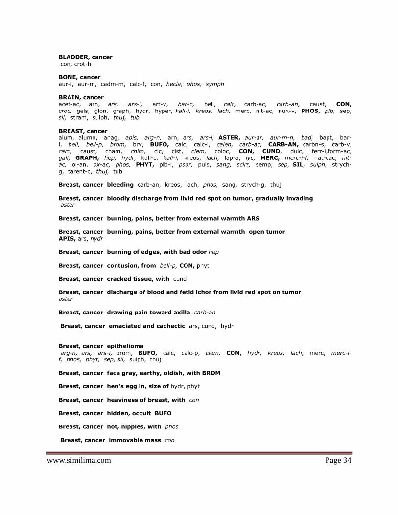
www.similima.com Page 34
BLADDER, cancer con, crot-h BONE, cancer aur-i, aur-m, cadm-m, calc-f, con, hecla, phos, symph
BRAIN, cancer acet-ac, arn, ars, ars-i, art-v, bar-c, bell, calc, carb-ac, carb-an, caust, CON, croc, gels, glon, graph, hydr, hyper, kali-i, kreos, lach, merc, nit-ac, nux-v, PHOS, plb, sep, sil, stram, sulph, thuj, tub BREAST, cancer alum, alumn, anag, apis, arg-n, arn, ars, ars-i, ASTER, aur-ar, aur-m-n, bad, bapt, bar-i, bell, bell-p, brom, bry, BUFO, calc, calc-i, calen, carb-ac, CARB-AN, carbn-s, carb-v, carc, caust, cham, chim, cic, cist, clem, coloc, CON, CUND, dulc, ferr-i,form-ac, gali, GRAPH, hep, hydr, kali-c, kali-i, kreos, lach, lap-a, lyc, MERC, merc-i-f, nat-cac, nit-ac, ol-an, ox-ac, phos, PHYT, plb-i, psor, puls, sang, scirr, semp, sep, SIL, sulph, strych-
g, tarent-c, thuj, tub Breast, cancer bleeding carb-an, kreos, lach, phos, sang, strych-g, thuj Breast, cancer bloodly discharge from livid red spot on tumor, gradually invading aster Breast, cancer burning, pains, better from external warmth ARS Breast, cancer burning, pains, better from external warmth open tumor APIS, ars, hydr
Breast, cancer burning of edges, with bad odor hep Breast, cancer contusion, from bell-p, CON, phyt Breast, cancer cracked tissue, with cund Breast, cancer discharge of blood and fetid ichor from livid red spot on tumor aster Breast, cancer drawing pain toward axilla carb-an
Breast, cancer emaciated and cachectic ars, cund, hydr Breast, cancer epithelioma arg-n, ars, ars-i, brom, BUFO, calc, calc-p, clem, CON, hydr, kreos, lach, merc, merc-i-f, phos, phyt, sep, sil, sulph, thuj Breast, cancer face gray, earthy, oldish, with BROM Breast, cancer hen's egg in, size of hydr, phyt Breast, cancer heaviness of breast, with con
Breast, cancer hidden, occult BUFO Breast, cancer hot, nipples, with phos Breast, cancer immovable mass con
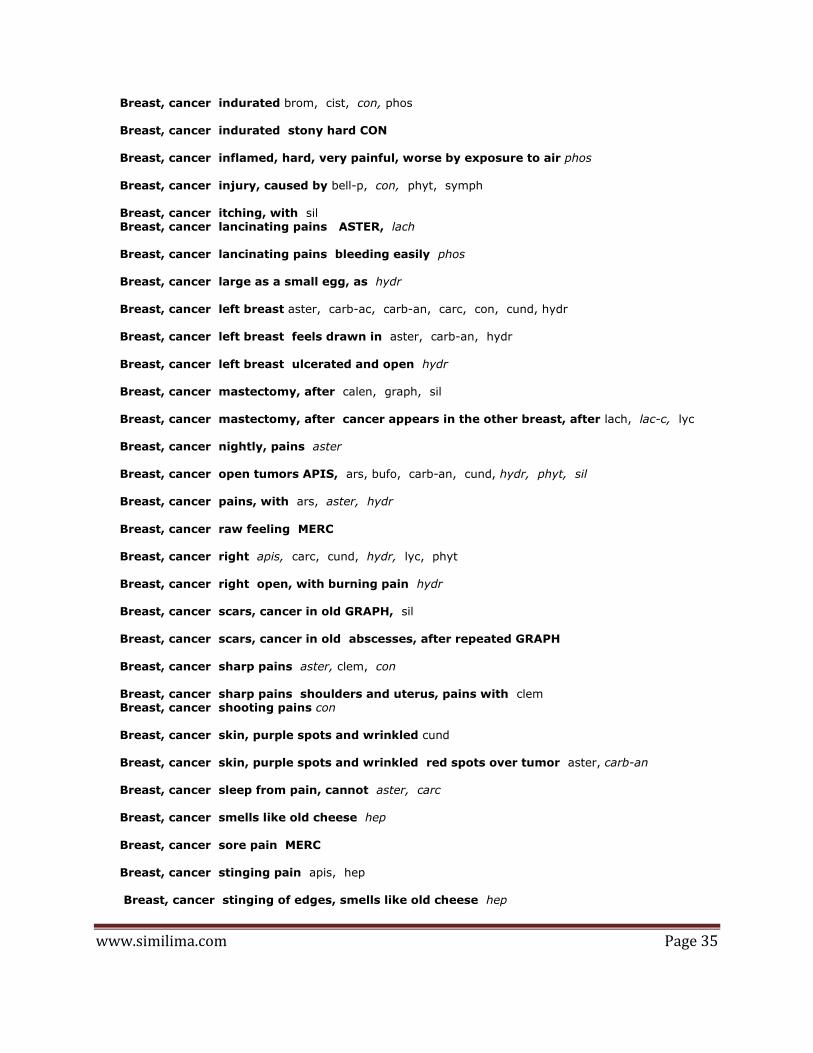
www.similima.com Page 35
Breast, cancer indurated brom, cist, con, phos Breast, cancer indurated stony hard CON Breast, cancer inflamed, hard, very painful, worse by exposure to air phos
Breast, cancer injury, caused by bell-p, con, phyt, symph Breast, cancer itching, with sil Breast, cancer lancinating pains ASTER, lach Breast, cancer lancinating pains bleeding easily phos Breast, cancer large as a small egg, as hydr Breast, cancer left breast aster, carb-ac, carb-an, carc, con, cund, hydr Breast, cancer left breast feels drawn in aster, carb-an, hydr
Breast, cancer left breast ulcerated and open hydr Breast, cancer mastectomy, after calen, graph, sil
Breast, cancer mastectomy, after cancer appears in the other breast, after lach, lac-c, lyc Breast, cancer nightly, pains aster Breast, cancer open tumors APIS, ars, bufo, carb-an, cund, hydr, phyt, sil Breast, cancer pains, with ars, aster, hydr
Breast, cancer raw feeling MERC Breast, cancer right apis, carc, cund, hydr, lyc, phyt Breast, cancer right open, with burning pain hydr Breast, cancer scars, cancer in old GRAPH, sil Breast, cancer scars, cancer in old abscesses, after repeated GRAPH
Breast, cancer sharp pains aster, clem, con Breast, cancer sharp pains shoulders and uterus, pains with clem Breast, cancer shooting pains con Breast, cancer skin, purple spots and wrinkled cund Breast, cancer skin, purple spots and wrinkled red spots over tumor aster, carb-an Breast, cancer sleep from pain, cannot aster, carc Breast, cancer smells like old cheese hep
Breast, cancer sore pain MERC
Breast, cancer stinging pain apis, hep Breast, cancer stinging of edges, smells like old cheese hep
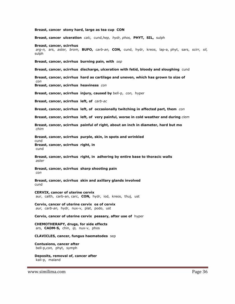
www.similima.com Page 36
Breast, cancer stony hard, large as tea cup CON Breast, cancer ulceration calc, cund,hep, hydr, phos, PHYT, SIL, sulph Breast, cancer, scirrhus arg-n, ars, aster, brom, BUFO, carb-an, CON, cund, hydr, kreos, lap-a, phyt, sars, scirr, sil,
sulph Breast, cancer, scirrhus burning pain, with sep Breast, cancer, scirrhus discharge, ulceration with fetid, bloody and sloughing cund Breast, cancer, scirrhus hard as cartilage and uneven, which has grown to size of con Breast, cancer, scirrhus heaviness con Breast, cancer, scirrhus injury, caused by bell-p, con, hyper
Breast, cancer, scirrhus left, of carb-ac Breast, cancer, scirrhus left, of occasionally twitching in affected part, them con Breast, cancer, scirrhus left, of very painful, worse in cold weather and during clem Breast, cancer, scirrhus painful of right, about an inch in diameter, hard but mo chim
Breast, cancer, scirrhus purple, skin, in spots and wrinkled cund Breast, cancer, scirrhus right, in
cund Breast, cancer, scirrhus right, in adhering by entire base to thoracic walls aster
Breast, cancer, scirrhus sharp shooting pain con Breast, cancer, scirrhus skin and axillary glands involved cund
CERVIX, cancer of uterine cervix aur, calth, carb-an, carc, CON, hydr, iod, kreos, thuj, ust Cervix, cancer of uterine cervix os of cervix aur, carb-an, hydr, nux-v, plat, podo, ust Cervix, cancer of uterine cervix pessary, after use of hyper CHEMOTHERAPY, drugs, for side effects ars, CADM-S, chin, ip, nux-v, phos CLAVICLES, cancer, fungus haematodes sep
Contusions, cancer after bell-p,con, phyt, symph
Deposits, removal of, cancer after kali-p, maland
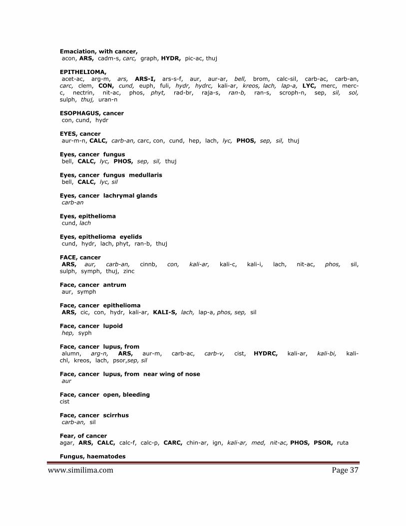
www.similima.com Page 37
Emaciation, with cancer, acon, ARS, cadm-s, carc, graph, HYDR, pic-ac, thuj EPITHELIOMA, acet-ac, arg-m, ars, ARS-I, ars-s-f, aur, aur-ar, bell, brom, calc-sil, carb-ac, carb-an, carc, clem, CON, cund, euph, fuli, hydr, hydrc, kali-ar, kreos, lach, lap-a, LYC, merc, merc-
c, nectrin, nit-ac, phos, phyt, rad-br, raja-s, ran-b, ran-s, scroph-n, sep, sil, sol, sulph, thuj, uran-n
ESOPHAGUS, cancer con, cund, hydr EYES, cancer aur-m-n, CALC, carb-an, carc, con, cund, hep, lach, lyc, PHOS, sep, sil, thuj Eyes, cancer fungus bell, CALC, lyc, PHOS, sep, sil, thuj
Eyes, cancer fungus medullaris bell, CALC, lyc, sil Eyes, cancer lachrymal glands carb-an Eyes, epithelioma cund, lach Eyes, epithelioma eyelids cund, hydr, lach, phyt, ran-b, thuj
FACE, cancer ARS, aur, carb-an, cinnb, con, kali-ar, kali-c, kali-i, lach, nit-ac, phos, sil, sulph, symph, thuj, zinc Face, cancer antrum aur, symph Face, cancer epithelioma ARS, cic, con, hydr, kali-ar, KALI-S, lach, lap-a, phos, sep, sil
Face, cancer lupoid
hep, syph Face, cancer lupus, from alumn, arg-n, ARS, aur-m, carb-ac, carb-v, cist, HYDRC, kali-ar, kali-bi, kali-chl, kreos, lach, psor,sep, sil
Face, cancer lupus, from near wing of nose aur
Face, cancer open, bleeding cist
Face, cancer scirrhus carb-an, sil Fear, of cancer agar, ARS, CALC, calc-f, calc-p, CARC, chin-ar, ign, kali-ar, med, nit-ac, PHOS, PSOR, ruta Fungus, haematodes
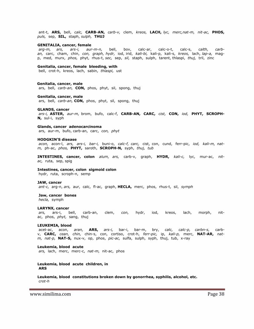
www.similima.com Page 38
ant-t, ARS, bell, calc, CARB-AN, carb-v, clem, kreos, LACH, lyc, merc,nat-m, nit-ac, PHOS, puls, sep, SIL, staph, sulph, THUJ GENITALIA, cancer, female arg-m, ars, ars-i, aur-m-n, bell, bov, calc-ar, calc-o-t, calc-s, calth, carb-an, carc, cham, chin, con, graph, hydr, iod, irid, kali-bi, kali-p, kali-s, kreos, lach, lap-a, mag-
p, med, murx, phos, phyt, rhus-t, sec, sep, sil, staph, sulph, tarent, thlaspi, thuj, tril, zinc Genitalia, cancer, female bleeding, with bell, crot-h, kreos, lach, sabin, thlaspi, ust
Genitalia, cancer, male
ars, bell, carb-an, CON, phos, phyt, sil, spong, thuj Genitalia, cancer, male ars, bell, carb-an, CON, phos, phyt, sil, spong, thuj
GLANDS, cancer ars-i, ASTER, aur-m, brom, bufo, calc-f, CARB-AN, CARC, cist, CON, iod, PHYT, SCROPH-N, sul-i, syph Glands, cancer adenocarcinoma ars, aur-m, bufo, carb-an, carc, con, phyt HODGKIN’S disease acon, acon-l, ars, ars-i, bar-i, buni-o, calc-f, carc, cist, con, cund, ferr-pic, iod, kali-m, nat-m, ph-ac, phos, PHYT, saroth, SCROPH-N, syph, thuj, tub
INTESTINES, cancer, colon alum, ars, carb-v, graph, HYDR, kali-c, lyc, mur-ac, nit-
ac, ruta, sep, spig Intestines, cancer, colon sigmoid colon hydr, ruta, scroph-n, semp
JAW, cancer ant-c, arg-n, ars, aur, calc, fl-ac, graph, HECLA, merc, phos, rhus-t, sil, symph Jaw, cancer bones hecla, symph
LARYNX, cancer ars, ars-i, bell, carb-an, clem, con, hydr, iod, kreos, lach, morph, nit-ac, phos, phyt, sang, thuj LEUKEMIA, blood acet-ac, acon, aran, ARS, ars-i, bar-i, bar-m, bry, calc, calc-p, carbn-s, carb-v, CARC, cean, chin, chin-s, con, cortiso, crot-h, ferr-pic, ip, kali-p, merc, NAT-AR, nat-m, nat-p, NAT-S, nux-v, op, phos, pic-ac, sulfa, sulph, syph, thuj, tub, x-ray Leukemia, blood acute ars, lach, merc, merc-c, nat-m, nit-ac, phos
Leukemia, blood acute children, in ARS Leukemia, blood constitutions broken down by gonorrhea, syphilis, alcohol, etc. crot-h
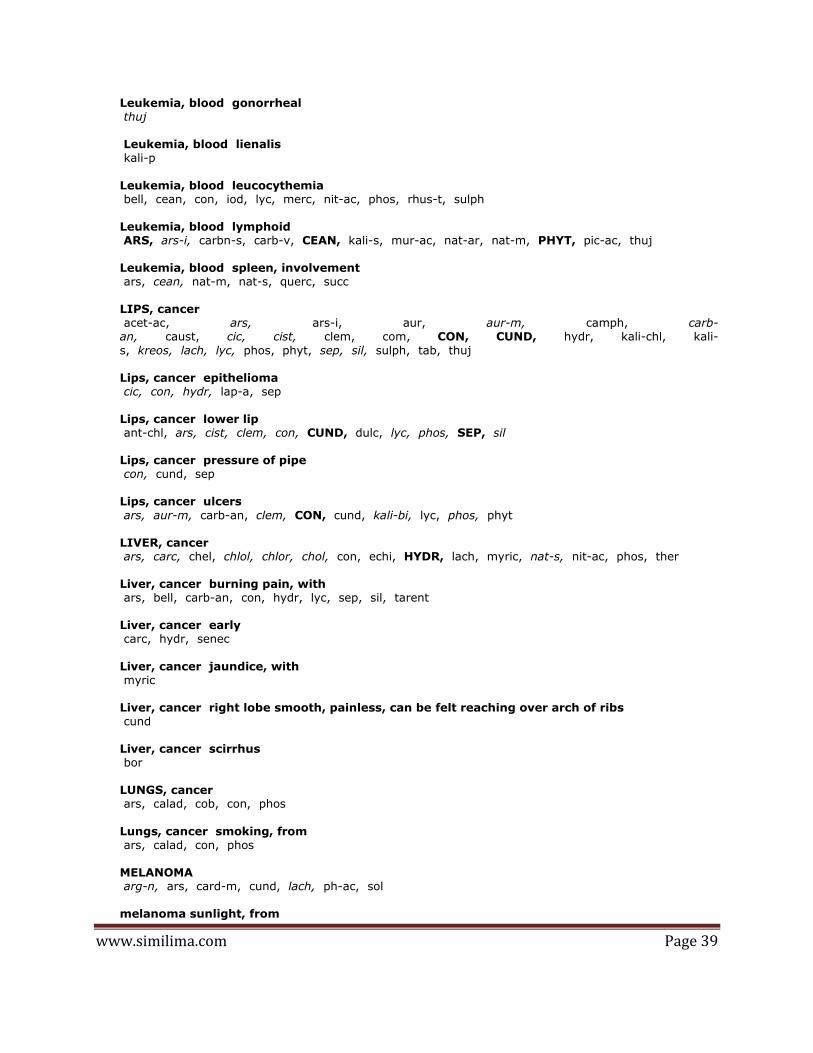
www.similima.com Page 39
Leukemia, blood gonorrheal thuj Leukemia, blood lienalis kali-p
Leukemia, blood leucocythemia bell, cean, con, iod, lyc, merc, nit-ac, phos, rhus-t, sulph Leukemia, blood lymphoid ARS, ars-i, carbn-s, carb-v, CEAN, kali-s, mur-ac, nat-ar, nat-m, PHYT, pic-ac, thuj Leukemia, blood spleen, involvement ars, cean, nat-m, nat-s, querc, succ LIPS, cancer acet-ac, ars, ars-i, aur, aur-m, camph, carb-an, caust, cic, cist, clem, com, CON, CUND, hydr, kali-chl, kali-
s, kreos, lach, lyc, phos, phyt, sep, sil, sulph, tab, thuj Lips, cancer epithelioma cic, con, hydr, lap-a, sep
Lips, cancer lower lip ant-chl, ars, cist, clem, con, CUND, dulc, lyc, phos, SEP, sil Lips, cancer pressure of pipe con, cund, sep Lips, cancer ulcers
ars, aur-m, carb-an, clem, CON, cund, kali-bi, lyc, phos, phyt LIVER, cancer ars, carc, chel, chlol, chlor, chol, con, echi, HYDR, lach, myric, nat-s, nit-ac, phos, ther Liver, cancer burning pain, with ars, bell, carb-an, con, hydr, lyc, sep, sil, tarent Liver, cancer early carc, hydr, senec
Liver, cancer jaundice, with myric Liver, cancer right lobe smooth, painless, can be felt reaching over arch of ribs cund Liver, cancer scirrhus bor LUNGS, cancer ars, calad, cob, con, phos
Lungs, cancer smoking, from ars, calad, con, phos
MELANOMA arg-n, ars, card-m, cund, lach, ph-ac, sol melanoma sunlight, from

www.similima.com Page 40
carc, sol Miasm, cancer ars, ars-i, CARC, cadm-s, con, hydr MOUTH, cancer, palate
aur, hydr Mouth, cancer, palate hardness, with hydr NECK, cancer cist, con, hydr, merc, scroph-n Neck, cancer glands, of cist, con, scroph-n
Noma, cancer alum, alumn, ars, calc, carb-v, con, elat, kali-chl, kali-p, merc, sil, sulph, tarent-c
NOSE, cancer alumn, ars, AUR, aur-m, calc, carb-ac, carb-an, cund, kreos, merc, phyt, sep, sulph Nose, cancer epithelioma ars, ars-i, carb-ac, cund, hydr, KALI-S, kreos Nose, cancer epithelioma nose wing med Nose, cancer flat, on right side euphr
Nose, cancer noli me tangere, on nose cist, jug-c, phyt, thuj OVARIES, cancer ars, CON, graph, kreos, lach, med, psor, thuj PAINS, from cancer acon, anthr, apis, ARS, aster, aur, bry, bufo, calc, calc-ar, cadm-s, carb-an, CARC, cedr, cinnam, cit-ac, coloc, con, cund, echi, euph, HYDR, mag-p, merc, morph, op, ph-ac, sil, sol
PALATE, cancer
aur, hydr Palate, cancer hardness, with hydr PANCREAS, cancer ars, calc-ar, CON, HYDR, phos PAROTID, cancer merc, phyt
PENIS, cancer, glans, epithelioma on excrescences arg-n, ars, con, thuj
PHARYNX, cancer cist PROSTATE, cancer
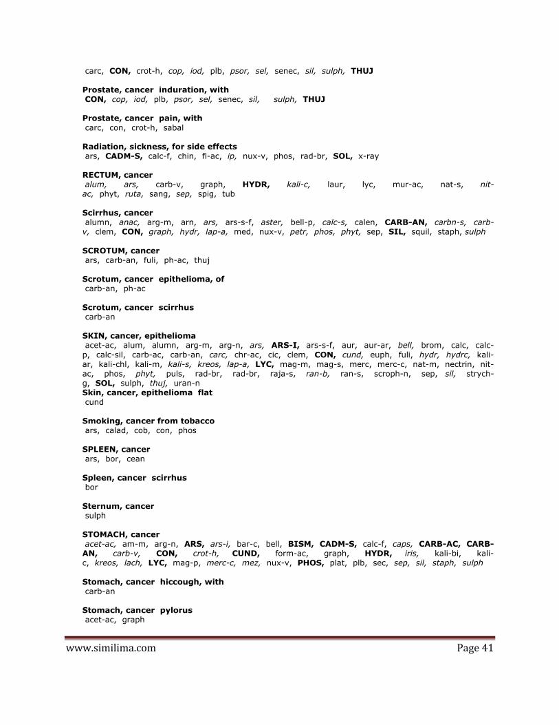
www.similima.com Page 41
carc, CON, crot-h, cop, iod, plb, psor, sel, senec, sil, sulph, THUJ Prostate, cancer induration, with CON, cop, iod, plb, psor, sel, senec, sil, sulph, THUJ Prostate, cancer pain, with
carc, con, crot-h, sabal Radiation, sickness, for side effects ars, CADM-S, calc-f, chin, fl-ac, ip, nux-v, phos, rad-br, SOL, x-ray RECTUM, cancer alum, ars, carb-v, graph, HYDR, kali-c, laur, lyc, mur-ac, nat-s, nit-ac, phyt, ruta, sang, sep, spig, tub Scirrhus, cancer alumn, anac, arg-m, arn, ars, ars-s-f, aster, bell-p, calc-s, calen, CARB-AN, carbn-s, carb-v, clem, CON, graph, hydr, lap-a, med, nux-v, petr, phos, phyt, sep, SIL, squil, staph, sulph
SCROTUM, cancer ars, carb-an, fuli, ph-ac, thuj Scrotum, cancer epithelioma, of carb-an, ph-ac Scrotum, cancer scirrhus carb-an SKIN, cancer, epithelioma acet-ac, alum, alumn, arg-m, arg-n, ars, ARS-I, ars-s-f, aur, aur-ar, bell, brom, calc, calc-
p, calc-sil, carb-ac, carb-an, carc, chr-ac, cic, clem, CON, cund, euph, fuli, hydr, hydrc, kali-ar, kali-chl, kali-m, kali-s, kreos, lap-a, LYC, mag-m, mag-s, merc, merc-c, nat-m, nectrin, nit-ac, phos, phyt, puls, rad-br, rad-br, raja-s, ran-b, ran-s, scroph-n, sep, sil, strych-g, SOL, sulph, thuj, uran-n Skin, cancer, epithelioma flat cund Smoking, cancer from tobacco ars, calad, cob, con, phos SPLEEN, cancer
ars, bor, cean Spleen, cancer scirrhus bor Sternum, cancer sulph STOMACH, cancer acet-ac, am-m, arg-n, ARS, ars-i, bar-c, bell, BISM, CADM-S, calc-f, caps, CARB-AC, CARB-AN, carb-v, CON, crot-h, CUND, form-ac, graph, HYDR, iris, kali-bi, kali-c, kreos, lach, LYC, mag-p, merc-c, mez, nux-v, PHOS, plat, plb, sec, sep, sil, staph, sulph
Stomach, cancer hiccough, with carb-an Stomach, cancer pylorus acet-ac, graph
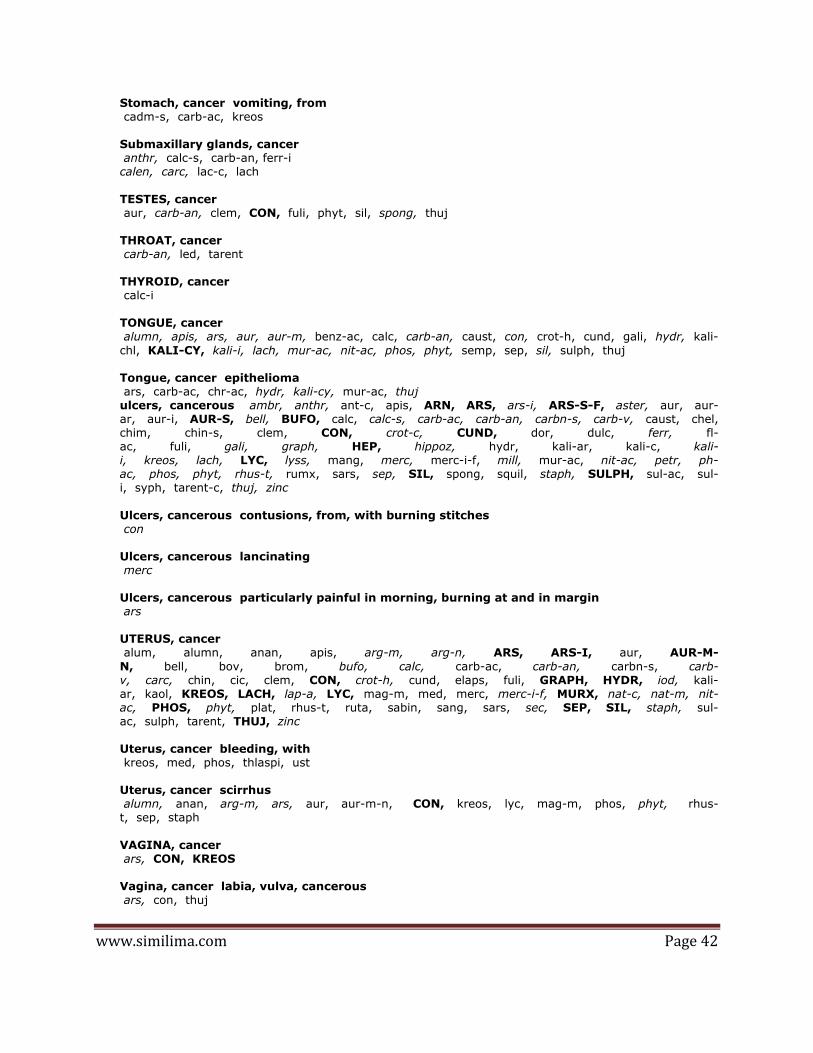
www.similima.com Page 42
Stomach, cancer vomiting, from cadm-s, carb-ac, kreos Submaxillary glands, cancer anthr, calc-s, carb-an, ferr-i calen, carc, lac-c, lach
TESTES, cancer aur, carb-an, clem, CON, fuli, phyt, sil, spong, thuj THROAT, cancer carb-an, led, tarent THYROID, cancer calc-i
TONGUE, cancer alumn, apis, ars, aur, aur-m, benz-ac, calc, carb-an, caust, con, crot-h, cund, gali, hydr, kali-
chl, KALI-CY, kali-i, lach, mur-ac, nit-ac, phos, phyt, semp, sep, sil, sulph, thuj Tongue, cancer epithelioma ars, carb-ac, chr-ac, hydr, kali-cy, mur-ac, thuj ulcers, cancerous ambr, anthr, ant-c, apis, ARN, ARS, ars-i, ARS-S-F, aster, aur, aur-ar, aur-i, AUR-S, bell, BUFO, calc, calc-s, carb-ac, carb-an, carbn-s, carb-v, caust, chel, chim, chin-s, clem, CON, crot-c, CUND, dor, dulc, ferr, fl-ac, fuli, gali, graph, HEP, hippoz, hydr, kali-ar, kali-c, kali-i, kreos, lach, LYC, lyss, mang, merc, merc-i-f, mill, mur-ac, nit-ac, petr, ph-ac, phos, phyt, rhus-t, rumx, sars, sep, SIL, spong, squil, staph, SULPH, sul-ac, sul-i, syph, tarent-c, thuj, zinc
Ulcers, cancerous contusions, from, with burning stitches con Ulcers, cancerous lancinating merc Ulcers, cancerous particularly painful in morning, burning at and in margin ars
UTERUS, cancer alum, alumn, anan, apis, arg-m, arg-n, ARS, ARS-I, aur, AUR-M-
N, bell, bov, brom, bufo, calc, carb-ac, carb-an, carbn-s, carb-v, carc, chin, cic, clem, CON, crot-h, cund, elaps, fuli, GRAPH, HYDR, iod, kali-ar, kaol, KREOS, LACH, lap-a, LYC, mag-m, med, merc, merc-i-f, MURX, nat-c, nat-m, nit-ac, PHOS, phyt, plat, rhus-t, ruta, sabin, sang, sars, sec, SEP, SIL, staph, sul-ac, sulph, tarent, THUJ, zinc
Uterus, cancer bleeding, with kreos, med, phos, thlaspi, ust
Uterus, cancer scirrhus alumn, anan, arg-m, ars, aur, aur-m-n, CON, kreos, lyc, mag-m, phos, phyt, rhus-t, sep, staph
VAGINA, cancer ars, CON, KREOS Vagina, cancer labia, vulva, cancerous ars, con, thuj

www.similima.com Page 43
Weakness, with cancer ars, aur, aur-m-n, CADM-S, CARC, CON, HYDR, kreos, phos, phyt Weakness, with cancer cancer, after ars, carc, hydr
Weakness, with cancer chemotherapy, after cadm-s Weakness, with cancer radiation therapy, after cadm-s EMACIATION, body stomach, cancer, with ars, cund, HYDR, mez FOOD APPETITE, diminished cancer patients, in
ALF, ars, cadm-s, hydr Appetite, loss, of appetite cancer patients, in ALF, ars, cadm-s, carc, hydr
GENERALS ANXIETY, (physical) pains, from the cancer, of ars, CARC
MIND
ANXIETY, pains, from the cancer, of ars, CARC DEPRESSION, (sadness), cancer, with carc, con FEAR, cancer, of agar, ARS, bar-c, CALC, calc-f, calc-p, CARC, chin-ar, ign, kali-ar, med, nit-ac, PHOS, PSOR, ruta FEAR, death, of cancer, from ars, carc
SUICIDAL, cancer history in family, with carc
(Bold Capital - 3 mark: Italics- 2 mark: Roman- 1mark ) Materials and Methods The present study was carried out at the Government Homoeopathic Medical College, Kozhikode, from August 2003 to February 2005. All patients with cancer pain coming to the O.P.D and I.P.D were selected for the study.
Inclusion criteria: Cancer patients who complaints of pain were taken for the study. The study was made in all age group The study was made in both sexes. Exclusion criteria: Cancer patients without pain were excluded The case history of the patients thus selected were taken according to homoeopathic philosophy with a detailed enquiry in to the pain characteristics like time of occurrence, severity and duration of pain, any radiation of pain, the quality of pain and finally the aggravating and ameliorating factors of pain . After

www.similima.com Page 44
completing the case the patients were then asked to rate their pain by using verbal rating scale and visual analog scale. Then they were assessed for the quality of life using degree of distress score and W.H.O performance status classification. In the selection of medicines the approach was to individualize each case based on homoeopathic principles. The miasmatic background was given due consideration while selecting the medicine. Repertorisation was based on Kent’s Repertory of the homoeopathic materia medica and Boenninghaussen’s
Charecterestics materia medica and repertory by C.M.Boger. Repetition and change in potency was based on homoeopathic principles. Pain, performance status and degree of distress had assessed objectively and subjectively and scored. Effectiveness has assessed on the basis of change in the scores. Out patients were reviewed every 2 weeks or until the pain get palliated, and the in patients were reviewed every day till they got discharged. The method of approach was a clinical study without the use of control. Assessment tool was developed after literature review. Three major areas were identified as important parameters and each item were rated. Assessment criteria include:
Ø Pain Sore
Ø Degree of distress score Ø W.H.O performance status score
Pain scoring: Pain was measured using verbal rating scale (Simple descriptive pain intensity scale) and visual analog scale or numeric analog scale. Each terminology on the verbal rating scale was explained to the patient before assessment, and they were asked to classify their intensity of pain they experienced daily. They were also explained how to use the visual analog scale to numerically rate their pain as already described. 0 for no pain and 1 to 10 for increasing severity as given by the patient25. Degree of distress score26:
Questions Response Point
1. Did you feel sick to day?
Not at all
Occasionally
A lot
All of the time
1
2
3
4
2. Did you vomit to day?
Not at all
Once
Twice
More than twice
1
2
3
4
3.How good has your
Appetite been today?
Good
Fair
Poor
Bad
1
2
3
4

www.similima.com Page 45
4.How much pain have you
had today?
None
A little
Quite a lot
A lot
1
2
3
4
5.How did you sleep last
Night?
Very well
Quite well
Badly
Not at all
1
2
3
4
6.How is your bowel
pattern today?
No distress
Low distress
Moderate distress
High distress
1
2
3
4
7.How happy have you
been today?
Happy
Fairly happy
Un happy
Very unhappy
1
2
3
4
8.How are you feeling
today?
Well
Fair
Poor
Very poor
1
2
3
4
9. What did you do today?
Fully active
Light work/house work
Got up but did nothing
Stayed in bed
1
2
3
4
W.H.O performance Status26
Performance Grade
Able to carry out all normal activity without restriction 0
www.similima.com Page 46
Restricted in physically strenuous activity but ambulatory &able to do
light work
1
Ambulatory &capable of self care but unable to carry out any work 2
Capable of only limited self care confined to bed or chair 50% or more
of the waking hours
3
Completely disabled &cannot carry on any self care 4
This study was carried out in the Govt.Homoeopathic Medical College, Kozhikode during the period from
Aug 2003 – to Feb 2005 in 30 patients. The individual observations were given in the distribution table no 1,
2,3,4,5,6,7,8 9 and 10.
The results were categorized under the following headings as follows,
Observation and Discussion
Age Wise Distribution of Patients:
Out of the 30 patients studied 2 patients (6.66%) were between the age group 30 -39. 5 patients (16.66%)
were between 40-49, 3 patients (10%) were between 50-59. 11 patients (36.66%) were between the age
groups 60-69, 7 patients (23.33%) were between the age group 70-79, and 2 patients (6.66%) were
between the age group 80-89.
Distribution of Patients According To Sex:
Chart: 3 Out of the 30 patients studied 16 patients (53.3%) were male patients and 14 patients (46.6%) were
female.
Distribution Of Patients According To Religion:
Out of the 30 patients studied 16 patients (53%) were of Hindu religion, 11 patients (37%) were of Muslim
religion and 3 patients (10%) belong to Christian religion.
Distribution of Patients according To Intensity of Pain Before and after treatment
Out of the 30 patients studied 8 patients (26.6%) presented with mild pain, 10 patients (33.3%) presented
with moderate pain, and 12 patients (40%) presented with severe pain before treatment. After treatment 12
patients (40%) have no pain, 14 patients (46.6%) have mild pain 3 patients (10%) have moderate pain and
1 patient (3.3%) has severe pain.
Distribution of Patients According To the Site of Cancer:
Out of the 30 patients studied 5 patients (16.66%) have esophageal carcinoma, 3 patients each (10%)
have bronchogenic carcinoma, bladder carcinoma, metastatic carcinoma with undetected primary and
carcinoma of the stomach, 2 patients each (6.66%) presented with oral cancer carcinoma pharynx, larynx

www.similima.com Page 47
and genital malignancy.1 patient each (3.33%) presented with lymphoma, leukemia, multiple myeloma,
liver carcinoma and rectal carcinoma.
Distribution of Medicines Used In the Study:
t of 30 patients studied Lycopodium was found effective in 7 (23.33%) cases. Sulphur and Phosphorous was
found effective in 6 (20%)cases each, Ars.alb was found effective in 3(10%) cases, Lachesis was found
effective in 2(6.66%)cases, Ars.iod, Calc.iod, Kali.carb, Phytolaca ,China and Hepar.sulph were found to
be effective in 1(3.33%) case each .
Distribution of Medicine Used For Acute Management:
Out of 30 patients studied Calc.carb was found to be effective in 9(30%) cases, Euphorbium and Nux.vom
was found to be effective in 4(13.33%) cases each, Ars.alb was found to be effective in 3(10%) cases,
Colocynth, Terebinth, and Carbo veg was found to be effective in 2 (6.66%) cases each, Spongia and
Phytolaca was found to be effective in 1(3.33%) cases each.
Distribution of miasm of the cases:
Out of the 30 patients studied 11 patients (36.66%) are of psoric miasm,9 patients (30%) are psoro
syphilitic,3 patients (10%) are psora sycotic and 7 patients (23.3%) have psoric+syphilitic+sycotic miasm.
Distribution of the miasm of Pain of the cases:
Out of the 30 patients studied, the miasm of pain of 27 patients (90%) are psoric, and the 3 patients (10%)
pain belongs to syphilitic miasm.
STATISTICAL ANALYSIS
Assessment criteria include:
- Pain Sore
- Degree of distress score
- WHO performance status score
Tests of significance: -
a) Questions to be answered. Is there any difference in the pain score, degree of distress score and
performance status score of the case before and after treatment?
b) Null hypothesis Ho: no difference in the pain score, degree of distress score and performance status
score of the case before and after treatment.
Test of significance determined by using paired t’ test.27
EQ / Sz/√n
Parameter Calculated t
value
Tabled t
value
Level of
significance
Significant or
not
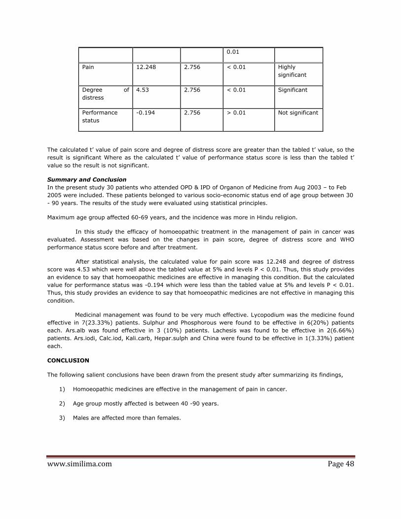
www.similima.com Page 48
0.01
Pain 12.248 2.756 < 0.01 Highly
significant
Degree of
distress
4.53 2.756 < 0.01 Significant
Performance
status
-0.194 2.756 > 0.01 Not significant
The calculated t’ value of pain score and degree of distress score are greater than the tabled t’ value, so the
result is significant Where as the calculated t’ value of performance status score is less than the tabled t’
value so the result is not significant.
Summary and Conclusion
In the present study 30 patients who attended OPD & IPD of Organon of Medicine from Aug 2003 – to Feb
2005 were included. These patients belonged to various socio-economic status end of age group between 30
- 90 years. The results of the study were evaluated using statistical principles.
Maximum age group affected 60-69 years, and the incidence was more in Hindu religion.
In this study the efficacy of homoeopathic treatment in the management of pain in cancer was
evaluated. Assessment was based on the changes in pain score, degree of distress score and WHO
performance status score before and after treatment.
After statistical analysis, the calculated value for pain score was 12.248 and degree of distress
score was 4.53 which were well above the tabled value at 5% and levels P < 0.01. Thus, this study provides
an evidence to say that homoeopathic medicines are effective in managing this condition. But the calculated
value for performance status was -0.194 which were less than the tabled value at 5% and levels P < 0.01.
Thus, this study provides an evidence to say that homoeopathic medicines are not effective in managing this
condition.
Medicinal management was found to be very much effective. Lycopodium was the medicine found
effective in 7(23.33%) patients. Sulphur and Phosphorous were found to be effective in 6(20%) patients
each. Ars.alb was found effective in 3 (10%) patients. Lachesis was found to be effective in 2(6.66%)
patients. Ars.iodi, Calc.iod, Kali.carb, Hepar.sulph and China were found to be effective in 1(3.33%) patient
each.
CONCLUSION
The following salient conclusions have been drawn from the present study after summarizing its findings,
1) Homoeopathic medicines are effective in the management of pain in cancer.
2) Age group mostly affected is between 40 -90 years.
3) Males are affected more than females.
www.similima.com Page 49
Bibliography
1. Malcom Alison &Catherine Saraf, Introduction to cancer (Ch 1), Cause of cancer (Ch 2),
Understanding cancer 1997,Cambride University press: Pp: 1,39-40
2. Nathan I.Cherny and Russell K.Portnenoy, Cancer pain: Principles of assessment and syndromes
(Ch.43): Text Book of Pain (4th Edn) Churchill livingston, Pp: 787-794
3. HarshMohan, Neoplasia (Ch.7) Text book of Pathology (4th Edn) JayPee brothers 2000,Pp: 177-215
4. Vinay Kumar & Cortan, Neoplasia, Pathological basis of disease, (7th Edn) Pp: 177-210
5. K.V.Krishna Das, Principles of oncology, Text book of Medicine (3rd Edn) Jay Pee Brothers 1997,Pp:
37
6. Robert Twycross, General topic, symptom management; Introducing palliative care (4th Edn)
;Institute of palliative medicine, Calicut 2003;Pp: 1-17,61-64
7. WHO Websites. Media center, press release, Global cancer report, April 3rd 2003,Geneva, Pp: 1-6
8. rcc.org.in; education,Pp:1-4
9. Medindia .net,health statistics,disease in India, prevalent cancers in India,Feb 10,2004
10. dep.iarc.fr (International agency for research on cancer) C15 I-VIII original, population based
11. Raynond D Adon, Maurice Victor, Pain (Ch 7) Principles of neurology (4th Edn.); Mc Grow –Hill
information service company (1989) Pp:103-110
12. Guyton &Hall, Somatic sensation II- Pain, headache and thermal sensation (Ch-48), Text book of
Physiology, Pp:552
13. Christopher G.Goetz &Eric J.Pappert:Pain &Temperature (Ch-20)Text book of clinical neurology:
W.B.Saunder’s Company Pp:333-337
14. Stedman’s Medical dictionary, (24th Edn.) Pain, William and Wilkins, Pp: 1282.
15. Roger woodruff, Cancer pain (2nd Edn) Introduction, Asperula Pty Ltd, Australia, Pp:1-10
16. Stuart Close, The scope of homoeopathy, Life, health and disease, The genius of homoeopathy,
B.Jain publishers. pvt.ltd, New Delhi, Pp:41-44,60-61
17. Dr.Samuel Hahnemann, Organon of Medicine, B.Jain publishers.pvt.ltd, New Delhi, 5th and 6th
edition,
Pp: 62,97-10104,126
18. H.A.Roberts,The law of Palliation ,Disease classification syphilis,Sycosis,The principles and art of
cure by Homoeopathy, Indian books and periodicals syndicate, Pp: 162,215,233
19. J.T.Kent, Why cancer is incurable, Lesser writings
20. Dr. Subrato Kumar Banerjea, Miasmatic diagnosis, B.Jain publishers Pvt.ltd, New Delhi, Pp: 53
21. Cancer _pain.org, understanding cancer pain, what is cancer pain, Pp: 4
www.similima.com Page 50
22. J.T.Kent,The difficult &incurable cases, Lectures on homoeopathic philosophy, B.Jain publishers
Pvt.ltd, New Delhi, Pp:. 288
23. Farokh J Master, Tumor and homoeopathy, Homoeopathy and cancer, (Ch-IX) B Jain Publishers,
(2002) Pp: 50-55
24. Robin Murphy, Diseases, Homoeopathic Medical Repertory (2nd Revised Edn.) B Jain Publishers
Pvt.ltd, New Delhi, (2002) Pp: 390-394
25. National cancer institute, Gathering information, Understanding cancer pain, Cancer.gov,
6.3.1999,Pp: 3-4.
26. .H.O performance status classification in cancer, symptoms and degree of distress in patients with
cancer.1.11.2005,medal.org
27.B.K.Mahajan, Significance of difference in means, Methods in bio-statistics for medical students and research workers, 6th edition, Jaypee brothers:Pp-148
Dr Rithesh.B. B.H.M.S MD(Hom) Department of Organon of Medicine Govt. Homeopathic Medical College, Calicut Kerala Email : [email protected] Mob : 09446063849