


The role of respiration in virulence gene expression of Vibrio cholerae
Page 1 of 30
1
2
Regulation of Virulence Gene Transcripts by the Francisella Orphan Response Regulator PmrA: 3
Role of Phosphorylation and Evidence of MglA/SspA Interaction 4
5
Brian L. Bell1, Nrusingh P. Mohapatra, and John S. Gunn* 6
Center for Microbial Interface Biology and Department of Molecular Virology, Immunology, and 7
Medical Genetics, The Ohio State University, Columbus, OH, USA 8
1 Current address: Air Force Research Laboratory at The University of Cincinnati; Medical Sciences
Building, Room 3308; Cincinnati, OH 45267
* Corresponding author: 460 W. 12th Ave.; Columbus, Ohio 43210 Rm. 1006; 614-292-60367; FAX:
614-292-5495; [email protected]
Copyright © 2010, American Society for Microbiology and/or the Listed Authors/Institutions. All Rights Reserved.Infect. Immun. doi:10.1128/IAI.00021-10 IAI Accepts, published online ahead of print on 15 March 2010
on March 17, 2018 by guest
http://iai.asm.org/
Dow
nloaded from

Page 2 of 30
Abstract 9
Francisella tularensis subspecies tularensis is the etiologic agent of tularemia and has been 10
designated a category A biothreat agent by the CDC. Tularemia is characterized by replication 11
and dissemination within host phagocytes. Intramacrophage growth is dependent upon the 12
regulation of Francisella Pathogenicity Island (FPI) virulence genes, which is poorly understood. 13
Two-component regulatory systems (TCS) are widely employed by Gram negative bacteria to 14
monitor and respond to environmental signals. Virulent strains of F. tularensis are devoid of 15
classical, tandemly arranged TCS genes, but orphaned members, such as that encoding the 16
response regulator PmrA, have been identified. In the F. novicida model system, previous work 17
has shown that a pmrA mutant shows decreased expression of FPI genes, is deficient for 18
intramacrophage growth and is avirulent in the mouse model. Here we determine that 19
phosphorylation aids PmrA binding to regulated promoters of pmrA and the FPI encoded pdpD, 20
and KdpD is the histidine kinase primarily responsible for phosphorylation of PmrA at the 21
aspartic acid at position 51 (D51). A strain expressing PmrA D51A retains some DNA binding 22
but exhibits reduced expression of the PmrA-regulon, is deficient for intramacrophage 23
replication and is attenuated in the mouse model. With regard to virulence gene induction, 24
PmrA co-precipitates with the FPI transcription factors MglA and SspA, which bind RNA 25
polymerase. Together these data suggest a model of Francisella gene regulation that includes 26
a TCS consisting of KdpD and PmrA. Once phosphorylated, PmrA binds to regulated gene 27
promoters recruiting free or RNA polymerase bound MglA and SspA to initiate FPI gene 28
transcription. 29
Introduction 30
Francisella tularensis subspecies tularensis (F. tularensis) is a Gram negative non-motile 31
facultative intracellular pathogen and the causative agent of tularemia. F. tularensis has been 32
on March 17, 2018 by guest
http://iai.asm.org/
Dow
nloaded from

Page 3 of 30
extensively researched as a biological weapon and has been designated a category A biothreat 33
agent by the Centers for Disease Control (CDC). There is a low frequency of tularemia in the 34
United States, but in those occurring cases, misdiagnosis can lead to a poor prognosis (22). F. 35
tularensis can be acquired from the bite of an infected arthropod, contact with an infected 36
animal, or ingestion of contaminated food, water or air (23). Different routes of entry can lead to 37
several forms of the disease, with pneumonic tularemia being most serious (22). There are four 38
subspecies of F. tularensis (tularensis, holarctica, mediasiatica, and novicida). Type A strains 39
(F. tularensis subspecies tularensis), recovered primarily from North America, are the most 40
virulent. Less virulent Type B (F. tularensis subspecies holarctica) strains are found in Europe. 41
F. tularensis subspecies novicida (F. novicida) is closely related to Type A F. tularensis (>96% 42
DNA homology) (13) and causes a tularemia-like disease in mice but does not cause disease in 43
immunocompetent humans. 44
Infections with F. tularensis are characterized by invasion of and replication within host 45
phagocytes. In fact, mutagenesis experiments have identified very few mutations that are 46
attenuated and yet replicate within macrophages (29, 17). Upon entry into macrophages, 47
Francisella modifies the endocytic pathway preventing phagolysosomal fusion. Once the 48
phagosomal maturation is halted, the bacteria escape to the cytosol where they replicate (7). It 49
is clear that in vivo expression of the Francisella Pathogenicity Island (FPI) is critical for the 50
ability of this pathogen to cause disease (25, 14). Mutations in the FPI result in attenuation, 51
inability to escape the phagosome, and deficient replication within macrophages (21). MglA, 52
SspA, FevR (also called PigR (6)), MigR, Hfq and PmrA have been shown to be necessary for 53
Francisella virulence and transcription of the FPI, and MglA and SspA have been shown to bind 54
to RNA polymerase (6, 19, 20, 15, 4). How these proteins coordinate regulation of the FPI is 55
not understood. 56
on March 17, 2018 by guest
http://iai.asm.org/
Dow
nloaded from

Page 4 of 30
Two-component regulatory systems (TCS) play a critical role in the regulation of virulence 57
determinants for many bacterial pathogens. TCS are composed of a sensor kinase and a 58
response regulator. Typically, response regulators are phosphorylated at a conserved 59
aspartate residue by the sensor kinase that has autophosphorylated at a conserved histidine 60
residue. Autophosphorylation occurs in response to an environmental signal that is detected by 61
the membrane bound sensor kinase. The phosphorylated response regulator then causes 62
changes in transcription by binding to gene promoters. Traditional TCS have tandemly 63
arranged genes in an operon transcribed from a single promoter (27). There are no tandemly 64
arranged TCS genes in virulent Francisella, but orphaned members including pmrA are present 65
(20). The pmrA gene was named as such because of the similarity of its product to the PmrA 66
protein of Salmonella spp.; however, PmrA also demonstrates similarity to other response 67
regulators including PhoP and QseB. 68
Our previous report showed that a F. novicida pmrA null mutant is defective for 69
intramacrophage survival and is attenuated in mice. Also, microarray and genetic analysis 70
indicates that PmrA positively regulates its own transcription and that of the FPI (20). Here we 71
demonstrate that PmrA binds to regulated gene promoters and that, though lacking a linked 72
kinase, DNA binding is enhanced by phosphorylation. The primary kinase phosphorylating 73
PmrA is KdpD, and this occurs at D51. In addition, data indicate that PmrA may physically 74
interact with MglA and SspA, suggesting a model in which PmrA promoter binding recruits MglA 75
and SspA to initiate FPI transcription. This work represents the first detailed characterization of 76
a regulated promoter and the first demonstration of DNA binding by a transcriptional activator in 77
Francisella. 78
Materials and Methods 79
on March 17, 2018 by guest
http://iai.asm.org/
Dow
nloaded from

Page 5 of 30
Protein Purification. The entire mglA gene was amplified by PCR from F. novicida genomic 80
DNA. The primers (JG1071, 5’ CGGGATCCGAGGATACAATCTTGCTTTTATACAC 3’ and 81
JG1072, 5’ AACTGCAGTTAAGCTCCTTTTGCTTTGATAGT 3’) were engineered to include a 82
BamHI restriction site on the forward primer and a PstI restriction site on the reverse (Table 1). 83
The PCR products were digested and cloned into the pQE30 His-tagged expression vector 84
(Qiagen, Valencia, CA). The presence of mglA was confirmed by DNA sequencing. His-tagged 85
proteins were purified using Immobilized metal affinity chromatography (IMAC) native affinity 86
and desalting columns with the Profinia protein purification system (Bio-Rad, Hercules, CA). 87
The concentration of the purified protein was determined using the BCA Protein Assay Kit 88
(Peirce, Rockford, IL). The purified protein was also analyzed by SDS-PAGE separation and 89
staining with Gel-Code Blue (Pierce Biotechnology Inc., Rockford, IL). 90
Site Directed Mutagenesis. The site directed mutant PmrA D51A was generated by using a 91
procedure with overlapping PCR primers to create a single base pair substitution that resulted in 92
the desired amino acid conversion (Table 1). The pKK214pgroEL plasmid carrying pmrA was 93
purified from JSG2846. Purified plasmid was methylated with CpG methyltransferase (New 94
England Biolabs, Ipswich, MA). The methylated plasmid DNA was used as a template for PCR 95
with primers JG1412 and JG1413. The resulting PCR products were transformed into F. 96
novicida where the methylated template was degraded, leaving the PCR produced plasmid DNA 97
with the mutated sequence. The constructed pmrA D51A gene was digested from the pKK214 98
plasmid and cloned into pQE30 His expression vector (Qiagen, Valencia, CA) using the 99
restriction sites BamHI and PstI. The pQE30 [pmrA D51A] plasmid was maintained in E. coli 100
JM109. The codon change in pmrA D51A was confirmed by DNA sequencing. 101
Primer Extension. These studies were performed essentially as described previously (16). 102
FAM labeled primers JG1514 and JG1515 were designed to bind downstream of the ATG of the 103
on March 17, 2018 by guest
http://iai.asm.org/
Dow
nloaded from

Page 6 of 30
gene of interest (Table 1). The primer was bound to a PCR product that contained the 104
fluorescent probe binding site and the predicted location of the promoter. The fluorescently 105
labeled fragment was then sequenced. One nanomole of the fluorescently labeled primer was 106
also bound to 50 µg of RNA and used as a template to generate an ssDNA using SuperScript III 107
reverse transcriptase (Invitrogen, Carlsbad, CA). Denatured single-stranded DNAs were 108
analyzed in an ABI 3770 capillary electrophoresis sequencer. The length of the fluorescently 109
labeled ssDNA fragment is equal to the distance from the primer to the start of transcription. 110
The fluorescent signal from the sequence reaction and the ssDNA reaction were aligned to 111
determine the exact base at which transcription was initiated. 112
EMSA. Promoter regions for pmrA and pdpD were amplified and labeled by PCR from F. 113
novicida wild-type genomic DNA. Standard PCR conditions were used and the reactions were 114
spiked with [γ-32P] dATP. Primer pairs JSG1508 and JSG1463 were used to amplify the pmrA 115
promoter region. Primers JG1510 and JG1511 were used to amplify the pdpD promoter region 116
(Table 1). Non-radioactive control reactions were used to estimate the concentration and purity 117
of the PCR products by NanoDrop spectrophotometry and gel electrophoresis followed by 118
staining with ethidium bromide. Purified His-tagged proteins were added to 12 ng of labeled 119
DNA and incubated for 30-40 minutes at room temperature in binding buffer (0.1 mM DTT, 2 120
mM MgCl2, 1% glycerol, 0.2 mM EDTA, 20 mM KCl and 2 mM Tris-HCl pH 7.5 and 1 µg 121
poly[d(I-C)]). Agarose loading buffer was added to each sample, then electrophoresed on pre-122
run 5% acrylamide TBE gels at 20 milliamps. Gels were dried and the DNA was detected by 123
on March 17, 2018 by guest
http://iai.asm.org/
Dow
nloaded from

Page 7 of 30
autoradiography. Purified protein was phosphorylated by incubating with acetyl phosphate (40 124
mM acetyl phosphate, 50 mM Tris-HCl, 20 mM MgCl2 and 0.1 mM DTT) for 30-40 minutes at 125
37°C prior to adding to the DNA. 126
Phosphotransfer with CheA. The conditions used for autophosphorylation and 127
phosphotransfer from CheA were previously described (30). Autophosphorylation of CheA was 128
performed with 50 µCi of [γ-32P] ATP at room temperature in 10 µl of phosphorylation buffer 129
(100 mM Tris-HCL [pH 7.4], 5 mM MgCl2, 50 mM KCl) for 1 hour. Phosphotransfer was 130
performed with 1 µg of purified His-PmrA or His-PmrA D51A with 100 ng of autophosphorylated 131
CheA in phosphorylation buffer at 37°C for 1 hour. The reaction was stopped with an equal 132
volume of Laemmli sample buffer (Bio-Rad, Hercules, CA) plus 0.5% β-mercaptoethanol (β-133
ME). Radiolabelled products were separated on a 12.5% SDS-PAGE gel and detected by 134
autoradiography. Duplicate reactions with non-radioactive ATP were electrophoresed through a 135
12.5% SDS-PAGE gel and proteins were detected by staining with GelCode Blue (Peirce, 136
Rockford, IL). Phophotransfer experiments with the C-terminal cytoplasmic phosphotransfer 137
domain of His-KdpD and His-PmrA was performed in an identical manner. The KdpD fragment 138
was amplified with primers JG2079 and JG2080, cloned into pTRC-Topo-2, and purified as 139
described above. 140
Phosphotransfer from bacteria membrane fractions. Phosphostransfer from membrane 141
fractions was performed as described by Gunn et al., 1996. F. novicida wild-type (JSG1819), 142
F.novicida ∆FTN1453 (JSG2890), F. novicida ∆FTN1617 (qseC, JSG2892), F. novicida 143
∆FTN1715 (kdpD, JSG2894), F. novicida ∆FTN1714 (kdpE, JSG2893), and F. tularensis Schu4 144
strains were grown in 100 ml of TSB + 0.1% cysteine to an OD600 of 1.0. Bacteria were 145
harvested by centrifugation at ~9,000 x g for 15 minutes at 4°C. Pellets were resuspended in 5 146
mL of Bugbuster protein extraction buffer (Novagen , Madison, WI) and sonicated for 2 minutes 147
on March 17, 2018 by guest
http://iai.asm.org/
Dow
nloaded from

Page 8 of 30
in 10 second bursts at an output of 50 using a Sonics VibraCell (Sonics and Materials, Inc., 148
Newtown, CT). Lysates were centrifuged at 100,000xg for 1 hour at 4°C. The pellet was 149
resuspended in 10% glycerol. Total protein was measured using the BCA Protein Assay Kit 150
(Peirce, Rockford, IL) and the NanoDrop spectrophotometer. Membranes (5 µg), His-PmrA or 151
His-PmrA D51A (1µg), and [γ-32P] ATP (3 pmol) were incubated at 37°C for 1 hour. The 152
reaction was stopped with an equal volume of Laemmli sample buffer (Bio-Rad, Hercules, CA) 153
plus 0.5% β-ME. Radiolabelled proteins were separated on a 12.5% SDS-PAGE gel and 154
detected by autoradiography or exposed to a phosphoscreen and imaged with a Typhoon 155
Variable Mode Imager (GE Healthcare, Pittsburgh, PA). Duplicate control reactions with an 156
equal amount of non-radioactive ATP were analyzed by Western blotting. The autoradiograph 157
from the Western blot was overlaid with the phosphotransfer assay to confirm the location of 158
His-PmrA. Growth and lysis of Type A Schu S4 strain was performed in CDC-approved 159
BSL3/aBSL3 suites with Biosafety Committee approved protocols at The Ohio State University. 160
Immunoprecipitation. Co-precipiation experiments were performed essentially as previously 161
described (2). F. novicida ∆pmrA (JSG2845), F. novicida ∆mglA (JSG2250), and F. novicida 162
SspA-His (JSB2970) were grown in TSB + 0.1% cysteine at 37°C to a OD600 of 1.0. The 163
bacteria were harvested by centrifugation at ~9000 x g for 15 minutes at 4°C. Pellets were 164
resuspended in 5 mL of Bugbuster protein extraction buffer (Novagen , Madison, WI) and 165
sonicated for 2 minutes in 10 second bursts at an output of 50 using a Sonics VibraCell 166
(Newtown, CT). Lysates were centrifuged at 100,000 x g for 1 hour at 4°C. The supernatant 167
was recovered for immunoprecipitation. Purified His-PmrA, His-MglA, or His-PhoP were added 168
to 1 ml of the appropriate soluble fractions and incubated for 1 hour at 4°C. His-tagged and 169
associated proteins were precipitated by incubating with 75 µl of His-Bind Resin (Novagen, 170
Madison, WI) overnight at 4°C with purification as per the manufacturer’s instructions. An equal 171
on March 17, 2018 by guest
http://iai.asm.org/
Dow
nloaded from

Page 9 of 30
volume of Laemmli sample buffer (Bio-Rad, Hercules, CA) plus 0.5% β-ME was added to the 172
samples and they were stored at -70°C. Proteins were detected by Western blotting. 173
Western Blotting. Proteins were separated on SDS-PAGE gels. The gels were transferred to 174
nitrocellulose using the Trans-Blot SD semi-dry transfer cell (Bio-Rad, Hercules, CA) for 1 hour 175
at 12 volts. Immuno-blots were blocked with 1% Casein (Novagen, Madison, WI). PmrA was 176
detected using anti-PmrA-His rabbit antisera. MglA was detected using anti-MglA-His rabbit 177
antisera. Antisera were produced by Alpha Diagnostic Intl. Inc. (San Antonio, TX). His-tagged 178
proteins were detected using a His-Tag monoclonal antibody (Novagen, Madison, WI). 179
Quantitative real-time PCR (qRT-PCR). Expression analysis was performed as described 180
previously (20). RNA was extracted from mid-log phase (OD600, 0.4 to 0.5) F. novicida 181
(JSG1819), F. novicida ∆pmrA (JSG2845), F. novicida ∆pmrA pKK214pgroEL(pmrA) (JSG2847) 182
and F. novicida ∆pmrA pKK 214pgroEL(pmrA D51A) (JSG3033) bacteria using the RNeasy Kit 183
(QIAGEN, Valencia, CA). The RNA quality and quantity were determined with the Experion 184
automated electrophoresis system (Bio-Rad, Hercules, CA) and NanoDrop spectrophotometry 185
(NanoDrop products. Wilmington, DE). One microgram of total RNA was reverse transcribed to 186
cDNA with Superscript II RNase H¯ reverse transcriptase (Invitrogen, Carlsbad, CA) and 187
normalized according to the concentration. Two nanograms of the converted cDNA was used 188
for quantitative PCR with the SYBR green PCR master mixture in the Bio-Rad iCycler apparatus 189
(Bio-Rad, Hercules, CA). Relative quantification was used to evaluate the expression of chosen 190
genes. All primers were designed to give 200- to 220-nucleotide amplicons, have a G+C range 191
of 30 of 50%, and a melting temperature of 58 to 60°C. Relative copy numbers and expression 192
ratios of selected genes were normalized to the expression of two housekeeping genes (the 193
16S rRNA gene and dnaK) and calculated as described by Gavrilin et al., 2006. 194
on March 17, 2018 by guest
http://iai.asm.org/
Dow
nloaded from
Page 10 of 30
Intramacrophage survival assay. Gentamicin protection assays were performed as 195
previously described (20). F. novicida (JSG1819), F. novicida ∆pmrA (JSG2845), F. novicida 196
∆pmrA pKK214pgroEL(pmrA) (JSG2847), and F. novicida ∆pmrA pKK 214pgroEL(pmrA D51A) 197
(JSG3033) were used to infect phorbol myristate acetate (PMA)-induced (10 ng/ml) THP-1 cells, 198
a human macrophage-like cell line, at a multiplicity of infection of 50:1. Wells were seeded with 199
2 x 10⁵ macrophages and 1.0 x 10⁷ bacteria were added to each well. After 2 hours of 200
incubation at 37°C and 5% CO2, gentamicin (50 µg/ml) was added to the medium to eliminate 201
extracellular organisms. After 30 minutes, wells were washed twice with PBS and incubated 202
with their respective media supplemented with 10 µg/ml gentamicin. The macrophages were 203
lysed with 0.1% sodium dodecyl sulfate (SDS) at 2 h, 12 h, and 24 h postinfection, and the 204
lysates were serially diluted in PBS and plated on chocolate II agar plates (BD, Franklin Lakes, 205
NJ) for determination of viable counts. 206
Mouse survival studies. Virulence experiments were performed as described in Mohapatra et. 207
al., 2007. Francisella strains were given intranasally to groups of five anesthetized female 4 to 208
6 week-old BALB/c mice (Harlan Sprague, Indianapolis, IN) at a dose of 1 x 10³ to 1 x 10⁶ 209
CFU/20 µl PBS. Actual bacterial counts delivered were determined by plate counts from the 210
inoculum. Mice were monitored for 5 weeks postinfection. 211
Results 212
PmrA binds to the promoter regions of regulated genes and binding is increased upon 213
treatment with a phosphate donor. 214
In order to perform DNA binding studies with PmrA, it was necessary to identify the promoter 215
regions of regulated genes. The transcriptional initiation sites for pmrA and pdpD were 216
determined by primer extension. Fluorescently labeled primers were designed to bind to the 217
on March 17, 2018 by guest
http://iai.asm.org/
Dow
nloaded from
Page 11 of 30
RNA 50-100 base pairs downstream of the start codon of the gene of interest (Table 1). These 218
primers were annealed to F. novicida wild-type RNA and used as a template for a reverse 219
transcription reaction. The genes pmrA and pdpD were chosen for initial analysis as pmrA is 220
positively autoregulated and unaffected by MglA/ SspA, while pdpD transcription required MglA, 221
SspA and PmrA (2, 6, 20). The start of transcription of pmrA was determined to be 40 base 222
pairs upstream of the ATG and pdpD 32 base pairs upstream of its ATG (Figures S1 and S2). 223
This confirmed the existence of promoters upstream for both of these genes, indicating they are 224
the first genes of their respective operons (24, 17). 225
The primer extension data was used to design PCR primers that would amplify a region of DNA 226
flanking the start of transcription. The pmrA promoter region PCR product is approximately 350 227
base pairs extending 190 base pairs upstream of the start of transcription. The pdpD probe is 228
200 base pairs in length extending approximately 128 base pairs upstream of the start of 229
transcription. PCR reactions incorporating radiolabeled nucleotides generated probes that were 230
incubated with purified His-PmrA in electrophoretic mobility shift assays (EMSA). The results 231
show that purified His-PmrA binds to the promoter regions of pmrA and pdpD in a dose 232
dependent manner (Figure 1A). Binding of His-PmrA was interrupted when unlabelled DNA 233
was added as a specific competitor; however, similar amounts of a non-specific competitor (an 234
internal region of the iglC open reading frame) did not affect binding (Figure 1B and 1C). 235
Purified His-tagged MglA was unable to bind to these same promoter regions. When His-MglA 236
and His-PmrA were added to the labeled promoter DNA, no additional shift (supershift) was 237
observed, suggesting that these two proteins do not physically associate under these conditions 238
(Figure 2). 239
Phosphorylation of response regulators typically results in an increase in DNA binding, providing 240
a mechanism for regulation by phosphotransfer. We treated His-PmrA with acetyl phosphate 241
on March 17, 2018 by guest
http://iai.asm.org/
Dow
nloaded from

Page 12 of 30
which has been shown to phosphorylate response regulators in vitro (18). The protein was then 242
added to radiolabeled DNA in EMSA. Treatment of His-PmrA with acetyl phosphate resulted in 243
a 15-20 percent increase in binding (as assessed by densitometry) versus untreated His-PmrA 244
to the promoter regions of pmrA and pdpD (Figure 2). 245
A mutation in the predicted site of PmrA phosphorylation (aspartate at position 51) was 246
constructed and the mutant protein was purified. The site directed mutant protein His-PmrA 247
D51A was also evaluated for its ability to bind DNA promoters by EMSA. Purified His-PmrA 248
D51A retains the ability to bind to both the pmrA and the pdpD promoters. In fact, binding 249
appears to be even more robust for His-PmrA D51A as compared to the same amount of His-250
PmrA (Figure 2). Using lower concentrations of His-PmrA D51A that did not result in a 100% 251
shift of the pmrA promoter fragment, the addition of acetyl phosphate did not enhance promoter 252
binding (data not shown). 253
PmrA is likely phosphorylated at aspartate 51. 254
To determine if D51 is the site of phosphorylation, we again utilized the site directed mutant, 255
His-PmrA D51A. The enteric histidine kinase CheA has been used to phosphorylate other 256
response regulators in vitro (30). The ability of CheA to phosphorylate His-PmrA and His-257
PmrA D51A was compared. CheA was autophosphorylated by incubation with [γ-32P] 258
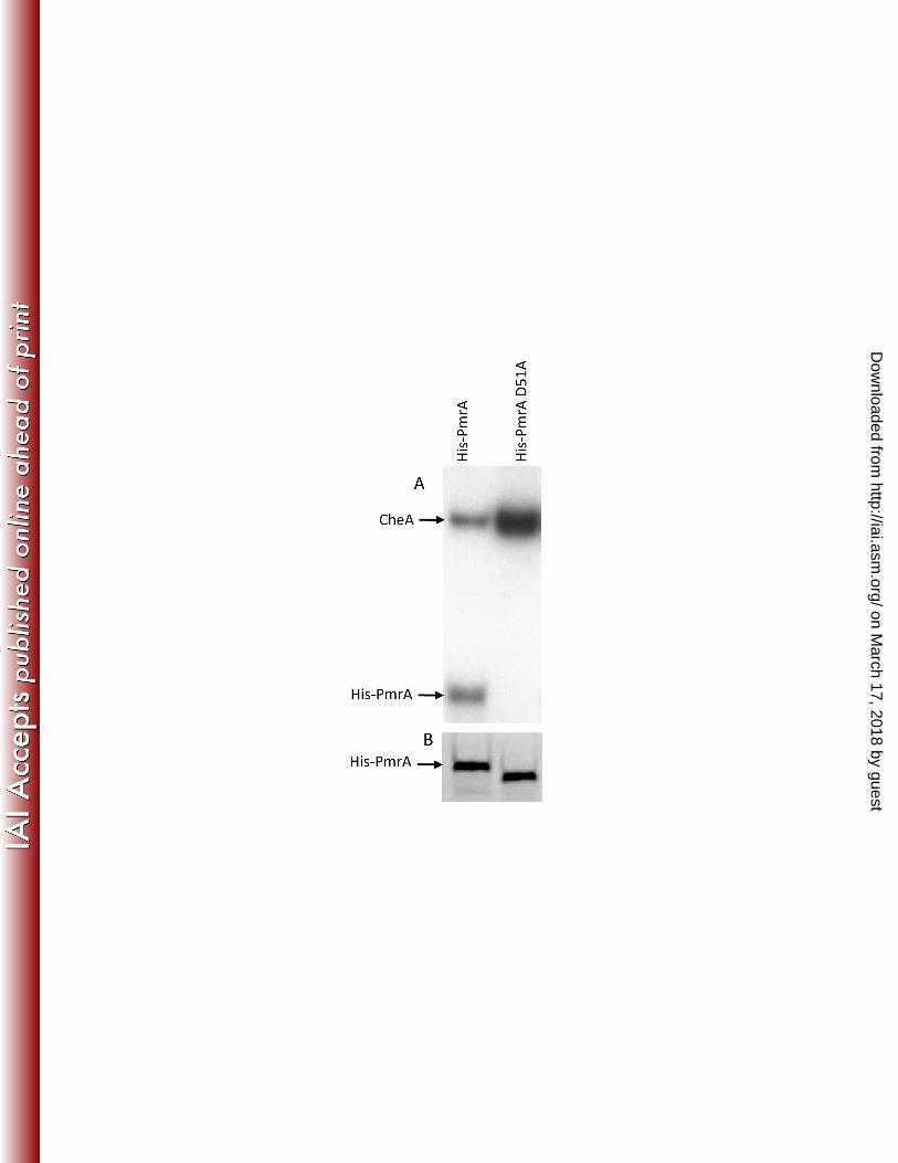
dATP. CheA was able to phosphorylate His-PmrA but not His-PmrA D51A (Figure 3A). A 259
duplicate SDS-PAGE gel stained for total protein showed equal amounts of His-PmrA and His-260
PmrA D51A, ruling out the possibility that PmrA D51A was degraded during the reaction (Figure 261
3B). It is important to note that His-PmrA D51A runs slightly faster on SDS-PAGE than His-262
PmrA even though the gene sequence is identical to pmrA except for the codon 51. In addition, 263
the His-tag is still present as the protein reacts with an anti-His antibody in Western blots (data 264
not shown). 265
on March 17, 2018 by guest
http://iai.asm.org/
Dow
nloaded from

Page 13 of 30
To determine if PmrA is phosphorylated by Francisella kinases, we prepared a membrane 266
fraction of F. novicida wild-type bacteria. This membrane fraction was incubated with His-PmrA 267
or His-PmrA D51A in the presence of [γ-32P] dATP. Phosphorylation was detected after 268
separation by SDS-PAGE. His-PmrA was phosphorylated while the mutated D51A protein was 269
not (Figure 4A, lanes 1 and 2). Duplicate reactions were run using unlabeled ATP and probed 270
for PmrA by Western blot to confirm that neither His-PmrA nor His-PmrA D51A were lost during 271
the reaction (Figure 4B, lanes 3 and 4). This data, combined with the results from the CheA 272
phosphorylation experiment, strongly suggests that the site of PmrA phosphorylation is 273
aspartate 51. 274
PmrA D51 is important for gene regulation. 275
If phosphorylation of PmrA increases binding to regulated promoters, it should have a positive 276
effect on the expression of regulated genes. Similarly, if the site of phosphorylation (D51) is 277
removed, then the expression of regulated gene promoters should decrease. To test if 278
phosphorylation of PmrA is important for expression of regulated genes, we performed qRT-279
PCR for iglC in F. novicida wild-type, F. novicida ∆pmrA (JSG2845), F. novicida ∆pmrA 280
complemented with pmrA carried on pKK214pgroEL (JSG2847), and F. novicida ∆pmrA 281
complemented with pmrA D51A on pKK 214pgroEL (JSG3033). Complemented strains 282
produce similar amounts of PmrA as the wild-type strain (data not shown). As expected, 283
expression of iglC was almost undetectable in the ∆pmrA (JSG2845) compared to wild-type and 284
transcript levels were restored in the ∆pmrA complemented strain (JSG2847). The ∆pmrA 285
strain complemented with PmrA D51A (JSG3033) had decreased expression of iglC compared 286
to wild-type and the strain complemented with native PmrA (JSG2847) (Figure 5). This data 287
indicates that expression of PmrA regulated genes is dependent upon the aspartate at position 288
51 and therefore, phosphorylation of PmrA is important for gene regulation. 289
on March 17, 2018 by guest
http://iai.asm.org/
Dow
nloaded from

Page 14 of 30
PmrA D51 is required for intramacrophage growth. 290
Host macrophages have been widely described as a site of replication for francisellae (8, 7, 9, 291
26). Previous data demonstrated that F. novicida ∆pmrA (JSG2845) is defective for replication 292
within macrophages (20). To determine the importance of phosphorylation of PmrA on 293
Francisella replication within macrophages, we infected PMA-induced THP-1 cells with F. 294
novicda wild-type, F. novicida ∆pmrA (JSG2845), F. novicida ∆pmrA complemented with native 295
PmrA (JSG2847), and F. novicida ∆pmrA complemented with pmrA D51A (JSG3033) (Table 1). 296
As expected, the wild-type and native complement strains were capable of replication within 297
THP-1 macrophages, increasing in number by more than two logs during 24 hours of infection. 298
Conversely the ∆pmrA strain and the strain complemented with the D51A mutant were largely 299
defective for replication within macrophages. Some replication was observed for the D51A 300
complemented strain, but at 24 hours post-infection the increase in CFU was less than one half 301
log (Figure 6A). These data indicate that PmrA aspartate 51 and likely phosphorylation of PmrA 302
is important for replication of F. novicida within macrophages. 303
PmrA D51 is required for mouse virulence. 304
Since PmrA D51 is required for intramacrophage survival and replication within macrophages is 305
closely tied to virulence, this residue may also be important for virulence. The lethality of F. 306
novicida ∆pmrA complemented with native PmrA (JSG2847), and F. novicida ∆pmrA 307
complemented with pmrA D51A (JSG3033) was compared. Groups of BALB/C mice were 308
infected via the intranasal route. F. novicida ∆pmrA complemented with pmrA (dose = 1000 309
CFU) resulted in 100% lethality within 4 days while no mice died after receiving 1000 CFU of the 310
pmrA mutant complemented with pmrA D51A. Another group of mice was infected with 106 311
pmrA D51A complemented bacteria. Eighty percent of these mice died within 10 days of 312
on March 17, 2018 by guest
http://iai.asm.org/
Dow
nloaded from
Page 15 of 30
infection (Figure 6B). Our previous report showed that the F. novicida ∆pmrA strain was 313
completely attenuated in mice at a dose of 108 bacteria delivered (20). Therefore, the pmrA 314
D51A complemented strain is attenuated but not to the extent of a strain that is completely 315
lacking PmrA. This data indicates that PmrA D51 and likely phosphorylation of PmrA is 316
important for F. novicida virulence. 317
PmrA is a primary target of the histidine kinase KdpD. 318
The aspartate at position 51 of PmrA is important for survival within macrophages and virulence 319
of F. novicida. Though the data suggests that this amino acid was the site of phosphotransfer 320
from CheA and F. novicida membrane extracts, the native Francisella kinase responsible for 321
phosphorylating PmrA at D51 has not been identified. Transposon mutants of the three putative 322
histidine kinase genes identified (http://go.francisella.org/cgi-bin/frangb/genomelist.cgi) in the F. 323
novicida genome: FTN1453 (JSG2890), FTN1617 (qseC, JSG2892), and FTN1715 (kdpD, 324
JSG2894) were used to attempt to identify which kinase is responsible for phosphorylation of 325
PmrA (10). Membrane fractions from these mutants were incubated with [γ-32P] dATP and 326
either His-PmrA or His-PmrA D51A. Phosphorylated proteins were separated on an SDS-327
PAGE gel and detected by autoradiography. Duplicate reactions were run without His-PmrA 328
and His-PmrA D51A as a control to identify phosphorylated proteins unrelated to PmrA. His-329
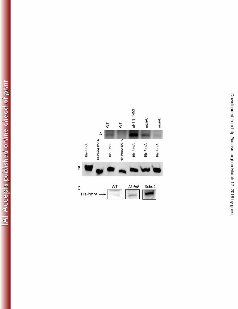
PmrA was phosphorylated to some degree by each of the membrane fractions. Densitometry 330
readings indicate that the FTN1453 mutant membrane fraction phosphorylated His-PmrA four 331
times as much as F. novicida wild-type membranes while the qseC mutant phosphorylated His-332
PmrA in a similar manner as wild-type. The kdpD mutant membrane preparations 333
phosphorylated His-PmrA only 20 percent of the level of wild-type membranes. This suggested 334
that the kinase primarily responsible for phosphorylation of PmrA was the histidine kinase KdpD 335
(Figure 4A, lanes 3-5). Duplicate reactions were run with cold ATP and analyzed by Western 336
on March 17, 2018 by guest
http://iai.asm.org/
Dow
nloaded from

Page 16 of 30
blot with anti-PmrA anti-sera to confirm that neither His-PmrA nor His-PmrA D51A were lost 337
during the reaction and that equal amounts of target proteins were present (Figure 4B, lanes 5-338
7). In addition, we purified the predicted C-terminal cytoplasmic portion (residues 499-893) of 339
KdpD as a His-tagged protein and examined its ability to mediate phosphotransfer with PmrA. 340
Phosphotransfer was observed, demonstrating that KdpD can phosphorylate PmrA (data not 341
shown). The F. novicida strain containing a transposon mutation in kdpD is also deficient for 342
intramacrophage survival (Figure 6), thus demonstrating the expected similarly to the pmrA null 343
mutation strain. 344
Further phosphorylation studies were conducted to determine the relative amount of PmrA 345
phosphorylation in different Francisella strains. Although there are no typical tandemly arranged 346
genes encoding two-component systems in human virulent francisellae, there is one in F. 347
novicida comprised of KdpD and KdpE. We hypothesized that in the absence of KdpE, 348
phosphorylation of PmrA by KdpD would be increased. Indeed, using a membrane fraction from 349
a KpdE (FTN_1714) transposon mutant (10), His-PmrA was phosphorylated 14-times more than 350
by F. novicida wild-type (Figure 4C). The fully virulent F. tularensis Schu4 strain does not have 351
homologs to either kdpE or FTN1453; therefore, we hypothesized that phosphorylation of PmrA 352
would be higher in this strain. When His-PmrA is incubated with a F. tularensis Schu4 353
membrane preparation in the presence of radiolabeled ATP, it was phosphorylated 354
approximately 50 times more compared to that observed with F. novicida wild-type membranes 355
(Figure 4C). 356
PmrA co-precipitates with MglA and SspA. 357
PmrA, MglA, SspA, MigR and FevR are required for Francisella intramacrophage survival and 358
regulate the FPI (2, 6, 1, 15, 4). FevR has poor homology to DNA binding proteins and appears 359
to act in a manner not involving direct promoter interaction (2). Recently, FevR (PigR) has also 360
on March 17, 2018 by guest
http://iai.asm.org/
Dow
nloaded from

Page 17 of 30
been demonstrated to interact with MglA and SspA, though there is conflicting data in the 361
literature regarding this (2, 5). One hypothesis to explain the involvement of PmrA, MglA, and 362
SspA is that PmrA binds to regulated promoters and interacts with MglA and/or SspA, which in 363
turn recruits RNA polymerase to the site (or interacts with MglA and/or SspA bound to RNA 364
polymerase), initiating transcription of regulated genes. For our model to be correct, this would 365
require PmrA to physically interact with MglA and/or SspA. We showed earlier that adding MglA 366
and PmrA together did not result in a supershift, which would have been an indication of an 367
interaction between the two proteins (Figure 2). This in vitro assay does not contain many of 368
the components that would be present in vivo (e.g. SspA and RNA polymerase), so we 369
attempted to co-precipitate PmrA with MglA and SspA from whole cell lysates. Soluble fractions 370
were prepared from lysates of F. novicida ∆pmrA (JSG2845), F. novicida ∆mglA (JSG2250), 371
and F. novicida His-SspA (JSG2970). His-PmrA was added to the soluble fraction of the F. 372
novicida ∆pmrA (Figure 7, lane 1). Once precipitated with His-bind resin, PmrA and associated 373
proteins were detected by Western blotting. PmrA was detected by an anti-PmrA anti-sera 374
(Figure 7A, lane 1) and by an anti-His antibody (Figure 7C, lane 1). MglA was detected in the 375
PmrA precipitated sample by anti-MglA anti-sera (Figure 7B, lane 1), demonstrating that MglA 376
co-precipitates with PmrA. Similarly, His-MglA was added to the soluble fraction from F. 377
novicida ∆mglA. Precipitation with His-bind resin allowed detection of MglA using an anti-MglA 378
anti-sera (Figure 7B, lane 2) and the anti-His antibody (Figure 7C, lane 2). PmrA precipitated 379
with MglA and was detected with anti-PmrA anti-sera (Figure 7A, lane 2). This confirms that 380
PmrA and MglA co-precipitate. Finally, SspA was precipitated from the soluble fraction of F. 381
novicida His-SspA and detected with the anti-His antibody (Figure 7C, lane 3). PmrA was 382
present in the SspA precipitated sample (Figure 7A, lane 3), as was MglA (Figure 7B, lane 3). 383
Using the identical protocol without adding His-tagged proteins did not result in 384
immunoprecipitation of PmrA, MglA or SspA (data not shown). Additionally, adding purified His-385
on March 17, 2018 by guest
http://iai.asm.org/
Dow
nloaded from

Page 18 of 30
tagged Salmonella PhoP to a F. novicida ∆pmrA whole cell soluble fraction and precipitating 386
using His-bind resin did not co-precipitate MglA or SspA (data not shown). The interaction of 387
PmrA, MglA, and SspA does not appear to be dependent on DNA because the proteins co-388
precipitate in the presence of DNase (data not shown). These data indicate that PmrA, MglA 389
and SspA are a part of the same protein complex. 390
Discussion 391
F. tularensis is an intracellular pathogen whose virulence is closely tied to the in vivo expression 392
of the genes encoded within a pathogenicity island. Though the regulation of this island is 393
poorly understood, it is clear that the FPI is important for escape from the phagosome and 394
replication within macrophages. PmrA regulates the FPI and is required for intramacrophage 395
survival and virulence (20). MglA and SspA are also regulators of the FPI and required for 396
virulence and FPI gene transcription by physically interacting with the RNA polymerase (6). The 397
data presented here shows that PmrA is a DNA binding protein whose interaction with DNA is 398
enhanced by Asp51 phosphorylation mediated primarily by the histidine kinase, KdpD. This 399
aspartate residue is required for phosphorylation of PmrA, normal replication within 400
macrophages, and full virulence. Furthermore, PmrA co-precipitates with MglA and SspA from 401
whole cell lysates. 402
Primer extension was used to determine the start of transcription for two PmrA regulated genes: 403
pmrA and pdpD. The pdpD gene is regulated by PmrA, MglA, and SspA; however, pmrA is 404
positively auto-regulated by PmrA but not affected by MglA or SspA. The start of transcription 405
for pdpD was identified as the thymine 32 bases upstream of its ATG. This identifies the 406
promoter to be located in the 101 base pair intergenic region between pdpD and FTN1326. Due 407
to the lack of significant intergenic spaces, it is likely that pdpD is the lead gene of the putative 408
operon including iglABCD. The identical 101 bases are upstream of pdpD in the fully virulent F. 409
on March 17, 2018 by guest
http://iai.asm.org/
Dow
nloaded from

Page 19 of 30
tularensis Schu4 strain, indicating the start of transcription and the promoter are the same for 410
both species. The start of transcription of pmrA is located 40 bases upstream of its ATG. There 411
is a 441 nucleotide intergenic region upstream of pmrA to the end of FTN1466, the first 324 412
bases of are identical in F. tularensis Schu4, suggesting that the pmrA promoter is conserved. 413
The pmrA operon contains five genes consisting of pmrA, lepB, rnc, truB, and rnr (24). To our 414
knowledge this is the first detailed characterization of a regulated promoter in Francisella. 415
His-PmrA binds to the promoter fragment of both pmrA and pdpD, and treating His-PmrA with 416
acetyl phosphate increased binding to both fragments. The pmrA promoter region required 417
approximately 45 nanomoles of purified protein to observe a shift in labeled DNA, while the 418
pdpD promoter region required a greater amount (~80 nanomoles) of protein to visualize 419
binding. This indicates that PmrA has a greater affinity for its own promoter than it does for the 420
promoter of pdpD, which may explain why MglA and SspA are not required for pmrA activation 421
(20, 6, 3). Perhaps this increased affinity obviates the need for interaction with MglA/SspA, 422
which may be required to recruit RNA polymerase to lower affinity promoters. EMSA 423
experiments performed with PmrA homologs have used similar amounts of protein to shift 424
labeled promoter fragments (28). From scanning the fragments and the likely binding locations 425
on these fragments, we were unable to identify a consensus binding site. The identification and 426
characterization of additional promoters will undoubtedly aid the defining of such a sequence. 427
Further complicating the issue was our finding that smaller, overlapping fragments of the shifted 428
promoters did not bind His-PmrA, suggesting the involvement of multiple, non-tandem binding 429
sites (Figure S3). We also tested the ability of His-MglA to bind to these same promoter 430
fragments, but no binding was observed with as much as 320 nanomoles of purified protein. A 431
supershift was not observed when both purified His-MglA and His-PmrA were used together in 432
these assays. This suggested either that PmrA and MglA do not physically interact or that their 433
on March 17, 2018 by guest
http://iai.asm.org/
Dow
nloaded from

Page 20 of 30
physical interaction is dependent upon other molecules (e.g. SspA, RNA polymerase or FevR). 434
Based on other data gained here and discussed below, we favor the latter hypothesis. 435
By amino acid alignment with known response regulators from other Gram negative bacteria, 436
the PmrA phosphorylation site was predicted to be D51. A His-PmrA D51A mutant protein was 437
constructed to test this prediction. We demonstrated that His-PmrA but not His-PmrA D51A is 438
phosphorylated by the enteric histidine kinase CheA and F. novicida wild-type membrane 439
fractions. The His-PmrA D51A mutant still bound DNA, demonstrating that the substitution did 440
not result in a non-functional protein. However, from our analysis, it is not known if PmrA and 441
PmrA D51A bind to the same promoter DNA sequence or if the binding of His-PmrA D51A is 442
specific. Regardless, His-PmrA D51A binding to promoter regions does not productively 443
stimulate gene transcription (Figure 5). The role of the putative histidine kinases in the 444
Francisella genome in phosphorylating PmrA was also investigated. This data indicated that 445
KdpD is the histidine kinase primarily responsible for phosphorylating PmrA; however, there 446
appeared to be some cross talk or target promiscuity as His-PmrA was phosphorylated to a 447
small degree in the absence of KdpD. In enterohemorrhagic E. coli, the sensor kinase QseC 448
has been shown to phosphorylate KdpE, demonstrating communication between the Kdp and 449
the Qse TCS (12). A kdpD transposon mutant, like a strain carrying a mutation in its 450
downstream target PmrA, is deficient for replication within macrophages (Figure 6A). In 451
addition, also like PmrA, microarray analysis comparing wild-type bacteria to the kdpD mutant 452
indicates that KdpD regulates the PmrA operon and the FPI (data not shown) and KdpD is a 453
virulence determinant uncovered using a in vivo negative selection screen (29). Membrane 454
fractions missing the putative sensor kinase FTN1453 resulted in increased PmrA 455
phosphorylation when compared to wild-type, suggesting that it regulated, or acted itself as a 456
phosphatase. Interestingly, F. tularensis Schu4 has no FTN1453 homolog. 457
on March 17, 2018 by guest
http://iai.asm.org/
Dow
nloaded from

Page 21 of 30
Growth of a pmrA null strain complemented with pmrA D51A within macrophages was 458
diminished as compared to a strain complemented with native pmrA; however, some replication 459
of the pmrA D51A complemented strain was observed as compared to a pmrA null mutant. 460
Similarly, the PmrA D51A complemented strain was more virulent in the mouse model than 461
strains devoid of PmrA. This is likely a result of PmrA D51A overexpression, such that 462
increased copy number coupled with the residual PmrA D51A DNA binding resulted in modest 463
expression of the PmrA regulon, resulting in some replication within macrophages and retention 464
of virulence. 465
Co-immunoprecipitation experiments reported here showed that PmrA, MglA, and SspA are a 466
part of the same protein complex. In addition to the F. novicida data presented, PmrA also co-467
precipitates with MglA and SspA from a F. tularensis Schu4 whole cell soluble fraction (data not 468
shown). This was predicted as the amino acid sequence of MglA and SspA are nearly identical 469
between F. novicida and F. tularensis Schu4, and the PmrA protein sequence is identical 470
between these subspecies. The data presented, however, does not indicate whether PmrA 471
binds to MglA, SspA, or to both proteins. We predict RNA polymerase is a part of this complex; 472
however, other binding partners/ complex components may also exist. We did not examine the 473
interaction of FevR (PigR) with PmrA as there is conflicting data in the literature concerning the 474
likelihood of its function as a DNA binding protein or its ability to interact with MglA/SspA. The 475
most recent data do suggest a physical interaction of FevR (PigR) with MglA/SspA that is 476
facilitated by the alarmone ppGpp, and thus present potential new interacting partners. The 477
sequence of protein binding for initiating transcription is also unclear. PmrA does bind 478
promoters in the absence of MglA and SspA, but the EMSA data suggest that MglA will not 479
interact with unbound or DNA bound PmrA in vitro without the presence of other factors. MglA 480
may need to interact with SspA before it can bind PmrA, or Francisella RNA polymerase may 481
need to interact with MglA and/ or SspA before binding to PmrA. 482
on March 17, 2018 by guest
http://iai.asm.org/
Dow
nloaded from

Page 22 of 30
The response regulator PmrA is the first characterized DNA binding protein in Francisella. 483
Phosphorylation of PmrA enhances promoter binding, affecting gene regulation. PmrA is 484
phosphorylated at Asp51 primarily by the putative histidine kinase KdpD, forming the first TCS 485
described for Francisella species. The KdpD/PmrA TCS system plays an important role in 486
regulating the FPI, affecting replication in macrophages and virulence. Phosphorylation of 487
PmrA is complicated in F. novicida by the presence of a secondary KdpD target, KdpE, and an 488
additional kinase FTN1453, both which negatively affect the phosphorylation state of PmrA. 489
Due likely to the absence of FTN1453 and KdpE in the fully virulent F. tularensis Schu4 strain, 490
phosphorylation of PmrA was increased, which may result in comparatively higher expression of 491
the PmrA regulon. Further investigation into the protein-protein interactions of factors at FPI 492
promoters, as well as PmrA-regulated expression of FPI genes in F. tularensis, will provide 493
further insight into the molecular pathogenesis of tularemia. 494
Acknowledgements 495
We thank Dr. Daniel Wozniak for providing purified CheA and Dr. Denise Monack of Stanford 496
University for generously providing the F. novicida His-sspA strain. Mike Zianni and the Plant 497
Microbe Genetics Facility at The Ohio State University were instrumental in performance of the 498
primer extension experiments. This work was supported by funding from The Region V “Great 499
Lakes” Regional Center of Excellence in Biodefense and Emerging Infectious Diseases 500
Consortium (NIH award 1-U54-AI-057153) and by NIH/NIAID award 1-T32-AI-065411, a NRSA 501
training grant administered by the Center for Microbial Interface Biology, at The Ohio State 502
University. 503
Reference List 504
505 1. Baron, G.S., and F.E. Nano. 1998. MglA and MglB are required for the intramacrophage 506
growth of Francisella novicida. Mol. Microbiol. 29: 247-259. 507
on March 17, 2018 by guest
http://iai.asm.org/
Dow
nloaded from

Page 23 of 30
2. Brotcke, A., and D.M. Monack. 2008. Identification of fevR, a novel regulator of virulence 508 gene expression in Francisella novicida. Infect. Immun. 76: 3473-3480. 509
3. Brotcke, A., D.S. Weiss, C.C. Kim, P. Chain, S. Malfatti, E. Garcia, and D.M. Monack. 510 2006. Identification of MglA-regulated genes reveals novel virulence factors in Francisella 511 tularensis. Infect. Immun. 74: 6642-6655. 512
4. Buchan, B.W., R.L. McCaffrey, S.R. Lindemann, L.A. Allen, and B.D. Jones. 2009. 513 Identification of migR, a regulatory element of the Francisella tularensis Live Vaccine 514 Strain iglABCD virulence operon required for normal replication and trafficking in 515 macrophages. Infect Immun 77: 2517-2529. 516
5. Charity, J.C., L.T. Blalock, M.M. Costante-Hamm, D.L. Kasper, S.L. Dove. 2009. 517 Small molecule control of virulence gene expression in Francisella tularensis. PLoS 518 Pathog 10: e1000641. 519
6. Charity, J.C., M.M. Costante-Hamm, E.L. Balon, D.H. Boyd, E.J. Rubin, and S.L. 520 Dove. 2007. Twin RNA polymerase-associated proteins control virulence gene expression 521 in Francisella tularensis. PLoS Pathog. 3: e84. 522
7. Clemens, D.L., B.Y. Lee, B, and M.A. Horwitz. 2005. Francisella tularensis enters 523 macrophages via a novel process involving pseudopod loops. Infect. Immun. 73: 5892-524 5902. 525
8. Ellis, J., P.C. Oyston, M. Green, and R.W. Titball. 2002. Tularemia. Clin. Microbiol. Rev. 526 15: 631-646. 527
9. Fortier, A.H., S.J. Green, T. Polsinelli, T.R. Jones, R.M. Crawford, D.A. Leiby, K.L 528 Elkins, M.S. Meltzer, and C.A. Nacy. 1994. Life and death of an intracellular pathogen: 529 Francisella tularensis and the macrophage. Immunol. Ser. 60: 349-361. 530
10. Gallagher, L.A., E. Ramage, M.A. Jacobs, R. Kaul, M. Brittnacher, and C. Manoil. 531 2007. A comprehensive transposon mutant library of Francisella novicida, a bioweapon 532 surrogate. Proc. Natl. Acad. Sci. U. S. A. 104: 1009-1014. 533
11. Gavrilin, M. A., I.J. Bouakl, N.L. Knatz, M.D. Duncan, M.W. Hall, J.S. Gunn, and M.D. 534 Wewers. 2006. Proc. Natl. Acad. Sci. U. S. A. 103: 141-146. 535
12. Hughes, D.T., M.B . Clarke, K. Yamamoto, D.A. Rasko, and V. Sperandio. 2009. The 536 QseC adrenergic signaling cascade in Enterohemorrhagic E. coli (EHEC). PLoS Pathog. 537 5: e1000553. 538
on March 17, 2018 by guest
http://iai.asm.org/
Dow
nloaded from
Page 24 of 30
13. Keim, P., A. Johansson, and D.M. Wagner. 2007. Molecular epidemiology, evolution, 539 and ecology of Francisella. Ann. N. Y. Acad. Sci. 1105: 30-66. 540
14. Lai, X.H., I. Golovliov, and A. Sjostedt. 2004. Expression of IglC is necessary for 541 intracellular growth and induction of apoptosis in murine macrophages by Francisella 542 tularensis. Microb. Pathog. 37: 225-230. 543
15. Lauriano, C.M., J.R. Barker, S.S. Yoon, F.E. Nano, B.P. Arulanandam, D.J. Hassett, 544 and K.E. Klose. 2004. MglA regulates transcription of virulence factors necessary for 545 Francisella tularensis intraamoebae and intramacrophage survival. Proc. Natl. Acad. Sci. 546 U. S. A. 101: 4246-4249. 547
16. Li, L.A., M.R. Zianni, and F.R. Tabita. 1999. Inactivation of the monocistronic rca gene in 548 Anabaena variabilis suggests a physiological ribulose bisphosphate 549 carboxylase/oxygenase activase-like function in heterocystous cyanobacteria. Plant Mol. 550 Biol. 40: 467-478. 551
17. Ludu, J.S., O.M. de Bruin, B.N. Duplantis, C.L Schmerk, A.Y. Chou, K.L Elkins, and 552 F.E. Nano. 2008. The Francisella Pathogenicity Island Protein PdpD is required for full 553 virulence and associates with homologues of the type VI secretion system. J. Bacteriol. 554 190: 4584-4595. 555
18. McCleary, W.R., and J.B. Stock. 1994. Acetyl phosphate and the activation of two-556 component response regulators. J. Biol. Chem. 269: 31567-31572. 557
19. Meibom, K.L., A.L. Forslund, K. Kuoppa, K. Alkhuder, I. Dubail, M. Dupuis, A. 558 Forsberg, A. Charbit. 2009. Hfq, a novel pleiotropic regulator of virulence associated 559 genes in Francisella tularensis. Infect. Immun. 77(5): 1866-1880. 560
20. Mohapatra, N.P., S. Soni, B.L. Bell, R. Warren, R.K. Ernst, A. Muszynski, R.W. 561 Carlson, and J.S. Gunn. 2007. Identification of an orphan response regulator required for 562 the virulence of Francisella spp. and transcription of pathogenicity island genes. Infect. 563 Immun. 75: 3305-3314. 564
21. Nano, F.E., N. Zhang, S.C. Cowley, K.E. Klose, K.K. Cheung, M.J. Roberts, J.S. Ludu, 565 G.W. Letendre, A.I. Meierovics, G. Stephens, and K.L Elkins. 2004. A Francisella 566 tularensis pathogenicity island required for intramacrophage growth. J. Bacteriol. 186: 567 6430-6436. 568
22. Nigrovic, L.E., and S.L. Wingerter. 2008. Tularemia. Infect. Dis. Clin. North Am. 22: 489-569 504, ix. 570
on March 17, 2018 by guest
http://iai.asm.org/
Dow
nloaded from

Page 25 of 30
23. Oyston, P.C., A. Sjostedt, and R.W. Titball. 2004. Tularaemia: bioterrorism defence 571 renews interest in Francisella tularensis. Nat. Rev. Microbiol. 2: 967-978. 572
24. Sammons-Jackson, W.L., K. McClelland, J.N. Manch-Citron, D.W. Metzger, C.S. 573 Bakshi, E. Garcia, A. Rasley, and B.E. Anderson. 2008. Generation and 574 characterization of an attenuated mutant in a response regulator gene of Francisella 575 tularensis live vaccine strain (LVS). DNA Cell. Biol. 27: 387-403. 576
25. Santic, M., M. Molmeret, K.E. Klose, and Y. Abu Kwaik. 2006. Francisella tularensis 577 travels a novel, twisted road within macrophages. Trends Microbiol. 14: 37-44. 578
26. Sjostedt, A. 2006. Intracellular survival mechanisms of Francisella tularensis, a stealth 579 pathogen. Microbes Infect. 8: 561-567. 580
27. Stock, A.M., V.L. Robinson, and P.N. Goudreau. 2000 Two-component signal 581 transduction. Annu. Rev. Biochem. 69: 183-215. 582
28. Tamayo, R., A.M. Prouty, and J.S. Gunn. 2005. Identification and functional analysis of 583 Salmonella enterica serovar Typhimurium PmrA-regulated genes. FEMS Immunol. Med. 584 Microbiol. 43: 249-258. 585
29. Weiss, D.S., A. Brotcke, T. Henry, J.J. Margolis, K. Chan, and D.M. Monack. 2007. In 586 vivo negative selection screen identifies genes required for Francisella virulence. Proc. 587 Natl. Acad. Sci. U. S. A. 104: 6037-6042. 588
30. Whitchurch, C.B., T.E. Erova, J.A. Emery, J.L Sargent, J.M. Harris, A.B. Semmler, 589 M.D. Young, J.S. Mattick, and D.J. Wozniak. 2002. Phosphorylation of the 590 Pseudomonas aeruginosa response regulator AlgR is essential for type IV fimbria-591 mediated twitching motility. J. Bacteriol. 184: 4544-4554. 592
on March 17, 2018 by guest
http://iai.asm.org/
Dow
nloaded from

Page 26 of 30
593
Table 1. Primers and strains used in this study.
Primer Sequence Description
JG1071 CGGGATCCGAGGATACAATCTTGCTTTTATACAC mglA forward primer BamHI
JG1072 AACTGCAGTTAAGCTCCTTTTGCTTTGATAGT mglA reverse primer PstI
JG1289 ATGATTATGAGTGAGATGATAACAAG iglC qRT-PCR forward primer
JG1290 CTATGCAGCTGCAATATATCC iglC qRT-PCR reverse primer
JG1412 GTATGATATAGTCGTCTTAGCCATTGGTATGCCAATAAAAAC
PmrA site directed mutant
Asp51 to Ala51 from GAT to
GCC forward primer
JG1413 TAAGACGACTATATCATACAATCCAGATTCTATAAAAGTTTGCGC
PmrA site directed mutant D51
to A51 from GAT to GCC
reverse primer
JG1462 ATATCTTATGGGCTTGGGCGAT pmrA primer extension PCR
forward primer
JG1463 ATAAAAGTTTGCGCTGCCTCACCA pmrA primer extension/ gel shift
reverse primer
JG1508 AAAAGTTTGATGTAACTTTAGAAAACATTTTCA pmrA gel shift forward primer
JG1510 GCAACCGGAGCAAAAAGTAG pdpD gel shift forward primer
JG1511 GAGGTCATCAGTATCATATAATAAATCGTT pdpD gel shift reverse primer
JG1514 FAM -- GCCTTCACCCAAATGAAGATC pmrA primer extension FAM
primer
JG1515 FAM -- TAGCCATGACATCCATCGTTT pdpD primer extension FAM
primer
JG2079 cgggatcccgGAGCTTTTACACTTCTAAAAGAACGAG kdpD C-terminal fragment
forward primer
JG2080 ggggtaccccGTGATCACTTGATAGCTAATTTGTAC kdpD C-terminal fragment
reverse primer
Strain Genotype and/ or relative phenotype Reference or source
JSG1819 F. novicida-U112 ATCC
JSG2250 F. novicida mglA::Erm (15)
JSG2845 JSG1819 with ∆pmrA::Kan (20)
JSG2846 E. coli DH5α with Francisella pmrA carried on pkk214pgroEL (20)
JSG2847 JSG2845 complemented with pmrA carried on pKK214pgroEL (20)
on March 17, 2018 by guest
http://iai.asm.org/
Dow
nloaded from

Page 27 of 30
JSG2894 F. novicida transposon T20 insertion at FTN1715 (kdpD) (10)
JSG3033 JSG2845 complemented with pmrA D51A carried on pKK214pgroEL This work.
JSG2970 sspA tagged with His and inserted in the chromosome of F. novicida
(single copy expression) (3)
JSG2890 F. novicida transposon T20 insertion at FTN1453 (10)
JSG2892 F. novicida transposon T20 insertion at FTN1617 (qseC) (10)
JSG2894 F. novicida transposon T20 insertion at FTN1715 (kdpD) (10)
JSG2895 F. novicida transposon T20 insertion at FTN1714 (kdpE) (10)
E. coli JM109 endA1, recA1, gyrA96, thi, hsdR17 (rk–, mk
+), relA1, supE44, ∆( lac-
proAB), [F´ traD36, proAB, laqIqZ∆M15]
Promega
JSG3031 E. coli JM109(pQE30[mglA]) This work.
JSG3032 E. coli XL-1 Blue MRF(pQE30XA[pmrA]) (20)
FT4 Francisella tularensis Schu4 BEI Resources via Dr. Rick
Lyons
on March 17, 2018 by guest
http://iai.asm.org/
Dow
nloaded from
Page 28 of 30
Figure 1. EMSA: PmrA Binding. Mobility shift assays were used to determine if purified 594
proteins bound to regulated gene promoters. Panel A: His-PmrA binds to the pmrA (12 ng 595
lanes 1-6) and pdpD (12 ng lanes 7-12) promoters in a dose dependent manner. Panel B: His-596
PmrA binding to the pmrA promoter is affected by specific but not non-specific competitor DNA. 597
Each lane contains 12 ng of labeled pmrA promoter. Panel C. His-PmrA binding to the pdpD 598
promoter is affected by specific but not non-specific competitor DNA. Each lane contains 12 ng 599
of labeled pdpD promoter. 600
601
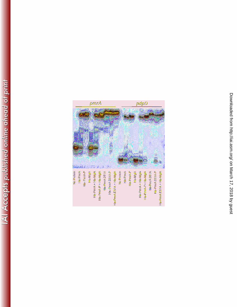
Figure 2. EMSA: PmrA Phosphorylation. Phosphorylating His-PmrA increases binding to 602
regulated promoters. His-MglA does not bind to either the pdpD or the pmrA promoters, the 603
combined addition of His-PmrA and His-MglA to promoters does not result in a supershift, and 604
His-PmrA D51A retains its ability to bind regulated promoters. Lanes 1-9 = 12 ng of labeled 605
pmrA promoter and 1 µg of each indicated protein. Lanes 10-18 = 12 ng of labeled pdpD 606
promoter and 150 nanomoles of each indicated protein. –P indicates the protein was treated 607
with acetyl phosphate. 608
609
Figure 3. Phosphotransfer from CheA. Panel A: PmrA but not PmrA D51A is 610
phosphorylated by the enteric histidine kinase CheA. Each lane consists of 0.1 ng 611
phosphorylated CheA and 1 µg His-PmrA or His-PmrA D51A. Panel B: Duplicate reactions 612
were stained for total protein to demonstrate the presence of His-PmrA and His-PmrA D51A 613
throughout the reaction. 614
615
on March 17, 2018 by guest
http://iai.asm.org/
Dow
nloaded from

Page 29 of 30
Figure 4. Phosphotransfer from membrane fractions. Panel A: Francisella membrane 616
fractions phosphorylate His-PmrA but not His-PmrA D51A, and the putative histidine kinase 617
KdpD is primarily responsible for phosphorylating His-PmrA. Each lane consists of 5 µg of 618
membrane fractions (lane designations above Panel A) and 1 µg His-PmrA or His-PmrA D51A 619
(lane designations directly above Panel B). Panel B. Western blot analysis detecting PmrA in 620
duplicate phosphotransfer reactions. Panel C. His-PmrA shows enhanced phosphorylation by 621
the ∆kdpE mutant and F. tularensis Schu4 membrane fractions. 622
623
Figure 5. RT-PCR analysis of iglC in F. novicida ∆pmrA complemented with PmrA or 624
PmrA D51A. Expression of iglC, which is within the PmrA regulated FPI, is significantly 625
reduced in an F. novicida ∆pmrA strain compared to wild-type bacteria. Complementation of F. 626
novicida ∆pmrA with PmrA results in restored expression of iglC while complementation with 627
PmrA D51A failed to restore iglC expression. * = p<0.05 628
629
Figure 6. PmrA D51A is important for intramacrophage replication and mouse virulence. 630
Panel A: PmrA D51 is required for replication within PMA-induced (10 ng/ml) THP-1 cells at a 631
multiplicity of infection of 50:1. Closed circles = F. novicida wild-type. Open circles = F. 632
novicida ∆pmrA. Open squares = F. novicida ∆pmrA pKK214pgroEL [pmrA]. Closed squares = 633
F. novicida ∆pmrA pKK214pgroEL [pmrA D51A]. Closed triangles = F. novicida ∆kdpD. Panel 634
B: PmrA D51 is required for maximal virulence of F. novicida in mice. Bacteria were given 635
intranasally to groups of five anesthetized female 4 to 6 week-old BALB/c mice at a dose of 1 x 636
103 or 1 x 10⁶ CFU. Closed circles = 1 x 10³ F. novicida ∆pmrA pKK214pgroEL [pmrA]. Closed 637
on March 17, 2018 by guest
http://iai.asm.org/
Dow
nloaded from

Page 30 of 30
squares with solid line = 1 x 10³ F. novicida ∆pmrA pKK214pgroEL [pmrA D51A]. Closed 638
squares with dashed line = 1 x 10⁶ F. novicida ∆pmrA pKK214pgroEL [pmrA D51A]. 639
640
Figure 7. Co-precipitation. PmrA, MglA and SspA co-precipitate. Western blot analysis of 641
soluble lysates to which His-PmrA or His-MglA were added and precipitated with His-Bind resin. 642
Panel A = anti-PmrA sera. Panel B = anti-MglA sera. Panel C = anti-His monoclonal antibody. 643
Lane 1 = His-PmrA added to an F. novicida ∆pmrA soluble fraction and precipitated with His-644
bind resin. Lane 2 = His-MglA added to an F. novicida ∆mglA soluble fraction and precipitated 645
with His-bind resin. Lane 3 = His-SspA from F. novicida with a chromosomal encoded His-646
tagged sspA precipitated with His-bind resin. 647
on March 17, 2018 by guest
http://iai.asm.org/
Dow
nloaded from