Paediatric Intensive Care Surge Standard Operating Procedure

Page 1 of 16 Title: Hypoplastic Left Heart Syndrome V: 1 Approved by: PICU Clinical Practice Group: April 2021 Next Review: April 2024 Trust Ref No: C16/2021 NB: Paper copies of this document may not be most recent version. The definitive version is held on INsite in the Policies and Guidelines Library
Paediatric Intensive Care Unit
Hypoplastic Left Heart Syndrome
Staff relevant to: PICU
Approval date: April 2021
Version: 1
Revision due: April 2024
Written by: S Neshat
Trust Ref: C16/2021
Contents
1. Introduction and Who Guideline applies to ...................................................................................... 1 2. Guideline Standards and Procedures .............................................................................................. 2
Preoperative management: ............................................................................................................. 3 Surgical Palliation ........................................................................................................................... 5 Post-Operative Management Strategies .......................................................................................... 7 Intervention by system .................................................................................................................... 8 Low cardiac out algorithm: cardiogenic shock ............................................................................... 10 Management strategies: ................................................................................................................ 10 Factors contributing to hypoxaemia and ventilator dependence after stage 1 palliation ................. 11 Signs of LCOS in patient’s status-post Norwood procedure .......................................................... 12 Important points for chest closure management ............................................................................ 13 Prognosis & major complications .................................................................................................. 13 Transfer to ward criteria ................................................................................................................ 14
3. Education and Training ................................................................................................................. 14 4. Monitoring Compliance ................................................................................................................. 14 5. Supporting References ................................................................................................................. 14 6. Key Words .................................................................................................................................... 15 Contact & review details.................................................................................................................... 15 Appendix 1 Post –Operative Norwood/Sano Management Algorithm ................................................ 16
1. Introduction and Who Guideline applies to
This guideline primarily concentrates on univentricular physiology of Hypoplastic Left Heart Syndrome (HLHS) on PICU. For the perinatal management of newborns with prenatally diagnosed duct dependent single ventricle circulation, please refer to Single Ventricle UHL Childrens Medical Guideline

Page 2 of 16 Title: Hypoplastic Left Heart Syndrome V: 1 Approved by: PICU Clinical Practice Group: April 2021 Next Review: April 2024 Trust Ref No: C16/2021 NB: Paper copies of this document may not be most recent version. The definitive version is held on INsite in the Policies and Guidelines Library
Related documents: Blalock-Taussig (BT) Shunt or Central Shunt UHL Paediatric Intensive Care Guideline The term hypoplastic left heart syndrome, initially proposed by Noonan and Nadas in 1958, describes
a spectrum of congenital cardiac abnormalities characterized by a functional single right ventricle with
marked hypoplasia of the left ventricle and some degree of hypoplasia of the ascending aorta or arch.
The aortic and mitral valves are atretic, hypoplastic, or stenotic. The right ventricle provides systemic
blood flow by a right-to-left shunt via a patent ductus arteriosus. A wide-open ductus arteriosus and
complete mixing of the venous returns at the atrial level are essential for postnatal survival.
HLHS represents 2–9% of congenital heart disease cases and accounts for 23% of neonatal deaths
from congenital heart malformations and remain, despite the vast progress in HLHS treatment over
the recent years, the highest risk and costliest group of lesions among congenital heart defects.
HLHS is more common in males than in females, with a 55-70% male predominance.
2. Guideline Standards and Procedures
The postnatal circulation in hypoplastic left heart syndrome depends on a delicate balance between the pulmonary and systemic blood flow in order to ensure adequate oxygenation and tissue perfusion. The systemic blood flow is dependent on the overall cardiac output from the functional single ventricle and the relative pulmonary and systemic vascular resistances. Blood flow is inversely proportional to resistance (Ohm’s law); thus whenever the resistance in blood vessels decreases, blood flow through those vessels increases. Following birth, as pulmonary vascular resistance decreases, a higher percentage of the fixed right ventricular output flows to the lungs instead of to the systemic circulation. This results in higher oxygen saturation on the cost of poor perfusion and metabolic acidosis and oliguria.
Coronary artery and cerebral perfusion also depends on blood flow through the ductus arteriosus and
then retrograde aortic arch and ascending aortic flow. Therefore, increased pulmonary blood flow
results in decreased flow to the coronary arteries and brain, with a risk of myocardial or cerebral
ischemia.
The term HLHS includes:
Hypoplastic or small left ventricle, usually not forming the apex of the heart.
Aorta and/or mitral hypoplasia or atresia. o 1. Mitral atresia (MA)/aortic atresia (AA) o 2. Mitral stenosis (MS)/aortic atresia (AA) o 3. Mitral stenosis (MS)/aortic stenosis (AS) o 4. MA/AS + ventricular septal defect (VSD)
AA/MA or AA/MS represent the most severe form of HLHS
AA/MS can be associated with ventriculo-coronary connections
(sinusoids), which adversely affect outcomes and survival
The ventricular septum is usually intact
Coarctation of the aorta is also commonly present
The specific subtype of HLHS and individual patient factors markedly affect surgical planning (type of Norwood procedure vs hybrid procedure with stenting of the patent ductus arteriosus and placement of pulmonary artery bands).

Page 3 of 16 Title: Hypoplastic Left Heart Syndrome V: 1 Approved by: PICU Clinical Practice Group: April 2021 Next Review: April 2024 Trust Ref No: C16/2021 NB: Paper copies of this document may not be most recent version. The definitive version is held on INsite in the Policies and Guidelines Library
It is vital that the pulmonary venous return is unobstructed by an unrestricted septum. The presence
of significant obstruction to pulmonary venous return from a restrictive atrial septum may cause
pulmonary venous & arterial hypertension and pulmonary oedema & hypoxia with systemic arterial
saturations below 75-80%. In severe cases there is in utero lung damage and lymphangiectasia.
These infants do not respond to oxygen and require prompt evaluation via echocardiography and
discussion as to whether urgent relief of obstruction by either percutaneous balloon septostomy or
operative atrial septectomy is appropriate. Venoarterial extracorporeal membrane oxygenation may
be acutely lifesaving but given the very poor prognosis for recovery, is not always appropriate.
Long-axis view of the aortic arch in a patient with HLHS. The ascending aorta is markedly hypoplastic, serving only to deliver blood in a retrograde fashion to the coronary arteries. An echo-bright coarctation shelf is seen at the insertion of the ductus arteriosus
4-chamber view of the heart in a patient with HLHS. A large right ventricle (RV) and hypoplastic left ventricle (star) are seen.
Preoperative management: Preoperative management of patients with HLHS aims to balance parallel circulations and maintain
an optimal equilibrium between oxygen supply and demand. One should aim for the lowest effective
HR and SVR to optimize diastolic filling time, with SaO2 maintained between 75% and 85% at the
lowest tolerated FiO2.
Management strategies are aimed at addressing the 3 major causes of desaturation:
Diminished pulmonary blood flow due to intact or highly restrictive atrial
septum
Low mixed venous oxygen saturation due to high Qp:Qs ratio attributing
to low cardiac output
Pulmonary venous desaturation (assuming a non-restrictive atrial
septum) due to increased PVR related to pulmonary infection or
pulmonary venous obstruction
Page 4 of 16 Title: Hypoplastic Left Heart Syndrome V: 1 Approved by: PICU Clinical Practice Group: April 2021 Next Review: April 2024 Trust Ref No: C16/2021 NB: Paper copies of this document may not be most recent version. The definitive version is held on INsite in the Policies and Guidelines Library
Stable neonate
Avoid excessive interventions (minimal handling in a tranquil environment)
Monitor RR and pattern, pre- and post-ductal pulse oximetry, continuous ECG and
intermittent non-invasive blood pressure (BP) in the right arm (term neonates > systolic
65 and diastolic >35 mmHg measured in the upper body)
Aim for spontaneous ventilation preferably in room air, accept 30-60/min, use non-
invasive ventilatory support to reduce work of breathing and oxygen consumption,
apply oxygen to maintain saturations in 75-85% pre-ductal
Aim for normal pH & avoid alkalosis
Continue Prostin @ 5-10ng/kg/min to maintain ductal patency, based on echo findings
and systemic perfusion. Use the lowest effective prostaglandin dose to minimize dose-
dependent side effects (hypotension and respiratory depression)
The ECG may be normal (120-160bpm), with the typical neonatal findings of right-axis
deviation and right ventricular hypertrophy, but in some cases T-wave inversion
appears across all precordial leads.
A complete echocardiogram to define cardiac anatomy
o HLHS subtype
o Ventricular function
o Size of the interatrial communication and indications for a
septostomy
o Status of the patent ductus arteriosus
o The presence and severity of tricuspid regurgitation
Monitor clinical signs of perfusion, urine output and lactate & cerebral Near-infrared
spectroscopy (NIRS)
Accept Saturations in the 90’s in room air if adequate urine output and low lactate.
Watch closely for signs of systemic hypoperfusion (SvO2 >lactate>organ perfusion) as
these neonates are in high risk for pulmonary overcirculation and need surgical repair
sooner than later.
Monitor fluid balance & administer fluids without restriction, according to standard
neonatal recommendations
Keep Hb 14-16 g/dl, increased concentration of haemoglobin decreases the ratio of
pulmonary flow to systemic flow by increasing PVR
Provision of nutrition to the neonate with HLHS is a controversial; EBM may be started
if there is adequate flow in descending aorta (Doppler), monitor abdominal distension
and stools. TPN would be an alternative if surgery needs to be delayed.
Avoid central lines (other than umbilical) unless the patient is becoming unstable
Unstable neonate
Patients with severe metabolic acidosis and cardiogenic shock require intubation and
ventilation to reduce O2 consumption and to decrease afterload, which subsequently
increases cardiac output.
The PGE1 infusion should be started at a higher dose 20-50ng/kg/min to ensure duct
patency
Use catecholamines as a rescue therapy (with high-dose PGE1), aim for normal BP
(systolic >65 and diastolic >35 mmHg) to support adequate perfusion of the coronary
and cerebral circulation; ensure adequate preload.
Coronary artery perfusion depends on blood flow through the duct. This is true for AA-
MA, AS-MA, AA-MS. But if there is forward flow then depends more on antegrade flow.
Page 5 of 16 Title: Hypoplastic Left Heart Syndrome V: 1 Approved by: PICU Clinical Practice Group: April 2021 Next Review: April 2024 Trust Ref No: C16/2021 NB: Paper copies of this document may not be most recent version. The definitive version is held on INsite in the Policies and Guidelines Library
Place appropriate central venous and arterial line
Adequate sedation and analgesia with morphine supplemented by regular
paracetamol, consider alpha-2 agonists such as dexmedetomidine or clonidine.
Monitor SvO2 & lactate & NIRS
Management of a low Qp:Qs ratio
o Systemic vasoconstriction with vasopressin/ or Noradrenaline
o Check for blood flow through ductus arteriosus and take measures to decrease
PVR
o Improve functional residual capacity with adequate positive end-expiratory
pressure (PEEP) and manipulating the PVR by using oxygen, inhaled nitric
oxide, alkalosis and aggressive sedation
o If there is evidence of low Qs with cyanosis and refractory hypoxaemia, despite
adequate flow through ductus arteriosus, the patient should be reassessed for
consideration of emergency atrial septostomy. Because due to restrictive ASD -
> increased LA pressure-> increased PV pressure-> high PVR-> increased PA
pressure low Qp.
Management of a high Qp:Qs ratio
o Milrinone to reduce afterload & increase cardiac output (be cautious as
Milrinone may also reduce PVR with a potential undesired risk of increasing the
Qp:Qs ratio in excess of total CO. Milrinone also uncovers any lack of volume ,
insufficient preload by hypotension which can drop to dangerous level, below
perfusion pressure, which can worsen cardiac failure due to coronary
hypoperfusion)
o Add Adrenaline to increase contractility to augment CO
o Keep ical>1.2
o Aim Hb >14g/dl (increases PVR),
Surgical Palliation HLHS is a uniformly lethal cardiac abnormality if not surgically addressed. The surgical palliation was
first described by Norwood in the early 1980s. Multi-stage palliation is the current management
strategy for the treatment of children with HLHS. In HLHS the single ventricle must be connected to
both the systemic and pulmonary circulations. The Norwood stage 1 procedure is usually carried out
around day 2-7 of age when pulmonary vascular resistance has dropped sufficiently. In patients who
present with comorbidities or in LCOS, this may be delayed.
Prognostic factors for a successful repair are:
o Ventricular function: poor RV function is a predictor of mortality throughout
surgical palliation
o Tricuspid valve function: significant TR is a poor prognostic factor
o Size of branch pulmonary arteries: Well-developed PAs, free of obstruction, are
essential for an effective SV circulation
o Pulmonary valve (PV) function: The PV (neo-Aortic Valve) should be
competent and non-stenotic. Significant stenosis and/or regurgitation (but not a
bicuspid PV alone) may preclude a Norwood operation
o Size of ascending aorta: size is of theoretical importance due to the retrograde
nature of coronary perfusion before and after Norwood procedure.

Page 6 of 16 Title: Hypoplastic Left Heart Syndrome V: 1 Approved by: PICU Clinical Practice Group: April 2021 Next Review: April 2024 Trust Ref No: C16/2021 NB: Paper copies of this document may not be most recent version. The definitive version is held on INsite in the Policies and Guidelines Library
Stage 1 Norwood procedure involves:
o Atrial septectomy – allowing unobstructed pulmonary and systemic venous
return and complete mixing in common atrium.
o Aortic arch reconstruction – allowing unobstructed, low resistance, systemic
outflow. The main pulmonary artery is separated from the pulmonary artery
branches and connected to the ascending aorta. The remainder of the aorta is
reconstructed using homograft material. Blood is now pumped from the single
right ventricle out the “neo-aorta” to the systemic circulation.
o Pulmonary circulation -since the pulmonary artery is now committed to the
systemic circulation, a systemic to pulmonary shunt or Sano shunt provides a
source of pulmonary blood flow. The Sano shunt is a Gore-Tex conduit that
connects the lungs to the single ventricle via an incision made in the anterior
wall of the right ventricle. The most common conduit size used is 5 mm for
patients between 2.5 and 4 kg. The Sano shunt is increasingly used due to its
better early haemodynamic stability; it eliminates the risk of diastolic run-off
from the systemic (and coronary) circulations. The need for ventriculotomy
associated with the Sano shunt is a theoretical concern due to the potential for
late ventricular dysfunction and dysrhythmias, and there remains controversy
over which type of shunt produces better long term pulmonary arterial ;growth’.
o The alternative to a Sano shunt; the BT (Blalock-Tassig) shunt, is usually a 3.5
- 4mm synthetic tube from a systemic artery (typically the right subclavian) to
the pulmonary artery. These infants have parallel circulations connected at the
arterial level, likely contributing to a better RPA growth versus the Sano shunt.
o Retrospective data comparing the two types of shunt suggest that early (<30
days) unfavourable events (death, acute shunt failure, cardiac arrest, ECMO,
unplanned cardiovascular reoperation or NEC) may be less common with the
Sano Shunt than the BT shunt; in 1 series [Ohye et al] 10% of infants in the
Sano conduit group versus 14% in the modified BT shunt group. From
postoperative day 30 to Glenn completion, events occurred in 12% in Sano
shunt group versus 22% in BT shunt group respectively. Fewer patients in the
Sano shunt group required early Glenn procedure for hypoxaemia after the
Norwood procedure, reflecting the less vulnerable physiology in Sano shunt
group.
The chest is routinely left splinted open for first 3 –5 days post Norwood, reflecting the
complexity of the surgery and the potential for haemodynamic instability in the early
postoperative period. Data from the multicentre study from the Society of Thoracic
Surgeons’ database shows that the chest was left open in 60% of cases [Hornik et al.].
The general aim of the 1st stage is to achieve the following four objectives:
o Unobstructed systemic cardiac output
o Controlled source of pulmonary blood flow
o Reliable source of coronary blood flow
o Unobstructed egress of pulmonary venous return across the atrial septum
Good development of the PAs in HLHS (central and peripheral) is crucial for the future
effectiveness of the Fontan circulation.

Page 7 of 16 Title: Hypoplastic Left Heart Syndrome V: 1 Approved by: PICU Clinical Practice Group: April 2021 Next Review: April 2024 Trust Ref No: C16/2021 NB: Paper copies of this document may not be most recent version. The definitive version is held on INsite in the Policies and Guidelines Library
Post-Operative Management Strategies
Cardiac output Monitoring
Cardiorespiratory monitor
CVP
SaO2
End-tidal CO2
Cerebral/Renal NIRS
Core/toe temperatures
SvO2
Lines and Tubes ·
Central venous catheter
Arterial line
PICC line
Foley catheter
Chest drains
NG tube
Atrial and ventricular pacing
wires
Assessment
Vitals per PICU routine, Interval
physical exam
Hourly urine output
Hourly chest tube output
Chest X-ray-admission
Daily ECG, Atrial wire study as
necessary
Daily assessment of possibility
for chest closure admission
Vascular US as needed if:
. Platelet count decreasing
. High volume chest tube output
. Signs of CVL malfunction
Labs on admission:
FBC, U&E, Mg, Phos, Clotting
(consider TEG if bleeding), SVO2
if IJ present: ABG, lactate, iCal,
glucose
Every 2 hours for 8 hours: SVO2,
ABG, lactate, iCal, glucose. Then
consider increasing interval, but
not less frequently than every 6
hours
Daily at 0500: FBC, U&E, Mg,
Clotting albumin, ABG, lactate,
iCal, glucose
A baseline TEG
(thromboelastograph test of
clotting) will be ideal if the drain
loss is sanguineous and
excessive (>4ml/kg/h).
BT Shunt Considerations:
Goal diastolic BP>35mmHg (coronary
perfusion)
Avoid hyper-oxygenation
Avoid hyperventilation and alkalosis
Blender on resuscitation bag to 50%
Please refer to Blalock-Taussig (BT) Shunt or Central Shunt UHL Paediatric Intensive Care Guideline
Sano Shunt Considerations:
Goal diastolic BP>35mmHg (coronary
perfusion)
Avoid hyper-oxygenation
Avoid hyperventilation and alkalosis
Blender on resuscitation bag to 50%

Page 8 of 16 Title: Hypoplastic Left Heart Syndrome V: 1 Approved by: PICU Clinical Practice Group: April 2021 Next Review: April 2024 Trust Ref No: C16/2021 NB: Paper copies of this document may not be most recent version. The definitive version is held on INsite in the Policies and Guidelines Library
o Intervention by system Respiratory:
Mechanical ventilation-SIMV/PC, Target: PaCo2 4.5-5.5 kPa, PaO2 5.3-6 kPa/pH 7.3-7.4,
ventilate in air with Saturation range 75-85%
If SaO2 < 75%or paO2<5.3 kPa: CXR to rule out pulmonary venous desaturation due to a
malpositioned endotracheal tube, pulmonary consolidation, pleural effusion/haemothorax or
pneumothorax, based on findings +/- ↑PEEP, ↑FiO2, +/- initiate iNO. If more profound
hypoxaemia (SaO2 < 70%), urgent Echo to rule out anatomic obstruction to pulmonary blood
flow such as blocked, compressed or kinked BTS or Sano shunt, and adequacy of
septectomy. This scenario requires emergency investigation and treatment to increase the
shunt perfusion pressure.
If SaO2 >85%: decrease FiO2 to 0.21, consider increasing Milrinone to reduce afterload
Cardiovascular:
Common atrial pressure (cardiac preload) should be maintained at an adequate level (aim
CVP around 10) with judicious volume administration (boluses of 5–10 ml/kg), tailored on a
case by case basis considering specific factors such as degree of TR
Milrinone infusion at 0.25 to 0.75 mg/kg/min to reduce systemic vascular resistance. A dose at
the lower end of the range should be considered as the starting point early after Norwood
procedure, because of reduced renal clearance of the drug in this setting
Low dose Adrenaline infusion 0.02 to 0.05 mcg/kg/min to support the myocardial contractility
of the single ventricle and to maintain a MAP (typically > 40 mmHg) to promote diuresis. Try
not to use high doses of Adrenaline if possible because of the increased myocardial oxygen
consumption and associated tachycardia
Noradrenalin or Vasopressin if diastolic pressures consistently below 25mmHg. It is important
to maintain adequate diastolic BP for adequate coronary perfusion and adequate filling
pressures. Patients with modified BTS have greater systolic and diastolic flow into the Qp and
therefore may have diastolic hypotension, requiring the addition of a noradrenaline infusion.
Calcium infusion is beneficial to increase contractility without further provoking tachycardia
Target iCal 1.2-1.3mmol/L
Early echo to assess for unobstructed Tricuspid valve and outlet flows to and from the single
ventricle. Rule out residual anatomic lesions.
Maintain Hb around 14 g/dl or HCT 0.4 to maintain adequate oxygen carrying capacity
For signs and symptoms of Low Cardiac Output, please follow LCOS algorithm below
Fluids, Electrolytes and Nutrition, please follow postop management guidelines:
Enteral feeds should be started by nasogastric tube when there is reasonable CO, follow high
risk feeding protocol
GI prophylaxis- standard IV dosing. Patient should be given GI prophylaxis until they are at
50% of their goal enteral feeds
Fluid overload and acute kidney injury are relatively common after Norwood procedure, due to
LCOS. In patients with preserved renal function, start low dose furosemide infusion at 0.2

Page 9 of 16 Title: Hypoplastic Left Heart Syndrome V: 1 Approved by: PICU Clinical Practice Group: April 2021 Next Review: April 2024 Trust Ref No: C16/2021 NB: Paper copies of this document may not be most recent version. The definitive version is held on INsite in the Policies and Guidelines Library
mg/kg/hr around 12 hours post op to avoid excessive fluid overload. Patients with fluid
overload who do not respond to diuretics and those with more severe acute kidney injury
should be managed with dialysis.
Consider TPN if not tolerating feeds and concerns with distended abdomen
Haemostasis:
Primary objective: Normalize coagulation profile to reduce bleeding
Normalize coagulation profile to reduce bleeding, target normal parameters for: INR, PTT,
Fibrinogen, Platelet count, and HCT
Monitor chest drain output. High volume chest tube output or tension of the membrane
covering the open chest. Bleeding is considered to be severe when drain/measured losses
are sanguineous and exceed:
o 4ml/kg/hr in the first hour or tension of the membrane covering the open chest
o 2ml/kg in the second hour
o 1ml/kg in subsequent hours or any bleeding below this threshold associated with
haemodynamic instability
A baseline TEG (thromboelastograph) will be ideal if the drain loss more than expected
and FFP, Cryo & Platelets administered accordingly. Ideally, if the bleeding is excessive
(>4ml/kg/h), the drain losses should be evaluated every 15 minutes.
Notify surgeon for bleeding > 4mL/kg/hr for 2 consecutive hours
If haemostasis achieved, begin low dose heparin infusion (10 units/kg/h) after
approximately 4 hours for shunt/line prophylaxis. Convert to aspirin 5mg/kg when
tolerating enteral feeds.
No need to hold prophylactic heparin prior to chest closure. Stopping therapeutic heparin
prior to chest closure is discussed with surgeons and usually held 2h before chest closure.
Neurology:
All opiate analgesics, the majority of sedatives and all muscle relaxing agents have a negative
inotropic effect, at the same time adequate pain control decreases oxygen demand, and
minimises need for additional sedatives. Fentanyl infusions are more appropriate for some
patients (those with pulmonary Hypertension)
Morphine infusion to titrate per ICU comfort protocol supplemented by regular paracetamol.
Spikes of lactate during cares and handling may suggest inadequate pain control.
Muscle relaxant: - Patients who are less stable or who have an open sternum may require
muscle relaxants to minimise their oxygen demand. Normal default is Atracurium to run at 10-
40 mcg/kg/min - dilute in sodium chloride 0.9%. As Atracurium causes histamine release,
some prefer to use Rocuronium instead.
Consider clonidine or dexmedetomidine infusion if no rhythm concerns.
Infectious Disease:
Antibiotic prophylaxis (ensure timing related to most recent dose in operating room)
1. Surgical antibiotic prophylaxis: in case of open chest to be continued 24 hours after chest
closure
Nystatin drops 100 000 units (= 1ml) 6 hourly orally to stop when extubated & lines out –
continue if on antibiotics.

Page 10 of 16 Title: Hypoplastic Left Heart Syndrome V: 1 Approved by: PICU Clinical Practice Group: April 2021 Next Review: April 2024 Trust Ref No: C16/2021 NB: Paper copies of this document may not be most recent version. The definitive version is held on INsite in the Policies and Guidelines Library
Lines
Usually managed with femoral central lines – if internal jugular central line present, try and
remove as soon as patient stable in view of importance to prevent any upper body veins
occlusion or narrowing which can affect future palliative pathway; ideally keep flushed with low
dose heparin infusion till in situ.
Low cardiac out algorithm: cardiogenic shock
Commonly after Norwood palliation, the nadir for Cardiac Output occurs up to 12 hrs after cardio
pulmonary bypass (CPB); therefore patients should be monitored very closely until this period has
passed. Monitoring includes a combination of clinical and laboratory parameters. It is important to
identify any existing or anticipated problems (bleeding, pulmonary hypertension crisis, arrhythmia).
Remember the desaturation may be the result of systemic venous desaturation (SvO2) or pulmonary
venous desaturation (SpvO2) or decreased pulmonary blood flow.
LCOS Definition: inadequate systemic O2 delivery, low mixed venous oxygen saturation with
SvO2 difference greater than 25%
Clinical picture: Tachycardia, hypotension, desaturation, mottling, oliguria and lactic acidosis,
elevated arterial oxygen saturation with poor perfusion, a wide pulse pressure, pulmonary
oedema, and high atrial pressure. Additionally, diminished right ventricular function may be
evident on echocardiography
Concerns: High lactate not responding to management, increasing inotropic support,
persistently low mixed venous oxygen saturations & low Systemic NIRS readings are warning
signs.
Common causes:
Anaemia/Haemorrhage
Ventricular dysfunction
Residual cardiac lesion
Arrhythmia
SIRS/Infection
Pericardial tamponade
Pneumothorax
Goal: to achieve normal systemic oxygen delivery. This requires that the Pulmonary to systemic blood
flow ratio (Qp:Qs= (SaO2 – SvO2) / (SpvO2 – SpaO2) is close to 1. SvO2 to SaO2 difference of
~25% suggests adequate O2 delivery, the O2 amount that the peripheral tissues will extract for their
aerobic metabolism. Based on the Fick principle, the amount of oxygen that the tissue consumes
(VO2 = (C0 x Ca)-(CO x Cv)) is the difference between the amount of O2 in the arteries and the
amount of O2 in the veins. Ca is O2 content in arterial blood, versus Cv is the O2 content in the
mixed venous blood. So if the demand increases, the SvO2 to SaO2 difference will increase. In
patients with low cardiac output post stage 1 procedure the oxygen extraction may be much higher
and the monitoring of mixed venous SmvO2 gives an accurate reflection of the adequacy of systemic
perfusion. Thus, a Low cardiac output state is the result of increased consumption & decreased O2
delivery, causing a decrease in SmvO2.
Management strategies:
Urgent echo: exclude anatomic lesions including
Obstruction to pulmonary venous return
Flow through the neo-aorta / aortic arch,
Shunt flow

Page 11 of 16 Title: Hypoplastic Left Heart Syndrome V: 1 Approved by: PICU Clinical Practice Group: April 2021 Next Review: April 2024 Trust Ref No: C16/2021 NB: Paper copies of this document may not be most recent version. The definitive version is held on INsite in the Policies and Guidelines Library
RV function and right atrio ventricular valve regurgitation (AVVR)
Minimize oxygen consumption. Consider aggressive sedation, paralysis and cooling (35 C)
to reduce metabolic demands if on higher dose inotropic support. Central temperature
should be monitored.
Increasing CO is the most effective way of improving oxygen delivery
Adequate O2 carrying capacity, Keep Hb around 14g/dl
Ensuring A-V synchrony
Reduce SVR with Milrinone up to 0.75 mcg/kg/min (remember
Milrinone accumulates in oliguria and has a half-life of 3-4 hrs)
Modest inotropic support with Adrenaline & add calcium chloride
infusion (keep ical>1.2) to optimize CO
If hypotension triggers the use of escalating catecholamine doses,
ECMO should be considered
Ensure adequate pulmonary gas exchange if hypoxaemic due to atelectasis.
Optimize lung recruitment with adequate positive end-expiratory
pressure of 6-8 to prevent pulmonary venous desaturation
Optimize ratios of Qp:Qs by manipulation of PVR with Oxygen and
consider NO
Consider Hydrocortisone in systemic inflammatory response secondary to the deleterious
effects of cardiopulmonary bypass with
Cardiac arrest, which is usually a complication of LCOS, is more common after Norwood
procedure than after most other operations, aim for early discussion with Surgeon and
ECMO team about ECMO initiation to rest the heart and the lungs.
Factors contributing to hypoxaemia and ventilator dependence after stage 1 palliation
Factor Possible cause Diagnostic measures
Pulmonary venous desaturation
Ventilation/perfusion mismatch: malpositioned endotracheal tube, pleural effusion, haemothorax, pneumothorax, chylothorax, pulmonary oedema, atelectasis, pneumonia, pneumononitis and AV malformation
Chest radiogram
Lung ultrasound
Bubble echocardiography/angio-CT/cardiac catheter for arteriovenous malformation
Systems venous desaturation
Low Do2: anaemia, LCOS, impaired coronary perfusion, ventricular dysfunction, RT or TS, neo AR or AS, CoA, pericardial effusion/tamponade, arrhythmia
Increased VO2: sepsis, hyperthermia, pain, agitation, seizures
NIRS, SVO2
Haemoglobin
Temperature
ECG, atrial EC, Holter monitor
ECHO
Cardiac catheter
Inadequate Qp Low SVR (in patient with MBTS): sepsis, hyperthermia, systemic vasodilators
High OVR: lung disease, pulmonary, vascular disease, pulmonary vein stenosis, r-FO/IAS, TR, TS, RV, arch obstruction
Obstruction shunt-conduit stenosis
PA branches: stenosis, kinking, tenting, thrombosis, thromboembolism
Small MBTS or RV-PA conduit
Echocardiogram
Cardiac catheter, angio-CT
Anatomical evaluation of MBTS, RV-PA conduit, PC branches, pulmonary veins
PVR study
Page 12 of 16 Title: Hypoplastic Left Heart Syndrome V: 1 Approved by: PICU Clinical Practice Group: April 2021 Next Review: April 2024 Trust Ref No: C16/2021 NB: Paper copies of this document may not be most recent version. The definitive version is held on INsite in the Policies and Guidelines Library
CVP > 8 mmHg
Consider an initial fluid bolus (10 mL/kg ), and reassess hemodynamics. If no improvement and CVP remains > 8 mmHg, consider increasing Adrenaline infusion
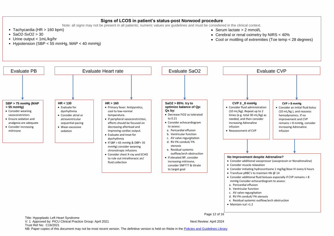
Signs of LCOS in patient’s status-post Norwood procedure Note: all signs may not be present in all patients; numeric values are guidelines and must be considered in the clinical context.
• Tachycardia (HR > 160 bpm) • SaO2-SvO2 > 30 • Urine output < 1mL/kg/hr • Hypotension (SBP < 55 mmHg, MAP < 40 mmHg)
Serum lactate > 2 mmol/L
Cerebral or renal oximetry by NIRS < 40%
Cool or mottling of extremities (Toe temp < 28 degrees)
Evaluate Heart rate Evaluate PB Evaluate CVP Evaluate SaO2
HR < 130
Evaluate for dysrhythmia
Consider atrial or atrioventricular sequential pacing
Wean excessive sedation
CVP ≤ _8 mmHg
Consider fluid administration (10 mL/kg). Repeat up to 2 times (e.g. total 30 mL/kg) as needed, and then consider increasing Adrenaline infusion
Reassessment of CVP
HR > 160
Primary fever: Antipyretics, cool to low-normal temperature.
If peripheral vasoconstriction, efforts should be focused on decreasing afterload and improving cardiac output.
Evaluate and treat for dysrhythmia
If SBP > 65 mmHg & DBP> 35 mmHg) consider weaning chronotropic infusions
Consider chest X-ray and ECHO to rule out intrathoracic air/ fluid collection
No Improvement despite Adrenaline?
Consider additional vasopressor (vasopressin or Noradrenaline)
Consider muscle relaxation
Consider initiating hydrocortisone 1 mg/kg/dose IV every 6 hours
Transfuse pRBC's to maintain Hb @ 14
Consider additional fluid boluses especially if CVP remains < 8 mmHg Consider echocardiogram to assess:
a. Pericardial effusion b. Ventricular function c. AV valve regurgitation d. RV-PA conduit/ PA stenosis e. Residual systemic outflow/arch obstruction
Maintain ical >1.2
SaO2 > 85%: try to optimize balance of Qp: Qs by:
Decrease FiO2 as tolerated to 0.21
Consider echocardiogram to assess:
a. Pericardial effusion b. Ventricular function c. AV valve regurgitation d. RV-PA conduit/ PA
stenosis e. Residual systemic
outflow/arch obstruction
If elevated BP, consider increasing milrinone, consider SNP??? & titrate to target goal
SBP > 75 mmHg (MAP > 55 mmHg)
Consider weaning vasoconstrictors
Ensure sedation and analgesia are adequate
Consider increasing milrinone

Page 13 of 16 Title: Hypoplastic Left Heart Syndrome V: 1 Approved by: PICU Clinical Practice Group: April 2021 Next Review: April 2024 Trust Ref No: C16/2021 NB: Paper copies of this document may not be most recent version. The definitive version is held on INsite in the Policies and Guidelines Library
Important points for chest closure management
Therapeutic heparin is witheld 2 hours prior to chest closure if surgeons agree. Prophylactic
heparin do not to be witheld prior to chest closure
At time of chest closure, be aware of desaturation and hypotension from compression of shunt
May need to reopen chest
Gently increase PIP (usually by 10- 20%) to maintain inspired tidal volume as chest
compliance usually reduced following sternal closure
Avoid overly negative fluid balance and assess fluid status frequently
Restart heparin one hour post chest closure
Prognosis & major complications
Overall survival to the time of hospital discharge after the Norwood procedure is approximately
75%. Success rates are higher (85%) in patients with low preoperative risk and lower (45%) in
patients with important risk factors. Survival after the bidirectional Glenn/hemi-Fontan and Fontan
operations is nearly 90-95%. The actuarial survival rate after staged reconstruction is 70% at 5 years.
Institutional success rates vary.
Important preoperative risk factors are:
Prematurity, birth during the early term period of 37 to 38 weeks’ gestation is
associated with worse outcome
Birth weight <2.5kg
Significant preoperative tricuspid insufficiency
Pulmonary venous hypertension
Associated major chromosomal or non-cardiac abnormalities
Preoperative mechanical ventilatory or circulatory support
Note the following:
Poor RV function is a predictor of mortality throughout surgical palliation
Low cerebral near-infrared spectroscopy oxygen saturations during the first 48 hours after
Norwood procedure are strongly associated with adverse outcomes
The need for mechanical preoperative ventilation has been shown to be a risk factor for poor
outcome
Severe preoperative lactic acidosis, and a need for inotropic support, which further increases
the risk for right ventricular dysfunction and tricuspid regurgitation are important risk factors for
survival of stage I Norwood palliation
Neurodevelopmental prognosis is not known; however, differences are well described.
-Major complications following the Norwood procedure include aortic arch obstruction at the site of
surgical anastomosis and progressive cyanosis caused by limited blood flow through the shunt. An
inadequate atrial communication contributes to progressive cyanosis.

Page 14 of 16 Title: Hypoplastic Left Heart Syndrome V: 1 Approved by: PICU Clinical Practice Group: April 2021 Next Review: April 2024 Trust Ref No: C16/2021 NB: Paper copies of this document may not be most recent version. The definitive version is held on INsite in the Policies and Guidelines Library
Transfer to ward criteria
SaO2 > 75% with stable respiratory status for >24 hours
Stable haemodynamic for >24 hours without need for vasoactive infusions
Tolerating intermittent diuretics
Tolerated 24 hours of feeding protocol
3. Education and Training
None
4. Monitoring Compliance
None identified at present
What will be measured to monitor compliance
How will compliance be monitored
Monitoring Lead
Frequency Reporting arrangements
5. Supporting References
Donnellan, Amy, and Lindsey Justice. "Preoperative stabilization of infants with hypoplastic left heart
syndrome before stage I palliation." Critical care nurse 36, no. 1 (2016): 52-59.
Roeleveld, Peter P., David M. Axelrod, Darren Klugman, Melissa B. Jones, Nikhil K. Chanani, Joseph W. Rossano, and John M. Costello. "Hypoplastic left heart syndrome: from fetus to fontan." Cardiology in the Young 28, no. 11 (2018): 1275-1288. Alphonso, Nelson, Annalisa Angelini, David J. Barron, Hannah Bellsham-Revell, Nico A. Blom, Katherine Brown, Deborah Davis et al. "Guidelines for the management of neonates and infants with hypoplastic left heart syndrome: the European Association for Cardio-Thoracic Surgery (EACTS) and the Association for European Paediatric and Congenital Cardiology (AEPC) Hypoplastic Left Heart Syndrome Guidelines Task Force." European Journal of Cardio-Thoracic Surgery 58, no. 3 (2020): 416-499. Ohye, Richard G., Lynn A. Sleeper, Lynn Mahony, Jane W. Newburger, Gail D. Pearson, Minmin Lu, Caren S. Goldberg et al. "Comparison of shunt types in the Norwood procedure for single-ventricle lesions." New England Journal of Medicine362, no. 21 (2010): 1980-1992.
Hornik, Christoph P., Xia He, Jeffrey P. Jacobs, Jennifer S. Li, Robert DB Jaquiss, Marshall L. Jacobs, Sean M. O'Brien, Eric D. Peterson, and Sara K. Pasquali. "Complications after the Norwood operation: an analysis of the society of thoracic surgeons congenital heart surgery database." The Annals of thoracic surgery 92, no. 5 (2011): 1734-1740. Schwartz, Steven M., Minmin Lu, Richard G. Ohye, Kevin D. Hill, Andrew M. Atz, Maryam Y. Naim, Ismee A. Williams et al. "Risk factors for prolonged length of stay after the stage 2 procedure in the single-ventricle reconstruction trial." The Journal of thoracic and cardiovascular surgery 147, no. 6 (2014): 1791-1798.

Page 15 of 16 Title: Hypoplastic Left Heart Syndrome V: 1 Approved by: PICU Clinical Practice Group: April 2021 Next Review: April 2024 Trust Ref No: C16/2021 NB: Paper copies of this document may not be most recent version. The definitive version is held on INsite in the Policies and Guidelines Library
Alsoufi, Bahaaldin. "Management of the single ventricle and potentially obstructive systemic ventricular outflow tract." Journal of the Saudi Heart Association 25, no. 3 (2013): 191-202.
6. Key Words
Congenital cardiac abnormalities, Norwood procedure, Ventricular
__________________________________________________________
The Trust recognises the diversity of the local community it serves. Our aim therefore is to provide a safe environment free from discrimination and treat all individuals fairly with dignity and appropriately according to their needs. As part of its development, this policy and its impact on equality have been reviewed and no detriment was identified.
Contact & review details
Guideline Lead (Name and Title) Samira Neshat – PICU Consultant
Executive Lead Chief Medical Officer
Details of Changes made during review: New guideline

Page 16 of 16 Title: Hypoplastic Left Heart Syndrome V: 1 Approved by: PICU Clinical Practice Group: April 2021 Next Review: April 2024 Trust Ref No: C16/2021 NB: Paper copies of this document may not be most recent version. The definitive version is held on INsite in the Policies and Guidelines Library
Appendix 1 Post –Operative Norwood/Sano Management Algorithm
Note: all signs may not be present in all patients; numeric values are guidelines and must be considered in the clinical context.
Cardiac output monitoring Goal: adequate CO
Cardiorespiratory monitor: CVP
SaO2 End-tidal CO2
Cerebral/Renal NIRS
Core/toe temperatures, SvO2
Sats 75%-85% avoid hyperoxygenation
HR 130-160 bpm
CVP 8-12
BP 65/35 mmHg, map >40mmHg
Ph 7.35-7.45, pCO2 4.5-5.5
Blender on resuscitation bag to 50%
Evaluate for Pulmonary Venous Desaturation Evaluate for Low Pulmonary Blood Flow Evaluate for Low SvO2
ABG/MVO2/Chest x-ray
Adequate chest rise? Tidal volume? ET CO2
trend?
Suction patient (after bolus sedation)
O2 challenge, if 10% increase in saturations, then lung disease is present, increase MAP + judicious increase in airway pressure to treat V/Q mismatch
Minimal response to O2 challenge
+/- hypoperfused chest x-ray/ET CO2
Usually normal BP
Low BP or evidence of compromised oxygen delivery, consumption or not optimised ventilation
Haemoglobin <14g/dl?
At risk: if high sats pre-op, <38 wks, lung disease, restrictive ASD, pulmonary vein obstruction
Start INO, alkalosis, sedation and paralysis, aim PaCO2 4.5, increase FiO2, increase milrinone
Shunt murmur?
Echo, contractility? Valvar competency? Already on adrenalin low dose? iCal?
Increase BP to drive shunt flow (Vasopressin or Noradrenaline)
Start Heparin
Evaluate PVR Small, Kinked or Thrombosed Shunt
No improvement