![JICA 201610B 10B 10B 13B 22 10B 11B 11B 26B M2:30 JTñ-ñ-3— … · 2016-10-13 · JICA 201610B 10B 10B 13B 22 10B 11B 11B 26B M2:30 JTñ-ñ-3— JD+ñ3—] (3) @ @ @ 201 2016 11](https://static.fdocuments.in/doc/165x107/5f7b7664c26e297ff6248b8f/jica-201610b-10b-10b-13b-22-10b-11b-11b-26b-m230-jt-3a-2016-10-13-jica.jpg)
pADL-10b Phagemid - abdesignlabs.com manual.pdf4 pADL-10b Phagemid Instruction Manual Description...
12
pADL-10b Phagemid INSTRUCTION MANUAL pADL-10b Phagemid Vector for Phage Display Catalog #: PD0105 Version: A1.7 – February 2017
Transcript of pADL-10b Phagemid - abdesignlabs.com manual.pdf4 pADL-10b Phagemid Instruction Manual Description...
pADL-10b Phagemid
INSTRUCTION MANUAL
pADL-10b Phagemid Vector for Phage Display
Catalog #: PD0105
Version: A1.7 – February 2017

2 pADL-10b Phagemid Instruction Manual

pADL-10b Phagemid Instruction Manual 3
Table of Contents
Description 4
Introduction 4
Content, Shipping & Storage 4
Limited Product Warranty 4
Vector Map 5
Cloning Site 5
Feature Table 6
Restriction Site Summary 6
Experimental Procedures 8
General Molecular Biology Techniques 8
Working with Filamentous Phage 8
Bacterial Strains and Helper Phage 8
Plasmid Maintenance 8
Cloning into pADL-10b 9
Sequencing of Inserts 10
Phagemid Virion Production 10
Appendix 12
MSDS Information 12
Quality Control 12
Technical Support 12
References 12
Legal and Disclaimers
Antibody Design Labs grants to the buyer with the sale of its phage and/or phagemid vectors (the “Product”) a non-
exclusive, non-transferable, royalty-free, commercial license to use Product in research conducted by the buyer (whether
the buyer is an academic or a for-profit entity). The buyer is NOT granted a license to (a) use Product for human or animal
therapeutic, diagnostic, or prophylactic purposes, (b) act as reseller or distributor of Product, or (c) resell, distribute, or
transfer Product without modification under any name. Antibody Design Labs does not warrant that the use or sale of
Product, the use thereof in combination with other products, or the use of Product in the operation of any process will not
infringe the claims of any United States or other patent(s). If the buyer is not willing to accept the limitations of this license,
without modification, buyer may refuse this license by returning Product unopened and unused. By keeping or using
Product, buyer agrees to be bound by the terms of this license.
4 pADL-10b Phagemid Instruction Manual
Description
Introduction
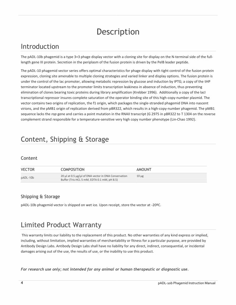
The pADL-10b phagemid is a type 3+3 phage display vector with a cloning site for display on the N-terminal side of the full-
length gene III protein. Secretion in the periplasm of the fusion protein is driven by the PelB leader peptide.
The pADL-10 phagemid vector series offers optimal characteristics for phage display with tight control of the fusion protein
expression, cloning site amenable to multiple cloning strategies and varied linker and display options. The fusion protein is
under the control of the lac promoter, allowing metabolic repression by glucose and induction by IPTG; a copy of the tHP
terminator located upstream to the promoter limits transcription leakiness in absence of induction, thus preventing
elimination of clones bearing toxic proteins during library amplification (Krebber 1996). Additionally a copy of the lacI
transcriptional repressor insures complete saturation of the operator binding site of this high-copy-number plasmid. The
vector contains two origins of replication, the f1 origin, which packages the single-stranded phagemid DNA into nascent
virions, and the pMB1 origin of replication derived from pBR322, which results in a high-copy-number phagemid. The pMB1
sequence lacks the rop gene and carries a point mutation in the RNAII transcript (G 2975 in pBR322 to T 1304 on the reverse
complement strand responsible for a temperature-sensitive very high copy number phenotype (Lin-Chao 1992).
Content, Shipping & Storage
Content
VECTOR COMPOSITION AMOUNT
pADL-10b 20 µl at 0.5 µg/µl of DNA vector in DNA Conservation Buffer (Tris-HCL 5 mM, EDTA 0.1 mM, pH 8.5)
10 µg
Shipping & Storage
pADL-10b phagemid vector is shipped on wet ice. Upon receipt, store the vector at -20ºC.
Limited Product Warranty
This warranty limits our liability to the replacement of this product. No other warranties of any kind express or implied,
including, without limitation, implied warranties of merchantability or fitness for a particular purpose, are provided by
Antibody Design Labs. Antibody Design Labs shall have no liability for any direct, indirect, consequential, or incidental
damages arising out of the use, the results of use, or the inability to use this product.
For research use only; not intended for any animal or human therapeutic or diagnostic use.
pADL-10b Phagemid Instruction Manual 5
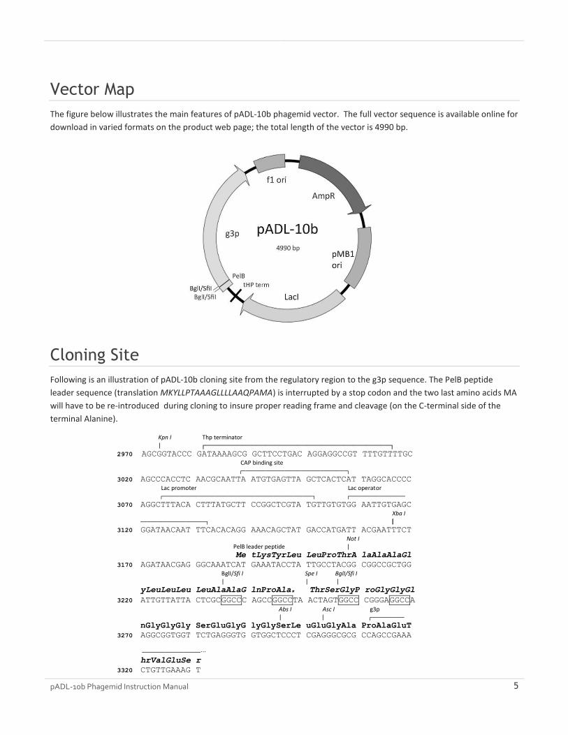
Vector Map
The figure below illustrates the main features of pADL-10b phagemid vector. The full vector sequence is available online for
download in varied formats on the product web page; the total length of the vector is 4990 bp.
Cloning Site
Following is an illustration of pADL-10b cloning site from the regulatory region to the g3p sequence. The PelB peptide
leader sequence (translation MKYLLPTAAAGLLLLAAQPAMA) is interrupted by a stop codon and the two last amino acids MA
will have to be re-introduced during cloning to insure proper reading frame and cleavage (on the C-terminal side of the
terminal Alanine).
Kpn I Thp terminator | ┌────────────────────────────────────────┐
2970 AGCGGTACCC GATAAAAGCG GCTTCCTGAC AGGAGGCCGT TTTGTTTTGC
CAP binding site ┌────────────────────────────────┐
3020 AGCCCACCTC AACGCAATTA ATGTGAGTTA GCTCACTCAT TAGGCACCCC
Lac promoter Lac operator ┌──────────────────────────────────────────────┐ ┌─────────────────
3070 AGGCTTTACA CTTTATGCTT CCGGCTCGTA TGTTGTGTGG AATTGTGAGC
Xba I ────────────────────┐ |
3120 GGATAACAAT TTCACACAGG AAACAGCTAT GACCATGATT ACGAATTTCT
Not I PelB leader peptide |
Me tLysTyrLeu LeuProThrA laAlaAlaGl
3170 AGATAACGAG GGCAAATCAT GAAATACCTA TTGCCTACGG CGGCCGCTGG
BglI/Sfi I Spe I BglI/Sfi I | | |
yLeuLeuLeu LeuAlaAlaG lnProAla* ThrSerGlyP roGlyGlyGl
3220 ATTGTTATTA CTCGCGGCCC AGCCGGCCTA ACTAGTGGCC CGGGAGGCCA
Abs I Asc I g3p | | ┌──────────
nGlyGlyGly SerGluGlyG lyGlySerLe uGluGlyAla ProAlaGluT
3270 AGGCGGTGGT TCTGAGGGTG GTGGCTCCCT CGAGGGCGCG CCAGCCGAAA
──────────────────∙∙∙
hrValGluSe r
3320 CTGTTGAAAG T

6 pADL-10b Phagemid Instruction Manual
Feature Table
The features of pADL-10b phagemid vector are highlighted in the following table.
FEATURE LOCATION DESCRIPTION
TEM1 beta-lactamase 126-986 Ampicillin resistance for selection in E. coli.
pMB1 origin 1141-1760 pBR322 origin for replication in E. coli with a high copy-number.
LacI 1888-2970 LacI transcriptional repressor for controlled expression of gene III fusion protein in E. coli strains lacking LacI gene.
tHP terminator 2981-3017 Transcriptional terminator (taken from the glutamine permease operon) to prevent transcriptional leakage into the lac operon.
CAP binding site 3039-3059 Mediate the catabolite repression of the lac operator in the presence of glucose >1% w/v.
-35 signal 3074-3079 Lac promoter -35 signal
-10 signal 3098-3103 Lac promoter -10 signal
PelB leader sequence 3188-3250 PelB leader sequence for export in the periplasm of the host bacteria. The missing terminal methionine and alanine will have to be added during the cloning to obtain a complete leader peptide (MKYLLPTAAAGLLLLAAQPAMA) necessary for proper removal of the leader during the export process.
g3p fusion coding sequence 3250-4533 Full-length gene III fusion protein coding sequence; the M13 g3p protein is fused on its N-terminal side to the linker GPGGQGGGSEGGGSLEGAP; the exact final sequence of the fusion depends on the cloning strategy (see cloning site).
oriF1 4661..4967 Origin of replication for phage f1
Restriction Site Summary
Enzyme Site Nb Position Strand Isoschizomers
AbsI CC^TCGAGG 1 3297
AloI (7/12)GAACNNNNNNTCC(12/7) 1 4695
ApaI GGGCC^C 1 2445 Bsp120I PspOMI
AscI GG^CGCGCC 1 3304 PalAI SgsI
AviII TGC^GCA 1 688 Acc16I FspI NsbI
BamHI G^GATCC 1 3900
BclI T^GATCA 1 2252 FbaI Ksp22I
BseRI GAGGAG(10/8) 1 3688
BsmI GAATGC(1/-1) 1 3426 BsaMI Mva1269I PctI
BspMI ACCTGC(4/8) 1 3938 Acc36I BfuAI BveI
BstAPI GCANNNN^NTGC 1 1915
BstEII G^GTNACC 1 2419 BstPI Eco91I EcoO65I PspEI
ClaI AT^CGAT 1 4207 BsuTUI BspDI BseCI Bsa29I BanIII
BshVI Bsu15I
DraIII CACNNN^GTG 1 4748 AdeI
EagI C^GGCCG 1 3210 BseX3I BstZI EclXI Eco52I
Eam1105I GACNNN^NNGTC 1 908 AhdI AspEI BmeRI DriI
Eco31I GGTCTC(1/5) 1 841 Bso31I BsaI
Esp3I CGTCTC(1/5) 1 2847 BsmBI
FalI (8/13)AAGNNNNNCTT(13/8) 1 4166
HindIII A^AGCTT 1 4536
HpaI GTT^AAC 1 2742 KspAI
KpnI GGTAC^C 1 2973 Acc65I Asp718I
MluI A^CGCGT 1 2238
NarI GG^CGCC 1 2877 DinI EgeI EheI KasI Mly113I SfoI

pADL-10b Phagemid Instruction Manual 7
SspDI
NdeI CA^TATG 1 4403 FauNDI
NotI GC^GGCCGC 1 3209 CciNI
PflMI CCANNNN^NTGG 1 1814 AccB7I BasI Van91I
PsiI TTA^TAA 1 4623 AanI
PspXI VC^TCGAGB 1 3297
PvuI CGAT^CG 1 541 Ple19I BpvUI MvrI
ScaI AGT^ACT 1 430 AssI BmcAI ZrmI
SmaI CCC^GGG 1 3258 Cfr9I TspMI XmaI
SpeI A^CTAGT 1 3250 AhlI BcuI
TstI (8/13)CACNNNNNNTCC(12/7) 1 2307
XbaI T^CTAGA 1 3167
XhoI C^TCGAG 1 3298 StrI TliI Sfr274I PaeR7I SlaI
AlwNI CAGNNN^CTG 2 1387 CaiI PstNI
2 3867
BaeI (10/15)ACNNNNGTAYC(12/7) 2 3475 -
2 3817
BauI CACGAG(-5/-1) 2 244 BssSI Bst2BI
2 1628
BcgI (10/12)CGANNNNNNTGC(12/10)2 386 -
2 2541
BglI GCCNNNN^NGGC 2 3236
2 3257
BpiI GAAGAC(2/6) 2 2377 BbsI BpuAI BstV2I
2 2716
BsePI G^CGCGC 2 2649 BssHII PauI PteI
2 3305
BsgI GTGCAG(16/14) 2 2104 -
2 2304
DrdI GACNNNN^NNGTC 2 1693 AasI DseDI
2 4702
Eam1104I CTCTTC(1/4) 2 119 - Bst6I EarI
2 1861
Eco57I CTGAAG(16/14) 2 226 AcuI
2 1274
NaeI GCC^GGC 2 3241 PdiI NgoMIV MroNI
2 4854
NmeAIII GCCGAG(21/19) 2 785 -
2 2463
PvuII CAG^CTG 2 2836
2 2929
SfiI GGCCNNNN^NGGCC 2 3235
2 3256
XmnI GAANN^NNTTC 2 309 Asp700I MroXI PdmI
2 4326
Absent Sites:
AarI, AatII, AflII, AgeI, AjuI, AlfI, ArsI, AsuII, AvrII, BalI, BarI, BbvCI,
BglII, BplI, Bpu10I, BsaBI, BsiWI, Bsp1407I, Bsp1720I, BssNAI, Bsu36I, BtrI,
CspCI, Eco47III, EcoNI, EcoRI, EcoRV, FseI, FspAI, KflI, MauBI, MfeI, MreI,
MroI, NcoI, NheI, NruI, OliI, PacI, PasI, PfoI, PmaCI, PmeI, PscI, PshAI, PsrI,
PstI, RsrII, SacI, SacII, SalI, SapI, SexAI, SgfI, SgrAI, SgrDI, SnaBI, SphI,
SrfI, Sse8387I, StuI, SwaI, Tth111I, Zsp2I.

8 pADL-10b Phagemid Instruction Manual
Experimental Procedures
General Molecular Biology Techniques
Molecular cloning and phage display should be conducted under the supervision of a qualified instructor trained to
standard safety practice in a molecular biology laboratory environment. Standard molecular biology procedures can be
found in a general molecular biology handbook such as Sambrook (1989).
Working with Filamentous Phage
Keep the bench clean and regularly wiped with 2% bleach to limit phage cross-contamination and only use filtered tips to
prevent aerosol contaminations. Phages are known to survive standard autoclaving conditions and are not removed by
0.22 µm filtration. Phages are either killed by heat-treating dry, autoclaved materials in an oven for 4 hours at 105ºC
(Phage Display (2001)) or by incubation in 2% bleach for at least 1 hour. We recommend to extensively wash with hot
water all glass and plastic-ware, then submerge (tubes) or incubate (flasks) with a 2% solution of bleach for at least one
hour. Heat-resistant glassware can then be autoclaved in an autoclave that is never used for biological waste while
sensitive plastic-ware can be used directly or at best heat-treated as described above.
Bacterial Strains and Helper Phage
Bacterial Strains
In theory, any K12 F+ E. coli strain is suitable for phage display using pADL-10b. Practically we recommend either SS320 or
TG1 bacterial strains; both of them have been widely used for phage display and are well documented in the literature.
SS320 derives from MC1061 by introduction of the F’ episome (Sidhu 2000). Like most derivatives of MC1061, SS320 can be
made highly competent for transformation by electroporation. TG1 suppresses amber codons and can also be made highly
competent for transformation by electroporation. Their respective phenotype is highlighted below:
SS320 hsdR2 mcrA0 araD139 Δ(araA-leu)7697 ΔlacX74 galK16 galE15(GalS) e14- rpsL150(Str
R) spoT1 thi
F'[proAB+lacIqlacZΔM15 Tn10 (tetr)]
TG1 supE thi-1 Δ(lac-proAB) Δ(mcrB-hsdSM)5, (rK-mK
-)
F' [traD36 proAB+ lacI
q lacZΔM15]
Helper Phage
We recommend CM13 helper phage. CM13 is made available by Antibody Design Labs under product number PH020L,
which offers a highly concentrated virion preparation, eliminating the need to generate and characterize your own helper
phage stocks. CM13 derived from M13KO7 by a single point mutation and produces on average twice more virions.
Plasmid Maintenance
Propagation and maintenance of pADL-10b is obtained on any recA1, endA1 E. coli strain using LB or 2xYT medium
supplemented with ampicillin 100 µg/ml as a selection marker, without glucose, and incubated at 37ºC with agitation.
Phagemid pADL-10b is a derivative of pBR322 with a high copy number origin of replication and usually gives high yields of
plasmid DNA with most standard laboratory strains such as XL1-blue or DH5α. Some DNA stabilizing strains are known to

pADL-10b Phagemid Instruction Manual 9
produce smaller amounts of plasmid DNA. In case of issues, we recommend using XL10-Gold® from Agilent Technologies,
Inc., on which pADL-10b plasmid DNA can be isolated in large quantities.
Cloning into pADL-10b
Primer Design and PelB Leader Sequence
A complete PelB leader sequence MKYLLPTAAAGLLLLAAQPAMA is necessary for export in the periplasm and proper
removal of the leader peptide by host proteases. In the following schema, where [NNN] represents the insert sequence and
[Xxx] the translated amino acid sequence, the short hexanucleotide ATGGCN must be appended immediately to the first
BglI/SfiI site to obtain a complete PelB leader encoding sequence; cleavage will occur on the C-terminal side of the terminal
alanine (codon GCN).
BglI/Sfi I Spe I BglI/Sfi I | | |
yLeuLeuLeu LeuAlaAlaG lnProAlaMe tAla [Xxx] ThrSerGlyP roGlyGlyGl
3220 ATTGTTATTA CTCGCGGCCC AGCCGGCCAT GGCN [NNN] ACTAGTGGCC CGGGAGGCCA
Retention of the SpeI site is optional during cloning and the encoded dipeptide ThrSer is not known to interfere with
display.
Cloning in pADL-10b Using BglI/SfiI Sites
Large libraries in the 1 x 10⁹ range and above can easily be constructed using the double BglI/SfiI cloning site.
WORKING WITH BGL I/SFI I SITES
The SfiI restriction enzyme recognizes rare 8-base-long interrupted palindromes GGCCNNNN/NGGCC and leaves 3-
nucleotide-long overhangs after digestion. The pADL-10b cloning site contains one SfiI site close to the end of the PelB
leader sequence and a second SfiI site 8 nucleotides apart from the first site. The PelB sequence of the empty vector has an
early termination by an ochre stop codon and no gene III protein is produced by the vector alone.
The SfiI restriction enzyme requires two copies of its recognition sequence for cleavage to occur; cleavage of the two sites
happens simultaneously through interaction of two SfiI tetramers (Wertzell 1995). Vectors bearing two sites very close to
each other are cut in trans and digestion might not complete. Therefore we strongly recommend opening pADL-10b with
the alternative BglI restriction enzyme, which cuts the shorter 6-base-long interrupted palindromes GCCNNNN/NGGC and
generates identical overhangs.
Sites open with BglI will re-ligate with sites open with SfiI as long as overhangs are complementary. Practically, the
pentanucleotide NNNNN must be identical to the original vector sequence to handle both ligation of the complementary
overhangs and conservation of the amino acid sequence (PelB sequence and linker to protein III). Since the overhang of the
two BglI/SfiI sites are non-palidromic and different, a cut empty vector cannot ligate onto itself; it is therefore possible to
follow a ligation reaction by minigel analysis since remaining unligated vector or unligated insert will migrate unchanged at
their expected size.
PREPARATION OF VECTOR DNA FOR CLONING
1. On ice add successively water, BglI buffer (1x final), pADL-10b vector and BglI enzyme 5 units/µg DNA; make sure the
enzyme volume does not to exceed 1/10 of the total reaction volume.
2. Incubate 4 h to overnight at 37ºC.
3. Inactivate for 20 min at 70ºC.

10 pADL-10b Phagemid Instruction Manual
4. Confirm the digestion by DNA analysis on a minigel.
5. Purify the cut vector.
For routine cloning, a standard DNA purification kit can be used directly after the digestion to remove the excess of buffer,
the small DNA stuffer and leftover restriction enzyme. For library construction, phenol/chloroform extraction and/or gel
purification may be required.
PREPARATION OF INSERTS
SfiI digestion should be rapid and complete in 4 hours especially for fragments longer or equal to 200 bp where sites are cut
in cis. BglI may be used when the insert sequence is known to be free of BglI site and therefore is not recommended for
building antibody libraries.
Cloning using NotI-SpeI sites
The NotI site located in the first half of the PelB leader encoding sequence may be used in conjunction with the SpeI site to
clone inserts. This strategy has been applied in some early phage display vectors. Consult your restriction enzyme
distributor resources to identify a buffer compatible with both enzymes and follow the concentration schema given above
to conduct the digestion. NotI and SpeI can be inactivated by heat before DNA purification.
Sequencing of Inserts
The following primers give both strong PCR amplification and sequencing traces.
Forward or Sense Primers
phiS3 5’-CACAGGAAACAGCTATGACCATGATTA
phiS2 5’-ATGAAATACCTATTGCCTACGG
Backward, Antisense or Reverse Primers
psiR2 5’-CGTTAGTAAATGAATTTTCTGTATGAGG
psiR3 5’-GCGTAACGATCTAAAGTTTTGTCG
Nested Sequencing Often it is easier to sequence an insert by PCR on the bacterial culture supernatant or directly from a colony rather than on
tediously isolated plasmids. Use the outward primers phiS3 and betaR3 together with a DNA polymerase not inhibited by
bacterial cultures such as TAQ polymerase for the PCR and sequence the insert with the nested reverse primer betaR2. Use
less than 1 µl of bacterial culture supernatant per 50 µl-PCR reaction or the touch of a toothpick on a colony as DNA
template.
Phagemid Virion Production
A superinfection by a helper phage is necessary for phagemid pADL-10b-containing bacteria to produce virions. Please,
consult the M13KO7 or CM13 helper phage manual for optimal conditions of superinfection. We recommend a rich
medium such as 2xYT medium supplemented with ampicillin 100 µg/ml, kanamycin 50 µg/ml (when M13KO7 or CM13
helper phages are used), IPTG 200 µM, no glucose or less than 0.1% w/v, and incubation from 8 h to overnight at 30ºC and
250 rpm. We recommend adding the helper phage when the bacterial culture reaches an optical density at 600 nm of 0.5
OD; large amounts of non-superinfected cells due to immunity to superinfection will decrease virion production above 0.5

pADL-10b Phagemid Instruction Manual 11
OD while disparities caused by differences in phage growth rates will be amplified at a lower OD. Immunity to
superinfection refers to the difficulty to transduce bacteria when protein III is expressed, as it is the case when with
phagemids expressing a full-length pIII fusion protein.
Notes
Supplementation with IPTG 200 µM is necessary to achieve display on the phage with pADL-10b. Virions produced in
absence of IPTG will not bear any antibodies and subsequent selection will not work.
Shorter incubation times 6 to 8 h will produce less virions; we have not seen improvement of display on shorter
incubation times or, inversely, we have not seen sign of proteolysis of the linker after overnight incubation. Always
use freshly prepared buffers from commercial concentrates during virion preparations to limit sources of proteolysis.
Proteolysis sometimes occurs on concentrated virions; always prepare virions quickly and on ice.
Kanamycin 50 µg is enough to ensure selection with derivatives of M13KO7. Higher concentration may be needed on
very rich culture media or if your culture medium contains phosphate salts.
Higher IPTG concentrations will give higher levels of display and is recommended for making initial libraries or
preparing single phages.
12 pADL-10b Phagemid Instruction Manual
Appendix
MSDS Information
MSDSs (Material Safety Data Sheets) are available on the Antibody Design Labs website at the corresponding product page.
Quality Control
Specifications and quality control are detailed on the online product page. Antibody Design Labs certifies that the product
will perform according to these specifications.
Technical Support
Visit Antibody Design Labs’ website at www.abdesignlabs.com for technical resources, including manuals, vector maps and
sequences, application notes, FAQs, etc.
FOR MORE INFORMATION OR TECHNICAL ASSISTANCE, CALL, WRITE, FAX, OR EMAIL US AT:
Antibody Design Labs Email: [email protected]
4901 Morena Blvd, Suite 203 Phone: 1-877-223-3104 (TOLL-FREE)
San Diego, CA 92117 Fax: 1-858-272-6007 (24 hour)
(Monday – Friday 9:00 AM – 5:00 PM PST)
References
1. KREBBER A, BURMESTER J, PLÜCKTHUN A. (1996). INCLUSION OF AN UPSTREAM TRANSCRIPTIONAL TERMINATOR IN PHAGE DISPLAY VECTORS ABOLISHES BACKGROUND EXPRESSION OF TOXIC FUSIONS WITH COAT PROTEIN G3P. GENE 178(1-2):71-4.
2. SAMBROOK, J., FRITSCH, E.F., AND MANIATIS, T. (1989). IN MOLECULAR CLONING: A LABORATORY MANUAL. COLD SPRING HARBOR LABORATORY PRESS, NY, VOL. 1, 2, 3.
3. SCOTT JK & BARBAS CF (2001). PHAGE-DISPLAY VECTORS (2.1-2.19) IN PHAGE DISPLAY: A LABORATORY MANUAL. EDITED BY C. F. BARBAS III, D. R. BURTON, J. K. SCOTT, AND G. J. SILVERMAN. COLD SPRING HARBOR, LABORATORY PRESS, COLD SPRING HARBOR, NY.
4. PHAGE DISPLAY: A LABORATORY MANUAL (2001). EDITED BY C. F. BARBAS III, D. R. BURTON, J. K. SCOTT, AND G. J. SILVERMAN. COLD SPRING HARBOR, LABORATORY PRESS, COLD SPRING HARBOR, NY.
5. SIDHU SS, LOWMAN HB, CUNNINGHAM BC, WELLS JA (2000). PHAGE DISPLAY FOR SELECTION OF NOVEL BINDING PEPTIDES. METHODS ENZYMOL.328:333-63.
6. WERTZELL L.M. ET AL., (1995). THE SFII RESTRICTION ENDONUCLEASE MAKES A FOUR-STRAND DNA BREAK AT TWO COPIES OF ITS RECOGNITION SEQUENCE. J. MOL. BIOL. 248:581-595.
7. LIN-CHAO S, CHEN WT, WONG TT (1992). HIGH COPY NUMBER OF THE PUC PLASMID RESULTS FROM A ROM/ROP-SUPPRESSIBLE POINT MUTATION IN RNA II. MOL MICROBIOL. 22:3385-93.
This product is subject to Antibody Design Labs Terms & Conditions of Sales available online at http://www.abdesignlabs.com/terms/.
© 2017 Antibody Design Labs. All rights reserved.