PACS for Bhutan: a cost effective open source architecture ... · PDF fileREPORT PACS for...
7
REPORT PACS for Bhutan: a cost effective open source architecture for emerging countries Osman Ratib 1,5 & Nicolas Roduit 1 & Dechen Nidup 2 & Gerard De Geer 3 & Antoine Rosset 1,2,3,4 & Antoine Geissbuhler 1 Received: 17 January 2016 /Revised: 27 June 2016 /Accepted: 4 July 2016 /Published online: 28 July 2016 # The Author(s) 2016. This article is published with open access at Springerlink.com Abstract This paper reports the design and implementation of an inno- vative and cost-effective imaging management infrastructure suitable for radiology centres in emerging countries. It was implemented in the main referring hospital of Bhutan equipped with a CT, an MRI, digital radiology, and a suite of several ultrasound units. They lacked the necessary infor- matics infrastructure for image archiving and interpretation and needed a system for distribution of images to clinical wards. The solution developed for this project combines sev- eral open source software platforms in a robust and versatile archiving and communication system connected to analysis workstations equipped with a FDA-certified version of the highly popular Open-Source software. The whole system was implemented on standard off- the-shelf hardware. The system was installed in three days, and training of the radiologists as well as the technical and IT staff was provided onsite to ensure full ownership of the system by the local team. Radiologists were rapidly ca- pable of reading and interpreting studies on the diagnos- tic workstations, which had a significant benefit on their workflow and ability to perform diagnostic tasks more efficiently. Furthermore, images were also made avail- able to several clinical units on standard desktop com- puters through a web-based viewer. Messages/teaching points • Open source imaging informatics platforms can provide cost-effective alternatives for PACS • Robust and cost-effective open architecture can provide ad- equate solutions for emerging countries • Imaging informatics is often lacking in hospitals equipped with digital modalities Keywords Medical imaging . Picture archiving and communication systems . Radiology information system . Teleradiology . Software tools Introduction Digital imaging modalities are becoming more widely available even in remote areas of emerging countries extending the capabilities of these areas to provide higher quality of care and diagnostic capabilities locally. While most of these centres can afford state-of-the-art imaging equipment, they often lack the needed IT infra- structures to manage and distribute the digital imaging data. Adding these informatics infrastructures will add a significant cost in the context of limited budgets, assigning priorities to medical equipment needed for pa- tient care and for diagnostic services. This often leaves the users and care providers with limited resources for * Osman Ratib [email protected] 1 University Hospital of Geneva, Geneve, Switzerland 2 Jigme Dorji Wangchuck National Referral Hospital, Thimphu, Bhutan 3 Imagerie & Développement (ID), Geneva, Switzerland 4 La Tour Réseau de Soins, Geneva, Switzerland 5 Department of Medical Imaging and Information Sciences, Division of Nuclear Medicine, Rue Gabrielle-Perret-Gentil 4, 1211 Geneve 14, Switzerland Insights Imaging (2016) 7:747–753 DOI 10.1007/s13244-016-0512-7
-
Upload
nguyentuyen -
Category
Documents
-
view
219 -
download
2
Transcript of PACS for Bhutan: a cost effective open source architecture ... · PDF fileREPORT PACS for...

REPORT
PACS for Bhutan: a cost effective open source architecturefor emerging countries
Osman Ratib1,5& Nicolas Roduit1 & Dechen Nidup2
& Gerard De Geer3 &
Antoine Rosset1,2,3,4 & Antoine Geissbuhler1
Received: 17 January 2016 /Revised: 27 June 2016 /Accepted: 4 July 2016 /Published online: 28 July 2016# The Author(s) 2016. This article is published with open access at Springerlink.com
AbstractThis paper reports the design and implementation of an inno-vative and cost-effective imaging management infrastructuresuitable for radiology centres in emerging countries. It wasimplemented in the main referring hospital of Bhutanequipped with a CT, an MRI, digital radiology, and a suiteof several ultrasound units. They lacked the necessary infor-matics infrastructure for image archiving and interpretationand needed a system for distribution of images to clinicalwards.
The solution developed for this project combines sev-eral open source software platforms in a robust andversatile archiving and communication system connectedto analysis workstations equipped with a FDA-certifiedversion of the highly popular Open-Source software.The whole system was implemented on standard off-the-shelf hardware.
The system was installed in three days, and trainingof the radiologists as well as the technical and IT staffwas provided onsite to ensure full ownership of the
system by the local team. Radiologists were rapidly ca-pable of reading and interpreting studies on the diagnos-tic workstations, which had a significant benefit on theirworkflow and ability to perform diagnostic tasks moreefficiently. Furthermore, images were also made avail-able to several clinical units on standard desktop com-puters through a web-based viewer.
Messages/teaching points• Open source imaging informatics platforms can providecost-effective alternatives for PACS
• Robust and cost-effective open architecture can provide ad-equate solutions for emerging countries
• Imaging informatics is often lacking in hospitals equippedwith digital modalities
Keywords Medical imaging . Picture archiving andcommunication systems . Radiology information system .
Teleradiology . Software tools
Introduction
Digital imaging modalities are becoming more widelyavailable even in remote areas of emerging countriesextending the capabilities of these areas to providehigher quality of care and diagnostic capabilities locally.While most of these centres can afford state-of-the-artimaging equipment, they often lack the needed IT infra-structures to manage and distribute the digital imagingdata. Adding these informatics infrastructures will add asignificant cost in the context of limited budgets,assigning priorities to medical equipment needed for pa-tient care and for diagnostic services. This often leavesthe users and care providers with limited resources for
* Osman [email protected]
1 University Hospital of Geneva, Geneve, Switzerland2 Jigme Dorji Wangchuck National Referral Hospital,
Thimphu, Bhutan3 Imagerie & Développement (ID), Geneva, Switzerland4 La Tour Réseau de Soins, Geneva, Switzerland5 Department of Medical Imaging and Information Sciences, Division
of Nuclear Medicine, Rue Gabrielle-Perret-Gentil 4, 1211 Geneve14, Switzerland
Insights Imaging (2016) 7:747–753DOI 10.1007/s13244-016-0512-7

p rope r managemen t o f d i g i t a l imag ing da t a .Furthermore, efficient image interpretation requires spe-cific software tools and imaging platforms that radiolo-gists need for their daily task of image interpretationand reporting.
In Bhutan, a small kingdom of a little over 700000inhabitants south of the chain of the Himalayans, theJigme Dorji Wangchuck National Referral Hospital inThimphu, the capital, is the only radiology centreequipped with a CT scanner, MRI scanner, digital ra-diology unit, and a suite of five ultrasound units. In afirst informal visit to the hospital and discussion withlocal radiologists, we identified that they lack the prop-er infrastructure to manage and review the imagingdata acquired from these imaging modalities. Imagesare interpreted directly on the scanner consoles locatedin the imaging suites, competing with the technolo-gist’s task of performing imaging procedures concur-rently. Besides the physical dispersion of these devicesrequiring radiologists to move from one unit to anotherto review and interpret the studies, it has a significantimpact on the workflow and efficiency of radiologistsin their daily tasks. Furthermore, the lack of properstorage and archiving of the images requires savingdigital data on individual CDs that are often hard toretrieve when a patient returns for a new study and theneed for comparing with previous studies is required.Finally, there is an increasing need for facilitating thecommunication between radiologists and referring phy-sicians and clinical units in the hospital. Distribution ofmedical images to clinical wards and directly to clini-cal partners is particularly difficult given that only avery limited number of images are being printed foreconomic reasons, reading images from CDs remainslimited and cumbersome for referring physicians.While the hospital is equipped with a secure local areanetwork, it lacks the proper software and infrastructureto provide access to imaging data in a cost effectiveand easy way.
In recent years a trend toward development of opensource software platforms in medicine have made greatprogress toward providing very robust and reliable alter-native solutions to commercial products [1]. Severalopen source solutions for image display and analysisare available. We have contributed in over 10 yearsnow to the development and evolution of the OsiriXimaging platform [2], a widely adopted image reviewand analysis software that is used by radiologists andclinicians around the world for interpretation of allkinds of digital images [3]. Several spin-offs of certifiedversions of the OsiriX software are now available fromdifferent vendors. Through a non-profit foundation wecontinue to encourage and sponsor young developers as
well as innovative initiatives based on open source plat-forms [4, 5].
Material and methods
The department of radiology at Wangchuck National ReferralHospital is equipped with a CT scanner (Brilliance 16 fromPhilips Medical), MRI scanner (Signa 1.5 T from GeneralElectric), and a digital radiology unit (IDC DR 1590 CURA)as well as five ultrasound units from different vendors (twoLogiq e series GE Medical, one Siemens Acuson X500, andtwo Philips Clear Vue 350). All these modalities have theability to export images digitally in DICOM compliant format.The hospital is also equippedwith an internal secured Ethernetlocal area network available across the hospital as well as arestricted Wi-Fi network. The radiology department benefitsfrom a separate sub-network allowing a better communicationbandwidth between different imaging equipment.
The PACS server was implemented on a Supermicro7048R-TR server running under the Linux operating system.The server specifications included:
& 1× Intel Xeon Processor E5-2650 v3 (25 M Cache,2.30 GHz)
& 2× DDR4 16GB 2133MHz (memory)& 2× 240GB HyperX Predator PCIe (SSD card)& 8× WD Green 2 TB Desktop SATA 3.5^& 2× RJ45 Gigabit Ethernet LAN ports 1× RJ45& 2× 920 W redundant power supplies
The software framework consisted of the followingcomponents:
& Operating system: openmediavault (http://www.openmediavault.org), a popular Linux distribution for anopen network attached storage solution. This distributionis based on Linux Debian and provides a Web-basedGraphical User Interface (Web-GUI).
& The open source dcm4che DICOM archive solution with aMySQL database (http://dcm4che.org). A specific Debianpackage has been built to facilitate the installation directlythrough the Web-GUI as a service of openmediavault(https://github.com/nroduit/openmediavault-dcm4chee).Part of this work is based on the open source CDMEDICsolution (http://cdmedicpacsweb.sourceforge.net) by Dr.Pablo Sau, Valencia.
& Java-based web viewer (Weasis developed by NicolasRodui t – Geneva) , h t tp : / /dcm4che .a t l ass ian .net/wiki/display/WEA/Home)
& Advanced image analysis platform (OsiriX developed byDr. Antoine Rosset and the OsiriX team - Geneva)
748 Insights Imaging (2016) 7:747–753

The PACS service component allows managing andenabling/disabling the PACS server through a dedicatedWeb-based graphic user interface (GUI). Likewise, thePACS activity can be monitored directly from the Web-GUI.
The operating system is installed on the SSD disks, whichare mirrored by a RAID-1 configuration, while all the ar-chived images are stored in a Raid-5 virtual partition (sixhotswap disks and two hotspare disks).
Two workstations based on 27^ retina display iMac work-stations from Apple Computer are equipped with DICOM-compliant OsiriX software for diagnostic interpretation andimage analysis. The general diagram of the system is shownin Fig. 1.
The volumes of images generated per year by thedifferent imaging modalities are listed in Table 1. Inaddition to the three main digital imaging modalities,there is a volume of selected images and dynamic se-quences from ultrasound examinations that are beingarchived for reference and follow-up of patients. Thestorage volume of the latter is hard to estimate giventhat only selected studies are archived on demand.
The clinical staff of the radiology departments consistof three board certified radiologists, two radiology tech-nologists (with bachelor's degrees), 14 radiographers(certificate degree), eight ultrasound technicians
(diploma and certificate degree), four CT technicians(diploma and certificate degree), three MRI technicians(diploma and certificate degrees) as well as supportfrom a local IT team for support and management ofIT infrastructure.
While the hospital is equipped with a secure intranetlocal area network, it does not offer any electronic pa-tient record or digital hospital information system.Likewise, the radiology department is not yet equippedwith a radiology information system (RIS) and all or-ders for radiological procedures as well as radiology
Fig. 1 The general diagram of the imaging network associated with the PACS server that was implemented showing the different imaging modalities,the primary interpretation workstations, and web-based applications that can run on any standard desktop computer
Table 1 Estimated volumes of image data generated by the three maindigital imaging modalities based on the current number of studiesperformed
Modality Studies/year MB/study Est Total vol. (GB)
CT 6000 250 1500
MR 4000 40 160
Dig RX 5000 15 75
Total 1735
These volumes are based on uncompressed data; lossless compressioncan reduce these volumes by half
Insights Imaging (2016) 7:747–753 749

reports are handled in paper format. While OsiriX soft-ware supports the option of importing orders and gen-erating diagnostic reports, it was implemented as astand-alone PACS network for image viewing and anal-ysis. It is, however, fully compatible with future deploy-ment of RIS and HIS systems.
Results and observations
A complete pre-configured server unit with all softwarecomponents installed and tested was transported toBhutan and installed by the Geneva team in collabora-tion with the local team (see Fig. 2). Technical supportengineers from the different imaging equipment compa-nies were available on site or remotely through phonecommunication for configuring the imaging modalitiesto connect to the PACS server and viewing workstations
through standard DICOM protocols. Proper preparationand setup of the network and equipment identities wascarried out prior to our arrival by the local IT team.
A special room was set up to serve as a readingroom for radiologists where the two OsiriX workstationswere installed (see Fig. 3). The full installation of thecomplete system was carried out in less than 48 h,followed by appropriate training of the technical andIT staff as well as the radiologists in managing andusing the system. A PACS manager was designatedamong the hospital IT team and was trained for man-agement, updates, and backup tasks of the system. Thelead technologists of each modality were trained tomonitor and perform regular quality control tasks ofthe images archived on the system. Remote support ofthe IT and technical teams was also provided by ourteam in Geneva. The ease of use and well-known userinterface of the OsiriX software allowed radiologists torapidly become familiar with its main features and adoptit for retrieving and reviewing studies from all the im-aging modalities.
The management of the PACS server as well as mon-itoring and supervision of the performance of the sys-tem is available through a simple Web-GUI that waspartially developed by our team specifically for thisproject. This allows technologists of the radiology de-partment, as well as members of the IT team, to remote-ly access the server and perform the basic managementand maintenance tasks. This user-friendly platformallows technologists to easily correct errors that mayoccur in demographic data and image identification da-ta. The main window of the web-portal to the PACSserver is shown in Fig. 4. It was implemented as anextension of an open source data storage platform(openmediavault) with an extended set of features
Fig. 2 The Geneva teamdelivering the system was greetedby the local team at arrival in theBhutan hospital
Fig. 3 The setup of the radiology reading room after installation of thetwo OsiriX workstations
750 Insights Imaging (2016) 7:747–753

and data management components that are accessiblethrough a simple graphic user interface, Fig. 5.
Furthermore, the IT staff was trained to extend the ac-cess to images to clinical wards through a web-basedviewer that can easily be launched on conventional desk-top computers around the hospital through standard webbrowsers. The only requirement is the installation at the
first launch of the Java Runtime Environment (JRE) onthe local operating system. The first units that were con-nected to the PACS were the emergency unit and theintensive care unit. Other workstations in physicians' andsurgeons' offices were also set up. This was progressivelyextended to different locations in the hospital without anyadditional cost or hardware.
Fig. 4 a The main window of the BOpenMediaVault^ software used fordata management. This basic framework was extended with a specificBPACS^ plugin designed for management of DICOM files and medicalimaging data, b The management and basic maintenance functions of the
PACS server are accessible through a simple user-friendly web interfacethat is accessible remotely to the ITsupport team as well as to the radiologytechnologists
Insights Imaging (2016) 7:747–753 751
At the time of submission of this paper, the systemwas operational without any interruption for over3 months and the local IT team was in charge of man-agement and maintenance of the system performing reg-ular backups and reporting of the system performance.The technologists and the radiologists were fully auton-omous in using the system in clinical routines. All stud-ies of the digital modalities of CT, MRI, and digitalradiography were archived automatically on the PACSserver, while selected sets of images from the differentultrasound units were archived and routed to the diag-nostic workstation on demand.
Discussion and conclusions
This pilot project of implementing a robust and cost-effective solution for image management and interpreta-tion allowed establishing a model of a system that caneasily be disseminated in a large number of institutionsaround the world that already have digital imaging mo-dalities but cannot afford the purchase and implementa-tion of image management infrastructure and PACS sys-tems. The PACS server architecture that we developedbased on existing open source components can easily bereplicated on standard off-the-shelf hardware [6]. Theglobal package is made available as free open sourcesoftware by us on the publicly accessible server(https://github.com/nroduit/openmediavault-dcm4chee).The system is also fully compliant with other
commercial platforms of radiology information systems(RIS) or electronic patient records in hospital-wide infor-mation systems (HIS). The dedicated high performanceOsiriX platform allowing advance image processing andvisualization is complemented by a web-based viewer(WEASIS) that is compatible with all operating systems[7]. This architecture follows a general trend in imaginginformatics aiming toward the development of open sourcecomponents in an open architecture that can easily beexpandable [8]. It can also be extended to wireless devicesand tablets through WiFi networks using existing mobileDICOM viewers.
The main feature of the system is its simple architecturethat facilitates the adoption by local teams that can easily betrained to use it, but also to manage it and extend it to adapt itto the evolving needs of the hospital. New modalities caneasily be added to the network with simple procedures forconfiguration of the PACS server. The goal being that eachinstitution should take ownership of such a system and bedirectly involved in its operation. We will continue to providethe necessary technical support and training for the local teamsto become fully independent and autonomous in maintainingthe system. In its current implementation, the PACS systemdoes not benefit from a RIS or HIS companion system. Tobenefit from more efficient and complete imaging workflow,integration of a RIS could provide the additional features ofmanagement and distribution of radiological reports. Theopen source software components allow implementing thesystem on any off-the-shelf hardware. The system is also fullycompliant with IHE (Integrated Healthcare Enterprise www.
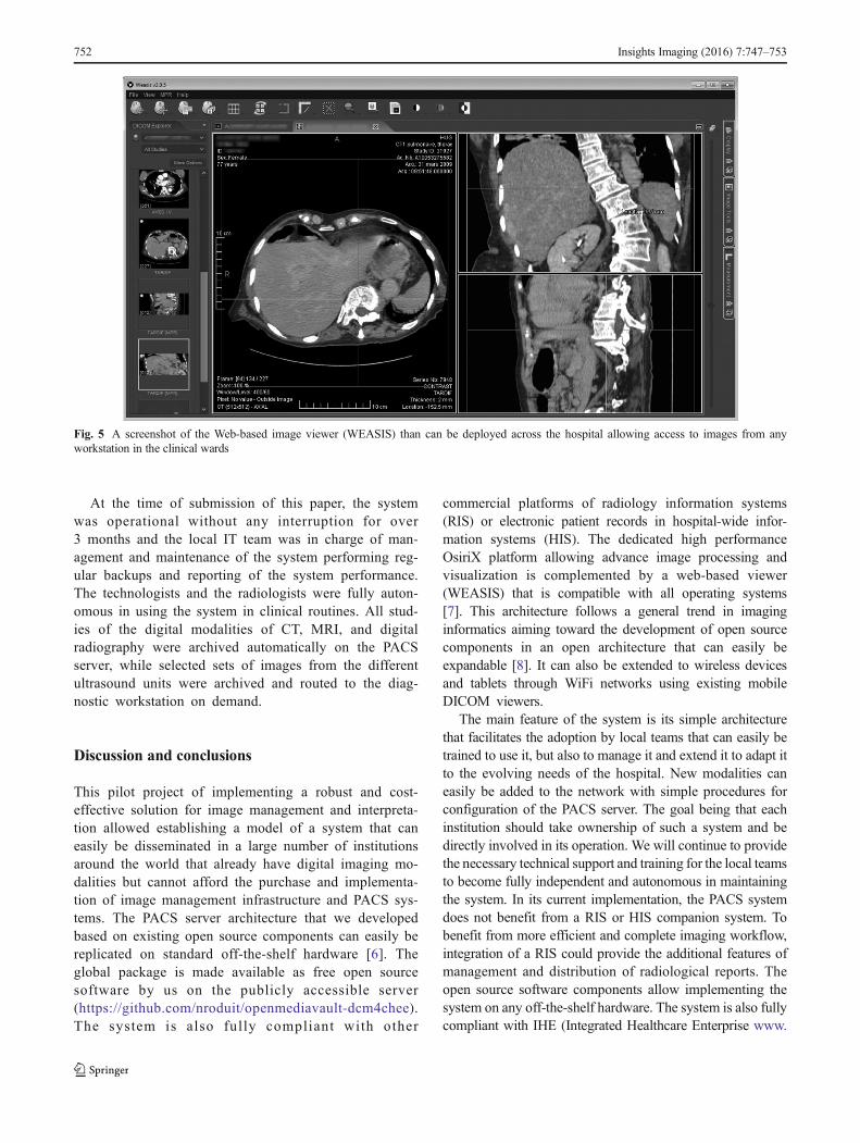
Fig. 5 A screenshot of the Web-based image viewer (WEASIS) than can be deployed across the hospital allowing access to images from anyworkstation in the clinical wards
752 Insights Imaging (2016) 7:747–753
ihe.net) profiles allowing easy integration with future hospitalinformation systems (HIS and RIS) and can easily link toelectronic medical records through a basic set of webservices. It also offers a flexible platform for teleradiology aswell as for setting up anonymized teaching and educationdatabases.
Open Access This article is distributed under the terms of the CreativeCommons Attribution 4.0 International License (http://creativecommons.org/licenses/by/4.0/), which permits unrestricted use, distribution, and reproduc-tion in any medium, provided you give appropriate credit to the originalauthor(s) and the source, provide a link to the Creative Commons license,and indicate if changes were made.
References
1. Ratib O (2009) Imaging informatics: from image management toimage navigation. Yearbook Med Inform 167–172
2. Rosset A, Spadola L, Ratib O (2004) OsiriX: an open-source soft-ware for navigating in multidimensional DICOM images. J DigitImaging 17(3):205–216
3. Ratib O, Rosset A, Heuberger J (2011) Open source software andsocial networks: disruptive alternatives for medical imaging. Eur JRadiol 78(2):259–265
4. Valeri G, Mazza FA, Maggi S, Aramini D, La Riccia L, Mazzoni G,Giovagnoni A (2015) Open source software in a practical approach forpost processing of radiologic images. Radiol Med 120(3):309–323
5. Yamauchi T, Yamazaki M, Okawa A, Furuya T, Hayashi K, SakumaT, Takahashi H, YanagawaN,KodaM (2010) Efficacy and reliabilityof highly functional open source DICOM software (OsiriX) in spinesurgery. J Clin Neurosci 17(6):756–759
6. Rosset C, Rosset A, Ratib O (2005) General consumer communica-tion tools for improved image management and communication inmedicine. J Digit Imaging 18(4):270–279
7. Haak D, Page C-E and Deserno TM (2015) A survey of DICOMviewer software to integrate clinical research and medical imaging. JDigit Imaging. (Online publication prior to print)
8. Valente F, Silva LAB, Godinho TM and Costa C (2015) Anatomy ofan extensible open source PACS. J Digit Imaging. (Online publica-tion prior to print)
Insights Imaging (2016) 7:747–753 753