P2X4 receptor controls microglia activation and favors...
20
Research Article P2X4 receptor controls microglia activation and favors remyelination in autoimmune encephalitis Alazne Zabala 1 , Nuria Vazquez-Villoldo 1 , Björn Rissiek 2 , Jon Gejo 1 , Abraham Martin 3 , Aitor Palomino 1 , Alberto Perez-Samartín 1 , Krishna R Pulagam 3 , Marco Lukowiak 2 , Estibaliz Capetillo-Zarate 1,4 , Jordi Llop 3 , Tim Magnus 2 , Friedrich Koch-Nolte 5 , Francois Rassendren 6 , Carlos Matute 1,* & María Domercq 1,** Abstract Microglia survey the brain microenvironment for signals of injury or infection and are essential for the initiation and resolution of pathogen- or tissue damage-induced inflammation. Understanding the mechanism of microglia responses during pathology is hence vital to promote regenerative responses. Here, we analyzed the role of purinergic receptor P2X4 (P2X4R) in microglia/macrophages during autoimmune inflammation. Blockade of P2X4R signaling exacerbated clinical signs in the experimental autoimmune encephalomyelitis (EAE) model and also favored microglia activa- tion to a pro-inflammatory phenotype and inhibited myelin phagocytosis. Moreover, P2X4R blockade in microglia halted oligodendrocyte differentiation in vitro and remyelination after lysolecithin-induced demyelination. Conversely, potentiation of P2X4R signaling by the allosteric modulator ivermectin (IVM) favored a switch in microglia to an anti-inflammatory phenotype, potentiated myelin phagocytosis, promoted the remyelination response, and ameliorated clinical signs of EAE. Our results provide evidence that P2X4Rs modulate microglia/macrophage inflamma- tory responses and identify IVM as a potential candidate among currently used drugs to promote the repair of myelin damage. Keywords microglia; myelin phagocytosis; P2X4 receptor; remyelination Subject Categories Immunology; Neuroscience; Pharmacology & Drug Discovery DOI 10.15252/emmm.201708743 | Received 4 December 2017 | Revised 5 June 2018 | Accepted 8 June 2018 EMBO Mol Med (2018)e8743 Introduction Multiple sclerosis (MS) is a chronic inflammatory disease of the brain and spinal cord leading to demyelination and neurodegenera- tion. The clinical disease course usually starts with reversible episodes of neurological disability (relapsing–remitting MS; RRMS), which later goes into a progressive stage with irreversible neurologi- cal decline (secondary progressive MS; SPMS; Dendrou et al, 2015). Demyelinated lesions, a hallmark of multiple sclerosis, are caused by immune cell infiltration across the blood–brain barrier (BBB) that promotes inflammation, demyelination, gliosis, and neuroaxonal degeneration (Dendrou et al, 2015; Lassmann & Bradl, 2017). Axonal loss occurs in both the acute and chronic phases of MS and its animal model experimental autoimmune encephalomyelitis (EAE), and the loss of compensatory central nervous system (CNS) mechanisms contributes to the transition from RRMS to SPMS (Ran- sohoff, 2012; Dendrou et al, 2015). Activated microglia and macro- phages are thought to contribute to neurodegeneration as their number correlates with the extent of axonal damage in MS lesions (Bitsch et al, 2000; Rasmussen et al, 2007; Fischer et al, 2013; Vogel et al, 2013). The activation of microglia/macrophages may repre- sent one of the initial steps in EAE pathogenesis, preceding and possibly triggering T-cell development and infiltration of blood- derived cells (Heppner et al, 2005; Ajami et al, 2011; Goldmann et al, 2013; Yamasaki et al, 2014; Yoshida et al, 2014). However, other studies indicate that microglia/macrophage activation counter- acts pathological processes by providing neurotrophic and immuno- suppressive factors and by promoting recovery (Kotter et al, 2006; Miron & Franklin, 2014; Lampron et al, 2015). Microglia/macrophages are highly heterogeneous immune cells with a continuous spectrum of activation states (Xue et al, 2014). The so-called classically activated or pro-inflammatory and the alter- natively activated or anti-inflammatory microglia/macrophages are 1 Achucarro Basque Center for Neurosciences, CIBERNED and Departamento de Neurociencias, Universidad del País Vasco, Leioa, Spain 2 Department of Neurology, University Medical Center, Hamburg, Germany 3 Molecular Imaging Unit, CIC biomaGUNE, San Sebastian, Spain 4 IKERBASQUE, Basque Foundation for Science, Bilbao, Spain 5 Institute of Immunology, University Medical Center, Hamburg, Germany 6 Institut de Génomique Functionnelle, CNRS UMR5203, Montpellier, France *Corresponding author. Tel: +34 94 6013244; Fax: +34 94 6015055; E-mail: [email protected] **Corresponding author. Tel: +34 94 6015681; Fax: +34 94 6015055; E-mail: [email protected] ª 2018 The Authors. Published under the terms of the CC BY 4.0 license EMBO Molecular Medicine e8743 | 2018 1 of 20 Published online: July 4, 2018
Transcript of P2X4 receptor controls microglia activation and favors...

Research Article
P2X4 receptor controls microglia activation andfavors remyelination in autoimmune encephalitisAlazne Zabala1, Nuria Vazquez-Villoldo1, Björn Rissiek2, Jon Gejo1, Abraham Martin3, Aitor Palomino1,
Alberto Perez-Samartín1, Krishna R Pulagam3, Marco Lukowiak2, Estibaliz Capetillo-Zarate1,4,
Jordi Llop3, Tim Magnus2, Friedrich Koch-Nolte5, Francois Rassendren6, Carlos Matute1,* &
María Domercq1,**
Abstract
Microglia survey the brain microenvironment for signals of injuryor infection and are essential for the initiation and resolution ofpathogen- or tissue damage-induced inflammation. Understandingthe mechanism of microglia responses during pathology is hencevital to promote regenerative responses. Here, we analyzed therole of purinergic receptor P2X4 (P2X4R) in microglia/macrophagesduring autoimmune inflammation. Blockade of P2X4R signalingexacerbated clinical signs in the experimental autoimmuneencephalomyelitis (EAE) model and also favored microglia activa-tion to a pro-inflammatory phenotype and inhibited myelinphagocytosis. Moreover, P2X4R blockade in microglia haltedoligodendrocyte differentiation in vitro and remyelination afterlysolecithin-induced demyelination. Conversely, potentiation ofP2X4R signaling by the allosteric modulator ivermectin (IVM)favored a switch in microglia to an anti-inflammatory phenotype,potentiated myelin phagocytosis, promoted the remyelinationresponse, and ameliorated clinical signs of EAE. Our results provideevidence that P2X4Rs modulate microglia/macrophage inflamma-tory responses and identify IVM as a potential candidate amongcurrently used drugs to promote the repair of myelin damage.
Keywords microglia; myelin phagocytosis; P2X4 receptor; remyelination
Subject Categories Immunology; Neuroscience; Pharmacology & Drug
Discovery
DOI 10.15252/emmm.201708743 | Received 4 December 2017 | Revised 5 June
2018 | Accepted 8 June 2018
EMBO Mol Med (2018) e8743
Introduction
Multiple sclerosis (MS) is a chronic inflammatory disease of the
brain and spinal cord leading to demyelination and neurodegenera-
tion. The clinical disease course usually starts with reversible
episodes of neurological disability (relapsing–remitting MS; RRMS),
which later goes into a progressive stage with irreversible neurologi-
cal decline (secondary progressive MS; SPMS; Dendrou et al, 2015).
Demyelinated lesions, a hallmark of multiple sclerosis, are caused
by immune cell infiltration across the blood–brain barrier (BBB) that
promotes inflammation, demyelination, gliosis, and neuroaxonal
degeneration (Dendrou et al, 2015; Lassmann & Bradl, 2017).
Axonal loss occurs in both the acute and chronic phases of MS and
its animal model experimental autoimmune encephalomyelitis
(EAE), and the loss of compensatory central nervous system (CNS)
mechanisms contributes to the transition from RRMS to SPMS (Ran-
sohoff, 2012; Dendrou et al, 2015). Activated microglia and macro-
phages are thought to contribute to neurodegeneration as their
number correlates with the extent of axonal damage in MS lesions
(Bitsch et al, 2000; Rasmussen et al, 2007; Fischer et al, 2013; Vogel
et al, 2013). The activation of microglia/macrophages may repre-
sent one of the initial steps in EAE pathogenesis, preceding and
possibly triggering T-cell development and infiltration of blood-
derived cells (Heppner et al, 2005; Ajami et al, 2011; Goldmann
et al, 2013; Yamasaki et al, 2014; Yoshida et al, 2014). However,
other studies indicate that microglia/macrophage activation counter-
acts pathological processes by providing neurotrophic and immuno-
suppressive factors and by promoting recovery (Kotter et al, 2006;
Miron & Franklin, 2014; Lampron et al, 2015).
Microglia/macrophages are highly heterogeneous immune cells
with a continuous spectrum of activation states (Xue et al, 2014).
The so-called classically activated or pro-inflammatory and the alter-
natively activated or anti-inflammatory microglia/macrophages are
1 Achucarro Basque Center for Neurosciences, CIBERNED and Departamento de Neurociencias, Universidad del País Vasco, Leioa, Spain2 Department of Neurology, University Medical Center, Hamburg, Germany3 Molecular Imaging Unit, CIC biomaGUNE, San Sebastian, Spain4 IKERBASQUE, Basque Foundation for Science, Bilbao, Spain5 Institute of Immunology, University Medical Center, Hamburg, Germany6 Institut de Génomique Functionnelle, CNRS UMR5203, Montpellier, France
*Corresponding author. Tel: +34 94 6013244; Fax: +34 94 6015055; E-mail: [email protected]**Corresponding author. Tel: +34 94 6015681; Fax: +34 94 6015055; E-mail: [email protected]
ª 2018 The Authors. Published under the terms of the CC BY 4.0 license EMBO Molecular Medicine e8743 | 2018 1 of 20
Published online: July 4, 2018

at the opposite ends of this spectrum (Mosser & Edwards, 2008;
Murray et al, 2014; but see also Ransohoff, 2016). Although such a
classification underestimates the complexity of macrophage/micro-
glia plasticity, the distinction nevertheless provides a useful frame-
work for exploring the diverse functions of the innate immune
system in disease pathogenesis. Anti-inflammatory macrophages
have been shown to play central roles in mediating Th2 immunity,
wound healing, and the suppression of effector T-cell function
(Mosser & Edwards, 2008). In MS, pro-inflammatory microglia exist
in all types of lesions and correlate with axonal damage, whereas
anti-inflammatory microglia are increased in acute active lesions
and in the rim of chronic active lesions where efficient remyelina-
tion occurs (Miron et al, 2013). Anti-inflammatory microglia secrete
anti-inflammatory cytokines and growth factors that promote oligo-
dendrocyte progenitor differentiation and that protect neurons from
damage (Butovsky et al, 2006; Mikita et al, 2011; Starossom et al,
2012; Miron et al, 2013; Yu et al, 2015). Finally, a block in the pro-
inflammatory-to-anti-inflammatory switch has been hypothesized to
contribute to remyelination failure in chronic inactive MS lesions
(Miron et al, 2013; Sun et al, 2017).
As key immune effector cells of the CNS, surveillant microglia
act as sensor of infection and pathologic damage of the brain, lead-
ing to a rapid plastic process of activation that culminates in the
endocytosis and phagocytosis of damaged tissue. Multiple signals
converge on microglial cells to actively maintain or alter their func-
tional state and orchestrate the specific repertoire of microglial func-
tions. In the absence of pathogens, microglia sense the injury by
recognizing the release of molecules that are normally located inside
the cell, known as damage-associated molecular patterns (DAMPs)
or “endogenous danger signals” (Di Virgilio, 2007). Recently, ATP
has been characterized as a danger signal implicated in innate and
adaptive immunity (Junger, 2011), leading to a plethora of
responses in microglia through its interaction with their purinergic
P2 receptors (Domercq et al, 2013). On the basis of their signaling
properties, P2 receptors can be further subdivided into metabotropic
P2Y receptors (P2YRs) that are G-protein-coupled, and ionotropic
P2X receptors (P2XRs) that are nucleotide-gated ion channels
(Domercq et al, 2013). We have previously observed that purinergic
P2X4R is highly expressed in activated microglia in EAE and in
human MS optic nerve samples (Vazquez-Villoldo et al, 2014).
Here, we identified P2X4R as a significant regulator of microglia
inflammatory cascade and the resultant repair response after
demyelination.
Results
P2X4R expression is upregulated during EAE
Following peripheral nerve injury, microglia in the spinal dorsal
horn exhibit a reactive phenotype and upregulate expression of a
variety of genes, including purinergic P2x4r (Tsuda et al, 2003;
Beggs et al, 2012). Accordingly, we detected previously an increase
in P2x4r mRNA expression in multiple sclerosis (MS) samples and
at the peak of the immune attack in the acute EAE model (Vazquez-
Villoldo et al, 2014). We have further analyzed the time course of
the P2x4r expression in EAE mice immunized with myelin oligoden-
drocyte glycoprotein (MOG). Levels of P2x4r expression were
increased at the peak of the disease and remained elevated during
the recovery phase (30 days; Fig 1A). Interestingly, there was a
strong correlation between P2x4r expression and the neurological
score, both at the peak and at recovery (Fig 1A; r2 = 0.99 and 0.61,
respectively). Previous data have demonstrated that interferon regu-
latory factor 8 (IRF8)–IRF5 transcriptional axis is a critical regulator
for shifting microglia toward a P2X4R+-reactive phenotype (Masuda
et al, 2014). Accordingly, we observed that Irf8 and Irf5 transcrip-
tion factors were upregulated at the peak and recovery phases of the
disease and their expression correlated well with P2x4r expression
(Fig 1B). P2x4r upregulation was also detected in FACS-isolated
microglia (Cd11b+CD45high) in the spinal cord at the EAE recovery
phase (Fig 1C).
P2X4R blockade exacerbates EAE
We then tested the role of P2X4R in EAE pathogenesis in mice
treated daily with P2X4R antagonist TNP-ATP (10 mg/kg) from the
onset of the disease at 10 days postimmunization (dpi). This time
window is coincident with microglia activation, as previously
reported (Ajami et al, 2011), and does not interfere with immune
priming. Microglia die at early stages of EAE induction, and this
population is replenished by infiltrating monocytes, promoting
progression to paralysis (Ajami et al, 2011). Because P2X4R block-
ade was previously demonstrated to prevent LPS-induced microglial
cell death (Vazquez-Villoldo et al, 2014), we reasoned that blockade
of microglial cell death by TNP-ATP would prevent replacement by
monocyte and improve clinical signs of EAE. In contrast, blockade
of P2X4R with TNP-ATP exacerbated EAE disease (Fig 2A). Accord-
ingly, the latency of the corticospinal tract was significantly
increased in TNP-ATP-treated mice (Fig 2A), indicative of higher
demyelination. We next stained spinal cord sections at the end of
the experiment for ionized calcium-binding adapter protein 1 (Iba1),
which is a marker commonly used to identify microglia/macro-
phages in the CNS. TNP-ATP-treated mice showed a significant
increase in Iba1+ cells in the white and gray matter of the spinal
cord versus vehicle-treated EAE mice (Fig 2B). The increase was
observed even at the same neurological score (Fig EV1), probably
indicating that the increase in microglia/macrophage number is not
only the consequence of the higher EAE severity after P2X4R
blockade.
We next confirmed the role played by P2X4R in EAE pathogene-
sis using P2X4�/� mice. We first checked whether P2X4R deficiency
could affect microglia and oligodendrocytes in normal conditions.
We did not detect any change in the number or morphology of
Iba1+ cells nor in the number of Olig2+ oligodendrocytes in the
spinal cord of 2-month-old P2X4�/� mice (Appendix Fig S1). Then,
we compared neurological score in WT and P2X4�/� MOG-injected
mice. In accordance with results obtained with TNP-ATP, P2X4�/�
mice showed an exacerbated EAE, higher latency of the corti-
cospinal tract, and an increase in the number of Iba1+ microglia/
macrophage cells (Fig 2C). To further assess that the effect of TNP-
ATP was P2X4R-dependent, we treated P2X4�/� mice with TNP-
ATP from the onset of the disease. TNP-ATP failed to alter the
course of EAE disease in P2X4�/� mice (Fig 2D). All these data con-
firmed the role played by P2X4R in EAE pathogenesis.
During EAE, T cells are primed in the mouse peripheral immune
system before the onset of the clinical signs (Stromnes & Goverman,
2 of 20 EMBO Molecular Medicine e8743 | 2018 ª 2018 The Authors
EMBO Molecular Medicine P2X4R and autoimmune demyelination Alazne Zabala et al
Published online: July 4, 2018
A
B
C
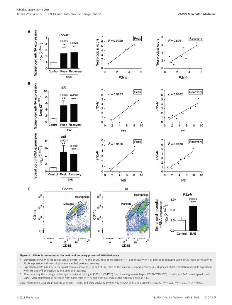
Figure 1. P2x4r is increased at the peak and recovery phases of MOG-EAE mice.
A Expression of P2x4r in the spinal cord of control (n = 7) and of EAE mice at the peak (n = 4) and recovery (n = 8) phases as analyzed using qPCR. Right, correlation ofP2x4r expression with neurological score at EAE peak and recovery.
B Expression of Irf8 and Irf5 in the spinal cord of control (n = 7) and of EAE mice at the peak (n = 4) and recovery (n = 8) phases. Right, correlation of P2x4r expressionwith Irf5 and Irf8 expression at EAE peak and recovery.
C Plots depicting the strategy to distinguish resident microglia (CD11b+/CD45low) from invading macrophages (CD11b+/CD45high) in naïve and EAE mouse spinal cords.Right, P2x4r expression in microglia from naïve mice (n = 4) and from EAE mice at the recovery phase (n = 6).
Data information: Data are presented as mean � s.e.m. and were analyzed by one-way ANOVA (A, B) and Student’s t-test (C). *P < 0.05, **P < 0.01, ***P < 0.001.
ª 2018 The Authors EMBO Molecular Medicine e8743 | 2018 3 of 20
Alazne Zabala et al P2X4R and autoimmune demyelination EMBO Molecular Medicine
Published online: July 4, 2018
A
B
C
E
F
D
Figure 2.
4 of 20 EMBO Molecular Medicine e8743 | 2018 ª 2018 The Authors
EMBO Molecular Medicine P2X4R and autoimmune demyelination Alazne Zabala et al
Published online: July 4, 2018

2006), suggesting that treatment with TNP-ATP starting after the
onset of the disease would have no impact on T-cell infiltration. In
order to corroborate this hypothesis, we analyzed immune cell infil-
tration (CD4+ T cells, CD8+ T cells, cd T cells, neutrophils, and
macrophages) in the brain (Fig EV1) and spinal cord (Fig 2E) at the
peak of disease in vehicle- and TNP-ATP-treated mice by flow cytom-
etry in another set of mice. We found similar proportions for all of
CNS-infiltrating CD45+ leukocytes in TNP-ATP- and vehicle-treated
mice at the peak of EAE except for CD8+ T cells in the spinal cord
(Fig 2E) and brain (Fig EV1) which were significantly increased in
TNP-ATP-treated mice. We further assessed the role of P2X4R on T-
cell function directly by stimulating isolated splenocytes in vitro with
anti-CD3/CD28 in the presence or absence of TNP-ATP for 3 days
and then measured proliferation of CD4+ and CD8+ T cells. Further-
more, we determined T-cell cytokine production via intracellular
cytokine staining of PMA/ionomycin-stimulated cells. Importantly,
in vitro assays showed that blockade of P2X4R on lymphocytes did
not alter T-cell proliferation or cytokine production (Fig 2F).
To further exclude the involvement of P2X4R in adaptive
immune system, we performed an additional EAE experiment and
treated mice with TNP-ATP during the priming phase (0–17 dpi).
TNP-ATP treatment did not affect disease development (Fig 3A). At
the peak, we quantified by flow cytometry the immune response in
periphery (spleen and lymph nodes) and in the spinal cord. Treat-
ment with TNP-ATP during the priming phase did not change the
number of CD4+ T cells, CD8+ T cells, and cd T cells in spleen,
lymph nodes, or the spinal cord (Fig 3B). To further assess the
CD4+ T-cell response, we measured mRNA for Foxp3 and Ror, tran-
scription factors that specify Tregs and Th17, respectively, and Ifng,
signature cytokine for Th1 cells. We did not detect any change in
transcript expression after TNP-ATP treatment (Fig 3C). Finally, to
check whether P2X4R blockage could alter infiltration of T cells
during the immune priming phase, we measured BBB disruption by
PET imaging using a radioligand that enabled tracking of matrix
metalloproteinase (MMP) activity as a marker of early lesions and
ongoing leukocyte infiltration (Gerwien et al, 2016). An increase in
MMP activity was detected in the lumbar spinal cord at the peak of
the disease; however, treatment with TNP-ATP during the immune
priming did not change the MMP-PET signal in EAE (Fig 3D). Alto-
gether, these data suggest that blockade of P2X4R did not interfere
with the efficacy of immunization and the T-cellular immune
response against MOG.
Role of P2X4R on microglia polarization
Since myelin clearance is necessary for remyelination and recovery
(Li et al, 2005; Kotter et al, 2006; Neumann et al, 2009) and phago-
cytosis and remyelination are modulated by microglia/macrophage
polarization (Miron et al, 2013), we hypothesized that P2X4R could
be involved in this process. We first checked the status of micro-
glia/macrophage in TNP-ATP- and vehicle-treated EAE mice. We
performed gene expression profiling from the lumbar spinal cord of
vehicle- and TNP-ATP-treated EAE mice at the peak and recovery
phases (Fig 4A and B). Expression of pro-inflammatory and anti-
inflammatory genes involved in microglia/macrophage activation
was analyzed using a 96.96 Dynamic Array™ integrated fluidic
circuit (Fluidigm). In accordance with previous data showing that
macrophages and microglia showed an intermediate activation
status in MS (Vogel et al, 2013), most pro-inflammatory and anti-
inflammatory genes were significantly increased at the EAE peak
(Fig EV2) and recovery phase (Fig 4B). Blockade of P2X4R with
TNP-ATP did not significantly alter anti-inflammatory gene expres-
sion, but it significantly increased pro-inflammatory gene expression
at the recovery phase (Fig 4B), but not at EAE peak (Fig EV2). A
higher increase in pro-inflammatory gene expression was also
detected on microglia FACS-sorted (Cd11b+CD45high; see gating in
Fig 1C) from EAE P2X4�/� mice versus EAE WT (Fig 4C). Accord-
ingly, we found an increase in iNOS expression in microglia/macro-
phage after EAE in P2X4�/� mice and in TNP-ATP-treated mice
(Fig 4D and E). These data suggest that P2X4R could be influencing
microglia/macrophage activation.
To further analyze the influence of P2X4R on microglia polariza-
tion in vitro, cells were primed with colony-stimulating factors to
differentiate into pro-inflammatory and anti-inflammatory microglia
according to a previous protocol (Fig 5A; see details in Materials
and Methods) and analyzed by immunocytochemistry using pro-
inflammatory (iNOS) and anti-inflammatory (mannose receptor;
MRC1) markers. Blockade of P2X4R with TNP-ATP induced a
significant increase in iNOS+ cells and a significant reduction in
MRC1+ cells (Fig 5B). Accordingly, qPCR analysis revealed an
increase in pro-inflammatory genes and a decrease in anti-inflam-
matory genes after TNP-ATP treatment during polarization
(Fig 5C). Similar results were obtained in P2X4�/� microglia
(Fig EV3). Altogether, these data suggest that P2X4Rs modulate
microglial polarization.
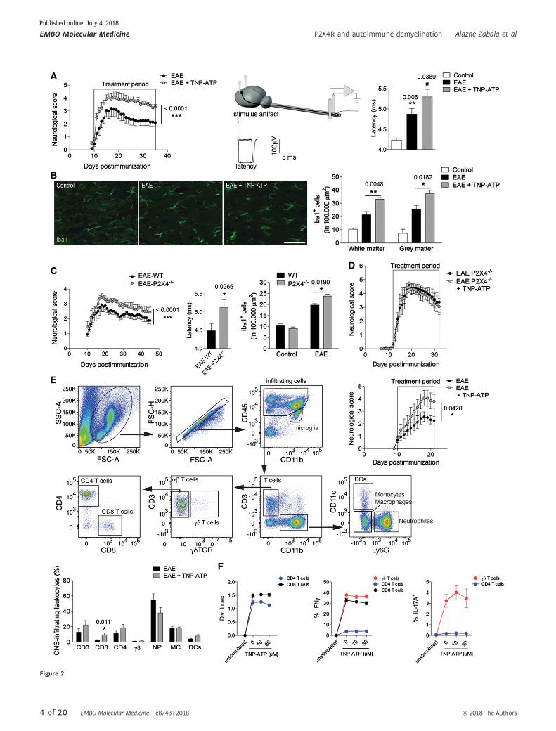
◀ Figure 2. P2X4R blockade increases autoimmune inflammation.
A Clinical score of vehicle (n = 10)- and TNP-ATP (10 mg/kg)-treated mice (n = 10) after EAE induction. Right, scheme, raw data, and histogram showing axonconduction latency in the corticospinal tract of control (n = 4), EAE (n = 10), and TNP-ATP-treated EAE mice (n = 10). Symbols indicate significance versus control (*)or versus EAE (#).
B Histology of spinal cord sections using Iba1 antibodies shows a significant increase in microglia/macrophage cell number in control mice (n = 3), in TNP-ATP-treatedEAE mice (n = 4), and in non-treated EAE mice (n = 5). Scale bar = 50 lm.
C Neurological score, axon conduction latency, and microglia/macrophage quantification in control WT (n = 3) and P2X4�/� mice (n = 3) and after EAE induction in WT(n = 10) and P2X4�/� mice (n = 10).
D Neurological score after EAE induction in P2X4�/� mice treated with vehicle (n = 4) or TNP-ATP (n = 5).E Left, flow cytometry gating strategy for analysis of infiltrates in the spinal cord of EAE mice at peak (18–21 dpi). Right, graph showing the neurological score of the
mice used for the analysis (top) (n = 8). Histogram showing flow cytometric quantification of CD3+, CD8+, CD4+, and cd T cells, neutrophils (NP), macrophages (MC),and dendritic cells (DC) in the spinal cord at EAE peak (bottom).
F Immune response analysis in vitro. T-cell proliferation assay and flow cytometric quantification of cytokine expression in T cells after PMA/ionomycin stimulation inthe absence or presence of TNP-ATP (10–30 lm, 3 days; n = 3 independent experiments).
Data information: Data are presented as mean � s.e.m. Statistics were performed with Mann–Whitney U-test (neurological score) and Student’s t-test. */#P < 0.05,**P < 0.01, ***P < 0.001.
ª 2018 The Authors EMBO Molecular Medicine e8743 | 2018 5 of 20
Alazne Zabala et al P2X4R and autoimmune demyelination EMBO Molecular Medicine
Published online: July 4, 2018

Effect of P2X4R on oligodendrocyte differentiationand myelination
A switch from a pro-inflammatory to an anti-inflammatory pheno-
type occurs in microglia and peripherally derived macrophages
during remyelination in MS, and this change is essential for efficient
remyelination (Miron et al, 2013). These data led us to the hypothe-
sis that P2X4R blockade in microglia could be indirectly affecting
oligodendrocyte differentiation and remyelination. We first charac-
terized the expression and function of P2X4R in oligodendrocytes
A
B
C
D
Figure 3. P2X4R does not interfere with immune priming.
A Neurological score of vehicle (n = 23 from three independent experiments)- and TNP-ATP-treated (n = 21 from three independent experiments) mice after EAE. Micewere treated daily with TNP-ATP from 0 dpi to EAE peak.
B Flow cytometric quantification of CD8+, CD4+, and cd T cells in lymph nodes (LN), spleen, and spinal cord at EAE peak of vehicle (n = 7)- and TNP-ATP-treated mice(n = 5) (gating strategy as described in Fig 2E).
C Relative mRNA expression of Foxp3, Ifng, and Ror in LN, spleen, and spinal cord at EAE peak of vehicle (n = 6)- and TNP-ATP-treated mice (n = 7).D Representative images of 18F-MMPi PET imaging in control mice (n = 6), EAE mice (n = 10), and EAE mice treated with TNP-ATP (n = 9) as described in (A).
18F-MMPi signal in the lumbar spinal cord was expressed as %ID/g.
Data information: Data are presented as mean � s.e.m. and were analyzed by one-way ANOVA. ***P < 0.001.
6 of 20 EMBO Molecular Medicine e8743 | 2018 ª 2018 The Authors
EMBO Molecular Medicine P2X4R and autoimmune demyelination Alazne Zabala et al
Published online: July 4, 2018

A
C
E
D
B
Figure 4.
ª 2018 The Authors EMBO Molecular Medicine e8743 | 2018 7 of 20
Alazne Zabala et al P2X4R and autoimmune demyelination EMBO Molecular Medicine
Published online: July 4, 2018
and microglia. Double-immunocytochemistry analysis showed that
P2X4R expression is virtually absent from Olig2+ oligodendrocytes
and highly enriched in isolectin B4+ microglial cells in microglia–
oligodendrocyte progenitor cell (OPC) mixed cultures (Fig 6A;
Appendix Fig S2). On the contrary, P2X7 receptors are expressed in
both cell populations. The function of P2X4R in microglia and oligo-
dendrocytes was further analyzed by electrophysiology in acute
slices from CXCR3-GFP and PLP-DsRed mice, respectively. When
ATP was applied to whole-cell-clamped microglia at �70 mV, it
elicited a massive inward current (619 � 100 pA; n = 8) that was
significantly reduced by A438079 (10 lM), antagonist of P2X7R
(172 � 69 pA; n = 8; Fig 6B). However, the remaining ATP-evoked
current was virtually abolished in the presence of TNP-ATP
(Fig 6B). These data suggest that P2X7R and P2X4R are the main
contributors to ATP-evoked currents in microglia. In contrast, ATP-
evoked currents in oligodendrocytes (80 � 21 pA) were totally
abolished in the presence of A438079 (Fig 6B), indicating the lack
of functional P2X4R on these cells. To further exclude any direct
role of P2X4R on oligodendrocyte differentiation, we stimulated
culture oligodendrocytes with ATPcS (10 lM, 3 days) at low
concentrations to avoid P2X7R activation. ATPcS did not induce any
change in oligodendrocyte differentiation (Fig EV4). Altogether,
these results suggested that oligodendrocytes lack functional
P2X4Rs.
Next, we analyzed whether microglia P2X4R could play a role
on oligodendrocyte differentiation. To this end, we conditioned
oligodendrocyte culture medium (SATO; see Materials and Meth-
ods) with control, pro-inflammatory, and anti-inflammatory micro-
glia for 24 h and then incubated oligodendrocyte progenitors with
microglia-conditioned media for 3 days (see cartoon in Fig 6C). All
microglia-conditioned media induced an increase in the number of
mature MBP+ oligodendrocytes as compared to polarization
factors alone (Fig 6C). However, anti-inflammatory microglia
induced a higher increase in oligodendrocyte differentiation, an
effect that was blocked in the presence of TNP-ATP (Fig 6C). Since
BDNF enhances oligodendrocyte differentiation and myelination
(Wong et al, 2013) and P2X4R stimulation in microglia has been
linked to BDNF release (Tsuda et al, 2003; Coull et al, 2005; see
also Fig EV4), we analyzed BDNF production in differentially
polarized microglia. Western blot analysis revealed a significant
increase in BDNF production in anti-inflammatory microglia,
which was significantly reduced in the presence of TNP-ATP
(Fig 6D). Accordingly, Bdnf mRNA expression during the recovery
phase of EAE was significantly reduced in the presence of TNP-
ATP, a fact that correlated with the downregulation of Mbp
expression (Fig 6E). Bdnf mRNA was also reduced in FACS-
isolated microglia from control and EAE P2X4�/� mice (Fig EV4).
These data suggested that P2X4R blockade in microglia inhibits
oligodendrocyte differentiation by shifting microglia toward a pro-
inflammatory phenotype, an effect that could impede remyelination.
To assess that possibility, we further checked the impact of P2X4R
on remyelination on the lysolecithin-induced demyelination model
using ex vivo organotypic cerebellar slice culture (Fig 6F), a model
independent of adaptive immune system. Indeed, slices treated with
TNP-ATP (10 lM) during the remyelination phase (7 days) showed
a significant decrease in their remyelination capacity, as revealed by
analyzing MBP expression by Western blot (Fig 6F).
Myelin clearance is modulated by P2X4Rs
Phagocytosis of myelin debris by microglia is essential for an effi-
cient regenerative response (Kotter et al, 2006; Ruckh et al, 2012).
We observed a higher accumulation of disrupted or fragmented
myelin after EAE induction in TNP-ATP-treated mice (Fig 7A) as
well as in P2X4�/� mice (Fig EV5). Fragmented myelin yielded
higher immunoreactivity likely due to additional exposed epitopes
of the MBP antibody. Myelin debris accumulation could be the
result of higher demyelination or a defect on myelin phagocytosis.
Indeed, we observed higher accumulation of myelin debris not
surrounded by microglia phagocytic processes in TNP-ATP-treated
mice (Fig 7A) at the recovery phase. Because of that, we challenged
the hypothesis that these features could be the consequence of a fail-
ure on microglia/macrophage phagocytosis. For that, myelin was
isolated from adult rat whole brain using sucrose gradient (Norton
& Poduslo, 1973), labeled with the dye Alexa 488, and added to
microglia cultures. In order to efficiently clear up myelin, microglia
should internalize myelin and deliver it to lysosomes to degrade it.
We monitored by confocal microscopy myelin endocytosis and
myelin degradation time course. At initial stages, myelin endocyto-
sis (1 h) was significantly increased in anti-inflammatory microglia
(Fig 7B), in accordance with recent data (Healy et al, 2016). Block-
ade of P2X4R with TNP-ATP significantly reduced the anti-inflam-
matory-dependent increase in myelin endocytosis (Fig 7B). We then
checked myelin degradation in the different polarized microglia
populations. After 1-h incubation with Alexa 488-myelin, microglia
were kept in label-free growth medium for up to 6 days in the pres-
ence of polarizing factors. We could not eliminate polarization
factors because microglia activation quickly reverses without them
(unpublished observations). We found that microglia retained Alexa
488-labeled myelin for up to 6 days and only about 30% of
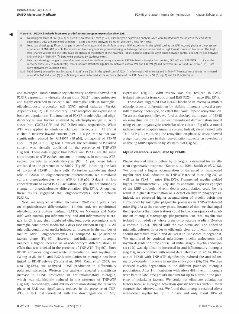
◀ Figure 4. P2X4R blockade increases pro-inflammatory gene expression after EAE.
A Neurological score of EAE (n = 4) or TNP-ATP-treated EAE mice (n = 4) used for gene expression analysis. Mice were treated from the onset to the end of theexperiment. Data are presented as mean � s.e.m. and were analyzed by Mann–Whitney U-test. *P < 0.05.
B Heatmap showing significant changes in pro-inflammatory and anti-inflammatory mRNA expression in the spinal cord at the EAE recovery phase in the presenceor absence of TNP-ATP (n = 3). The expression levels of genes are presented using fold-change values transformed to Log2 format compared to control. The Log2(fold-change values) and the color scale are shown at the bottom of the heatmap. Tables indicate statistical significance between control and EAE (*) and betweenEAE and EAE + TNP-ATP (#). Data were analyzed by Student’s t-test.
C Heatmap showing changes in pro-inflammatory and anti-inflammatory markers in FACS-isolated microglia from control, EAE WT, and EAE P2X4�/� mice at therecovery phase (n = 2 in duplicate). Tables indicate statistical significance between control WT and EAE WT (*) and between EAE WT and EAE P2X4�/� (#). Datawere analyzed by Student’s t-test.
D, E iNOS (green) expression was increased in Iba1+ cells (red) in the spinal cord of P2X4�/� mice versus WT mice (D) and in TNP-ATP-treated mice versus non-treatedmice after EAE induction (E) (n = 3). Analysis was performed at the recovery phase of the EAE. Scale bar = 50 (D, top; E) and 25 (D, bottom) lm.
8 of 20 EMBO Molecular Medicine e8743 | 2018 ª 2018 The Authors
EMBO Molecular Medicine P2X4R and autoimmune demyelination Alazne Zabala et al
Published online: July 4, 2018

internalized myelin was degraded in that time in control microglia
(Fig 7C). Anti-inflammatory microglia showed a higher capacity to
degrade myelin at 3 and 6 days, whereas pro-inflammatory micro-
glia degraded myelin less efficiently (Fig 7C). Moreover, P2X4R
blockade by TNP-ATP significantly attenuated the increase on
myelin degradation observed in anti-inflammatory microglia and
exacerbated the deficits on myelin degradation characteristic of pro-
inflammatory microglia (Fig 7D). These results suggest that P2X4R
blockade could contribute to the failure on myelin phagocytosis
necessary prior to regeneration.
P2X4R potentiation ameliorates EAE
We then explored the therapeutic potential of P2X4R potentiation.
Ivermectin (IVM) is an FDA-approved semi-synthetic macrocyclic
lactone used in veterinary and clinical medicine as an anti-parasitic
agent. IVM allosterically modulates both ion conduction and chan-
nel gating of P2X4Rs (Priel & Silberberg, 2004). We first analyzed
the role of P2X4R potentiation in EAE pathogenesis in mice treated
daily with IVM (1 mg/kg) after the onset of the disease (14 dpi).
IVM induced a rapid and significant amelioration of the motor
A
B
C
Figure 5. P2X4R modulates microglia polarization.
A Schematic representation of microglia polarization protocol.B Staining for iNOS (red) (n = 7) and mannose receptor (MRC1, green) (n = 4) in different activated microglia in the absence or presence of TNP-ATP (10 lM). Scale
bar = 50 lm.C qPCR quantification of pro-inflammatory genes (Ccl2 and Nos2) and anti-inflammatory genes (Arg1 and Mrc1) in different activated microglia in the absence or
presence of TNP-ATP (10 lM) (n = 3).
Data information: Data are presented as mean � s.e.m. and were analyzed by one-way ANOVA. */#P < 0.05, **/##P < 0.01, ***P < 0.001 versus control (*) or versus pro-/anti-inflammatory microglia (#).
ª 2018 The Authors EMBO Molecular Medicine e8743 | 2018 9 of 20
Alazne Zabala et al P2X4R and autoimmune demyelination EMBO Molecular Medicine
Published online: July 4, 2018

A
C
EF
B
D
Figure 6.
10 of 20 EMBO Molecular Medicine e8743 | 2018 ª 2018 The Authors
EMBO Molecular Medicine P2X4R and autoimmune demyelination Alazne Zabala et al
Published online: July 4, 2018
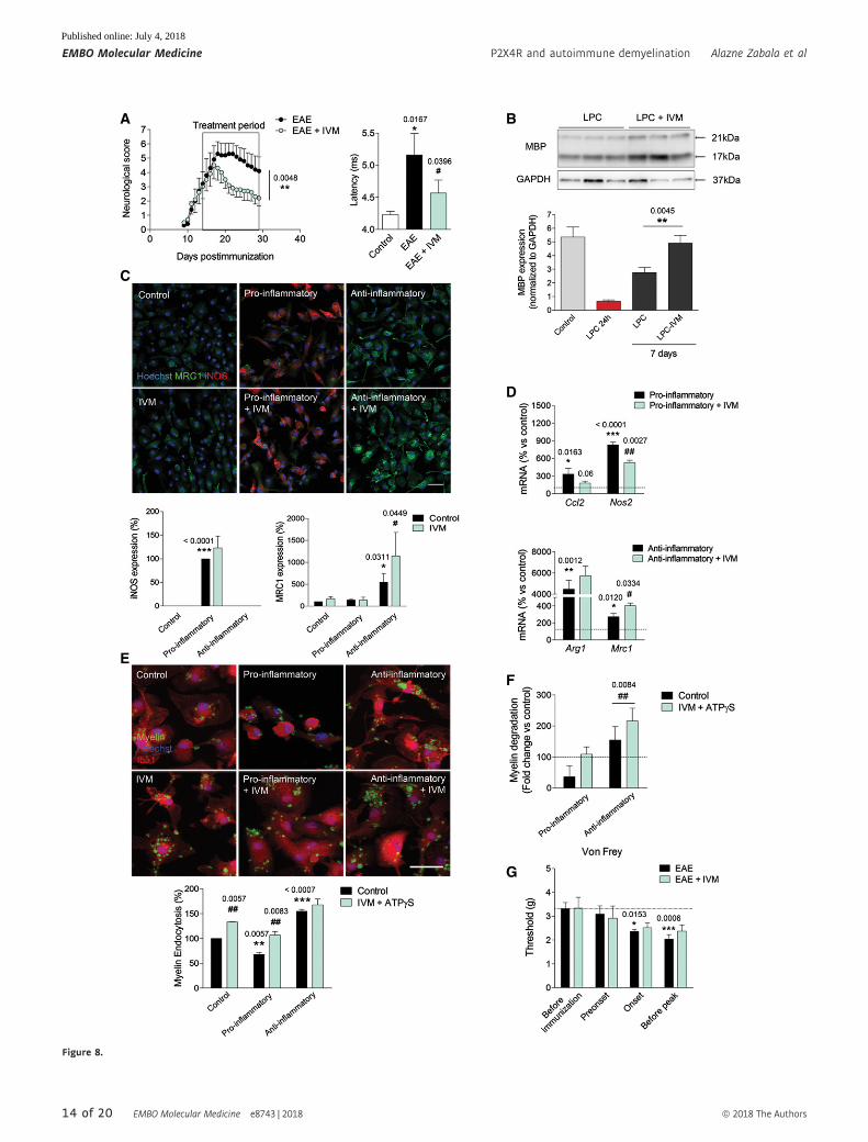
deficits of EAE (Fig 8A) that lead to an improvement in corticospinal
tract function in the recovery phase, as revealed by the decrease in
the latency of corticospinal tract (Fig 8A). These data indicate that
IVM treatment effectively promoted myelin recovery. Accordingly,
IVM significantly enhanced remyelination in the lysolecithin-
induced demyelination model in organotypic cerebellar slices
(Fig 8B).
Then, we analyzed the effect of IVM on microglial polarization
and phagocytosis in vitro. IVM (3 lM) induced a significant increase
in polarization to an anti-inflammatory phenotype (Fig 8C). Accord-
ingly, qPCR analysis revealed a decrease in pro-inflammatory genes
and an increase in anti-inflammatory genes after IVM treatment
during polarization (Fig 8D). Finally, we analyzed the effect of IVM
on myelin phagocytosis. We did not detect a potentiation of myelin
endocytosis (1 h) in anti-inflammatory microglia, perhaps because
myelin endocytosis capacity is already saturated. However, myelin
endocytosis was significantly increased in pro-inflammatory micro-
glia and even in control microglia in the presence of IVM (Fig 8E).
Regarding myelin degradation, long-term treatment with ivermectin
potentiated myelin degradation in control (Fig EV5), pro-inflamma-
tory, and anti-inflammatory microglia (Fig 8F). The effect of IVM in
myelin phagocytosis was absent in P2X4�/� microglia (Fig EV5).
Previous studies have described P2X4R to be located intracellularly
in late endosomes and lysosome membranes where they form
functional ATP-activated cation channels regulated by luminal ATP
in a pH-dependent manner (Huang et al, 2014). We observed that
IVM induced lysosome fusion, an effect dependent on P2X4R
(Fig EV5). Because fusion of lysosomes with late endosomes gener-
ates acidic endolysosomes with high cathepsin activity for phago-
cytic degradation, we then measured intralysosomal pH in
microglia. Anti-inflammatory microglia showed an acidic shift of
lysosomes, which correlates with its higher phagocytic capacity.
Moreover, IVM treatment (3 lM, 16 h) induced a significant acidifi-
cation of lysosomes (Fig EV5). Altogether, data suggested that
P2X4R potentiation by IVM could favor microglia switch necessary
to efficient remyelination.
The therapeutic potential of ivermectin in MS could be compro-
mised by its possible influence in neuropathic pain. Thus, a recent
study reported the anti-allodynic effect of a novel P2X4R antagonist
in mice with traumatic nerve damage, although the antagonist did
not affect acute nociceptive pain and motor function (Matsumura
et al, 2016). We therefore measured the effects of IVM in mechani-
cal allodynia at different stages of EAE before the appearance of
severe motor deficits. Withdrawal thresholds were significantly
diminished in both vehicle- and IVM-treated mice at EAE onset and
before EAE peak relative to baseline responses, but there were no
significant differences between the two cohorts at any stage
(Fig 8G).
Discussion
Brain injury induces an upregulation of P2X4R and shifts microglia
toward a P2X4R-expressing reactive state through an IRF8–IRF5
transcriptional axis (Beggs et al, 2012). In the present study, we
showed that IRF8–IRF5–P2X4R is upregulated in the peak and recov-
ery phases of EAE. Moreover, we demonstrated that blockade of
P2X4R exacerbates EAE, whereas potentiation with IVM ameliorates
this experimental disease. Mechanistically, P2X4 receptor signaling
potentiation in microglia/macrophages favors a switch to an anti-
inflammatory phenotype that, by secreting factors such as BDNF
and increasing myelin phagocytosis, leads to higher remyelination.
Altogether, data here suggest that P2X4R upregulation could be a
marker of the neuroinflammatory response in MS and that potentia-
tion of signaling by P2X4R has therapeutic potential to treat
demyelinating disorders.
Microglial P2X4R upregulation through IRF8–IRF5 transcription
factors, the P2X4R+ state of microglia, seems to be common in most
acute and chronic neurodegenerative diseases associated with
inflammation (reviewed in Domercq et al, 2013). IRF5 drives de
novo expression of P2X4R by directly binding to the promoter region
(Masuda et al, 2014). Here, we showed that Irf5, Irf8, and P2x4r
mRNA expression is increased and is correlated at the peak as well
as in the recovery phase of the EAE. Recent genome-wide SNP anal-
ysis has identified IRF8 as a susceptibility factor for multiple sclero-
sis (De Jager et al, 2009). In addition, genetic polymorphisms in
human IRF5 that lead to the expression of various unique isoforms
or higher expression of Irf5 mRNA have been linked to autoimmune
diseases, including MS (Kristjansdottir et al, 2008). IRF5 and IRF8
play a key role in the induction of pro-inflammatory cytokines,
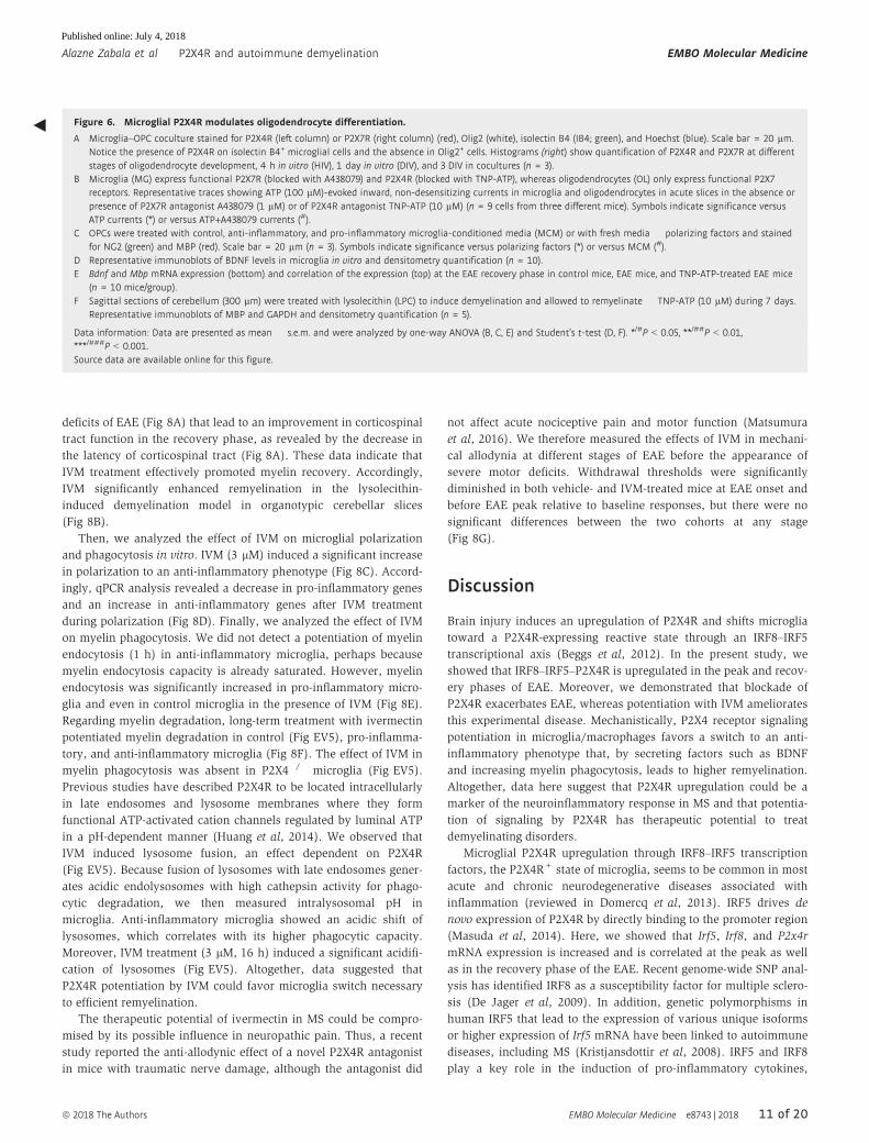
◀ Figure 6. Microglial P2X4R modulates oligodendrocyte differentiation.
A Microglia–OPC coculture stained for P2X4R (left column) or P2X7R (right column) (red), Olig2 (white), isolectin B4 (IB4; green), and Hoechst (blue). Scale bar = 20 lm.Notice the presence of P2X4R on isolectin B4+ microglial cells and the absence in Olig2+ cells. Histograms (right) show quantification of P2X4R and P2X7R at differentstages of oligodendrocyte development, 4 h in vitro (HIV), 1 day in vitro (DIV), and 3 DIV in cocultures (n = 3).
B Microglia (MG) express functional P2X7R (blocked with A438079) and P2X4R (blocked with TNP-ATP), whereas oligodendrocytes (OL) only express functional P2X7receptors. Representative traces showing ATP (100 lM)-evoked inward, non-desensitizing currents in microglia and oligodendrocytes in acute slices in the absence orpresence of P2X7R antagonist A438079 (1 lM) or of P2X4R antagonist TNP-ATP (10 lM) (n = 9 cells from three different mice). Symbols indicate significance versusATP currents (*) or versus ATP+A438079 currents (#).
C OPCs were treated with control, anti-inflammatory, and pro-inflammatory microglia-conditioned media (MCM) or with fresh media � polarizing factors and stainedfor NG2 (green) and MBP (red). Scale bar = 20 lm (n = 3). Symbols indicate significance versus polarizing factors (*) or versus MCM (#).
D Representative immunoblots of BDNF levels in microglia in vitro and densitometry quantification (n = 10).E Bdnf and Mbp mRNA expression (bottom) and correlation of the expression (top) at the EAE recovery phase in control mice, EAE mice, and TNP-ATP-treated EAE mice
(n = 10 mice/group).F Sagittal sections of cerebellum (300 lm) were treated with lysolecithin (LPC) to induce demyelination and allowed to remyelinate � TNP-ATP (10 lM) during 7 days.
Representative immunoblots of MBP and GAPDH and densitometry quantification (n = 5).
Data information: Data are presented as mean � s.e.m. and were analyzed by one-way ANOVA (B, C, E) and Student’s t-test (D, F). */#P < 0.05, **/##P < 0.01,***/###P < 0.001.Source data are available online for this figure.
ª 2018 The Authors EMBO Molecular Medicine e8743 | 2018 11 of 20
Alazne Zabala et al P2X4R and autoimmune demyelination EMBO Molecular Medicine
Published online: July 4, 2018

contributing to the plasticity and polarization of macrophages to a
pro-inflammatory phenotype and initiation of a potent T(H)1-T(H)
17 response that boost EAE disease progression (Krausgruber et al,
2011; Yoshida et al, 2014). In accordance, in vitro polarization of
microglia toward a pro-inflammatory phenotype, not to an anti-
inflammatory phenotype, upregulated P2X4R expression and
A
B
C
D
Figure 7.
12 of 20 EMBO Molecular Medicine e8743 | 2018 ª 2018 The Authors
EMBO Molecular Medicine P2X4R and autoimmune demyelination Alazne Zabala et al
Published online: July 4, 2018

function (Appendix Fig S3). However, the risk factor for MS of IRF5
and IRF8 contrasts with the protective role described here of P2X4R.
Thus, although this receptor is activated during pro-inflammatory
polarization, it is conceivable that P2X4R overexpression may help
to resolve or counterbalance the inflammatory reaction by priming a
subsequent anti-inflammatory response. Indeed, the presence of
pro-inflammatory macrophages is a prerequisite for the successive
emergence of anti-inflammatory macrophages and tissue homeosta-
sis during wound healing and Listeria monocytogenes infections
(Chazaud, 2014; Bleriot et al, 2015).
Antigen-presenting cells, including CNS microglia and perivas-
cular macrophages, play pivotal roles in initiating Th17-cell devel-
opment and transmigration through the BBB leading to EAE
(Bartholomaus et al, 2009; Goldmann et al, 2013; Xiao et al,
2013; Yoshida et al, 2014). However, our results did not substan-
tiate a direct (T-cell-mediated) or indirect (APC-dependent) role of
P2X4R for the development of T-cell response and recruitment to
the CNS, excluding any role of P2X4R on the onset of EAE
disease. A role of P2X4R in recovery is also supported by the
beneficial/detrimental effect of P2X4R manipulation in remyelina-
tion in LPC-treated slices, a model lacking adaptive immune
activation.
The role of inflammation in promoting neural repair is gaining
increasing recognition. Products of macrophages as well as of
microglia, their CNS counterparts, facilitate the regeneration of
axons (David et al, 1990; Yin et al, 2006) and promote remyelina-
tion in animal models of demyelination as their deficiency retards
the process of remyelination (Kotter et al, 2005; Kondo et al, 2011;
Miron et al, 2013; Sun et al, 2017; Cantuti-Castelvetri et al, 2018).
However, the innate immune system capacity to restore myelina-
tion in the context of MS depends on microglia/macrophage
polarization state. Thus, pro-inflammatory microglia/macrophage
deactivation suppresses EAE acute phase (Starossom et al, 2012),
whereas microglia/macrophage polarization to an anti-inflamma-
tory phenotype is essential for efficient remyelination later on
(Butovsky et al, 2006; Miron et al, 2013; Sun et al, 2017). Thus, a
switch from a pro-inflammatory to an anti-inflammatory dominant
polarization of microglia/macrophage is critical in the repair
process, and therefore, manipulating polarization phenotypes of
microglia/macrophage might be a promising therapeutic strategy
for treating MS. We here demonstrate that blocking P2X4R exacer-
bated a switch to a pro-inflammatory phenotype and increased
neurological deterioration in the recovery phase, whereas its
potentiation with IVM increased anti-inflammatory polarization
and ameliorated clinical signs. Resident microglia and monocytes
contribute differentially to EAE induction (Ajami et al, 2011;
Yamasaki et al, 2014), whereas few studies have addressed their
specific contribution to remyelination (Lampron et al, 2015). The
experiments described in this paper do not allow us to discrimi-
nate between microglia and monocyte-derived macrophages, and
further experiments are necessary to define the role played by
P2X4R in the two cell populations.
The benefits of microglia/macrophage may be attributed to being
required in clearing myelin debris after a demyelinating episode
(Kotter et al, 2006; Neumann et al, 2009; Lampron et al, 2015;
Cantuti-Castelvetri et al, 2018), as well as their release of a variety
of growth factors into the injured CNS that favor oligodendrocyte
differentiation (Miron et al, 2013). Phagocytosis of myelin is more
robust in anti-inflammatory microglia than in pro-inflammatory
microglia (Durafourt et al, 2012; Healy et al, 2016). We also
detected an increase in myelin endocytosis as well as in the subse-
quent myelin degradation in anti-inflammatory microglia, and a
decrease in pro-inflammatory microglia. Moreover, we demon-
strated here that P2X4R blockade or potentiation modulates the
effect of polarization on phagocytosis. However, the opposite inter-
pretation is also possible. Thus, phagocytosis of myelin controls
microglia/macrophage inflammatory response (Kroner et al, 2014).
Recently, it has been described that phagocytosis of myelin in aged
microglia/macrophages after demyelination results in cholesterol
accumulation in these cells, leading to a maladaptive inflammatory
response with inflammasome activation that impairs remyelination
(Cantuti-Castelvetri et al, 2018).
Our data showed that IVM also potentiates myelin engulfment
and degradation in control microglia. Previous studies have
described that P2X4R-mediated endolysosomal Ca2+ release is
involved in vacuolation and endolysosomal membrane fusion with
lysosomes (Cao et al, 2015) which could control phagocytosis. In
accordance, we observed that P2X4R induces endosome–lysosome
fusion and lysosome pH acidification, a pivotal step for enzymatic
degradation of material delivered by phagocytic pathways. Thus, it
is possible that these strategically located P2X4Rs could directly
modulate myelin phagocytosis. Whether IVM potentiation of phago-
cytosis is the mechanism controlling microglia polarization or the
opposite requires further studies.
On the other hand, previous data on literature demonstrated
that OPC differentiation and myelination in the CNS are controlled
by highly regulated sequences of molecular interactions with
neurotransmitters released by axons, growth factors, neuregulins,
integrins, and cell adhesion molecules. Among all, it is well known
that BDNF enhances oligodendrocyte differentiation and myelina-
tion (Wong et al, 2013). A source of BDNF promoting oligodendro-
genesis after white matter ischemic insults is astrocytes (Miyamoto
◀ Figure 7. P2X4R modulates myelin phagocytosis.
A Spinal cord sections of control mice, EAE mice, and EAE mice treated with TNP-ATP at the recovery phase stained for MBP (red) and Iba1 (green). Scale bar = 50 lm.Representative images from n = 6 mice/group from two independent experiments.
B Alexa 488-labeled myelin endocytosis (1 h, 37°C) in Iba1+ microglia polarized in the absence or presence of TNP-ATP (10 lM). Scale bar = 50 lm.C Degradation of Alexa 488-labeled myelin by control, pro-inflammatory, and anti-inflammatory microglia at chase time 0, 3, and 6 days. Left, representative images at
0 and 6 days. Scale bar = 50 lm. Right, histogram shows the percentage of 488-myelin retained in the cells after the 3- and 6-day chase periods with respect to the488-myelin at 0-h chase time.
D Histogram represents the effect of TNP-ATP (10 lM), applied during differentiation and chase time (6 days), on myelin degradation. Myelin degradation at 6 dayswas expressed as fold change versus control microglia at the same chase time.
Data information: Data are presented as mean � s.e.m. and were analyzed by Student’s t-test. */#P < 0.05, ##P < 0.01. Symbols indicate significance versus control (*) orversus pro-/anti-inflammatory microglia (#). In vitro data come from n = 4 independent experiments performed in duplicate.
ª 2018 The Authors EMBO Molecular Medicine e8743 | 2018 13 of 20
Alazne Zabala et al P2X4R and autoimmune demyelination EMBO Molecular Medicine
Published online: July 4, 2018
A
C
E
B
D
F
G
Figure 8.
14 of 20 EMBO Molecular Medicine e8743 | 2018 ª 2018 The Authors
EMBO Molecular Medicine P2X4R and autoimmune demyelination Alazne Zabala et al
Published online: July 4, 2018

et al, 2015). However, microglia are also another important source
of BDNF in physiological conditions and after injury (Dougherty
et al, 2000; Parkhurst et al, 2013), and microglia P2X4R activation
has been linked to BDNF release, causing tactile allodynia (Ferrini
et al, 2013). We showed here that BDNF production by microglia
was increased in anti-inflammatory microglia, an effect signifi-
cantly reduced by TNP-ATP treatment. In addition, Mbp levels
after EAE strongly correlated with Bdnf levels and were dramati-
cally reduced in the recovery phase of EAE after TNP-ATP treat-
ment. These data are only correlative, so we not exclude the role
of other factors secreted by microglia after P2X4R activation to
EAE remyelination.
Manipulating innate immune system to promote repair might
be a promising therapeutic strategy for treating MS. The results of
our study identify P2X4R as a key modulator of microglia/macro-
phage polarization and support the use of IVM to potentiate a
microglia/macrophage switch that favors remyelination in MS. It is
important to note that anti-helminthic host responses are based on
anti-inflammatory macrophage polarization (Satoh et al, 2010),
and thus, it is conceivable that the mechanism described here
could be added to the IVM therapeutic effects against helminths.
The fact that IVM is already used as an anti-parasitic agent in
humans will facilitate challenging this drug in clinical trials in that
demyelinating disease.
Materials and Methods
Animals
All experiments were performed according to the procedures
approved by the Ethics Committee of the University of the Basque
Country (UPV/EHU). Animals were handled in accordance with
the European Communities Council Directive. Mice were kept
under conventional housing conditions (22 � 2°C, 55 � 10%
humidity, and 12-h day/night cycle) at the University of the
Basque Country animal facilities. All possible efforts were made
to minimize animal suffering and the number of animals used.
Generation of P2X4�/� mice was described previously (Sim et al,
2006).
Cell cultures
Microglia and OPC culture
Primary mixed glial cultures were prepared from the cerebral cortex
of neonatal rats and mice (P0–P2) as previously described (Domercq
et al, 2007). After 10–15 days in culture, microglia were isolated by
mechanical shaking (400 rpm, 1 h) as previously described
(Domercq et al, 2007). The remaining oligodendrocyte progenitor
cells (OPCs) present on the top of the confluent monolayer of astro-
cytes in the flasks were dislodged by shaking flasks overnight at
400 rpm. The final cell suspension was collected, centrifuged, and
resuspended in a chemically defined high-glucose Dulbecco’s modi-
fied Eagle’s medium supplemented with 100 lg/ml transferrin,
60 ng/ml progesterone, 40 ng/ml sodium selenite, 5 lg/ml insulin,
16 lg/ml putrescine, and 100 lg/ml BSA.
Microglial cells were polarized according to previous protocols
(Durafourt et al, 2012) with minor modifications. To generate pro-
inflammatory microglia, cells were treated with GM-CSF (5 ng/ml;
Peprotech) for 5 days followed by 24-h treatment with LPS
(10 ng/ml) and IFNc (20 ng/ml; Peprotech). To generate anti-
inflammatory microglia, cells were treated with M-CSF (20 ng/ml;
Peprotech) for 5 days followed by 24-h treatment with IL-4
(20 ng/ml; Peprotech) and IL-13 (50 ng/ml; Peprotech). Microglia-
conditioned medium was collected and centrifuged (270 g, 5 min).
OPCs were treated with microglia-conditioned medium for 3 days at
37°C. Polarizing factors alone were directly applied to OPCs as a
control. Calcium measurements were performed at 37°C using Fluo-
4 calcium indicator in a Leica LCS SP2 AOBS confocal microscope.
T cells
Mouse splenocytes were obtained by mashing the disintegrated
organs through cell strainers into PBS. Cell suspension was freed
from erythrocytes by incubation with ACK lysing buffer (155 mM
NH4Cl, 10 mM KHCO3, 100 lM EDTA, pH ~7.2). Cytokine measure-
ment and cell proliferation assay are described in Appendix.
Cerebellar organotypic cultures
Cultures were prepared from cerebellar sections of P5–P7 Sprague
Dawley rat pups according to previously described procedures
(Cavaliere et al, 2010). Slices (350 lm) were maintained in medium
◀ Figure 8. Potentiation of P2X4R favors remyelination and ameliorates EAE.
A Left, neurological score of EAE (n = 5) and ivermectin (IVM)-treated EAE mice (1 mg/kg) (n = 5 mice/group; one representative experiment of two independentexperiments). Right, axon conduction latency in the corticospinal tract of control and vehicle- and IVM-treated EAE mice. Symbols indicate significance versus control(*) or versus EAE (#).
B Effect of IVM (3 lM)- on lysolecithin (LPC)-induced demyelination in cerebellum organotypic slices, as analyzed by MBP densitometry (n = 5).C Staining for iNOS (red) and MRC1 (green) in control and differently activated microglia in the absence or presence of IVM (3 lM) (n = 3). Scale bar = 50 lm. Symbols
indicate significance versus control (*) or versus anti-inflammatory microglia (#).D qPCR quantification of pro-inflammatory genes (Ccl2 and Nos2) and anti-inflammatory genes (Arg1 and Mrc1) in different activated microglia in the absence or
presence of IVM (3 lM) (n = 3). Symbols indicate significance versus control (*) or versus pro-/anti-inflammatory microglia (#).E Control, pro-inflammatory, and anti-inflammatory microglia endocytosis (1 h) of Alexa 488-labeled myelin in the absence or presence of IVM (3 lM) plus ATPcS
(10 lM) (n = 4). Scale bar = 40 lm. Symbols indicate significance versus control (*) or between IVM + ATPcS-treated and non-treated cells (#).F Myelin degradation at 6 days, expressed as fold change versus control microglia at the same chase time, in the absence and in the presence of IVM (3 lM) + ATPcS
(10 lM) (n = 4).G Withdrawal threshold (grams) of mechanical stimulation by von Frey filaments applied to the mouse hind paw in non-treated (n = 6) and IVM-treated mice (n = 7)
at specific stages of EAE progression.
Data information: Data are presented as mean � s.e.m. Statistics were performed with Mann–Whitney U-test (neurological score), one-way ANOVA (A, D, E, F), andStudent’s t-test (B, C). */#P < 0.05, **/##P < 0.01, ***P < 0.001.Source data are available online for this figure.
ª 2018 The Authors EMBO Molecular Medicine e8743 | 2018 15 of 20
Alazne Zabala et al P2X4R and autoimmune demyelination EMBO Molecular Medicine
Published online: July 4, 2018
consisting in 50% basal medium with Earle’s salt, 25% HBSS, 25%
inactivated horse serum, 5 mg/ml glucose (Panreac), 0.25 mM
L-glutamine (Sigma-Aldrich) at 37°C in an atmosphere of humidified
5% CO2. Demyelination was induced at 14 days in vitro by lysole-
cithin (0.5 mg/ml; 17 h). Slices were allowed to remyelinate during
1 week, and remyelination was analyzed by Western blot using
antibodies to MBP (#SMI-99P; Covance).
EAE induction
EAE was induced in 8- to 10-week-old female P2X4�/� (backcrossed
for > 15 generations onto C57Bl/6 background; Sim et al, 2006) and
their wild-type littermate C57Bl/6 mice by subcutaneous immuniza-
tion with 300 ll of myelin oligodendrocyte glycoprotein 35–55
(MOG; 200 lg; Sigma) in incomplete Freund’s adjuvant supple-
mented with 8 mg/ml Mycobacterium tuberculosis H37Ra. Pertussis
toxin (500 ng; Sigma) was injected on the day of immunization and
again 2 days later. Motor function was recorded daily and scored
from 0 to 8 following standard scales (Matute et al, 2007). Mice
were daily treated with TNP-ATP (10 mg/kg, i.p.; Tocris), iver-
mectin (1 mg/kg, i.p.; Sigma), or vehicle from EAE onset to the end
of the experiment except for the experiments designed to check the
effect of TNP-ATP on immune priming, where TNP-ATP was admin-
istered daily from day 0 to EAE peak. All mice were randomized
before the immunization and before the appearance of EAE symp-
toms. Conduction velocity of the corticospinal tract was assessed at
the end of the experiment in anesthetized mice with tribro-
moethanol (240 mg/kg, i.p.; Sigma) using stimulatory and recording
electrodes placed in the primary motor cortex and in the vertebral
canal at the L2 level, respectively (Matute et al, 2007). Neurological
score and latency recording were undertaken by readers blinded to
the study. Disease phases were assigned according to days after
onset as follows: peak, 6–10 days after onset; and recovery, score
stabilized and 18–30 days after onset.
Pain assessment
Mechanical allodynia was assessed by an e-VF Electronic von Frey
aesthesiometer (Ugo Basile SRL) at different stages of EAE: before
immunization, EAE preonset (5 dpi), onset (10–11 dpi), and before
peak (15 dpi). Mice, placed upon an elevated wire mesh surrounded
by a Perspex box, were exposed to increasing mechanical pressure
to the plantar hind paw through a metal filament. Withdrawal
threshold was measured automatically from the initiation of
mechanical stimulus to withdrawal of the paw three times in both
left and right hind paws separated by at least 10 min between each
stimulus. Mean results for each animal were calculated.
Immunochemistry
Cells were fixed in 4% PFA in PBS for 20 min and processed for
conventional immunocytochemistry. For tissue, adult mice were
deeply anesthetized with chloral hydrate (500 mg/kg, i.p.) and tran-
scardially perfused with 0.1 M sodium phosphate buffer, pH 7.4,
followed by 4% PFA in the same buffer. Antibodies used are
described in Appendix.
Images were acquired using a Leica TCS STED SP8 confocal
microscope or a Zeiss AxioVision microscope with the same settings
for all samples within one experimental group. Olig2+ cells in corpus
callosum and in longitudinal sections of spinal cord and Iba1+ cells
in longitudinal sections of spinal cord were counted blindly using a
40× objective in an AxioVision microscopy (Zeiss). At least four dif-
ferent fields from three slices per animal were counted from each
mouse. To quantify microglia polarization, immunoreactivity of
iNOS and MRC1 was calculated with the ImageJ software (NIH) and
normalized to the number of cells (eight fields per coverslip from at
least four different experiments performed in triplicate). To analyze
the effect on OPC differentiation, MBP+ cells were counted and the
results were expressed in percentage versus total cells (15 fields per
coverslip were analyzed by two observers from n = 3 different exper-
iments performed in triplicate). To quantify microglia and oligoden-
drocyte expression of P2X receptors, regions of interest (ROIs) were
generated with ImageJ software (NIH) in IB4+ and Olig2+ cells (12–
15 cells per culture from three independent experiments).
Western blot
Total protein was extracted from microglia by scraping the cells in
SDS/sample buffer. Tissue from cerebellar organotypic slices was
directly heated at 100°C in sample buffer (7 min). Samples were
loaded and size-separated by electrophoresis using Criterion TGX
Precast 12% gels and transferred to Trans-Blot Turbo Midi PVDF
Transfer Packs (Bio-Rad, Hercules, USA). Membranes were blocked
in 5% skimmed milk and 5% serum in Tris-buffered saline/0.05%
Tween-20 (TBS-T) and proteins detected by specific primary anti-
bodies to BDNF (#sc-547, 1:200; Santa Cruz), to MBP (#SMI-99P,
1:2,000, Covance), to GAPDH (#MAB374, 1:2,000; Millipore), and to
b-actin (#A2066, 1:1,000; Sigma), followed by secondary peroxi-
dase-coupled goat anti-rabbit antibodies (#A6154, 1:2,000; Sigma)
or sheep anti-mouse antibodies (#A6782, 1:2,000; Sigma). After
washing, blots were developed using an enhanced chemilumines-
cence detection kit according to the manufacturer’s instructions
(SuperSignal West Dura or Femto, Pierce). Images were acquired
with a ChemiDoc MP system (Bio-Rad) and quantified using ImageJ
software. Values of BDNF and MBP were normalized to correspond-
ing b-actin and GAPDH signal, respectively.
Myelin phagocytosis and lysosomal pH measurement
Rat and mouse myelin was isolated as previously described (Norton
& Poduslo, 1973). Briefly, brain was mechanically homogenized in
0.32 M sucrose and subjected to repeated sucrose gradient centrifu-
gation and osmotic shocks to separate myelin from other cellular
components. Myelin was incubated with Alexa 488-NHS dye (A2000
Life Technologies) for 1 h 45 min at RT in PBS (pH 8). Dyed myelin
was dialyzed for removing dye excess, resuspended in PBS (pH
7.4), vortexed for 60 s for fragmentation in homogeneous size
aggregates, and added to microglia culture medium (1:100 dilution).
To evaluate myelin endocytosis, microglia were incubated with
Alexa 488-NHS-labeled myelin for 1 h at 37°C, rinsed, and fixed. To
evaluate myelin degradation, cells were subsequently chased at 3
and 6 days for rat microglia and at 3 days for mouse microglia. Cells
were fixed with 4% PFA and stained using antibodies to Iba1 (#019-
19741, 1:500; Wako) and Hoechst 33258. Myelin was quantified on
Iba1+ cells using ImageJ on individual microglial cells outlined with
the Iba1 immunostaining as the defining parameter for the ROIs (at
16 of 20 EMBO Molecular Medicine e8743 | 2018 ª 2018 The Authors
EMBO Molecular Medicine P2X4R and autoimmune demyelination Alazne Zabala et al
Published online: July 4, 2018

least 50 cells were analyzed in each experiment). Identical acquisi-
tion parameters were used for image capture of individual experi-
ments.
The measurement of lysosomal/endosomal pH by confocal
microscopy is based on the use of the ratio of the pH-sensitive fluo-
rescein fluorescence to pH-insensitive rhodamine fluorescence as
previously described (Majumdar et al, 2007). The same dye was
used to measure lysosome area. Briefly, cells were incubated for
16 h with 5 mg/ml dextran conjugated to both fluorescein and
rhodamine (70,000 mol. wt.; ThermoFisher), washed thoroughly,
and then further incubated for 2 h to chase dextran into lysosomes.
Cells were then examined by confocal imaging at 37°C. Lysosome
fluorescence and lysosome area were quantified in defined ROIs
corresponding to individual lysosomes using Fiji software. For pH
measurements, the ratio of fluorescein to rhodamine fluorescence
was determined. For all experimental sets, cross-talk of the fluo-
rophores was negligible. Calibration curves were generated after fix-
ing and equilibrating the fluorescein–rhodamine–dextran-loaded
cells to a range of buffer pH values. We quantified about 40 ROIs
per field (63× photographs) from at least 10 fields per condition in
two independent experiments performed in duplicate.
Electrophysiology
Cortical slices (300 lm thick) were prepared from the brain of P15–
P20 PLP-DsRed and CXCR3-GFP mice to record oligodendrocytes and
microglia, respectively. Slices were obtained in ice-cold solution
containing (in mM): 215 sucrose, 2.5 KCl, 26 NaHCO3, 1.6 NaH2PO4,
1 CaCl2, 4 MgCl2, 4 MgSO4, 20 glucose, and 1.3 ascorbic acid bubbled
with 95% O2/5% CO2, pH 7.4. Slices were then stored for at least 1 h
at 32°C in artificial cerebrospinal fluid (aCSF) that contained (in mM)
124 NaCl, 2.5 KCl, 25 NaHCO3, 1.2 NaH2PO4, 1.25 CaCl2, 2.6 MgCl2,
and 10 glucose. Standard whole-cell recordings of microglia and
oligodendrocytes (Vh = �70 mV) were performed at 37°C on a Leica
microscope (Leica DM LFSA) using the MultiClamp 700B amplifier
(Axon). Recordings were low-pass-filtered at 2 kHz, digitized at
5 kHz, and stored as data files on a computer using the pClamp 8.2
program (Axon Instruments, CA) for later analysis. The extracellular
bath solution contained (in mM) 124 NaCl, 26 NaHCO3, 1 NaH2PO4,
2.5 KCl, 2 MgCl2, 2.5 CaCl2, and 10 glucose, bubbled with 95% O2/
5% CO2, pH 7.4. ATP was applied through a 5242 microinjector
(Eppendorf) in divalent cation-free extracellular solutions to maxi-
mize purinergic currents. Patch clamp pipettes (3–5 MΩ) were filled
with a solution containing (in mM) 135 KCl, 4 NaCl, 0.7 CaCl2, 4
MgATP, 10 HEPES, 10 BAPTA, and 0.5 Na2-GTP, pH 7.3.
FACS
For flow cytometric analyses, cells were stained in FACS buffer
containing 0.1% BSA and 1 mM EDTA with fluorochrome-conju-
gated monoclonal antibodies. Details about antibodies used are
described in Appendix. Stained cells were washed and resuspended
in 300 ll FACS buffer. Cells were measured using a BD LSRFortessa,
and data were analyzed with FlowJo software (Tree Star).
For microglia sorting, samples were sorted using CD11b
(#101205, 1:100; Biolegend), CD45 (#103134, 1:100; Biolegend),
Ly6C (#128012, 1:100; Biolegend), Ly6G (#127622, 1:100; Biole-
gend), and CCR2 (#FAB5538, 1:100; R&D Systems) to distinguish
between resident microglia (CD11b+/CD45low) and invading macro-
phages (CD11b+/CD45high; Szulzewsky et al, 2015). Cells were
sorted using a FACSAria IIIu (BD Bioscience).
qPCR and gene expression profiling
Total RNA from control and EAE lumbar spinal cords and from
microglia cultures was isolated using TRIzol (Invitrogen) according
to the manufacturer’s instructions. RNA from sorted microglia was
isolated using RNeasy Plus Micro Kit (Qiagen). Subsequently, cDNA
synthesis was conducted using SuperScript III retrotranscriptase
(200 U/ll; Invitrogen) or AffinityScript Multiple Temperature cDNA
Synthesis Kit (Agilent Technologies Inc.) and random hexamers as
primers (Promega). cDNA from EAE experiments was processed via
Fluidigm real-time PCR analysis and GenEx software, and the results
were depicted as relative gene expression according to the DDCq
method (2�DDCt ) and expressed in base 2 logarithmic scale. For
in vitro experiments, real-time quantitative PCRs were performed
with SYBR Green using a Bio-Rad CFX96 Real-Time PCR Detection
System as described previously (Domercq et al, 2016).
PET imaging
[18F]BR-351, the matrix metalloproteinase inhibitor used for PET
imaging (18F-MMPi), was prepared from its tosylate precursor
according to a previously reported procedure (Wagner et al, 2011)
using a TRACERlab FXFN synthesis module (GE Healthcare). Radio-
chemical yields (non-decay-corrected) were in the range 12–16%,
and radiochemical purity was always > 95% at the time of injection.
Naıve control mice and EAE mice � TNP-ATP (treatment from day 0
to day 17 postimmunization) at the peak of the EAE were subjected
to lPET imaging using the radiotracer [18F]BR-351 for assessment of
MMP activity. Thirty-minute static lPET scans were acquired for
100 min after intravenous injection of 10 MBq. CT acquisitions were
performed to provide anatomical information and the attenuation
map for reconstruction using 2DOSEM. PET images were co-regis-
tered to the anatomical data of the CT of the same mouse, and
volumes of interest (VOIs) were manually drawn in the inner part of
the brain and spinal cord. The PET signal was expressed as percent-
age of injected dose per gram of tissue (%ID/g).
Statistical analysis
Data are presented as mean � s.e.m. with sample size and number
of repeats indicated in the figure legends. Comparisons between two
groups were analyzed using paired Student’s two-tailed t-test for data
coming from in vitro experiments and unpaired Student’s two-tailed
t-test for data coming from in vivo experiments, except in MOG-EAE
experiments where statistical significance in neurological score was
determined by Mann–Whitney U-test. Comparisons among multiple
groups were analyzed by one-way analysis of variance (ANOVA)
followed by Bonferroni’s multiple comparison tests for post hoc
analysis. Statistical significance was considered at P < 0.05.
Study approval
Organotypic cultures, primary cultures, electrophysiology protocols,
and EAE experiments in wild-type or P2X4�/� mice were approved
ª 2018 The Authors EMBO Molecular Medicine e8743 | 2018 17 of 20
Alazne Zabala et al P2X4R and autoimmune demyelination EMBO Molecular Medicine
Published online: July 4, 2018

by the Comite de Etica y Bienestar Animal (Animals Ethics and
Welfare Committee) of the University of the Basque Country. All the
experiments were conducted in accordance with the Directives of
the European Union on animal ethics and welfare.
Expanded View for this article is available online.
AcknowledgementsThis work was supported by Merck Serono (a business of Merck KGaA, Darm-
stadt, Germany) grants (to M.D. and C.M. and to T. M.); Spanish Ministry of
Education and Science (SAF2013-45084-R and SAF2016-75292-R); Basque
Government (fellowship to N.V-V.); University of the Basque Country (UPV/
EHU; fellowship to A.Z. and J.G.); and Centro de Investigación Biomédica en
Red, Enfermedades Neurodegenerativas (CIBERNED). The authors would like to
thank to S. Marcos and L. Colás for technical assistance in culture and in PET
imaging, respectively.
Author contributionsAZ conducted the microglia polarization experiments and the EAE experi-
ments, and analyzed and interpreted the data; NV-V performed the first EAE
experiments; AZ, BR and ML performed the FACS study and the in vitro
analysis on T cells; JG performed the myelin phagocytosis study; AP performed
the remyelination studies on organotypic slices; AM, KRP, and JL performed the
synthesis of the radioligand [18F]BR-351 and the PET imaging study; AP-S and
MD performed the electrophysiology recordings in slices; EC-Z, TM, and FK-N
contributed to designing phagocytosis studies and immune cell analysis; FR
developed the P2X4�/� mice; CM designed the study, conducted and super-
vised EAE experiments, and wrote the manuscript; and MD designed the study,
supervised all experiments, analyzed and interpreted the data, and wrote the
manuscript.
Conflict of interestThe authors declare that they have no conflict of interest.
References
Ajami B, Bennett JL, Krieger C, McNagny KM, Rossi FM (2011) Infiltrating
monocytes trigger EAE progression, but do not contribute to the resident
microglia pool. Nat Neurosci 14: 1142 – 1149
Bartholomäus I, Kawakami N, Odoardi F, Schläger C, Miljkovic D, Ellwart JW,
Klinkert WE, Flügel-Koch C, Issekutz TB, Wekerle H et al (2009) Effector T
cell interactions with meningeal vascular structures in nascent
autoimmune CNS lesions. Nature 462: 94 – 98
Beggs S, Trang T, Salter MW (2012) P2X4R+ microglia drive neuropathic pain.
Nat Neurosci 15: 1068 – 1073
Bitsch A, Schuchardt J, Bunkowski S, Kuhlmann T, Brück W (2000) Acute
axonal injury in multiple sclerosis. Correlation with demyelination and
inflammation. Brain 123: 1174 – 1183
Bleriot C, Dupuis T, Jouvion G, Eberl G, Disson O, Lecuit M (2015) Liver-
resident macrophage necrosis orchestrates type 1 microbicidal
inflammation and type-2 mediated tissue repair during bacterial infection.
Immunity 42: 145 – 158
Butovsky O, Landa G, Kunis G, Ziv Y, Avidan H, Greenberg N, Schwartz A,
Smirnov I, Pollack A, Jung S et al (2006) Induction and blockade of
oligodendrogenesis by differently activated microglia in an animal model
of multiple sclerosis. J Clin Invest 116: 905 – 915
Cantuti-Castelvetri L, Fitzner D, Bosch-Queralt M, Weil MT, Su M, Sen P,
Ruhwedel T, Mitkovski M, Trendelenburg G, Lütjohann D et al (2018)
Defective cholesterol clearance limits remyelination in the aged central
nervous system. Science 359: 684 – 688
Cao Q, Zhong XZ, Zou Y, Murrell-Lagnado R, Zhu MX, Dong XP (2015)
Calcium release through P2X4 activates calmodulin to promote
endolysosomal membrane fusion. J Cell Biol 209: 879 – 894
Cavaliere F, Vicente ES, Matute C (2010) An organotypic culture model to
study nigro-striatal degeneration. J Neurosci Methods 188: 205 – 212
Chazaud B (2014) Macrophages: supportive cells for tissue repair and
regeneration. Immunobiology 219: 172 – 178
Coull JA, Beggs S, Boudreau D, Boivin D, Tsuda M, Inoue K, Gravel C, Salter
MW, De Koninck Y (2005) BDNF from microglia causes the shift in
neuronal anion gradient underlying neuropathic pain. Nature 438:
1017 – 1021
The paper explained
ProblemPromoting endogenous remyelination represents an attractive targetfor regeneration in multiple sclerosis (MS). A major component ofremyelination in the CNS is a robust innate immune response.Although microglia and macrophages are implicated in CNS autoim-mune disease via secretion of toxic molecules and antigen presenta-tion to cytotoxic lymphocytes, they also exhibit regenerativeproperties through the phagocytosis of myelin debris and secretion ofgrowth and neurotrophic factors that promote oligodendrocyteprogenitors differentiation and myelination. Thus, identifying molecu-lar targets that could switch microglia/macrophage activation/polar-ization could be therapeutically relevant. We have here analyzed therole of P2X4 receptor, as we detected previously P2X4R+ reactivemicroglia/macrophages to be present in EAE as well as in MS demyeli-nating lesions.
ResultsTo study the role played by P2X4R in MS pathophysiology, we testedthe effects of pharmacological or genetic blocking of P2X4 as well asthe effect of the allosteric modulator ivermectin, an enhancer ofP2X4R function, in the experimental autoimmune encephalitis mice(EAE), a chronic animal model of MS. Treating mice with TNP-ATP, anantagonist of P2X4R, or knockout for the P2X4 gene showed exacer-bated EAE neurological symptoms, whereas administration of iver-mectin, an enhancer of P2X4R function, ameliorated neurologicaldamage. Detailed FACS immune analysis in combination with PETimaging demonstrated that P2X4R modulation did not interfere withthe efficacy of immunization and the cellular adaptive immuneresponse against MOG. Rather, the beneficial effect of ivermectin,administered after the onset of the disease, was clear in the recoveryphase and was associated with a switch in microglia polarization thatfavors myelin phagocytosis and remyelination. This conclusion is alsosupported by the results obtained in another model of MS, which lackthe adaptive immune activation, the lysolecithin-induced demyelina-tion. Thus, we propose that ivermectin could have potential benefit topromote remyelination in MS lesions.
ImpactAvailable therapies for MS include immunomodulators that reducerelapses, but they do not halt the neuronal damage and the remyeli-nation. Understanding chronic progression of MS, more than autoim-mune-driven relapses, and understanding the disease pathways thatallow recovery in the animal models is certainly relevant to designalternative interventions to treat specifically primary and secondaryprogressive MS.
18 of 20 EMBO Molecular Medicine e8743 | 2018 ª 2018 The Authors
EMBO Molecular Medicine P2X4R and autoimmune demyelination Alazne Zabala et al
Published online: July 4, 2018

David S, Bouchard C, Tsatas O, Giftochristos N (1990) Macrophages can
modify the nonpermissive nature of the adult mammalian central nervous
system. Neuron 5: 463 – 469
De Jager PL, Chibnik LB, Cui J, Reischl J, Lehr S, Simon KC, Aubin C, Bauer D,
Heubach JF, Sandbrink R et al (2009) Integration of genetic risk factors
into a clinical algorithm for multiple sclerosis susceptibility: a weighted
genetic risk score. Lancet Neurol 8: 1111 – 1119
Dendrou CA, Fugger L, Friese MA (2015) Immunopathology of multiple
sclerosis. Nat Rev Immunol 15: 545 – 558
Di Virgilio F (2007) Liaisons dangereuses: P2X(7) and the inflammasome.
Trends Pharmacol Sci 28: 465 – 472
Domercq M, Sánchez-Gómez MV, Sherwin C, Etxebarria E, Fern R, Matute C
(2007) System xc- and glutamate transporter inhibition mediates
microglial toxicity to oligodendrocytes. J Immunol 178: 6549 – 6556
Domercq M, Vázquez-Villoldo N, Matute C (2013) Neurotransmitter signaling
in the pathophysiology of microglia. Front Neurosci 7: 49
Domercq M, Szczupak B, Gejo J, Gómez-Vallejo V, Padro D, Gona KB, Dollé F,
Higuchi M, Matute C, Llop J et al (2016) PET imaging with [(18)F]FSPG
evidences the role of system xc(-) on brain inflammation following
cerebral ischemia in rats. Theranostics 6: 1753 – 1767
Dougherty KD, Dreyfus CF, Black IB (2000) Brain-derived neurotrophic factor
in astrocytes, oligodendrocytes, and microglia/macrophages after spinal
cord injury. Neurobiol Dis 7: 574 – 585
Durafourt BA, Moore CS, Zammit DA, Johnson TA, Zaguia F, Guiot MC, Bar-Or
A, Antel JP (2012) Comparison of polarization properties of human adult
microglia and blood-derived macrophages. Glia 60: 717 – 727
Ferrini F, Trang T, Mattioli TA, Laffray S, Del’Guidice T, Lorenzo LE,
Castonguay A, Doyon N, Zhang W, Godin AG et al (2013) Morphine
hyperalgesia gated through microglia mediated disruption of neuronal Cl-
homeostasis. Nat Neurosci 16: 183 – 192.
Fischer MT, Wimmer I, Höftberger R, Gerlach S, Haider L, Zrzavy T, Hametner
S, Mahad D, Binder CJ, Krumbholz M et al (2013) Disease-specific
molecular events in cortical multiple sclerosis lesions. Brain 136:
1799 – 1815
Gerwien H, Hermann S, Zhang X, Korpos E, Song J, Kopka K, Faust A, Wenning
C, Gross CC, Honold L et al (2016) Imaging matrix metalloproteinase
activity in multiple sclerosis as a specific marker of leukocyte penetration
of the blood-brain barrier. Sci Transl Med 8: 364ra152
Goldmann T, Wieghofer P, Müller PF, Wolf Y, Varol D, Yona S, Brendecke SM,
Kierdorf K, Staszewski O, Datta M et al (2013) A new type of microglia
gene targeting shows TAK1 to be pivotal in CNS autoimmune
inflammation. Nat Neurosci 16: 1618 – 1626
Healy LM, Perron G, Won SY, Michell-Robinson MA, Rezk A, Ludwin SK, Moore
CS, Hall JA, Bar-Or A, Antel JP (2016) MerTK is a functional regulator of
myelin phagocytosis by human myeloid cells. J Immunol 196: 3375 – 3384
Heppner FL, Greter M, Marino D, Falsig J, Raivich G, Hövelmeyer N, Waisman
A, Rülicke T, Prinz M, Priller J et al (2005) Experimental autoimmune
encephalomyelitis repressed by microglial paralysis. Nat Med 11: 146 – 152
Huang P, Zou Y, Zhong XZ, Cao Q, Zhao K, Zhu MX, Murrell-Lagnado R, Dong
XP (2014) P2X4 forms functional ATP-activated cation channels on
lysosomal membranes regulated by luminal pH. J Biol Chem 289:
17658 – 17667
Junger WG (2011) Immune cell regulation by autocrine purinergic signalling.
Nat Rev Immunol 11: 201 – 212
Kondo Y, Adams JM, Vanier MT, Duncan ID (2011) Macrophages counteract
demyelination in a mouse model of globoid cell leukodystrophy. J Neurosci
31: 3610 – 3624
Kotter MR, Zhao C, van Rooijen N, Franklin RJ (2005) Macrophage-depletion
induced impairment of experimental CNS remyelination is associated with
a reduced oligodendrocyte progenitor cell response and altered growth
factor expression. Neurobiol Dis 18: 166 – 175
Kotter MR, Li WW, Zhao C, Franklin RJ (2006) Myelin impairs CNS
remyelination by inhibiting oligodendrocyte precursor cell differentiation. J
Neurosci 26: 328 – 332
Krausgruber T, Blazek K, Smallie T, Alzabin S, Lockstone H, Sahgal N, Hussell
T, Feldmann M, Udalova IA (2011) IRF5 promotes inflammatory
macrophage polarization and TH1-TH17 responses. Nat Immunol 12:
231 – 238
Kristjansdottir G, Sandling JK, Bonetti A, Roos IM, Milani L, Wang C,
Gustafsdottir SM, Sigurdsson S, Lundmark A, Tienari PJ et al (2008)
Interferon regulatory factor 5 (IRF5) gene variants are associated with
multiple sclerosis in three distinct populations. J Med Genet 45: 362 – 369
Kroner A, Greenhalgh AD, Zarruk JG, Passos Dos Santos R, Gaestel M, David S
(2014) TNF and increased intracellular iron alter macrophage polarization
to a detrimental M1 phenotype in the injured spinal cord. Neuron 83:
1098 – 1116
Lampron A, Larochelle A, Laflamme N, Préfontaine P, Plante MM, Sánchez
MG, Yong VW, Stys PK, Tremblay MÈ, Rivest S (2015) Inefficient clearance
of myelin debris by microglia impairs remyelinating processes. J Exp Med
212: 481 – 495
Lassmann H, Bradl M (2017) Multiple sclerosis: experimental models and
reality. Acta Neuropathol 133: 223 – 244
Li WW, Setzu A, Zhao C, Franklin RJ (2005) Minocycline-mediated inhibition
of microglia activation impairs oligodendrocyte progenitor cell responses
and remyelination in a non-immune model of demyelination. J
Neuroimmunol 158: 58 – 66
Majumdar A, Cruz D, Asamoah N, Buxbaum A, Sohar I, Lobel P, Maxfield FR
(2007) Activation of microglia acidifies lysosomes and leads to
degradation of Alzheimer amyloid fibrils. Mol Biol Cell 18: 1490 – 1496
Masuda T, Iwamoto S, Yoshinaga R, Tozaki-Saitoh H, Nishiyama A, Mak TW,
Tamura T, Tsuda M, Inoue K (2014) Transcription factor IRF5 drives
P2X4R+-reactive microglia gating neuropathic pain. Nat Commun 5: 3771
Matsumura Y, Yamashita T, Sasaki A, Nakata E, Kohno K, Masuda T, Tozaki-
Saitoh H, Imai T, Kuraishi Y, Tsuda M et al (2016) A novel P2X4 receptor-
selective antagonist produces anti-allodynic effect in a mouse model of
herpetic pain. Sci Rep 6: 32461
Matute C, Torre I, Pérez-Cerdá F, Pérez-Samartín A, Alberdi E, Etxebarria E,
Arranz AM, Ravid R, Rodríguez-Antigüedad A, Sánchez-Gómez M et al
(2007) P2X(7) receptor blockade prevents ATP excitotoxicity in
oligodendrocytes and ameliorates experimental autoimmune
encephalomyelitis. J Neurosci 27: 9525 – 9533
Mikita J, Dubourdieu-Cassagno N, Deloire MS, Vekris A, Biran M, Raffard G,
Brochet B, Canron MH, Franconi JM, Boiziau C et al (2011) Altered M1/M2
activation patterns of monocytes in severe relapsing experimental rat
model of multiple sclerosis. Amelioration of clinical status by M2 activated
monocyte administration. Mult Scler 17: 2 – 15
Miron VE, Boyd A, Zhao JW, Yuen TJ, Ruckh JM, Shadrach JL, van Wijngaarden
P, Wagers AJ, Williams A, Franklin RJ et al (2013) M2 microglia and
macrophages drive oligodendrocyte differentiation during CNS
remyelination. Nat Neurosci 16: 1211 – 1218
Miron VE, Franklin RJ (2014) Macrophages and CNS remyelination. J
Neurochem 130: 165 – 171
Miyamoto N, Maki T, Shindo A, Liang AC, Maeda M, Egawa N, Itoh K, Lo EK,
Lok J, Ihara M et al (2015) Astrocytes promote oligodendrogenesis after
ª 2018 The Authors EMBO Molecular Medicine e8743 | 2018 19 of 20
Alazne Zabala et al P2X4R and autoimmune demyelination EMBO Molecular Medicine
Published online: July 4, 2018
white matter damage via brain-derived neurotrophic factor. J Neurosci 35:
14002 – 14008
Mosser DM, Edwards JP (2008) Exploring the full spectrum of macrophage
activation. Nat Rev Immunol 8: 958 – 969
Murray PJ, Allen JE, Biswas SK, Fisher EA, Gilroy DW, Goerdt S, Gordon S,
Hamilton JA, Ivashkiv LB, Lawrence T et al (2014) Macrophage activation
and polarization: nomenclature and experimental guidelines. Immunity 41:
14 – 20
Neumann H, Kotter MR, Franklin RJ (2009) Debris clearance by microglia: an
essential link between degeneration and regeneration. Brain 132: 288 – 295
Norton WT, Poduslo SE (1973) Myelination in rat brain: method of myelin
isolation. J Neurochem 21: 749 – 757
Parkhurst CN, Yang G, Ninan I, Savas JN, Yates JR III, Lafaille JJ, Hempstead BL,
Littman DR, Gan WB (2013) Microglia promote learning-dependent synapse
formation through brain-derived neurotrophic factor. Cell 155: 1596 – 1609
Priel A, Silberberg SD (2004) Mechanism of ivermectin facilitation of human
P2X4 receptor channels. J Gen Physiol 123: 281 – 293
Ransohoff RM (2012) Animal models of multiple sclerosis: the good, the bad
and the bottom line. Nat Neurosci 15: 1074 – 1077
Ransohoff RM (2016) A polarizing question: do M1 and M2 microglia exist?
Nat Neurosci 19: 987 – 991
Rasmussen S, Wang Y, Kivisäkk P, Bronson RT, Meyer M, Imitola J, Khoury SJ
(2007) Persistent activation of microglia is associated with neuronal
dysfunction of callosal projecting pathways and multiple sclerosis-like
lesions in relapsing remitting experimental autoimmune
encephalomyelitis. Brain 130: 2816 – 2829
Ruckh JM, Zhao JW, Shadrach JL, van Wijngaarden P, Rao TN, Wagers AJ,
Franklin RJ (2012) Rejuvenation of regeneration in the aging central
nervous system. Cell Stem Cell 10: 96 – 103
Satoh T, Takeuchi O, Vandenbon A, Yasuda K, Tanaka Y, Kumagai Y, Miyake T,
Matsushita K, Okazaki T, Saitoh T et al (2010) The Jmjd3-Irf4 axis
regulates M2 macrophage polarization and host responses against
helminth infection. Nat Immunol 11: 936 – 944
Sim JA, Chaumont S, Jo J, Ulmann L, Young MT, Cho K, Buell G, North RA,
Rassendren F (2006) Altered hippocampal synaptic potentiation in P2X4
knock-out mice. J Neurosci 26: 9006 – 9009
Starossom SC, Mascanfroni ID, Imitola J, Cao L, Raddassi K, Hernandez SF,
Bassil R, Croci DO, Cerliani JP, Delacour D et al (2012) Galectin-1
deactivates classically activated microglia and protects from
inflammation-induced neurodegeneration. Immunity 37: 249 – 263
Stromnes IM, Goverman JM (2006) Active induction of experimental allergic
encephalomyelitis. Nat Protoc 1: 1810 – 1819
Sun D, Yu Z, Fang X, Liu M, Pu Y, Shao Q, Wang D, Zhao X, Huang A, Xiang Z
et al (2017) LncRNA GAS5 inhibits microglial M2 polarization and
exacerbates demyelination. EMBO Rep 18: 1801 – 1816
Szulzewsky F, Pelz A, Feng X, Synowitz M, Markovic D, Langmann T, Holtman
IR, Wang X, Eggen BJ, Boddeke HW et al (2015) Glioma-associated
microglia/macrophages display an expression profile different from M1
and M2 polarization and highly express Gpnmb and Spp1. PLoS One 10:
e0116644
Tsuda M, Shigemoto-Mogami Y, Koizumi S, Mizokoshi A, Kohsaka S, Salter
MW, Inoue K (2003) P2X4 receptors induced in spinal microglia gate
tactile allodynia after nerve injury. Nature 424: 778 – 783
Vázquez-Villoldo N, Domercq M, Martín A, Llop J, Gómez-Vallejo V, Matute C
(2014) P2X4 receptors control the fate and survival of activated microglia.
Glia 62: 171 – 184
Vogel DY, Vereyken EJ, Glim JE, Heijnen PD, Moeton M, van der Valk P, Amor
S, Teunissen CE, van Horssen J, Dijkstra CD (2013) Macrophages in
inflammatory multiple sclerosis lesions have an intermediate activation
status. J Neuroinflammation 10: 35
Wagner S, Faust A, Breyholz HJ, Schober O, Schäfers M, Kopka K (2011) The
MMP inhibitor (R)-2-(N-benzyl-4-(2-[18F]fluoroethoxy)
phenylsulphonamido)-N-hydroxy-3-methylbutanamide: improved
precursor synthesis and fully automated radiosynthesis. Appl Radiat Isot
69: 862 – 868
Wong AW, Xiao J, Kemper D, Kilpatrick TJ, Murray SS (2013) Oligodendroglial
expression of TrkB independently regulates myelination and progenitor
cell proliferation. J Neurosci 33: 4947 – 4957
Xiao Y, Jin J, Chang M, Chang JH, Hu H, Zhou X, Brittain GC, Stansberg C,
Torkildsen Ø, Wang X et al (2013) Peli1 promotes microglia-mediated
CNS inflammation by regulating Traf3 degradation. Nat Med 19:
595 – 602
Xue J, Schmidt SV, Sander J, Draffehn A, Krebs W, Quester I, De Nardo D,
Gohel TD, Emde M, Schmidleithner L et al (2014) Transcriptome-based
network analysis reveals a spectrum model of human macrophage
activation. Immunity 40: 274 – 288
Yamasaki R, Lu H, Butovsky O, Ohno N, Rietsch AM, Cialic R, Wu PM, Doykan
CE, Lin J, Cotleur AC et al (2014) Differential roles of microglia and
monocytes in the inflamed central nervous system. J Exp Med 211:
1533 – 1549
Yin Y, Henzl MT, Lorber B, Nakazawa T, Thomas TT, Jiang F, Langer R,
Benowitz LI (2006) Oncomodulin is a macrophage-derived signal for axon
regeneration in retinal ganglion cells. Nat Neurosci 9: 843 – 852
Yoshida Y, Yoshimi R, Yoshii H, Kim D, Dey A, Xiong H, Munasinghe J, Yazawa
I, O’Donovan MJ, Maximova OA et al (2014) The transcription factor IRF8
activates integrin-mediated TGF-b signaling and promotes
neuroinflammation. Immunity 40: 187 – 198
Yu Z, Sun D, Feng J, Tan W, Fang X, Zhao M, Zhao X, Pu Y, Huang A, Xiang Z
et al (2015) MSX3 switches microglia polarization and protects from
inflammation-induced demyelination. J Neurosci 35: 6350 – 6365
License: This is an open access article under the
terms of the Creative Commons Attribution 4.0
License, which permits use, distribution and reproduc-
tion in any medium, provided the original work is
properly cited.
20 of 20 EMBO Molecular Medicine e8743 | 2018 ª 2018 The Authors
EMBO Molecular Medicine P2X4R and autoimmune demyelination Alazne Zabala et al
Published online: July 4, 2018