P h y s i ol gy: Curent Anatomy & Physiology: Current y m ... · Krishna Chaitanya. Anat Physiol...
3
ISSN:2161-0940 Physiol, an open access journal Physiology & Pharmacology Anat Physiol Open Access Research Article Krishna Chaitanya. Anat Physiol 2012, S7 DOI: 10.4172/2161-0940.S7-001 Keywords: e celiac trunk (CT); e leſt phrenic artery (LPA); Right phrenic artery (RPA); Leſt gastric artery (LGA); Splenic artery (SA); Common hepatic artery (CHA) Introduction e celiac trunk (CT) takes origin from the antero-lateral surface of the abdominal aorta. Altogether, there were five branches, including three classic branches of CT. e leſt phrenic artery (LPA), right phrenic artery (RPA) was the first two branches of the CT. e remaining three branches were leſt gastric artery (LGA), splenic artery (SA), and common hepatic artery (CHA). e classic description of normal celiac trunk anatomy is that the main trunk trifurcates into the leſt gastric, splenic and common hepatic arteries, additional branches of the coeliac trunk other than its usual branches are referred to as collaterals. We found Inferior phrenic arteries as collateral branches. e celiac trunk is the first ventral branch of abdominal aorta, arises just below the aortic hiatus at the level of T12/L1 vertebral body. It is 1.5-2 cm long and passes almost horizontally forwards and divides into the leſt gastric, common hepatic and splenic arteries. Celiac trunk supplies the parts of the foregut. Variations in the branching pattern of the celiac trunk are therefore having immense surgical importance. e celiacomesenteric system develops from six sets of paired leſt and right vessels -subphrenic, upper ventricular, middle ventricular, lower ventricular, upper intestinal and lower intestinal arteries- which are modified during the later stages of fetal development. Collaterals may be the result of either the persistence of some parts of the longitudinal channels normally disappear or disappearance of parts that normally persist. e knowledge of the arterial anatomic variations is very important for the clinical, radiological and surgical diagnosis. Regarding inferior phrenic arteries, which irrigate the diaphragm, it is known that they vary in relation to their origin. e purpose of the present study is to verify these variations. Method e branching patterns of the celiac trunk of 50 cadavers (40 male, 10 female) were recorded and analysed during routine dissection by undergraduate students from the Department of Anatomy, Kamineni Institute of Medical Sciences, Narketpally, Nalgonda, India, from 2007 to 2010. Observations of important variations of the celiac trunk were noted. We followed Cunningham’s dissection manual method. Observation We described an unusual branching pattern of the celiac trunk (Figure 1) during routine educational dissection of a 50-years-old male cadaver. e inferior phrenic arteries usually arise from the abdominal aorta, just above the level of the celiac trunk. Each inferior phrenic artery gives origin to superior suprarenal artery. Inferior phrenic arteries also occur as branches originated directly from the celiac trunk (Figure 2), independently at the right and leſt sides. Although descriptions of the right inferior phrenic artery (RIPA) and leſt inferior phrenic artery (LIPA) are typically very brief and lacking in detail in anatomy textbooks, they have received increased attention in recent years in the clinical literature. Result e celiac trunk arose from the abdominal aorta at the level of the twelſth thoracic vertebra. e leſt Inferior phrenic and the Right Inferior Phrenic arteries arose from the celiac trunk at its origin, instead of from the abdominal aorta. We found origin of the leſt inferior phrenic artery and right inferior phrenic artery from the celiac trunk (Figure 2) directly. e knowledge of this type of variation is important for the surgeons performing kidney transplants and suprarenal surgeries. e celiac trunk had its usual branching in 49 cases but in one case celiac trunk given origin to right and leſt inferior phrenic arteries. Discussion Anatomical variations in the branching pattern of the celiac trunk *Corresponding author: K. Krishna Chaitanya, Department of Anatomy, Kamineni Institute of Medical Sciences, Dr. NTR University of Health Sciences -Vijayawada, Andhrapradesh, India, E-mail: [email protected] Received January 16, 2012; Accepted February 17, 2012; Published February 24, 2012 Citation: Krishna Chaitanya K, Sharada HR, Suseelamma D (2012) Pentafurca- tion of the Celiac Trunk. Anat Physiol S7:001. doi:10.4172/2161-0940.S7-001 Copyright: © 2012 Krishna Chaitanya K. This is an open-access article distributed under the terms of the Creative Commons Attribution License, which permits unrestricted use, distribution, and reproduction in any medium, provided the original author and source are credited. Abstract “Variability is the law of life” Sir William Osler – Father of modern medicine. The aim is to illustrate the variation in the branching pattern of the celiac trunk. The study was conducted in the Department of Anatomy, Kamineni Institute of medical sciences Narketpally, Nalgonda Dt. Andhra Pradesh, India. A case of unusual origin of Inferior phrenic arteries from the celiac trunk in addition to its usual left gastric, splenic and common hepatic arteries were recorded. The pattern of the branching of the celiac trunk was observed to vary from classical trifurcation to an abnormal pentafurcation. This type of rare variation has significant importance in surgical and radiological procedures. Knowledge of variations of the celiac trunk is important as to do an appropriate vascular ligation and anastomosis. Pentafurcation of the Celiac Trunk K. Krishna Chaitanya*, HR. Sharada and D. Suseelamma Department of Anatomy, Kamineni Institute of Medical Sciences, Dr. NTR University of Health Sciences -Vijayawada, Andhrapradesh, India. Anatomy & Physiology: Current Research A n a t o m y & P h y s i o l o g y : C u r r e n t R e s e a r c h ISSN: 2161-0940
Transcript of P h y s i ol gy: Curent Anatomy & Physiology: Current y m ... · Krishna Chaitanya. Anat Physiol...

ISSN:2161-0940 Physiol, an open access journal Physiology & PharmacologyAnat Physiol
Open AccessResearch Article
Krishna Chaitanya. Anat Physiol 2012, S7 DOI: 10.4172/2161-0940.S7-001
Keywords: The celiac trunk (CT); The left phrenic artery (LPA);Right phrenic artery (RPA); Left gastric artery (LGA); Splenic artery (SA); Common hepatic artery (CHA)
IntroductionThe celiac trunk (CT) takes origin from the antero-lateral
surface of the abdominal aorta. Altogether, there were five branches, including three classic branches of CT. The left phrenic artery (LPA), right phrenic artery (RPA) was the first two branches of the CT. The remaining three branches were left gastric artery (LGA), splenic artery (SA), and common hepatic artery (CHA).
The classic description of normal celiac trunk anatomy is that the main trunk trifurcates into the left gastric, splenic and common hepatic arteries, additional branches of the coeliac trunk other than its usual branches are referred to as collaterals. We found Inferior phrenic arteries as collateral branches.
The celiac trunk is the first ventral branch of abdominal aorta, arises just below the aortic hiatus at the level of T12/L1 vertebral body. It is 1.5-2 cm long and passes almost horizontally forwards and divides into the left gastric, common hepatic and splenic arteries.
Celiac trunk supplies the parts of the foregut. Variations in the branching pattern of the celiac trunk are therefore having immense surgical importance. The celiacomesenteric system develops from six sets of paired left and right vessels -subphrenic, upper ventricular, middle ventricular, lower ventricular, upper intestinal and lower intestinal arteries- which are modified during the later stages of fetal development. Collaterals may be the result of either the persistence of some parts of the longitudinal channels normally disappear or disappearance of parts that normally persist.
The knowledge of the arterial anatomic variations is very important for the clinical, radiological and surgical diagnosis. Regarding inferior phrenic arteries, which irrigate the diaphragm, it is known that they vary in relation to their origin. The purpose of the present study is to verify these variations.
Method The branching patterns of the celiac trunk of 50 cadavers (40 male,
10 female) were recorded and analysed during routine dissection by undergraduate students from the Department of Anatomy, Kamineni Institute of Medical Sciences, Narketpally, Nalgonda, India, from 2007 to 2010. Observations of important variations of the celiac trunk were
noted. We followed Cunningham’s dissection manual method.
ObservationWe described an unusual branching pattern of the celiac trunk
(Figure 1) during routine educational dissection of a 50-years-old male cadaver.
The inferior phrenic arteries usually arise from the abdominal aorta, just above the level of the celiac trunk. Each inferior phrenic artery gives origin to superior suprarenal artery. Inferior phrenic arteries also occur as branches originated directly from the celiac trunk (Figure 2), independently at the right and left sides.
Although descriptions of the right inferior phrenic artery (RIPA) and left inferior phrenic artery (LIPA) are typically very brief and lacking in detail in anatomy textbooks, they have received increased attention in recent years in the clinical literature.
ResultThe celiac trunk arose from the abdominal aorta at the level of the
twelfth thoracic vertebra. The left Inferior phrenic and the Right Inferior Phrenic arteries arose from the celiac trunk at its origin, instead of from the abdominal aorta. We found origin of the left inferior phrenic artery and right inferior phrenic artery from the celiac trunk (Figure 2) directly. The knowledge of this type of variation is important forthe surgeons performing kidney transplants and suprarenal surgeries.
The celiac trunk had its usual branching in 49 cases but in one case celiac trunk given origin to right and left inferior phrenic arteries.
Discussion Anatomical variations in the branching pattern of the celiac trunk
*Corresponding author: K. Krishna Chaitanya, Department of Anatomy, Kamineni Institute of Medical Sciences, Dr. NTR University of Health Sciences -Vijayawada, Andhrapradesh, India, E-mail: [email protected]
Received January 16, 2012; Accepted February 17, 2012; Published February 24, 2012
Citation: Krishna Chaitanya K, Sharada HR, Suseelamma D (2012) Pentafurca-tion of the Celiac Trunk. Anat Physiol S7:001. doi:10.4172/2161-0940.S7-001
Copyright: © 2012 Krishna Chaitanya K. This is an open-access article distributedunder the terms of the Creative Commons Attribution License, which permits unrestricted use, distribution, and reproduction in any medium, provided the original author and source are credited.
Abstract“Variability is the law of life” Sir William Osler – Father of modern medicine. The aim is to illustrate the variation in
the branching pattern of the celiac trunk. The study was conducted in the Department of Anatomy, Kamineni Institute of medical sciences Narketpally, Nalgonda Dt. Andhra Pradesh, India. A case of unusual origin of Inferior phrenic arteries from the celiac trunk in addition to its usual left gastric, splenic and common hepatic arteries were recorded. The pattern of the branching of the celiac trunk was observed to vary from classical trifurcation to an abnormal pentafurcation.
This type of rare variation has significant importance in surgical and radiological procedures. Knowledge of variations of the celiac trunk is important as to do an appropriate vascular ligation and anastomosis.
Pentafurcation of the Celiac TrunkK. Krishna Chaitanya*, HR. Sharada and D. SuseelammaDepartment of Anatomy, Kamineni Institute of Medical Sciences, Dr. NTR University of Health Sciences -Vijayawada, Andhrapradesh, India.
Anatomy & Physiology: CurrentResearchAn
atom
y&
Physiology: Current Research
ISSN: 2161-0940
Citation: Krishna Chaitanya K, Sharada HR, Suseelamma D (2012) Pentafurcation of the Celiac Trunk. Anat Physiol S7:001. doi:10.4172/2161-0940.S7-001
Page 2 of 3
ISSN:2161-0940 Physiol, an open access journal Anat Physiol Physiology & Pharmacology
are of considerable importance in liver transplants, laparoscopic surgery, radiological abdominal interventions and penetrating injuries to the abdomen.
Adachi & Michels et al. [1,2] have classified the coeliac trunk into six different types. The types of coeliac trunk according to Michels [2] classification are as follows:
Type 1: Normal branching –Trifurcation.
Type 2: Hepatosplenic trunk and left gastric artery from aorta.
Type 3: Hepatosplenomesentric trunk and left gastric from aorta.
Type 4: Hepatogastric trunk and splenic artery from superior mesenteric artery.
Type 5: Splenogastric type; splenic and left gastric from the celiac trunk and common hepatic artery from superior mesenteric artery.
Type 6: Celiacomesentric trunk; splenic, left gastric, common hepatic and superior
Mesenteric arteries arise from a common trunk
The same types were observed as 8%, 3% and 0.5%, respectively, in the study conducted by Adachi [1].
The hepatosplenic trunk and separate left gastric artery from the aorta and the accessory left hepatic artery from the left gastric artery in a cadaver have been reported by Loukas et al. [3].
An anomalous hepatic arterial anatomy causing the traumatic false
aneurysm of the left gastric artery has been described by Allorto et al. [4].
The incidences of the various types of celiac trunk found in different studies have been provided by Branco via Poynter [5].
Celiac artery compression syndrome has been studied in detail by Loukas et al. [6]. According to Bergman et al., the trunk may have more than three branches [7].
The incidence of gastroduodenal artery arising from the coeliac trunk was 2% in the present study. Daseler et al. quoted the incidence of the gastroduodenal artery from the coeliac trunk as 0.4% in their study [8].
The incidence of a middle colic artery arising from the celiac trunk in the present study was 4%. A middle colic artery originating from a coeliac trunk is con sidered as evidence for the ventral longitudinal anastomosis of the primitive vitelline arteries in the embryo. An anomalous middle colic artery originating from the common hepatic artery has been reported by Wadhwa et al. [9].
Inferior phrenic arteries arose from the celiac trunk at a rate of 40% in the study by Loukas et al. [10].
The patterns of branching of the celiac trunk were observed to vary from classical trifurcation to abnormal trifurcation, bifurcation, quadrifurcation, pentafurcation and even hexafurcation of the celiac trunk.
ConclusionIn the current case five branches arose from the celiac trunk
(Pentafurcation) (Figure 2). Knowledge of this type of rare variation is very useful in surgical, oncologic or interventional procedures and should be kept in mind to avoid complications.
The IPA usually originates from the aorta, and less frequently
Figure 1: Photograph showing normal trifurcation branching of the coeliac trunk.
Figure 2: Photograph showing abnormal - pentafurcation of the celiac trunk.
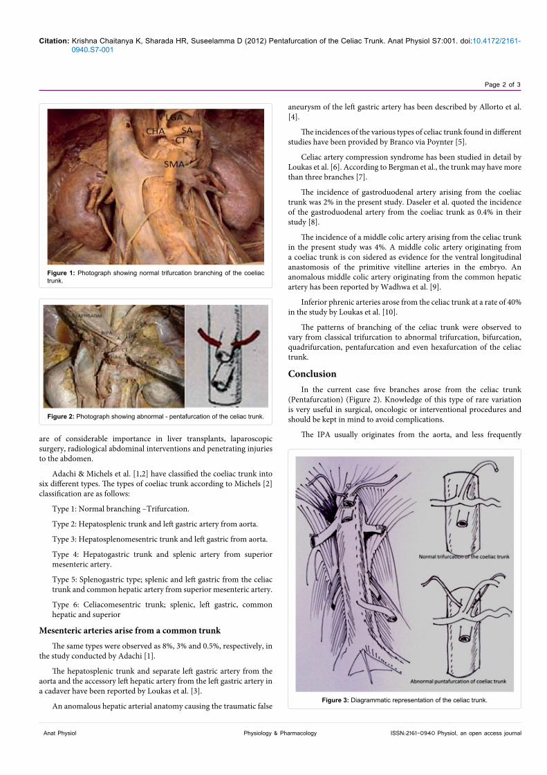
Figure 3: Diagrammatic representation of the celiac trunk.

Citation: Krishna Chaitanya K, Sharada HR, Suseelamma D (2012) Pentafurcation of the Celiac Trunk. Anat Physiol S7:001. doi:10.4172/2161-0940.S7-001
Page 3 of 3
ISSN:2161-0940 Physiol, an open access journal Anat Physiol Physiology & Pharmacology
from the celiac trunk, renal, hepatic or left gastric arteries. The inferior phrenic arteries are one of the common extrahepatic collateral arteries that supply hepatocellular carcinomas (HCCs). Literature on the IPA origin and clinical implications of variation in its origin has been reviewed in this article.
References
1. Adachi B (1928) Das Arteriensystem der Japaner. Vol. 2. Verlag der Kaiserlich-Japanischen Universitat zu Kyoto, Japan.
2. Michels NA (1955) Blood supply and anatomy of the upper abdominal organs with descriptive atlas. Philadelphia: Lippincott, USA.
3. Loukas M, Fergurson A, Louis RG Jr, Colborn GL (2006) Multiple variations of the hepatobiliary vasculature including double cystic arteries, accessory left hepatic artery and hepatosplenic trunk: a case report. Surg Radiol Anat 28: 525-528.
4. Allorto NL, Royston D, Hadley GP (2009) Traumatic false aneurysm of the left gastric artery. Pediatr Surg Int 25:455-457.
5. Poynter CWM (1922) Congenital anomalies of the arteries and veins of the human body with bliography. The University Studies of the University of Nebraska, USA. 22: 1-106.
6. Loukas M, Pinyard J, Vaid S, Kinsella C, Tariq A, et al. (2007) Clinical anatomy of celiac artery compression syndrome: a review. Clin Anat 20: 612-617.
7. Bergman RA, Thompson SA, Afif i AK, Saadeh FA (1988) Compendium of human anatomic variations. Baltimore: Urban Schwarzenberg, USA.
8. Daseler EH, Anson BJ, Hambley WC, Reimann AF (1947) Variations in Origin of Gastroduodenal Artery. Illustrated Encyclopedia of Human Anatomic Variation: Opus II: Cardiovascular System.
9. Wadhwa S, Barua MP (2008) Anomalous middle colic artery originating from common hepatic artery: a case report. Clin Anat 21: 798-799.
10. Loukas M, Hullett J, Wagner T (2005) Clinical anatomy of the inferior phrenic artery. Clin Anat 18: 357-365.
Thisarticlewasoriginallypublishedinaspecialissue,Physiology & Phar-macology handled by Editor(s). Anil Kumar Jain, University of ColoradoDenver,USA.