Oxime Amides as a Novel Zinc Binding Group in … · Oxime Amides as a Novel Zinc Binding Group in...
18
Published: March 18, 2011 r2011 American Chemical Society 2165 dx.doi.org/10.1021/jm101373a | J. Med. Chem. 2011, 54, 2165–2182 ARTICLE pubs.acs.org/jmc Oxime Amides as a Novel Zinc Binding Group in Histone Deacetylase Inhibitors: Synthesis, Biological Activity, and Computational Evaluation Cinzia B. Botta, ‡ Walter Cabri,* ,^ Elena Cini, † Lucia De Cesare, || Caterina Fattorusso,* ,|| Giuseppe Giannini, ^ Marco Persico, || Antonello Petrella, ‡ Francesca Rondinelli, || Manuela Rodriquez,* ,‡,§ Adele Russo, † and Maurizio Taddei † † Dipartimento Farmaco Chimico Tecnologico, Universit a degli Studi di Siena, Via A. Moro 2, I-53100 Siena, Italy ‡ Dipartimento di Scienze Farmaceutiche e Biomediche, Universit a di Salerno, Via Ponte don Melillo, I-84084 Fisciano (SA), Italy § Department of Chemistry and Chemical Biology, Harvard University, 12 Oxford Street, Cambridge, Massachusetts 02138, United States ) Dipartimento di Chimica delle Sostanze Naturali, Universit a di Napoli, Via D. Montesano, 49 I-80131 Napoli, Italy ^ Chemistry and Analytical Development, R&D Sigma-Tau S.p.A., Via Pontina, km 30,400 I-00040 Pomezia (RM), Italy b S Supporting Information ’ INTRODUCTION Histone deacetylases (HDACs) are enzymes involved in the remodeling of chromatin and have a key role in the epigenetic regulation of gene expression, as they remove the acetyl moiety from the ε-amino group of lysine side chains of histone and non- histone proteins. High levels of histone acetylation are associated with increased transcriptional activity, whereas low levels of acetylation are associated with repression of gene expression. Eighteen HDACs, subdivided into four classes, have been identi- fied in humans. Class I (HDACs 1, 2, 3, and 8), class IIa (HDACs 4, 5, 7, and 9), class IIb (HDACs 6 and 10), and class IV (HDAC 11) operate as Zn dependent enzymes, have distinct gene expression patterns, and have different cellular location and function. Inhibition of HDACs causes histone hyperacetylation with contemporary transcriptional activation of genes associated with cell cycle arrest or apoptosis in tumor cells. Indeed, several types of HDAC inhibitors have been found to have potential anticancer activity with remarkable tumor specificity. 1 A number of HDAC inhibitors (HDACIs) reached clinical trials, and two of them were lately approved by FDA: suberoylanilide hydroxamic acid (SAHA) from Merck and depsipeptide (FK228) from Gloucester Pharmaceuticals, both for use in cutaneous T-cell lymphoma (CTCL). 2 Moreover, the prospects of using HDACIs as therapeutic interventions for diseases other than cancer are evidenced by the growing number of papers detailing the use of HDACIs in immunomodulation, neurodegeneration, protozoan infections, inflammatory, and heart diseases. 3 The largely accepted pharmacophore model for most of the known hydroxamic acid derivatives inhibitors consists of (a) a capping group that interacts with the residues at the active site entrance (cap), (b) a zinc binding group (ZBG) that coordinates to the catalytic metal atom within the active site, and (c) a linker that binds to the hydrophobic channel and helps Cap and ZBG to find the correct position. Although SAHA contains a hydroxamic acid moiety and is considered a pan-inhibitor, some data suggest that selectivity toward a specific HDAC might be useful to find more effective inhibitors with limiting side effects. Modifications Received: October 21, 2010 ABSTRACT: Several oxime containing molecules, characterized by a SAHA-like struc- ture, were explored to select a potentially new biasing binding element for the zinc in HDAC catalytic site. All compounds were evaluated for their in vitro inhibitory acti- vity against the 11 human HDACs isoforms. After identification of a “hit” molecule, a programmed variation at the cap group and at the linker was carried out in order to increase HDAC inhibition and/or paralogue selectivity. Some of the new derivatives showed increased activity against a number of HDAC isoforms, even if their overall activity range is still far from the inhibition values reported for SAHA. Moreover, different from what was reported for their hydroxamic acid analogues the new R-oxime amide derivatives do not select between class I and class II HDACs; rather they target specific isoforms in each class. These somehow contradictory results were finally rationalized by a computa- tional assisted SAR, which gave us the chance to understand how the oxime derivatives interact with the catalytic site and justify the observed activity profile.
Transcript of Oxime Amides as a Novel Zinc Binding Group in … · Oxime Amides as a Novel Zinc Binding Group in...

Published: March 18, 2011
r 2011 American Chemical Society 2165 dx.doi.org/10.1021/jm101373a | J. Med. Chem. 2011, 54, 2165–2182
ARTICLE
pubs.acs.org/jmc
Oxime Amides as a Novel Zinc Binding Group in Histone DeacetylaseInhibitors: Synthesis, Biological Activity, and ComputationalEvaluationCinzia B. Botta,‡Walter Cabri,*,^ Elena Cini,† Lucia De Cesare,|| Caterina Fattorusso,*,|| Giuseppe Giannini,^
Marco Persico,|| Antonello Petrella,‡ Francesca Rondinelli,|| Manuela Rodriquez,*,‡,§ Adele Russo,† andMaurizio Taddei†
†Dipartimento Farmaco Chimico Tecnologico, Universit�a degli Studi di Siena, Via A. Moro 2, I-53100 Siena, Italy‡Dipartimento di Scienze Farmaceutiche e Biomediche, Universit�a di Salerno, Via Ponte don Melillo, I-84084 Fisciano (SA), Italy§Department of Chemistry and Chemical Biology, Harvard University, 12 Oxford Street, Cambridge, Massachusetts 02138, UnitedStates
)Dipartimento di Chimica delle Sostanze Naturali, Universit�a di Napoli, Via D. Montesano, 49 I-80131 Napoli, Italy^Chemistry and Analytical Development, R&D Sigma-Tau S.p.A., Via Pontina, km 30,400 I-00040 Pomezia (RM), Italy
bS Supporting Information
’ INTRODUCTION
Histone deacetylases (HDACs) are enzymes involved in theremodeling of chromatin and have a key role in the epigeneticregulation of gene expression, as they remove the acetyl moietyfrom the ε-amino group of lysine side chains of histone and non-histone proteins. High levels of histone acetylation are associatedwith increased transcriptional activity, whereas low levels ofacetylation are associated with repression of gene expression.Eighteen HDACs, subdivided into four classes, have been identi-fied in humans. Class I (HDACs 1, 2, 3, and 8), class IIa (HDACs4, 5, 7, and 9), class IIb (HDACs 6 and 10), and class IV (HDAC11) operate as Zn dependent enzymes, have distinct geneexpression patterns, and have different cellular location andfunction. Inhibition of HDACs causes histone hyperacetylationwith contemporary transcriptional activation of genes associatedwith cell cycle arrest or apoptosis in tumor cells. Indeed, severaltypes of HDAC inhibitors have been found to have potentialanticancer activity with remarkable tumor specificity.1 A numberof HDAC inhibitors (HDACIs) reached clinical trials, and two ofthem were lately approved by FDA: suberoylanilide hydroxamic
acid (SAHA) from Merck and depsipeptide (FK228) fromGloucester Pharmaceuticals, both for use in cutaneous T-celllymphoma (CTCL).2 Moreover, the prospects of using HDACIsas therapeutic interventions for diseases other than cancer areevidenced by the growing number of papers detailing the use ofHDACIs in immunomodulation, neurodegeneration, protozoaninfections, inflammatory, and heart diseases.3
The largely accepted pharmacophore model for most of theknown hydroxamic acid derivatives inhibitors consists of (a) acapping group that interacts with the residues at the active siteentrance (cap), (b) a zinc binding group (ZBG) that coordinatesto the catalytic metal atom within the active site, and (c) a linkerthat binds to the hydrophobic channel and helps Cap and ZBG tofind the correct position. Although SAHA contains a hydroxamicacid moiety and is considered a pan-inhibitor, some data suggestthat selectivity toward a specific HDAC might be useful to findmore effective inhibitors with limiting side effects. Modifications
Received: October 21, 2010
ABSTRACT: Several oxime containing molecules, characterized by a SAHA-like struc-ture, were explored to select a potentially new biasing binding element for the zinc inHDAC catalytic site. All compounds were evaluated for their in vitro inhibitory acti-vity against the 11 human HDACs isoforms. After identification of a “hit” molecule, aprogrammed variation at the cap group and at the linker was carried out in order toincrease HDAC inhibition and/or paralogue selectivity. Some of the new derivativesshowed increased activity against a number ofHDAC isoforms, even if their overall activityrange is still far from the inhibition values reported for SAHA. Moreover, different fromwhat was reported for their hydroxamic acid analogues the newR-oxime amide derivativesdo not select between class I and class II HDACs; rather they target specific isoforms ineach class. These somehow contradictory results were finally rationalized by a computa-tional assisted SAR, which gave us the chance to understand how the oxime derivatives interact with the catalytic site and justify theobserved activity profile.

2166 dx.doi.org/10.1021/jm101373a |J. Med. Chem. 2011, 54, 2165–2182
Journal of Medicinal Chemistry ARTICLE
of the Cap, linker, or ZBG have been demonstrated to increaseselectivity.4 Moreover, hydroxamic acid, a strong zinc chelator,presents metabolic and pharmacokinetic issues such as glucur-onidation, sulfation, and enzymatic hydrolysis that result in ashort in vivo half-life.5
Consequently, there are many ongoing research activities tofind replacement groups of hydroxamic acid with the dual targetof reducing side effects and increasing paralogue selectivity.Many different moieties have been proposed as hydroxamic acidsurrogates yielding encouraging results.6
Following our interest in this field,7 we focused on the replace-ment of the hydroxamic group to find some potent non-hydro-xamate small-molecule HDAC inhibitors and to investigate onthe paralogue selectivity in order to identify key elements forselective HDAC inhibition. In addition, in terms of chemicalbiology research, the discovery of novel ZBGs may lead to newtypes of HDAC isozyme selective inhibitors that are useful toolsfor probing the biology of the enzyme.8
Oxime-containing molecules caught our attention, as theyappear to be amenable to biotransformation and conjugationswith organic and inorganic molecules. The properties of theseclasses of compounds have been recently exploited with the aimto design and develop novel therapeutic agents that can displayacyl group transfer capabilities and serve for the evaluation ofnovel candidate drugs for the treatment of various diseases. Forexample, furan oximes were found to inhibit DNA, RNA, andprotein synthesis in lipoid leukemia cells.9 Derivatives of quino-line oximes were also shown to possess antitumor activity,10 andglucosinolates were suggested as cancer preventive agents. Thestability of oxime complexes with various metals has been shownto result in promising compounds with antitumor activity, suchas cis and trans platinum complexes11 and homo- and hetero-nuclear Cu(II) and Mn(II) oxime complexes.12 A complex oftechnetium with hexamethylpropyleneamine oxime was alsoused to monitor photodynamic therapy of prostate tumors.13
On the basis of these considerations, several oxime containingmolecules characterized by a SAHA-like structure were exploredselecting those where a mimetic of the natural substrates chelat-ing features were possible, such as R-oxime esters and R-oximeamides (compounds 2-5 in Chart 1). After identification of 5 as
a “hit”molecule, a programmed variation at the cap group and atthe linker was carried out in order to increase the activity and/orparalogue selectivity. The results were finally rationalized by acomputational assisted SAR, which gave us the chance to under-stand how the oxime derivatives interact with the catalytic siteand justify the observed activity profile.
’CHEMISTRY
Oxime derivatives employed in this study were preparedfollowing the different routes described in Schemes 1-4. Subericacid (6) was acetylated with Ac2O and further reacted withaniline to generate monoamide 7 (Scheme 1). Activation of thecarboxylic group with CDI followed by reaction with themagnesium salt of diethyl malonate gave keto ester 8 whichwas eventually oxidized with NaNO2 in acetic acid to form ketooxime ester 2 in good overall yields.
For the synthesis of other oximes correlated to structures 3-5,commercially available ethyl 7-bromo-heptanoate 9 underwenthydrolysis, benzylation, and direct halogen exchange, yieldingbenzyliodo derivative 10 (Scheme 2). Reaction with diethyl mal-onate and NaH afforded compound 11 in 88% yield. The benzylprotection was removed by hydrogenolysis (Pd/C, H2, 1 atm) togive acid 12. This compound was the key intermediate for thesynthesis of 3-5 and 14-24, as it enables the introduction ofdifferent arylamides at the carboxylic position in order to optimizethe Cap. After transformation of 12 into the corresponding acylchloride, reaction with aniline generated compound 13 in overall80% yield. The amide was then transformed into the correspondingR-oxime ester 3 with ethyl nitrite and sodium ethoxide. Thistransformation passes through the formation of the nitroso deriva-tive, which is further cleaved by sodium ethoxide to give the sodiumsalt of the oxime ester and diethyl carbonate.14 This reaction gaveexclusively one isomer (NMRandHPLCanalysis). X-ray analysis ofthe crystals obtained from compound 3 showed that the oxime wasin the E configuration (Figure 1SI in Supporting Information).Since all the other oximes were obtained as single diastereomers, theconfiguration of the oxime moiety of all the other compounds wasconsequently assigned. The ethyl ester 3 was then transformed intothe corresponding amide 4 or methylamide 5 in good yields byreaction with aqueous ammonia or methylamine in ethanol respec-tively. Amides 14-19 were then prepared by coupling of 12 withthe corresponding arylamines followed by transformation of themalonic ester into the oxime ester and final amidation.
Chart 1. SAHA (1) and Oxime Derivatives 2-5 Scheme 1. Synthesis of Keto Oxime Ester 2a
aReagents and conditions: (i) (1) Ac2O, 140 �C, 39%; (2) aniline, THF,room temp, 74%; (ii) CDI, room temp, THF, CH2(CO2Et)2, Mg-(OEt)2, room temp, THF, 91%; (iii) NaNO2, AcOH, room temp, 80%.

2167 dx.doi.org/10.1021/jm101373a |J. Med. Chem. 2011, 54, 2165–2182
Journal of Medicinal Chemistry ARTICLE
Amide 19 was used to introduce a substituent in the metaposition through a Suzuki coupling in order to increase the size ofthe cap group and to get more data useful for a comprehensiveSAR study. It was possible to carry out the coupling on the freeoxime amide with a selected catalyst and conditions as reportedin Table 1SI in Supporting Information.
It is interesting to note that the use of microwave dielectricheating (in combination with Pd(PPh3)2Cl2) provides exclu-sively compounds 20-22, while for the preparation of amides23-24 conventional long time heating is required.
Oxime amides 30-33 were prepared by coupling of aminoderivative 29 with arylisoxazolecarboxylic acids15 as reported inScheme 3. Commercially available 6-aminohexanol 25 was firsttransformed into the aldehyde 26 and immediately submitted toKnoevenagel reaction with diethyl malonate with piperidine/DMFfollowed by hydrogenation of the double bond giving 27 in 53%overall yield. The malonic ester was treated with ethyl nitrite andsodiumethoxide, affording theR-oximeester28. After transformationof the ethyl ester into the corresponding methylamide, the Bocprotecting groupwas removedwithTFA togive the amine29 in goodyields. In order to carry out the reaction between the previouslydescribed 5-(3-tert-butoxycarbonylaminophenyl)isoxazole-3-carbox-ylic acid and 5-(4-tertbutoxycarbonylaminophenyl)isoxazole-3-
carboxylic acid with the amine 29, carrying unprotected oximegroup, several coupling agents were then explored.15
The most selective reaction occurred using DMTMM16 in thepresence of 1-methylmorpholine, with the formation of com-pounds 30 and 31 in good yields. Final removal of the Boc withTFA gave the aniline derivatives 32 and 33 in good yields.
The introduction of a stereocenter (with S configuration)close to the carboxyamido group was often required in order toobtain a more potent inhibitor.17 This goal was accomplishedstarting from (S)-glutamic acid (Scheme 4) protected as thetetrabenzyl derivative and further transformed into aldehyde 34as described in the literature.18
Treatment with vinylmagnesium bromide gave the allylicalcohol with contemporary formation of lactone 35 as a mixtureof diastereoisomers. The lactone was opened with aniline in thepresence of AlMe3 and the hydroxyl group acetylated. The allylacetate 36 was submitted to a Tsuji-Trost reaction with sodiummalonate in the presence of Pd(PPh3)2Cl2 that gave compounds37a and 37b in a 2:1 ratio. Complete hydrogenation/hydro-genolysis of the mixture followed by reductive aminationwith p-methoxybenzaldehyde in the presence of NaBH(OAc)3yielded compound 38, whichwas separated from the correspondingregioisomer originating from 37b and isolated in 46% overall yield.
Scheme 2. Synthesis of Oximes 3-5 and 14-24a
aReagents and conditions: (i) (1) LiOH, EtOH/THF/H2O 1:1:1, room temp; (2) BnOH, EDC, DMAP, 0 �C to room temp; (3) NaI, acetone, roomtemp, overall 87%; (ii) NaCH(CO2Et)2, THF, 0 �C to room temp, 88%; (iii) H2 (1 atm), Pd/C, CHCl3, 96%; (iv) (1) (COCl)2, DMF, CH2Cl2, roomtemp, 2 h; (2) aniline, Et3N, CH2Cl2, 0 �C to room temp, overall 80%; (v) C2H5NO2, NaOEt, EtOH,-5 �C, 92%. (vi) 4: NH4OH conc, room temp,90%. 5: MeNH2 (33 wt % in ethanol), room temp, 80%.

2168 dx.doi.org/10.1021/jm101373a |J. Med. Chem. 2011, 54, 2165–2182
Journal of Medicinal Chemistry ARTICLE
Attempts to carry out the direct transformation of 38 into thecorresponding oxime ester gave exclusively degradation of thestarting material. Consequently, the amino group was protectedwith Boc2O and compound 39 was then transformed into oximeamide 41 after deprotection.
Alternatively, double bond reduction with Pt2O in MeOH wascarried out in a mixture of 37a and 37b, giving the malonate 42 in60% yield as a single isomer after column chromatography separation.On 42 the standard sequence of oximation and transformation of theethyl ester into methylamide gave compound 43 in 84% isolatedyields (Scheme 4).HPLC/MS analysis (Chiralpack column) and 19FNMR analysis of the amides derived from 41 with (S)- and (R)-
Mosher acids confirmed that racemization did not occur along the fullsynthetic pathway of chiral oxime amides (Scheme 4).
’RESULTS AND DISCUSSION
HDAC Isoform Inhibition Assay. All the compounds pre-pared in this study were evaluated for their in vitro inhibitoryactivity against the 11 humanHDACs isoforms in order to obtaina complete potency profile. The inhibitory activity was carriedout as previously described using SAHA (1) as the referencecompound.17a,19
Scheme 3. Synthesis of Oxime Amides 30-33a
aReagents and conditions: (i) 1) Boc2O, Et3N, DCM, room temp, 12 h, 88%; (2) (COCl)2, DMSO, Et3N, CH2Cl2, -78 �C, 30 min, 82%; (ii) (1)CH2(COOEt)2 DMF, piperidine, room temp, 1 h, 65%; (2) H2 (1 atm), PtO2, MeOH, room temp, 1 h, 92%; (iii) C2H5NO2, NaOEt, EtOH,-15 �C,5 h, 61%; (iv) (1) NH2Me, EtOH, room temp, 12 h, 75%; (2) TFA, DCM, room temp, 12 h, 67%; (v) 5-(3-tert-butoxycarbonylaminophenyl)isoxazole-3-carboxylic acid and 5-(4-tert-butoxycarbonylaminophenyl)isoxazole-3-carboxylic acid, respectively, DMTMM, NMM, THF, room temp, 12 h.
Scheme 4. Synthesis of Chiral Oxime Amides 40, 41, and 43a
aReagents and conditions: (i) see ref 18; (ii) CH2CHMgBr, THF, 0 �C to room temp, 60%; (iii) (1) aniline, AlMe3, CH2Cl2, 0 �C to room temp, 95%;(2) Ac2O, NEt3, DMAP, CH2Cl2, 0 �C to room temp, 92%; (iv) CH2(CO2Et)2, NaH, Pd[PPh3]2Cl2, PPh3, THF, 0 �C to room temp, 80%; (v) (1) H2
(1 atm), Pd/C, CH3OH, room temp; (2) 4-methoxybenzaldehyde, NaBH(OAc)3, AcOHcat, CH2Cl2, room temp, chromatographic separation, overall72%; (vi) (1) Boc2O, NEt3, H2O/THF, room temp; (2) C2H5NO2, NaOEt, EtOH,-5 �C, 50%; (vii) (1) MeNH2 (33 wt % in ethanol), room temp,12 h, 70%; (2) CH2Cl2/TFA (4:1), 82%; (viii) H2 (1 atm), PtO2, CH3OH, room temp, chromatographic separation 60%; (ix) (1) C2H5NO2, NaOEt,EtOH, -5 �C, 76%; (2) MeNH2 (33 wt % in ethanol), room temp, 84%.
2169 dx.doi.org/10.1021/jm101373a |J. Med. Chem. 2011, 54, 2165–2182
Journal of Medicinal Chemistry ARTICLE
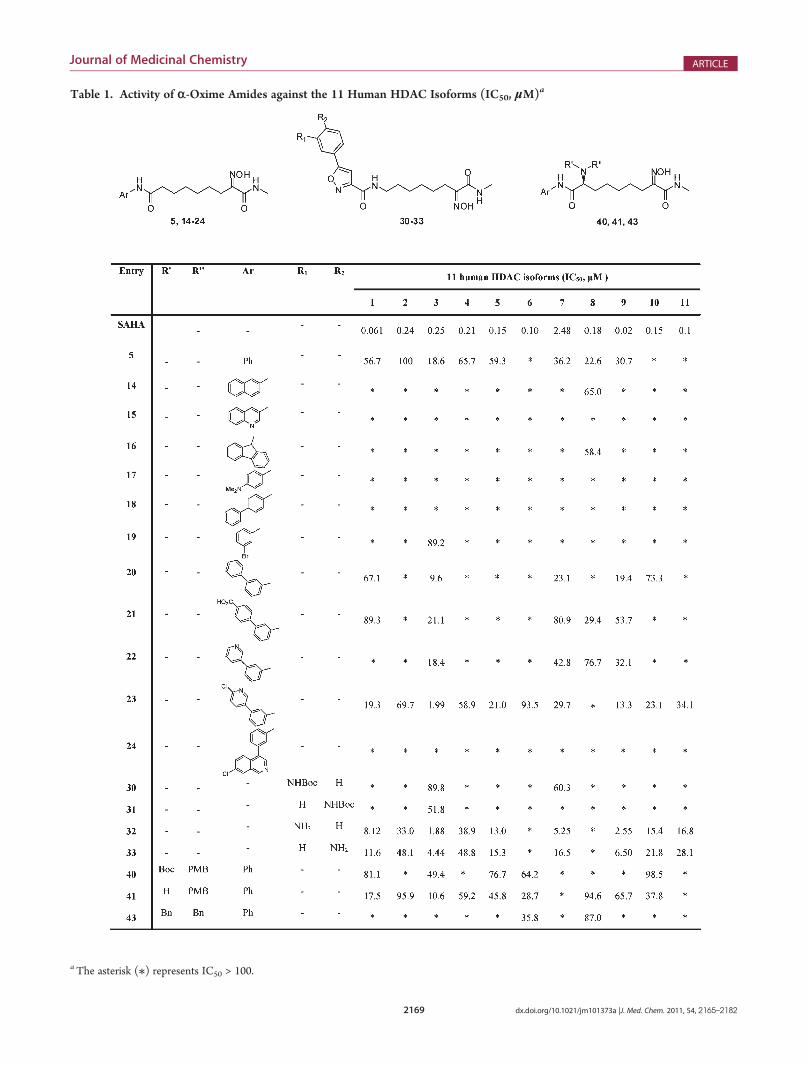
Table 1. Activity of r-Oxime Amides against the 11 Human HDAC Isoforms (IC50, μM)a
aThe asterisk (/) represents IC50 > 100.

2170 dx.doi.org/10.1021/jm101373a |J. Med. Chem. 2011, 54, 2165–2182
Journal of Medicinal Chemistry ARTICLE
Structure-Activity Relationships (SARs). At first, com-pounds 2-5 (Chart 1) were tested to select a potentially newbiasing binding element for the zinc in HDAC catalytic site(namely, ZBG). The R-keto oxime ester function of 2, as well asthe R-oxime ester of 3, was inactive, while the comparison of therelative activities of compounds 4 and 5 indicated that the abilityof the R-oxime amide function to inhibit HDAC catalytic site isrelated to the nature of the substituent on the amide nitrogen(Table 1). Interestingly, the same SAR trend was reported for a
series of SAHA analogues characterized by the R-keto amidefunction as ZBG.20 Since the HDAC natural substrate alsocontains an acetamidemoiety, it is likely that amethyl substitutedamide is able to reproduce substrate interaction with the catalyticsite, thus leading to a gain of inhibitory activity (4 vs 5, Table 1).Although not comparable with SAHA, these data were encoura-ging, suggesting an effective binding of theR-oximemethylamideto the catalytic zinc. Consequently, with the aim of increasinginhibitory activity, different cap groups on themolecular skeletonof compound 5 were explored (14-24, Scheme 2). Somederivatives of this series showed increased activity against anumber of HDAC isoforms (20-23, Table 1), even if theiractivity range is still far from the inhibition values of the referencecompound. The shape of the steric hindrance around the phenylring was critical for inhibitory activity (compounds 14-19 and24 vs compounds 20-23, Table 1), while the presence of an acidor a basic substituent modulated activity (23 > 20 > 22 > 21,Table 1) and selectivity (21, 22 > 20 > 23, Table 1). Still in theattempt to increase inhibitory activity, a Cap containing anarylisoxazole moiety was introduced (30-33, Scheme 3), as thismodification has been described to generate very potent andselective hydroxamic acid inhibitors.15 Compounds 30 and 31carrying a hindered N-Boc group did not show any activity,whereas the m- and p-aniline derivatives 32 and 33 were moreactive, the former being the most active compound of the wholeseries. Thus, different from what was reported for their hydro-xamic acid analogues,15 the activity and the selectivity of com-pounds 30-33 were driven by the steric hindrance at the Capregion. Finally, to further explore SARs in the Cap, a stereogeniccenter carrying a nitrogen atom was introduced, leading tobranched cap groups (40, 41, and 43; Scheme 4). Compound41 still retained activity on HDACs 1, 3, and 9, while a wider lossof activity was again observed by further increasing the sterichindrance at the Cap, as in the case of the N-Boc derivative 40and the bis-benzyl derivative 43 (Table 1).Thus, in the search of new ZBGs, the R-oxime amide function
on one hand showed an overall poorer ability to fulfill HDACcatalytic site pharmacophore compared to hydroxamic acid. Onthe other hand, it conferred to our compounds a peculiarparalogue selectivity profile. In order to rationalize these some-how contradictory results in terms of HDAC activity andselectivity, a computational approach was carried out.Computational Studies. The accommodation of the ZBG
into the HDAC catalytic site is a crucial step of the inhibitionprocess and is finely controlled by its zinc coordination abilityand by key interactions with the surrounding protein residues.First, we analyzed the ability of the R-oxime amide function tocoordinate the zinc atom by using a computational protocol,which included a dynamic (SA) conformational search followedby molecular mechanic (MM) and semiempirical (PM6) geo-metry optimization to generate input structures for calculationsat the density functional theory (DFT) level (see ExperimentalSection for details). Our results indicated the putative formationof several zinc coordination complexes, involving different R-oxime amide heteroatoms. In particular, our analysis identifiedfour putative complexes, two of which were characterized by afive-membered metal coordination ring and two by a six-mem-bered metal coordination ring, named according to their Oox-Nox-Cox-Cam and Oam-Cam-Cox-Nox dihedral angles values(C = 0�; T = 180�) and zinc coordination atoms (NO =Nox andOam; NN =Nox andNam; OO=Oox andOam; ON=Oox andNam) (Figure 1, Table 2).
Figure 1. 5/Zn2þ complexes optimized at DFT level: (a) TC-NOcomplex; (b) TT-NN complex; (c) CC-OO complex; (d) CT-ONcomplex. Only zinc binding groups are shown for the sake of clarity. Theconformers are colored by atom type [C = (a) yellow, (b) orange, (c)green, and (d) cyan; H = white; N = blue; O = red; Zn = magenta].
Table 2. Dihedral Angles and ZBG Distances of the r-OximeMethylamide/Zn Complex
τ1 (deg) τ2 (deg) D1 (Å) D2 (Å)
DFT
TC-NOa -0.37b -1.29c 2.02d 2.23e
TT-NNa -39.13f 7.74c 2.20g 2.22e
CC-OOa 32.17b 41.56h 2.00d 2.15i
CT-ONa 77.49f 18.95h 2.27g 2.15i
Docking
32j 1.75b 90.74h 2.63d 2.49i
32k 7.84b 17.00c 2.47d 2.99e
41 10.49b 86.73h 2.68d 2.49i
aDFT complexes as defined in the text and Figure 1. bCox-Cam-Oam-Zndihedral angle values. cCam-Cox-Nox-Zn dihedral angle values. dDis-tance between the Oam and Zn. eDistance between the Nox and Zn.
fCox-Cam-Nam-Zn dihedral angle values. gDistance between Nam andZn. hCox-Nox-Oox-Zn dihedral angle values. iDistance betweenthe Oox and Zn. jFirst set of docking results. k Second set of dockingresults.

2171 dx.doi.org/10.1021/jm101373a |J. Med. Chem. 2011, 54, 2165–2182
Journal of Medicinal Chemistry ARTICLE
All structures presented a planar coordination geometry inwhich the atoms involved in zinc coordination tend to lie on thesame plane of the metal. A fragment based search was performedin the Cambridge Structural Database (CSD) using as query anR-oxime amide function in complex with zinc, and the structuralanalysis of the results (CSD codes BICFUP, CABTUU, GIFSAF,GEPMET, IXEBIW, LOVQUI, MEHZUU, WASXEV, ZUDL-UF) confirmed our computational data. Thus, starting from thecalculated zinc coordination geometries (Figure 1), compounds32 and 41 were introduced into the human HDAC 7 (PDB code3C0Y) active site and several dynamic docking procedures werecarried out considering either a bi- or a monodentate zincinteraction mode. In particular, (a) a six-membered bidentateO-O interaction mode, (b) a monodentate interaction mode bythe amide oxygen, and (c) a monodentate interaction mode bythe oxime oxygen were considered for the CC-OO DFT
conformer, and (d) a five-membered bidentate N-N interactionmode, (e) a monodentate interaction mode by the oximenitrogen, and (f) a monodentate interaction mode by the amidenitrogen were considered for the TT-NN DFT conformer (seeExperimental Section for details). All the other DFT conformers(Figure 1, Table 2) when inserted in the enzyme catalytic sitecaused a steric clash with enzyme residues and/or zinc. Each ofthe six docking starting structures produced 20 results, whichwere selected on the bases of inhibitor conformations (solutionspresenting cis alkyl chain or amide bond were discarded) andgrouped according to enzyme-inhibitor interactions. In parti-cular, the coordination geometry of the ZBG and the positions of(i) the hydroxyl group, (ii) the methyl group, (iii) the alkyl chain,(iv) the H-bond group at the end of the alkyl chain, and (v) thearomatic Cap have been analyzed and compared to thosereported in all known inhibitors/HDAC X-ray complexes pre-sent in the Brookhaven PDB (Table 2SI in the Supportinginformation). This analysis provided an explanation for theinhibitory activity profile of our R-oxime amide derivatives onclasses I and IIa HDAC enzymes.Compound 32 produced twomain sets of results (Figures 2a,b
and 3a,b). In the first set of docking results (binding mode 1,Figures 2a and 3a) the ZBG binds His669 through its hydroxylhydrogen and interacts with the zinc atom through the amideoxygen and the oxime oxygen, thus forming a six-memberedmetal coordination ring. The resulting coordination geometry isvery different from that calculated through DFT methods (CC-OO complex, Figure 1, Table 2), revealing a reduced zinc bindingability of the R-oxime amide function when introduced intoHDAC catalytic site. The N-methylamide group reproducestrifluoromethyl ketone (TFMK, PDB code 2VQJ; Table 2SI inthe Supporting information) interactions with Pro667, Gly678,Phe679, and Gly841 (human HDAC 7 numbering), while thevolume of the aliphatic linker occupies a region similar to thatcovered by X-ray determined inhibitors in complex with HDAC(Figure 3a, Table 2SI in the Supporting information). Finally, theamide function at the end of the linker interacts with Thr625through its nitrogen and the aromatic cap group is surrounded byCys535, Asp537, Pro617, Cys618 (structural Zn coordinationsite; Figures 2a and 3a).The second set of docking results obtained for compound
32 (binding mode 2, Figures 2b and 3b) still presents the N-methylamide group positioned in the same enzyme cleft occu-pied by TFMK (Table 2SI in the Supporting information), asoccurred for binding mode 1. Nevertheless, the R-oxime amidefunction is differently oriented with the hydroxyl hydrogenbinding Asp801 and the amide oxygen and oxime nitrogeninteracting with zinc through a five-membered ring coordinationgeometry (Figure 3b). In this case, the docked structure is similarto the one obtained by DFT calculations (TC-NO complex,Figure 1) even if, because of the presence of the protein Zncoordinators, it showed an increased distance between the oximenitrogen and the zinc atom (D2, Table 2). The aromatic amidefunction at the end of the linker interacts through an H-bondwith the backbone of Ala808, while the Cap is positioned withinthe region Asp801-Gly812 and loop Arg731-Pro739 (namedpocket 2 in Figure 2b). A comparison of this set of docking resultswith X-ray determined HDAC inhibitor structures (Table 2SI inthe Supporting information) revealed that the only X-ray deter-mined HDAC complex in which the ligand projects the capregion in pocket 2 (Figure 2) is that of APHA in complex withHDAC 8 (PDB code 3F07). All the other cocrystallized ligands
Figure 2. Overall view of HDAC 7 in complex with compound 32[binding mode 1 (a) and binding mode 2 (b)] and compound 41 (c).vdW volumes of the ligands (colored in green) are displayed. Connollysurface of the enzyme is shown and colored on the base of active sitesubstructures. The region V623-I628 (blue) and loop H732-S741(yellow) set up pocket 1. Loop H732-S741 (yellow) and the regionE804-G812 (red) set up pocket 2. The region H531-H544 (orange)and region E804-G812 (red) set up pocket 3.

2172 dx.doi.org/10.1021/jm101373a |J. Med. Chem. 2011, 54, 2165–2182
Journal of Medicinal Chemistry ARTICLE
occupy the regions between loop Arg731-Pro739 and regionPro617-Thr627 (human HDAC 7 numbering; pocket 1 inFigure 2) and/or between loop region Asp537-Arg547 and loopAsp801-Gly812 (human HDAC 7 numbering; pocket 3 inFigure 2). Interestingly, similar to 32, APHA is characterizedby a lower (i.e., in the micromolar range) in vitro HDACinhibition constant21 compared to other classes of knowninhibitors, including SAHA. Thus, with adoption of binding
mode 2, the R-oxime amide function is better accommodatedin the catalytic site. However, the volume of the alkyl chain of 32does not fit the volume occupied by any ligand cocrystallized incomplex with HDAC (Figure 2b, Table 2SI in the Supportinginformation), thus projecting the Cap in a surface pocketdifferent from that occupied by SAHA and accounting for thepeculiar SARs and selectivity profile of the R-oxime amidederivatives. Indeed, the oxazole ring of 32 is involved in π-πinteraction with Phe737 (loop Arg731-Pro739) and the anilinemoiety is embedded between Asn736 and Leu765, Asp766 (loopGly763-Pro767) (Figure 3b). The side chains of Leu765 andAsp766 surround the aromatic amine, thus explaining theinactivity of sterically hindered derivatives, such as 30 and 31(Scheme 3, Table 1). The presence of the bulky Met274 inHDAC 8 (Table 3SI in the Supporting information), replacingHDAC 7 Leu810 (Figure 3b), may account for the inactivity of32 against this HDAC isoform. Indeed, the less hinderedmonophenyl analogue 5 resulted the most active derivative ofthe series on HDAC 8 (Table 1). The structural superimpositionof low energy conformers of 5 and 14-24 on low energyconformers of 30-33 pointed out that the steric bulk thatunfavorably affects their HDAC inhibitory activity (14-19, 24vs 5, 20-23; 30, 31 vs 32 and 33; Table 1) partially overlays(Figure 4), suggesting a similar binding mode for the two seriesof compounds.In this view, the complete loss of activity of compound 21
bearing a p-carboxylic function on the phenyl ring (Scheme 2,Table 1) is in agreement with the presence of a negativelycharged residue (Asp766) on the loop Gly763-Gly770, con-served in all HDAC isoforms (Table 3SI and Figure 2SI in theSupporting Information). Moreover, the activity of the mono-phenyl derivative 5 toward HDAC 3 could be explained by thedifferent amino acid composition of pocket 2 (Figure 2b).Indeed, only HDAC 3 presents an additional aromatic residuein this region (Tyr198, human HDAC 3 numbering), which,replacing HDAC 7 Asn736 (Figure 3b), could establish favorableπ-π interactions with the phenyl ring of 5. On the contrary, allother HDAC isoforms lack residues able to interact with the Capof 5 (Table 3SI in the Supporting information).In the case of compound 41 all input structures led to only one
set of docking results (Figures 2c and 3c). Compared to 32compound 41 loses activity against HDAC 7, accordingly; thestrong (ionic) interaction between the protonated nitrogen atthe Cap level and Asp624 in region Pro617-Thr627 (loop 2,Figure 2c) seems to drive enzyme binding, inducing a distortion
Figure 4. Superimposition of cap groups of low energy conformers of14 (yellow), 18 (pink), 30 (cyan), and 31 (orange) by fitting thecarbonyl group atoms and the amide and isoxazole nitrogens. Structuresare displayed as sticks (a) and as Connolly surfaces (b). In (a) allhydrogens, except those of amide function, have been omitted for thesake of clarity.
Figure 3. (a) Binding mode 1 of 32 in HDAC 7. (b) Binding mode 2 of32 inHDAC7. (c)Docked complex of 41/HDAC7.HDAC7 active sitesubstructures are colored: orange (D537-R547); blue (P617-T627);yellow (loop R731-P739); red (D801-G812). Carbon atoms of theligands are colored in green. vdW volumes of catalytic and structural zinc(magenta) are displayed. Heteroatoms are colored by atom type (O =red; N = blue). Hydrogen bonds are highlighted by green dashed lines.Hydrogens and water molecules are omitted for clarity except thoseinvolved in hydrogen bond interactions.

2173 dx.doi.org/10.1021/jm101373a |J. Med. Chem. 2011, 54, 2165–2182
Journal of Medicinal Chemistry ARTICLE
in 41 alkyl chain conformation and reducing ZBG accommoda-tion in the catalytic site. The zinc atom in the catalytic site iscoordinated through the amide oxygen and the oxime oxygen, asin the case of binding mode 1 of 32, presenting a six-memberedmetal coordination geometry, which was very different from thatcalculated through DFT methods (CC-OO complex, Figure 1,Table 2). The amide methyl group of 41 accommodates in thecleft formed by Pro667, Phe679, and Gly841, reproducingHDAC cocrystallized ligand positioning, as was also for com-pound 32 in both sets of docking results. Moreover, thethreonine residue present in HDAC 7 (Thr625) on loop 2, justafter Asp624, hinders the accommodation of the branched capgroup of 41. This residue is replaced by a negatively chargedresidue in HDACs 1 and 3 (Glu98 and Asp92, respectively). Thiscould account for the higher inhibitory activity of 41 towardthese enzyme isoforms and for the complete lack of activity ofcompounds 40 and 43, bearing a bulkyN-Boc group and a benzylgroup on the amine nitrogen, respectively (Scheme 4, Table 1).The p-OMe phenyl ring is accommodated in the same region(pocket 2 in Figure 2c) occupied by the 32 cap group in thesecond set of docking results (Figure 2b). On the other hand, thephenyl ring of 41 interacts with region Asp537-Arg547 (loop 1;Figures 2c and 3c) similar to 32 Cap, according to the bindingmode obtained in the first set of docking results (Figure 2a).The hypothesized binding modes also relate with the low
activity of all our derivatives toward HDAC 8. Indeed, in thisisoform, HDAC 7 Pro667 and Pro542 (Figure 3) are replaced byTrp141 and Lys33, respectively (Table 3SI in the Supportinginformation), thus preventing the accommodation of the R-oxime amide group in the catalytic site. On the other hand, thelack of activity against HDAC 4 can be due to the presence ofPro809, corresponding to Ala676 in HDAC 7 (Table 3SI in theSupporting information), which limits the structural flexibility ofa loop undergoing a significant conformational change duringour docking calculations (Ala676-Cys680 loop, rms value on allCR atoms of 2.6 Å).In summary, our computational results illustrated how the R-
oxime amide moiety scarcely adapts into HDAC catalytic site,being not able to reproduce any known Zn binding mode exceptfor the position of the amide methyl group, which indeed wascritical for the inhibitory activity (4 vs 5, Table 1). The posi-tioning of the ZBG affects the placement of the Cap on theenzyme surface pockets, whose different amino acid compositionamong HDAC isoforms is mainly responsible for inhibitorselectivity. Accordingly, the introduced modifications at theCap of our R-oxime amide derivatives did not produce the sameHDAC inhibitor profile reported for their hydroxamic acidanalogues.The rationalization of the peculiar binding profile of oxime
derivatives provides us with a useful tool to continue ourinvestigation on HDAC inhibitors. Indeed, the information onthe mutual relation between ZBG and Cap positioning will beexploited to sample the enzyme pockets at the entrance of thechannel where isoform selectivity stands. Moreover, our studydisclosed interesting insights into the molecular determinants forZBG accommodation into the catalytic site, which will be furtherexplored through the synthesis of new optimized derivatives.
’CONCLUSIONS
Adopting a different binding mode with respect to thatreported for hydroxamic acid and substrate-like HDAC
inhibitors, R-oxime amide derivatives show in vitro inhibitoryactivity in the micromolar range and a peculiar paralogueselectivity profile. Different from results observed for some classI selective inhibitors, our R-oxime amides do not select betweenclass I and class II HDAC; rather they target specific isoforms in eachclass. Our computational results indicate that despite its ability tocoordinate the zinc ion, the R-oxime amide function of compounds32 and 41 was able to reproduce known inhibitor-substrate ZBGinteractions with the HDAC catalytic site. The unusual accom-modation of the ZBG was also critical for the positioning of thelinker and the projection of the Cap toward the different surfacepockets of the enzyme. Accordingly, the SARs of the newderivatives differ from those of other classes of active com-pounds. The rationalization of the activity profile of the newoxime amide derivatives lays the base for future structuralmodifications.
’EXPERIMENTAL SECTION
Chemistry. Reagents were purchased from commercial suppliersand used without further purification. Anhydrous reactions were rununder (a positive pressure) dry N2. Flash chromatography employedMerck silica gel 60 (23-400 mesh). Microwave dielectric heating wasapplied using a CEM Discover microwave oven for chemical synthesis.NMR spectra were acquired at 300 K using Bruker AC200F and BrukerAdvance DPX400 spectrometers. Data are reported as chemical shifts (δppm), multiplicity (s = singlet, bs = broad singlet, d = doublet, dd =doublet of doublets, t = triplet, m = multiplet), coupling constants (J,Hz), and relative integral. Chemical shifts are reported relative totetramethylsilane at 0.00 ppm. Mass spectral (MS) data were obtainedusing an Agilent 1100 LC/MSD VL system (G1946C) with a 0.4 mL/min flow rate using a binary solvent system of 95:5 methanol/water. UVdetection wasmonitored at 254 nm.Mass spectra were acquired either inpositive or in negative mode scanning over the mass range of 105-1500.Microwave irradiations were conducted using a CEMDiscover synthesisunit (CEM Corp., Matthews, NC). Elemental analyses were performedon a Perkin-Elmer PE 2004 elemental analyzer, and the data for C, H,and N are within 0.4% of the theoretical values. The chemical purity ofthe target compounds was determined using the following conditions:an Agilent 1100 series LC/MS with a EC 125/4.6 NUCLEODUR 100-5C18 reversed phase column. The binary solvent system (A/B) was asfollows: 0.1% TFA in water (A) and acetonitrile (B). The absorbancewas detected at 254 nm, and the flow rate was 1 mL/min. The purity ofeach compound was g95% in either analysis.Ethyl 2-(Hydroxyimino)-3,10-dioxo-10-(phenylamino)de-
canoate (2). β-Keto ester 8 (0.207 g, 0.649 mmol) was dissolved inglacial acetic acid (5 mL) and cooled to 0 �C in ice bath. A solution ofNaNO2 (0.089 g, 1.30 mmol) in H2O (2 mL) was added. The reactionmixture was allowed to warm to room temperature and stirred overnight.Dry Et2O was added, and the organic layer was separated, washed withsaturated NaHCO3, dried (anhydrous Na2SO4), filtered, and concen-trated in vacuo. The crude residue was purified by flash chromatography(99:1 dichloromethane/methanol) to afford the title compound 2 as acolorless oil (0.180 g, 80%). 1H NMR (400 MHz, CDCl3): δ 1.3 (t, J =6.8Hz, 3H), 1.34 (m, 4H), 1.59 (m, 2H), 1.72 (m, 2H), 2.3 (t, J = 7.6Hz,2H), 2.71 (t, J = 6.8 Hz, 2H), 4.31 (q, J = 7.2 Hz, 2H), 7.05 (t, J= 8.8 Hz,1H), 7.25 (m, 2H), 7.5 (2H, J = 8 Hz, 2H). MS(ESI) m/z: 371.1 [MþNa]þ. Anal. Calcd for C18H24N2O5: C, 63.73; H, 7.55; N, 8.74. Found:C, 63.93; H, 7.49; N, 8.69.General Procedure To Convert Alkylated Malonic Ester to
r-Oxime Ester. Ethyl 2-(Hydeoxyimino)-9-oxo-9, 8-phenyla-mino)nonanoate (3). Diester 13 (0.473 g, 1.30 mmol) was placed ina 50 mL flask equipped with a magnetic stirring bar and N2 inlet. The

2174 dx.doi.org/10.1021/jm101373a |J. Med. Chem. 2011, 54, 2165–2182
Journal of Medicinal Chemistry ARTICLE
flask was immersed in an ice bath, and a solution of ethyl nitrite inethanol (1.31 mL, 2.61 mmol) was added to the stirred solution. Themixture was then cooled to -5 �C in ice-salt bath, and 1.31 mmol ofsodium ethoxide (prepared from 0.030 g of sodium in 2 mL of dryethanol) was added slowly with stirring. The resulting reaction mixturewas allowed to warm to room temperatue during the night. The solventwas concentrated under reduced pressure. To the residue equal volumesof ice-water and cold concentrated hydrochloric acid were added untilpH 4 was obtained. The aqueous solution was extracted three times withdiethyl ether. The combined ether extracts were dried (anhydrousNa2SO4), filtered, and evaporated to dryness. The crude was purifiedby flash chromatography (100% dichloromethane) to afford the titlecompound 3 as a white solid (0.384 g, 92%). Mp 118-120 �C. 1HNMR(400 MHz, CDCl3): δ 1.3 (t, J = 7.2 Hz, 3H), 1.35 (m, 4H), 1.52(m, 2H), 1.70 (m, 2H), 2.32 (t, J = 3.8 Hz, 2H), 2.59 (t, J = 7.2 Hz, 2H),4.28 (q, J = 7.2 Hz, 2H), 7.05 (t, J = 8.8 Hz, 1H), 7.25 (m, 2H), 7.5 (d,J = 8 Hz, 2H). MS(ESI) m/z: 343.1 [M þ Na]þ. Anal. Calcd forC17H24N2O4: C, 63.73; H, 7.55; N, 8.74. Found: C, 63.79; H, 7.51;N, 8.70.8-(Hydroxyimino)-N1-phenylnonanediamide (4). To a solu-
tion of 3 (0.050 g, 0.16 mmol) in ammonium hydroxide solution (28-30%, 5 mL) a catalytic amount of ammonium chloride was added. Theresulting mixture was stirred at room temperature for 18 h. The reactionmixture was diluted with ethyl acetate, and the organic layer wasseparated, dried (anhydrous Na2SO4), filtered, and concentrated invacuo. The crude was purified by flash chromatography (ethyl acetate/petroleum ether 6:4) to afford the desired compound 4 as a yellow solid(0.042 g, 90%).Mp 134-136 �C. 1HNMR (400MHz, CD3OD):δ 1.36(m, 4H), 1.48 (m, 2H), 1.66 (m, 2H), 2.32 (t, J = 3.8Hz, 2H), 2.52 (t, J =7.2 Hz, 2H), 7.05 (t, J = 8.8 Hz, 1H), 7.25 (m, 2H), 7.5 (d, J = 8Hz, 2H).MS(ESI) m/z: 314.1 [M þ Na]þ. Anal. Calcd for C15H21N3O3: C,61.84; H, 7.27; N, 14.42. Found C, 61.79; H, 7.23; N, 14.40.General Procedure To Convert the Oximino Ester to
r-Oxime Amide. 2-(Hydroxyimino)-N1-methyl-N9-phenylno-nanediamide (5). Compound 3 (0.170 g, 0.302 mmol) was dissolvedin 7 mL of methylamine solution (33 wt % in ethanol), and the reactionmixture was stirred at room temperature overnight. The solvent wasevaporated to dryness and the residue purified by flash chromatography(dichloromethane/methanol 95:5) to afford the desired compound 5 as awhite solid (0.074 g, 80%). Mp 138-140 �C. 1H NMR (200 MHz,CDCl3): δ 1.34 (m, 4H), 1.50 (m, 2H) 1.72 (m, 2H), 2.24 (t, J = 6.8 Hz,2H), 2.7 (t, J = 7.2 Hz, 2H), 2.75 (s, 3H), 7.03 (t, J = 8.8 Hz, 1H), 7.13(m, 2H), 7.48 (d, J = 8 Hz, 2H). MS(ESI) m/z: 328.2 [MþNa]þ. Anal.Calcd for C16H23N3O3: C, 62.93; H, 7.59; N, 13.76. Found: C, 62.88; H,7.55; N, 13.73.8-Oxo-8-(phenylamino)octanoic Acid (7). Acetic anhydride
(25 mL) was placed in a 250 mL flask equipped with a magnetic stirrerand a refrigerator and warmed to 180 �C. While the mixture wasrefluxing, suberic acid (10 g, 57mmol) was added. The resulting solutionwas warmed for 2 h and then concentrated. After concentration, theproduct was crystallized from DCM. After drying the product weighed3.1 g and a second crop was obtained from the mother liquor to obtainthe intermediate anhydride in 39% of combined yield. To a solution ofacetylated suberic acid in anhydrous THF (20 mL) was added aniline(0.56 mL, 6.2 mmol) at room temperature. The reaction mixture wasstirred for 2 h and then treated with 150 mL of H2O and 3 g of K2CO3.The basic resulting aqueous solution was stirred for 20 min and thenextracted three times with EtOAc. The combined organic phasescontained only the secondary product bis-anilide, whereas the aqueousphase was treated with HCl until pH 3-4 was obtained. The mono-anilide 7, precipitating during the night at 4 �C, was filtered and driedover P2O5 (74% yield, the product is a white solid). Mp 114-116 �C. 1HNMR (200MHz, CDCl3): δ 1.29 (m, 4H), 1.55 (m, 2H), 1.65 (m, 2H),2.30 (t, J = 3.8 Hz, 2H), 2.35 (t, J = 3.8 Hz, 2H), 7.05 (t, J = 8.8 Hz, 1H),
7.25 (m, 2H), 7.5 (d, J = 8.0 Hz, 2H). MS(ESI)m/z: 272.1 [MþNa]þ.Anal. Calcd for C14H19NO3: C, 67.45;H, 7.68; N, 5.62. Found: C, 67.40;H, 7.65; N, 5.60.Ethyl 3,10-Dioxo-10-(phenylamino)decanoate (8). To a
solution of 7 (1.08 g, 4.36 mmol) in anhydrous THF (25 mL) wasadded carbonyldiimidazole (0.777 g, 4.79 mmol) at room temperature.The reaction mixture was stirred for 6 h. Magnesium salt of monoethylmalonate (prepared from 4.03 g of monoethyl malonate and 1.74 g ofMg(OEt)2) was added, and the resulting mixture was stirred overnight.After concentration under reduced pressure, the residue was purified byflash chromatography (petroleum ether/diethyl ether 1:9) to afford 8(1.27 g, 91%) as a colorless oil. 1H NMR (400 MHz, CDCl3): δ 1.25 (t,J = 6.8 Hz, 3H), 1.34 (m, 4H), 1.58 (m, 2H), 1.68 (m, 2H), 2.3 (t, J =7.6Hz, 2H), 2.49 (t, J = 6.8Hz, 2H), 3.4 (s, 2H), 4.15 (q, J = 7.2Hz, 2H),7.05 (t, J = 8.8 Hz, 1H), 7.25 (m, 2H), 7.5 (d, J = 8 Hz, 2H). MS(ESI)m/z: 342.1 [MþNa]þ. Anal. Calcd for C18H25NO4: C, 67.69; H, 7.89;N, 4.39. Found: C, 67.72; H, 7.84; N, 4.34.Benzyl 7-Iodoheptanoate (10). To a solution of ethyl 7-bro-
moheptanoate (1.3 g, 6.3 mmol) in THF/H2O/EtOH 1:1:1 (15 mL),LiOH (0.343 g, 8.19 mmol) was added. The reaction mixture was stirredfor 3 h. The solvent was removed, and the residue was dissolved inCH2Cl2. The reactionmixture was acidified with 4MHCl until pH 1wasobtained. The organic layer was separated, washed with NaClss, driedover sodium sulfate, filtered, and concentrated in vacuo. The crude wassubmitted to the next step without additional purification (98%). Thecrude 7-bromoheptanoic acid (1.3 g, 6.3 mmol) and DMAP (0.077 g,0.63 mmol) were then added to a solution of benzyl alcohol (1.3 mL,12.6 mmol) and EDC (2.4 g, 12.6mmol) in CH2Cl2 (3mL) at 0 �C. Theresulting mixture was stirred at room temperature for 7 h. Then it waspoured into water and extracted with CH2Cl2� 3. The organic layer waswashed with saturated aqueous NaHCO3 � 2, water, and brine, dried(anhydrous Na2SO4), filtered, and concentrated. The crude was purifiedby flash column chromatography (petroleum ether/ethyl acetate 9:1) toafford the benzyl 7-bromoheptanoate as a colorless oil (91%). A solutionof this bromide (0.3 g, 1.0 mmol) in acetone (1 mL) was added to asolution of NaI (0.45 g, 3.01 mmol) in acetone (1 mL), and the resultingmixture was stirred overnight (18 h) at room temperature. The pre-cipitate NaBr was filtered off, and the filtrate was concentrated underreduced pressure. The pulpy yellow residue was purified by flash chro-matography (petroleum ether/ethyl acetate 9:1) to yield iodide 10(0.336 g, 97%) as a colorless oil. 1H NMR (400 MHz, CDCl3): δ 1.49-1.22 (m, 4H), 1.70-1.55 (m, 4H), 2.29 (t, J= 7.6Hz, 2H), 3.41 (t, J = 6.8Hz, 2H), 5.06 (s, 2H), 7.29-7.24 (m, 5H). 13C NMR (100 MHz,CDCl3): δ 24.3, 26.0, 27.8, 33.9, 33.6, 44.4, 65.5, 127.7 (�3), 128.05(�2), 135.8, 172.6. MS(ESI) m/z: 368.7 [M þ Na]þ
7-Benzyl 1,1-Diethylheptane-1,1,1-tricarboxylate (11).NaH(1.05 g, 26.2 mmol, 60% dispersion in oil) was added to a two-necked round-bottom flask closed with a septum to which dry hexane(4 mL) was added, and the mixture was stirred for 5 min to dissolve themineral oil. The hexane was removed by a syringe, THF (10 mL) wasadded, and the resulting mixture was stirred at 0 �C for 15 min. Asolution of diethyl malonate (6.7 mL, 44 mmol) in THF (12 mL) wasthen slowly added over 30 min at 0 �C to the resulting suspension. Thesolution became clear after 10min of stirring. The stirring was continuedfor another 30 min. Iodide 10 (5.05 g, 14.6 mmol) in THF (15 mL) wasadded dropwise over 30 min to the stirred solution, and this mixture wasstirred at room temperature for 4 h. The reaction mixture was quenchedwith NH4Clss. The aqueous phase was extracted three times with ether,and the combined organic phases were dried (anhydrous Na2SO4),filtered and evaporated in vacuo. The crude residue was purified by flashchromatography (hexanes/ethyl acetate 9:1) to afford 11 as a colorlessoil (4.84 g, 88%). 1H NMR (200 MHz, CDCl3): δ 1.23 (t, J = 6.9 Hz,6H), 1.30 (m, 4H), 1.7 (m, 4H), 1.87 (m, 2H), 2.3 (t, J = 3.8 Hz, 2H),3.30 (t, J = 7.2 Hz, 1H), 4.20 (q, J = 6.8 Hz, 4H), 5.09 (s, 2H), 7.05 (t, J =

2175 dx.doi.org/10.1021/jm101373a |J. Med. Chem. 2011, 54, 2165–2182
Journal of Medicinal Chemistry ARTICLE
8.8 Hz, 1H), 7.25 (m, 2H), 7.5 (d, J =8.0 Hz, 2H). 13C NMR (50 MHz,CDCl3): δ 13.7, 24.5, 26.9, 28.3, 28.4, 28.5, 33.8, 41.3, 51.6, 60.9 (�2),61.1, 65.7, 127.6, 127.6, 128.2, 135.6, 166.3, 169.1 (�2), 173.1. MS(ESI)m/z: 386.1 [M þ Na]þ. Anal. Calcd for C21H30O6: C, 66.65; H, 7.99.Found: C, 66.60; H, 7.95.9-Ethoxy-8-(ethoxycarbonyl)-9-oxononanoic Acid (12).
Pd-C (0.32 g, 10% in mmol, 10% w/w) was added to a solution of11 (0.993 g, 2.6 mmol) in CHCl3 (30 mL), and the mixture was stirredunder H2 (1 bar) for 15 h. After full conversion, the reaction mixture wasfiltered and concentrated in vacuo. Flash chromatography (petroleumether/ethyl acetate 7:3) gave 12 (96%) as a colorless oil. 1H NMR (400MHz, CDCl3): δ 1.21 (t, J = 7.2 Hz, 6H), 1.57 (m, 2H), 1.29 (m, 6H),1.83-1.82 (m, 2H), 2.28 (t, J= 7.2Hz, 2H), 3.25 (t, J = 7.6Hz, 1H), 4.15(q, J = 7.2 Hz, 4H). 13CNMR (100 MHz, CDCl3) δ: 13.6 (�2), 24.0,26.6, 28.2, 28.2, 28.4, 33.5, 51.5, 60.8 (�2), 169.1 (�2), 179.5. MS(ESI)m/z: 310.8 [M þ Na]þ. Anal. Calcd for C14H24O6: C, 58.32; H, 8.39.Found: C, 58.27; H, 8.35.General Amidation Reaction Procedure via Acyl Chloride
Intermediate. Diethyl 2-(7-Oxo-7-(phenylamino)heptyl)ma-lonate (13). To a suspension of 12 (0.3 g, 1.04 mmol) in CH2Cl2(2.5 mL) were added oxalyl chloride (0.13 mL, 1.5 mmol) and a catalyticamount of DMF. The mixture was stirred at room temperature for 2 h.The solvent was removed by evaporation in vacuo to give acid chlorideintermediate. A solution of this chloride in CH2Cl2 (1.3 mL) was addeddropwise to a solution of aniline (0.53 g, 5.7 mmol) and triethylamine(0.43 mL, 3.12 mmol) in CH2Cl2 (3.2 mL) cooled in an ice-water bath.The mixture was stirred at room temperature for 18 h. Then it wasdiluted with CH2Cl2 and washed with HCl 1M, water, and brine, beforebeing dried over anhydrous Na2SO4. Filtration and concentration invacuo and purification by flash chromatography (petroleum ether/ethylacetate 8:2) gave the title compound 13 as a white solid (80%). Mp 65-67 �C. 1H NMR (400 MHz, CDCl3): δ 1.23 (t, J = 6.8 Hz, 6H), 1.33(m, 6H), 1.67 (m, 2H), 1.85 (m, 2H), 2.28 (t, J = 7.2Hz, 2H), 3.27 (t, J =7.2 Hz, 1H), 4.14 (q, J = 6.8 Hz, 4H), 7.04 (m, 1H), 7.24 (t, J = 7.6 Hz,2H), 7.48 (d, J = 8.0 Hz, 2H), 7.68 (bs, 1H). 13C NMR (100 MHz,CDCl3): δ 14.06 (�2), 25.2, 26.9, 29.1, 28.5, 28.7, 37.3, 51.9, 61.3 (�2),119.9 (�2), 124.0, 128.9 (�2), 138.1, 169.5 (�2), 171.5.MS(ESI)m/z:371.8 [M þ Na]þ. Anal. Calcd for C20H29NO5: C, 66.09; H, 8.04; N,3.85. Found: C, 66.01; H, 8.00; N, 3.83.2-(Hydroxyimino)-N1-methyl-N9-(naphthalene-2-yl)non-
anediamide (14). Compound 14 was prepared from the correspond-ing R-oxime ester using the procedure described for 5 in 76% yield as ayellow solid. Mp 153-155 �C. 1HNMR (400MHz, CD3OD): δ 1.57-1.23 (m, 9H), 1.72-1.67 (m, 2H), 2.39 (t, J = 7.2Hz, 2H), 2.54 (t, J = 7.6Hz, 2H), 2.75 (s, 3H), 7.42-7.30 (m, 2H), 7.52 (d, J = 8.8 Hz, 1H),7.77-7.72 (m, 3H), 8.17 (s, 1H). 13CNMR (50MHz, CD3OD): δ 22.4,24.5, 24.9, 25.0, 28.1, 28.5, 28.7, 36.2, 115.9, 119.4, 123.9, 125.4, 126.6,127.5, 129.9, 133.2, 135.3, 153.1, 165.0, 172.9. MS(ESI) m/z: 355.8[M þ H]þ. Anal. Calcd for C20H25N3O3: C, 67.58; H, 7.09; N, 11.82.Found: C, 67.50; H, 7.04; N, 11.80.2-(Hydroxyimino)-N1-methyl-N9-(quinolin-3-yl)nonaned-
iamide (15). Compound 15 was prepared from the correspondingR-oxime ester using the procedure described for 5 in 80% yield as a whitesolid. Mp 208-210 �C. 1HNMR (400MHz, CD3OD): δ 1.22 (m, 4H),1.34 (m, 2H), 1.57 (m, 2H), 2.26 (t, J = 7.2 Hz, 2H), 2.39 (t, J = 7.6 Hz,2H), 2.64 (s, 3H), 7.36 (t, J = 7.6 Hz, 1H), 7.45 (t, J = 6.8 Hz, 1H), 7.63(d, J = 8 Hz, 1H), 7.79 (d, J = 8.4 Hz, 1H), 8.61 (bs, 2H). 13CNMR (50 MHz; CDCl3): δ 22.9, 25.1, 25.4 (�2), 28.4, 28.9, 36.7,124.1, 127.0, 127.5 (�2), 128.1, 128.2, 132.3, 143.7, 143.8, 153.7,164.9, 173.6. MS(ESI) m/z: 378.8 [M þ Na]þ. Anal. Calcd for C19-H24N4O3: C, 64.03; H, 6.79; N, 15.72. Found: C, 64.95; H, 6.73;N, 15.68.N9-(9H-Fluoren-9-yl)-2-(hydroxyimino)-N1-methylnona-
nediamide (16).Compound 16was prepared from the corresponding
R-oxime ester using the procedure described for 5 in 70% yield as ayellow oil. 1H NMR (400 MHz, CDCl3): δ 1.42-1.19 (m, 4H), 1.47-1.43 (m, 2H), 1.66-1.60 (m, 2H), 2.22 (t, J = 7.6Hz, 2H), 2.50 (t, J = 7.2Hz, 2H), 2.73 (s, 3H), 6.14 (bs, 1H), 7.26-7.22 (m, 2H), 7.35-7.31(m, 2H), 7.48 (d, J = 7.2 Hz, 2H), 7.62 (d, J = 7.2 Hz, 2H). 13C NMR(50MHz, CDCl3): δ 23.1, 25.6, 28.5, 28.9, 36.6 (�2), 54.5, 119.9 (�3),125.0 (�4), 127.7 (�2), 128.6 (�2), 140.5 (�2), 144.3 (�2), 174.7.MS(ESI)m/z: 392.1 [M-H]-. Anal. Calcd for C23H27N3O3: C, 70.21;H, 6.92; N, 10.68. Found; C, 70.14; H, 6.90; N, 10.65.N9-(4-(Dimethylamino)phenyl)-2-(hydroxyimino)-N1-me-
thylnonanediamide (17). Compound 17 was prepared from thecorrespondingR-oxime ester using the procedure described for 5 in 87%yield as a yellow oil. 1H NMR (400 MHz, CD3OD): δ 1.36 (m, 4H),1.48 (m, 2H), 1.65 (m, 2H), 2.29 (t, J = 7.5 Hz, 2H), 2.53 (t, J = 7.2 Hz,2H), 2.7 (s, 3H), 2.85 (s, 6H), 6.72 (d, J = 8.8 Hz, 2H), 7.31 (d, J = 8.8Hz, 2H). 13C NMR (50 MHz; CDCl3): δ 24.2, 26.1, 26.9, 30.0, 30.4,37.8, 41.4 (�2), 114.5 (�2), 123.0 (�2), 130.0, 149.5, 150.2, 155.1,166.9, 174.3. MS(ESI) m/z: 370.8 [M þ Na]þ. Anal. Calcd forC18H28N4O3: C, 62.05; H, 8.10; N, 16.08. Found: C, 61.98; H, 8.07;N, 16.01.N9-([1,10-Biphenyl]-4-yl)-2-(hydroxyimino)-N1-methylno-
nanediamide (18). Compound 18 was prepared from the corre-spondingR-oxime ester using the procedure described for 5 in 52% yieldas a white solid. Mp 177-180 �C. 1HNMR (400MHz, CD3OD): δ 1.36(m, 4H), 1.47 (m, 2H), 1.67 (m, 2H), 2.33 (t, J = 7.6 Hz, 2H), 2.52 (t, J =7.2 Hz, 2H), 2.76 (s, 3H), 7.25 (t, J = 7.2 Hz, 1H), 7.35 (t, J = 7.2 Hz,2H), 7.50 (t, J = 7.6 Hz, 4H), 7.57 (d, J = 7.6 Hz, 2H). 13C NMR (100MHz, CDCl3/CD3OD) δ: 22.6, 24.8, 25.1 (�2), 28.3, 28.7, 36.4, 119.7,120.0, 125.9, 126.2, 126.4, 126.5, 126.8 (�2), 128.0, 136.3, 137.4, 140.1,153.4, 164.9, 172.9. MS(ESI) m/z: 403.8 [M þ Na]þ. Anal. Calcd forC22H27N3O3: C, 69.27; H, 7.13; N, 11.02. Found: C, 69.20; H, 7.10;N, 10.99.N9-(3-Bromophenyl)-2-(hydroxyimino)-N1-methylnona-
nediamide (19).Compound 19was prepared from the correspondingR-oxime ester using the procedure described for 5 in quantitative yield asa white solid. Mp 142-145 �C. 1H NMR (400 MHz, CD3OD): δ 1.35(m, 4H), 1.47 (m, 2H), 1.66-1.64 (m, 2H), 2.32 (t, J = 7.2 Hz, 2H),2.53 (t, J = 6.8 Hz, 2H), 2.69 (s, 3H), 7.17-7.13 (m, 2H), 7.43-7.41(m, 1H), 7.85 (bs, 1H). 13C NMR (100 MHz, CD3OD): δ 22.4, 24.4,24.8, 25.1, 28.2, 28.6, 36.2, 117.7, 121.4, 122.0, 129.5, 125.9, 139.7, 153.3,165.1, 172.9. MS(ESI) m/z: 405.7 [M þ Na]þ. Anal. Calcd forC16H22BrN3O3: C, 50.01; H, 5.77; Br, 20.79; N, 10.94. Found: C,49.95; H, 5.74; N, 10.90.General Procedure of Suzuki Coupling with Pd[PPh3]2Cl2
as Catalyst. N9-([1,10-Biphenyl]-3-yl)-2-(hydroxyimino)-N1-methylnonanediamide (20). To a solution of 19 (0.026 g, 0.068mmol) in THF (2 mL), phenylboronic acid (0.025 g, 0.204 mmol),K2CO3 (0.056 g, 0.41 mmol), and Pd(PPh3)2Cl2 (0.002 g, 0.034 mmol)were added. The suspension obtained was submitted to microwavedielectric heating 2 � 20 min at 110 �C at 200 W. H2O was used forquenching, and the organic layer was separated. The aqueous phase wasextracted several times with ethyl acetate; the organic fractions werecollected and dried over anhydrous Na2SO4. The solvent was thenremoved in vacuo. The residue was purified by flash chromatography(petroleum ether/ethyl acetate 4:6) to give the title compound 20 as awhite solid (0.015 g, 60%). Mp 128-130 �C. 1H NMR (200 MHz,CDCl3): δ 1.47-1.25 (m, 6H), 1.67 (m, 2H), 2.32 (t, J = 7.6 Hz, 2H),2.56 (t, J = 7.6 Hz, 2H), 2.76 (d, J = 4.8 Hz, 3H), 5.90 (bs, 1H), 6.82-6.79 (m, 1H), 7.63-7.24 (m, 4H), 7.89-7.69 (m, 2H), 8.26-8.19 (m,2H), 9.68 (bs, 1H). 13C NMR (50 MHz, CD3OD): δ 25.0, 26.9, 27.6,27.7, 30.9, 31.2, 38.8, 120.6, 120.9, 124.5, 128.8 (�2), 129.3, 130.6 (�3),131.1, 135.6, 141.2, 143.9, 155.9, 175.6. MS(ESI) m/z: 403.8 [M þNa]þ. Anal. Calcd for C22H27N3O3: C, 69.27; H, 7.13; N, 11.02. Found:C, 69.21; H, 7.09; N, 11.00.

2176 dx.doi.org/10.1021/jm101373a |J. Med. Chem. 2011, 54, 2165–2182
Journal of Medicinal Chemistry ARTICLE
30-(8-(Hydroxyimino)-9-(methylamino)-9-oxononanami-do)-[1,10-biphenyl]-4-carboxylic Acid (21). Compound 21 wasprepared from 19 using the procedure described for 20 and obtained in45% yield as a yellow oil. 1H NMR (400 MHz, CD3OD): δ 1.55-1.24(m, 6H), 1.70-1.65 (m, 2H), 2.37 (t, J = 7.2 Hz, 2H), 2.54 (t, J = 7.2 Hz,2H), 2.79 (s, 3H), 7.38 (d, J = 8.4 Hz, 2H), 7.56-7.53 (m, 2H), 7.98 (d,J = 8.4 Hz, 2H), 8.19 (d, J = 8.4 Hz, 2H). 13C NMR (100 MHz,CD3OD): δ 22.3, 24.3, 24.9, 25.1, 28.2, 28.6, 36.2, 118.1, 119.0, 122.0,126.1 (�2), 127.8, 128.6, 129.5 (�2), 132.6, 138.7, 140.2, 144.5,153.3, 165.1, 173.0. MS(ESI) m/z: 424.1 [M - H]-. Anal. Calcd forC23H27N3O5: C, 64.93; H, 6.40; N, 9.88. Found: C, 64.99; H, 6.35;N, 9.84.2-(Hydroxyimino)-N1-methyl-N9-(3-(pyridine-3-yl)phen-
yl)nonanediamide (22). Compound 22 was prepared from 19 usingthe procedure described for 20 in 50% yield (the product is a yellow oil).1H NMR (400 MHz, CD3OD): δ 1.52-1.43 (m, 6H), 1.72-1.65 (m,2H), 2.37 (t, J= 7.6Hz, 2H), 2.53 (t, J = 7.6Hz, 2H), 2.75 (s, 3H), 7.36-7.34 (m, 1H), 7.43-7.39 (m, 1H), 7.51-7.48 (m, 1H), 7.57-7.55 (m,1H), 7.90 (s, 1H), 8.05 (d, J = 8.0 Hz, 1H), 8.50 (s, 1H), 8.76 (s, 1H).13CNMR (100MHz, CD3OD): δ 22.4, 24.3, 24.9, 25.1, 28.2, 28.6, 36.2,117.9, 119.3, 121.8, 123.7, 128.9, 134.7, 136.5, 137.2, 139.0, 146.5, 147.1,153.3, 165.0, 173.0. MS(ESI) m/z: 404.8 [M þ Na]þ. Anal. Calcd forC21H26N4O3: C, 65.95; H, 6.85; N, 14.65. Found: C, 65.90; H, 6.82;N, 14.68.N9-(3-(6-Chloropyridin-3-yl)phenyl)-2-(hydroxyimino)-
N1-methylnonanediamide (23).Compound 23was prepared from19 using the procedure described for 24 in 56% yield (the product is ayellow oil). 1H NMR (400 MHz; CD3OD): δ 1.38 (m, 4H), 1.48(m, 2H), 1.68 (m, 2H), 2.37 (t, J = 7.2 Hz, 2H), 2.53 (t, J = 7.6 Hz, 2H),3.28 (s, 3H), 7.35-7.51 (m, 5H), 7.9 (bs, 1H), 8.2 (m, 1H), 8.6 (bs,1H). 13C NMR (100 MHz; CDCl3): δ 22.4, 24.3, 24.9, 25.1, 28.1, 28.6,36.2 117.8, 119.4, 121.8, 123.8 (�2), 129.0 (�2), 135.4, 136.3, 137.3,146.4, 153.3, 165.0 172.9. MS(ESI) m/z: 415.1 [M-H]-. Anal. Calcdfor C21H25ClN4O3: C, 60.50; H, 6.04; Cl, 8.50; N, 13.44. Found C,60.57; H, 6.01; N, 13.40.General Procedure of Suzuki Coupling with Pd2(dba)3 as
Catalyst. N9-(3-(7-Chloroisoquinolin-4-yl)phenyl)-2-)hydro-xyimino)-N1-methylnonanediamide (24). A vial containing amagnetic stir bar was charged with Pd2(dba)3 (0.012 g, 1.0 mol %),P(tBu)3 (0.01 g, 2 mol %), 7-chloroquinoline-4-boronic acid pinacol(0.075 g, 0.26 mmol), and K3PO4 (0.055 g, 0.26 mmol). The vial wassealed with a Teflon-coated cap and then evacuated and backfilled withargon (three times). The aryl halide 19 (0.05 g, 0.13 mmol) and tBuOHwere added sequentially via syringe through the septum. Then the vialwas hermetically sealed and the reaction mixture was vigorously stirredat 100 �C for 18 h. The solvent was removed by evaporation in vacuo andthe residue was purified by flash chromatography (dichloromethane/methanol 9:1) to yield 24 as a yellow oil (0.028 g, 47%). 1HNMR (400MHz, CDCl3): δ 1.44-1.31 (m, 4H), 1.49-1.45 (m, 2H), 1.69-1.65(m, 2H), 2.36 (t, J = 7.2 Hz, 2H), 2.52 (t, J = 7.2 Hz, 2H), 7.21 (d, J = 7.2Hz, 1H), 2.74 (s, 3H), 7.5-7.45 (m, 3H), 7.52 (d, J= 2Hz, 1H), 7.55 (d,J= 1.6Hz, 1H), 7.65 (m, 1H), 7.78 (bs, 1H), 7.94 (d, J= 9.2Hz, 1H), 8.06(d, J= 1.6 Hz, 1H), 8.86 (d, J= 4.4 Hz, 1H). 13C NMR (100 MHz;CDCl3): δ 22.4, 24.3, 24.9, 25.1, 28.1, 28.5, 36.1, 119.7, 120.4, 121.1,124.2, 126.6, 127.1, 127.2, 128.6, 135.1, 137.1, 138.6, 147.8, 148.9, 150.3(�2), 153.3, 165.1, 173.1. MS(ESI)m/z: 489.2 [MþNa]þ. Anal. Calcdfor C25H27ClN4O3: C, 64.30; H, 5.83; Cl, 7.59; N, 12.00. Found: C,64.34; H, 5.80; N, 11.97.tert-Butyl 6-Oxohexylcarbamate (26). Compound 26 was
prepared following a reported procedure.22
Diethyl 2-(6-(tert-Butoxycarbonylamino)hexyl)malonate(27). To a solution of the aldehyde 26 (2.05 g, 9.52 mmol) in DMF(10 mL), dimethyl malonate (3.05 g, 2.89 mL, 19.04 mmol) andpiperidine (81 mg, 94 μL, 0.952 mmol) were added. The mixture was
stirred at room temperature for 1 h. The solvent was removed in vacuoand the mixture was purified by flash chromatography (petroleumether/ethyl acetate 70:30) to give the expected compound as a colorlessoil (2.21 g, 65% yield). This latter was dissolved in MeOH (10 mL), andPtO2 (28mg, 0.120mmol) was added. Themixture was stirred underH2
at room temperature for 30min. The catalyst was filtered off and washedwith MeOH to give 27 as colorless oil (2.05 g, 92% yield). 1H NMR(CDCl3, 200MHz): δ 1.12-1.44 (m, 23H), 1.71-1.89 (m, 2H), 2.94-1.05 (m, 2H), 3.21 (t, J = 7.6Hz, 1H), 4.12 (q, J = 7.2Hz, 4H). 13CNMR(50 MHz, CDCl3): δ 14.0 (�2), 26.3, 27.1, 28.3 (�3), 28.5, 28.8, 29.8,51.9 (�2), 61.1 (�2), 78.9, 157.7, 169.4 (�2). MS(ESI)m/z: 382.1 [MþNa]þ. Anal. Calcd for C18H33NO6: C, 60.14; H, 9.25; N, 3.90. Found:C, 60.10; H, 9.23; N, 3.88.Ethyl 8-(tert-Butoxycarbonylamino)-2-(hydroxyimino)oc-
tanoate (28). Compound 28 was prepared from 27 using theprocedure described for 3 in 61% yield (the product is a colorless oil).1H NMR (CDCl3, 200 MHz): δ 1.25-1.46 (m, 20H), 2.53 (t, J =7.6 Hz, 2H), 3.02 (t, J = 6.8 Hz, 2H), 4.22 (q, J = 7.2 Hz, 2H). 13C NMR(100 MHz, CDCl3): δ 13.6, 24.1, 25.3, 25.9, 27.9 (�3), 28.7, 29.4, 40.2,61.1, 78.4, 152.2, 157.1, 163.3. MS(ESI) m/z: 339.1 [M þ Na]þ. Anal.Calcd for C15H28N2O5: C, 56.94; H, 8.92; N, 8.85. Found: C, 56.99; H,8.95; N, 8.83.8-Amino-2-(hydroxyimino)-N-methyloctanamide (29). The
R-oxime ester 28 was first converted to the corresponding R-oxime amideusing the procedure described for 5 in 75% yield. The product (0.40 g, 1.33mmol) was then dissolved inCH2Cl2 (4mL), andTFA (1mL, 13.1mmol)was added. The mixture was stirred at room temperature for 12 h, thesolvent was removed in vacuo, and the mixture was purified by flashchromatography (CHCl3/MeOH 9:1) to give 29 as a yellow oil (0.240 g,67% yield). 1H NMR (CDCl3, 400 MHz): δ 1.33-1.40 (m, 4H), 1.46-1.50 (m, 2H), 1.59-1.64 (m, 2H), 2.53 (t, J = 7.6 Hz, 2H), 2.76 (s, 3H),2.89 (t, J = 7.6 Hz, 2H). 13C NMR (100 MHz, CDCl3): δ 22.2, 24.4, 24.9,25.2, 26.5, 28.2, 38.9, 153.1, 165.1. MS(ESI) m/z: 202.1 [Mþ H]þ. Anal.Calcd for C9H19N3O2: C, 53.71; H, 9.52; N, 20.88. Found: C, 53.77; H,9.55; N, 20.84.tert-Butyl 3-(3-(7-(Hydroxyimino)-8-(methylamino)-8-ox-
ooctylcarbamoyl)isoxazol-5-yl)phenylcarbamate (30). To asolution of isoxazolecarboxylic acid15 (0.101 g, 0.33 mmol) in dry THF(3 mL), the amine 29 (0.100 g, 0.49 mmol) was added. The reactionmixture was cooled to 0 �C, and DMTMM (0.164 g, 0.59 mmol) andNMM (0.100 g, 102 μL, 0.99 mmol) were added. The cold bath wasremoved and the mixture allowed to warm up to room temperature andstirred for 12 h. The white solid was filtered off and the solvent removedin vacuo. The crude mixture was then purified by flash chromatography(CHCl3/MeOH 9:1) to give the title compound 30 as a white solid(0.097 g, 60% yield). Mp 110-112 �C. 1H NMR (CDCl3, 400MHz): δ1.37-1.61 (m, 17H), 2.53 (t, J = 7.6 Hz, 2H), 2.75 (s, 3H), 3.27-3.37(m, 2H), 6.98 (s, 1H), 7.34-7.48 (m, 3H), 7.94 (m, 1H). 13C NMR(100 MHz, CDCl3): δ 24.1, 26.2, 26.9, 27.7, 28.7 (�3), 30.2, 30.3, 40.5,81.2, 99.9, 116.5, 120.9, 121.7, 128.5, 130.7, 141.6, 155.0, 155.1, 160.6,161.2, 166.9, 172.7. MS(ESI) m/z: 510.1 [M þ Na]þ. Anal. Calcd forC24H33N5O6: C, 59.12; H, 6.82; N, 14.36. Found: C, 59.19; H, 6.80;N, 14.34.tert-Butyl 4-(3-(7-Hydroxyimino-8-(methylamino)-8-oxo-
octylcarbamoyl)isoxazol-5-yl)phenylcarbamate (31). Com-pound 31 was prepared from isoxazole carboxylic acid15 and 29 using theprocedure described for 30 in 65% yield (the product is a white solid). Mp170-172 �C). 1HNMR (CDCl3, 400MHz): δ 1.38-1.61 (m, 17H), 2.54(t, J= 7.2Hz, 2H), 2.75 (s, 3H), 3.35 (t, J= 7.2Hz, 2H), 6.98 (m, 1H), 7.54(d, J=8.8Hz, 2H), 7.73 (d, J= 8.8Hz, 2H). 13CNMR(100MHz,CDCl3):δ 24.2, 26.2, 26.9, 27.7, 28.6 (�3), 30.2, 30.3, 40.5, 81.3, 98.6, 116.5, 119.6,122.1, 127.7 (�2), 143.3, 154.8, 155.1, 160.6, 161.4, 166.9, 172.7. MS(ESI)m/z: 510.1 [MþNa]þ. Anal. Calcd forC24H33N5O6:C, 59.12;H, 6.82;N,14.36. Found: C, 59.07; H, 6.80; N, 14.34.

2177 dx.doi.org/10.1021/jm101373a |J. Med. Chem. 2011, 54, 2165–2182
Journal of Medicinal Chemistry ARTICLE
5-(3-Aminophenyl)-N-(7-(hydroxyimino)-8-(methylami-no)-8-oxooctyl)isoxazole-3-carboxamide (32). Compound 30(0.050 g, 0.102 mmol) was dissolved in CH2Cl2 (400 μL), and TFA(100 μL, 1.31 mmol) was added. The mixture was stirred at roomtemperature for 12 h, the solvent was removed in vacuo, and the mixturewas purified by flash chromatography (CHCl3/MeOH 9:1) to give 32 asa yellow oil (0.033 g, 85%). 1H NMR (CDCl3, 400 MHz): δ 1.35-1.60(m, 8H), 2.53 (t, J = 7.6 Hz, 2H), 2.75 (s, 3H), 3.28-3.36 (m, 2H),6.77-6.79 (m, 1H), 6.91 (s, 1H), 7.08-7.20 (m, 3H). 13C NMR (100MHz, CDCl3): δ 22.3, 24.3, 25.1, 25.8, 28.4, 28.5, 38.7, 97.6, 111.0,114.3, 116.7, 126.9, 129.1, 148.1, 153.4, 158.7, 159.6, 165.1, 171.6.MS(ESI) m/z: 410.1 (M þ Na)þ. Anal. Calcd for C19H25N5O4: C,58.90; H, 6.50; N, 18.08. Found: C, 58.93; H, 6.49; N, 18.06.5-(4-Aminophenyl)-N-(7-(hydroxyimino)-8-(methylamin-
o)-8-oxooctyl)isoxazole-3-carboxamide (33). Compound 33was prepared from 31 using the procedure described for 32 in 81%yield (the product is a yellow solid). Mp 114-116 �C. 1H NMR (CD-Cl3, 400 MHz): δ 1.36-1.60 (m, 8H), 2.54 (t, J = 7.6 Hz, 2H), 2.75(s, 3H), 3.34 (t, J = 7.2 Hz, 2H), 6.73 (s, 1H), 6.74 (t, J = 9.6 Hz, 2H),7.55 (d, J = 8.8 Hz, 2H). 13C NMR (100 MHz, CDCl3): δ 22.3, 24.3,25.1, 25.8, 28.4, 28.5, 38.7, 94.9, 114.1 (�2), 115.4, 126.5 (�2), 149.7,153.3, 158.6, 159.8, 165.1, 171.9. MS(ESI) m/z: 410.1 [M þ Na]þ.Anal. Calcd for C19H25N5O4: C, 58.90; H, 6.50; N, 18.08. Found: C,58.96; H, 6.52; N, 18.06.(3S)-3-(Dibenzylamino)-6-vinyltetrahydro-2H-pyran-2-
one (35). To a solution of aldehyde 34 (1.4 g, 3.5 mmol) in THF(28 mL) vinylmagnesium bromide (1.9 mL, 1.9 mmol, solution 1 M inTHF) was added dropwise cooling in an ice-water bath. After theaddition, the reaction mixture was allowed to slowly warm to roomtemperature overnight. The mixture was quenched with saturatedaqueous NH4Cl solution and diluted with ethyl acetate. The organiclayer was separated and washed with brine, dried (anhydrous Na2SO4),filtered, concentrated, and purified by flash chromatography (petroleumether/ethyl acetate 9:1) to give a mixture of diastereomeric lactones 35as colorless oil in 60% yield. 1H NMR (400MHz, CDCl3): δ 2.04-1.58(m, 4HRþβ), 3.52-3.38 (m, 1HRþβ), 3.79 (d, J = 14Hz, 2HRþβ), 4.07-3.99 (m, 2HRþβ), 4.69 (m, 1HRþβ), 5.34-5.15 (m, 2HRþβ), 5.83-5.76(m, 1HRþβ), 7.40-7.23 (m, 10HRþβ). 13CNMR (400MHz, CDCl3):δ22.4, 26.3, 27.5, 29.2, 54.9, 55.4, 56.6, 58.1, 81.1, 116.9, 117.2, 127.1,128.4, 128.5, 128.7, 136.0, 136.1, 139.7, 104.0, 171.1, 172.3. MS(ESI)m/z: 343.8 [MþNa]þ. Anal. Calcd for C21H23NO2: C, 78.47; H, 7.21;N, 4.36. Found: C, 78.40; H, 7.18; N, 4.34.(6S)-6-(Dibenzylamino)-7-oxo-7-(phenylamino)hept-1-
en-3-yl Acetate (36). To a solution of aniline (1.32 mL, 16.35 mmol)in CH2Cl2 (57 mL), trimethylaluminum (8.2 mL of a 2 M solution intoluene, 16.40 mmol) was added at 0 �C, and the resulting mixture wasstirred for 30 min. A solution of 35 (1.75 g, 5.45 mmol) in CH2Cl2(30 mL) was added at 0 �C. After the addition, the reaction mixture wasstirred overnight at room temperature. The mixture was quenched withH2O (3 mL), and an amount of 5 g of anhydrous Na2SO4 was added.The resulting mixture was stirred for 30 min, filtered through Celite,washed with ethyl acetate, and concentrated in vacuo. The residue waspurified by flash chromatography (petroleum ether/ethyl acetate 7:3) togive a mixture of diastereomeric alcohols in 95% yield. To a solution ofthis mixture of alcohols (1.72 g, 4.15 mmol) in CH2Cl2 (10 mL),triethylamine (2.3 mL, 16.6 mmol), a catalytic amount of DMAP, andacetic anhydride (0.59 mL, 6.2 mmol) were added at 0 �C. The resultingmixture was stirred overnight at room temperature. The reaction wasquenched with aqueous NH4Clss and extracted with CH2Cl2. Theorganic extracts were dried (anhydrous Na2SO4), filtered, and concen-trated in vacuo. Flash chromatography (petroleum ether/ethyl acetate8:2) gave 36 as a mixture of two diastereoisomers and as a colorless oil(92%). 1H NMR (200 MHz, CDCl3): δ 2.1-1.6 (m, 4HRþβ, 3H,overlapped), 3.31-3.25 (m, 1H), 3.65-3.54 (m, 3H), 3.83 (d, J = 13.0
Hz, 2H), 4.15 (m, 1HRþβ), 5.33-5.20 (m, 2H), 5.84 (m, 1H), 7.10(m, 1H), 7.40-7.24 (m, 12H), 7.53 (m, 2H), 8.90 (d, J = 16 Hz, 1H).13C NMR (400 MHz, CDCl3): δ 14.0, 19.9, 20.4, 20.5, 20.9, 21.1, 21.1,32.6, 32.6, 54.4, 60.3, 62.3, 62.5, 74.4, 74.6, 116.8, 116.9, 119.0, 119.0,123.8, 127.4, 127.4, 127.8, 128.0, 128.3, 128.6, 128.9, 136.0, 136.1, 137.6,137.6, 138.4, 138.5, 170.3, 170.4, 171.1, 171.2, 171.3, 175.6. MS(ESI)m/z: 478.8 [MþNa]þ. Anal. Calcd for C29H32N2O3: C, 76.29; H, 7.06;N, 6.14. Found: C, 76.36; H, 7.04; N, 6.10.(S)-Diethyl 2-(6((4-Methoxybenzyl)amino)-7-oxo-7-(phe-
nylamino)heptyl)malonate (38). NaH (0.29 g, 7.6 mmol, 60%dispersion in oil) was added to a two-necked round-bottom flask closedwith a septum to which dry hexane (2 mL) was added, and the mixturewas stirred for 5min to dissolve themineral oil. The hexane was removedby a syringe, THF (15 mL) was added, and the resulting mixture wasstirred at 0 �C for 10 min. A solution of diethyl malonate (1.7 mL,11.5 mmol) in THF (15 mL) was then slowly added over 30 min at 0 �Cto the resulting suspension. The solution became clear after 10 min ofstirring. The stirring was continued for another 30 min. A solution ofPdCl2(PPh3)2 and PPh3 in THF (7 mL) was added dropwise at roomtemperature. After 20 min of stirring, a solution of 36 in THF (15 mL)was slowly added. This mixture was stirred at room temperatureovernight. The reaction mixture was concentrated and the residuedissolved in ethyl acetate. A solution of aqueous NH4Cl was added,and the organic phase was separated and washed with water and brine.The organic extracts were dried (anhydrous Na2SO4), filtered, andconcentrated in vacuo to give the desired product as a mixture of tworegioisomers 37a and 37b as colorless oil (80%, ratio 37a/37b is equal to66:34 from 1H NMR integration analysis of distinct vinyl protons). Theproduct was dissolved in methanol (5 mL) and submitted to H2 (1 atm)over Pd/C (5%) at room temperature for 2 h. The solvent was evaporatedand the residue dissolved in CH2Cl2 (6 mL). NaBH(OAc)3 (0.054 g, 0.40mmol) and acetic acid (few drops) were added, and the mixture was stirredat room temperature for 3 h. The reactionmixturewas quenchedwithH2O,extracted with CH2Cl2, and the organic layer was dried (anhydrousNa2SO4), filtered, and concentrated in vacuo. Flash chromatography(petroleum ether/ethyl acetate 9:1) gave 38 as a colorless oil (0.19 g,46% yield). 1H NMR (200 MHz, CDCl3): δ 1.29-1.19 (m, 10 H), 1.5-1.92 (m, 6H), 3.1-3.4 (m, 2H), 3.68 (d, J = 7.2Hz, 2H), 3.77 (s, 3H), 4.16(q, J=7.2Hz, 4H), 6.83 (d, J=7.6Hz, 2H), 7.11-7.04 (m, 1H), 7.38-7.19(4H), 7.55 (d, 7.6Hz, 2H), 9.34 (bs, 1H).MS(ESI)m/z: 498.8 [MþH]þ.Anal. Calcd for C28H38N2O6: C, 67.45; H, 7.68; N, 5.62. Found: C, 67.40;H, 7.64; N, 5.59. [R]D -22.5 (c 1.0, CHCl3).(S)-Ethyl 8-((tert-Butoxycarbonyl)(4-methoxybenzyl)ami-
no)-2-(hydroxyimino)-9-oxo-9-(phenylamino)nonanoate (39).Compound 39 was prepared from the corresponding N-protected deriva-tive using the procedure described for 3 in 50% yield (the product is acolorless oil). 1H NMR (200 MHz, CDCl3): δ 0.85-2.15 (m, 22H), 3.83(bs, 3H), 4.49-4.22 (m, 5H), 7.45-6.75 (m, 9H). MS(ESI) m/z: 577.8[M þ Na]þ. Anal. Calcd for C30H41N3O7: C, 64.85; H, 7.44; N, 7.56.Found: C, 64.89; H, 7.41; N, 7.53. [R]D -63.6 (c 1.0, CHCl3).(S)-tert-Butyl (8-(Hydroxyimino)-9-(methylamino)-1,9-di-
oxo-1-(phenylamino)nonan-2-yl)(4-methoxybenzyl)carba-mate (40). Compound 40 was prepared from 39 using the proceduredescribed for 5 in 70% yield (the product is colorless oil). 1HNMR (200MHz, CDCl3):δ 1.42-1.23 (m, 13H), 1.76 (m, 2H), 2.03 (m, 2H), 2.57(m, 2H), 2.81 (d, J = 4.8 Hz, 3H), 3.75 (bs, 3H), 4.37-4.88 (m, 3H),7.26-6.68 (m, 9H), 8.64 (bs, 1H). MS(ESI) m/z: 539.2 [M - H]-.Anal. Calcd for C29H40N4O6: C, 64.42; H, 7.46; N, 10.36. Found: C,64.36; H, 7.44; N, 10.34.(S)-2-(Hydroxyimino)-8-((4-methoxybenzyl)amino)-N1m-
ethyl-N9-phenylnonanediamide (41). To a solution of 40 (0.015xg, 0.03 mmol) in CH2Cl2, TFA (1 mL) was added. The reactionmixture was stirred for 1 h at room temperature. The solvent wasremoved under reduced pressure and the residue purified by flash

2178 dx.doi.org/10.1021/jm101373a |J. Med. Chem. 2011, 54, 2165–2182
Journal of Medicinal Chemistry ARTICLE
chromatography (dichloromethane/methanol 9:1) to yield 41 as acolorless oil (82%). 1H NMR (400 MHz, CDCl3): δ 1.47-1.23 (m,6H), 1.66 (m, 1H), 1.81 (m, 1H), 2.55 (m, 2H), 2.80 (bs, 2H), 3.33 (m,1H), 3.75 (bs, 5H), 6.68 (bs, 1H), 6.84 (d, J = 8.0Hz, 2H), 7.08 (m, 1H),7.23 (m, 2H), 7.30 (m, 3H), 7.57 (d, J = 7.6 Hz, 2H), 9.50 (bs, 1H). 13CNMR (100 MHz, CDCl3): δ 23.1, 25.3, 25.6, 26.0, 29.0, 33.1, 52.2, 55.3,62.7, 114.1 (�2), 119.6 (�2), 124.3, 129.0 (�4), 129.7, 137.6, 154.2,158.9, 160.9, 172.0. MS(ESI) m/z: 439.2 [M - H]-. Anal. Calcd for(C24H32N4O4: C, 65.43; H, 7.32; N, 12.72. Found: C, 65.49; H, 7.34; N,12.68. [R]D -18.0 (c 0.1, CHCl3). The enantiomeric integrity the finalproduct was determined via conversion of 41 to the Mosher amide usingcommercially available (S)-(þ)-R-methoxy-R-trifloromethylphenylacetylchloride and (R)-(-)-R-methoxy-R-trifloromethylphenylacetyl chlorideunder Schotten-Baumann conditions. (S)-amide: 19F NMR (215 MHz,CDCl3) δ -69.5. (R)-amide: 19F NMR (215 MHz, CDCl3) δ -71.3.(S)-Diethyl 2-(6-(Dibenzylamino)-7-oxo-7-(phenylamino)-
heptyl)malonate (42). To a solution of two regioisomers 37a and37b (0.1 g, 0.18 mmol) in MeOH (5 mL) was added PtO2 (0.05 equiv),and themixture was stirred underH2 (1 bar) for 10min. The catalyst wasfiltered off, and the filtrate was concentrated in vacuo. Flash chroma-tography (petroleum ether/ethyl acetate 85:15) gave pure 42 as color-less oil (60%). 1HNMR (200MHz, CDCl3): δ 1.5-1.2 (m, 10H), 2.1-1.57 (m, 6H), 3.38-3.16 (m, 2H), 3.6 (d, J = 13.2 Hz, 2H), 3.85 (d, J =13.2 Hz, 2H), 4.2 (q, J = 7.2 Hz, 4H), 7.1 (m, 1H), 7.48-7.2 (m, 13H),7.55 (m, 1H), 9.0 (bs, 1H).MS(ESI)m/z: 558.8 [MþH]þ. Anal. Calcdfor C34H42N2O5: C, 73.09; H, 7.58; N, 5.01. Found: C, 73.19; H, 7.55;N, 4.98. [R]D -153.6 (c 1.0, CHCl3).(S)-2-(Dibenzylamino)-8-(hydroxyimino)-N9-methyl-N1-
phenylnonanediamide (43). Compound 43 was prepared fromappropriate R-oxime ester using the procedure described for 5 in 84%yield (the product is a colorless oil). 1H NMR (400 MHz, CDCl3): δ1.42 (m, 2H), 1.65-1.54 (m, 5H), 1.95 (m, 1H), 2.62 (m, 2H), 2.83 (d, J= 5.2 Hz, 3H), 3.23-3.2 (m, 1H), 3.56 (d, J = 13.6 Hz, 2H), 3.79 (d, J =13.6 Hz, 2H), 6.15 (bs, 1H), 7.07 (m, 1H), 7.35-7.24 (m, 12H), 7.49(d, J = 8.0 Hz, 2H), 8.2 (bs, 1H), 8.94 (s, 1H). 13C NMR (100 MHz,CDCl3): δ 23.4, 24.5, 25.8, 26.0, 28.3, 29.9, 54.6 (�2), 62.8, 119.2 (�2),123.9, 127.5 (�2), 128.7 (�4), 128.8 (�4), 129.0 (�2), 137.9, 138.8(�2), 155.7, 163.9, 172.0. MS(ESI)m/z: 523.3 [MþNa]þ. Anal. Calcdfor C30H36N4O3: C, 71.97; H, 7.25; N, 11.19, Found: C, 72.05; H, 7.23;N, 11.16. [R]D -80.5 (c 0.2, CHCl3).Molecular Modeling Studies. Molecular modeling calculations
were performed on SGI Origin 200 8XR12000 and E4 Server Twin 2�Dual Xeon 5520, equipped with two nodes. Each node was 2� Intel XeonQuadCore E5520, 2.26 GHz, 36 GB RAM. The molecular modelinggraphics were carried out on SGI Octane 2 workstations. Experimentallydetermined structures of human histone deacetylase HDAC 2 (PDB code3MAX); HDAC 4 (PDB codes 2VQW, 2VQV, 2VQJ, 2VQM, 2VQO, and2VQQ); HDAC 7 (PDB codes 3C0Y, 3C0Z, and 3C10) and HDAC 8(PDB codes 3EW8, 3EWF, 3EZP, 3EZT, 3F06, 3F07, 3F0R, 2V5W, 2V5X,1W22, 1VKG, 1T69, 1T67, and 1T64) were downloaded from the ProteinData Bank (PDB, http://www.rcsb.org/pdb/) and analyzed using thehomology module of Insight 2005 (Accelrys, San Diego, CA). Hydrogenswere added to all the PDB structures assuming a pH of 7.2. Sequencealignments of all human histone deacetylase were performed using PRO-MALS3D server (http://prodata.swmed.edu/promals3d/promals3d.php).23
Conformational Analysis.The apparent pKa values of newly designedcompounds 2-5, 14, 17, 18, 20, 22-23, 32, 33, 40, 41, and 43, in theirtautomeric forms, were estimated using the pharma algorithms of ACD/pKa DB, version 12.00, software (Advanced Chemistry DevelopmentInc., Toronto, Canada). Accordingly, the percentage of neutral/ionizedforms was computed at pH 7.2 (cytoplasm) using the Handerson-Hasselbalch equation. Compounds 2-5, 14, 17, 18, 20, 22-23, 32, 33,40, 41, and 43were built taking into account the prevalent ionic forms ofeach tautomer at the considered pH value (i.e., 7.2) using the Insight
2005 Builder module. Partial charges were assigned by using the CVFFforce field.24 The conformational space of compounds was sampledthrough 200 cycles of simulated annealing. An initial temperature of1000 K was applied to the system for 1000 fs with the aim ofsurmounting torsional barriers. Successively, temperature was linearlyreduced to 300 K with a decrement of 0.5 K/fs. The resulting structureswere subjected to an energy minimization protocol within the Insight2005 Discovery module (CVFF force field, conjugate gradient algo-rithm; maximum rms derivative of <0.001 kcal/Å; ε = 1). In order toproperly analyze the electronic properties, all conformers, obtained frommolecular dynamics and mechanics calculations, were subjected to a fullgeometry optimization through semiempirical calculations, using thequantum mechanical method PM6 in the Mopac2009 package.25,26 TheEF (eigenvector following routine) algorithm of geometry optimizationwas used, with a GNORM value set to 0.01. To reach a full geometryoptimization, the criterion for terminating all optimizations was in-creased by a factor of 100, using the keyword PRECISE. All resultingPM6 conformers were subsequently ranked by (i) conformationalenergy (ΔE < 5 kcal), (ii) interatomic distance between the oximecarbon of ZBG and the Cap centroid (distance: <5 Å�, 5-7.5 Å�, 7.5-10Å�, >10 Å�), (iii) torsional angles (amide bonds conformation (cis andtrans), oxime configuration (E and Z)), and (iv) intramolecular hydro-gen bonds. Since all inhibitors crystallized into HDAC enzymes show anextended conformation of the linker, in order to analyze both theconformational preferences and the ability to coordinate zinc of newlycompounds R-oxime amide ZBG, the most stable stereoisomers of themain tautomer of 5with an extended conformation (distance ZBG-CAPof >7.5 Å) were selected for DFT calculations. Moreover, X-raystructures of metal oximes complexes (CSD codes BICFUP, CABTUU,CAYCIP, CAYCOV, CAYDEM, EYOTER, FIDWOF, FIDWUL, GIF-SAF, GEPMET, IXEBIW, LOVQUI, MEHZUU,WASXEV, ZUDLUF)and metal-amide complexes (CSD codes BUNWEN, LUHBUL) wereselected and downloaded from the Cambridge Structural Database(CSD) using the CSDS (Cambridge Structural Database System)software Conquest 1.12. These complexes were analyzed using Insight2005 (Accelrys, San Diego, CA).
Density Functional Calculations.The selected PM6 conformers of 5,both in their basic conformation and in complex with Zn2þ cation, wereoptimized at the density functional level of theory by B3 exchange,27 Lee,Yang, and Parr (LYP) correlation functionals.28 6-311þþG** basis setfunctions were used for nonmetal atoms,29 while LanL2DZ pseudopo-tential and related basis set were chosen for zinc.30 The presence ofdiffuse functions allowed an adequate representation of anionic speciesmolecular orbitals. Any geometrical constraint was imposed during theoptimization process. Vibrational frequency calculations at the samelevel of theory were ensured to verify that every structure represents aminimum on the potential energy surface and zero point correction wasincluded in all energies. The metal complexes binding energies (BE)were determined by considering the coordination of zinc to the ZBGportion of oximes as
BE ¼ ∑products
iEi - ∑
reactants
jEj
The BE evaluation was considered in absolute value in order to classifythe coordination compounds according to the ZBG-metal bond strength.Zn2þ and oximes represent the reactants that give rise to the product asmetal complex. As we were interested in the metal-ligands interaction, weperformed the natural bond orbital (NBO) analysis of every minimum toelucidate the features of the connection to zinc.31 The consequences of themetal retrodonation to ligands, according to the electroneutrality principle,were highlighted through the atomic charges evaluations, both in oximesand in zinc complexes. The methodology used to calculate the atomiccharges was the atomic polar tensor (APT),32 which provides values more

2179 dx.doi.org/10.1021/jm101373a |J. Med. Chem. 2011, 54, 2165–2182
Journal of Medicinal Chemistry ARTICLE
reliable thanMulliken and natural population analysis approaches especiallyfor organometallic compounds.33 All calculations were carried out by theGaussian 09 suite of programs.34
Docking Procedure. In order to find the bioactive conformation,docking studies were carried out on 32 and 41 in complex with humanhistone deacetylase HDAC 7 (PDB code 3C0Y), using a dockingmethodology (Affinity, SA_Docking; Insight2005, Accelrys, San Diego,CA) which considers all the systems flexible (i.e., ligand and protein).Atomic potentials were assigned using the esff.frc,35 a force fieldincluding zinc parameters, while the atomic partial charges were assignedusing the CVFF force field. Although in the subsequent dynamicdocking protocol all the systems were perturbed by means of MonteCarlo and simulated annealing procedures, nevertheless the dynamicdocking procedure formally requires a reasonable starting structure. Inorder to define the ZBG starting conformation of 32 and 41, allconformers of 5 in complex with Zn2þ cation, obtained from DFTcalculations, were placed in the active site of experimentally determinedstructures of human HDACs and superimposed on both the substrateand the inhibitors, taking into account the coordination atoms and thecatalytic zinc ion. The conformations that did not show any stericoverlap with catalytic-site amino acids were selected as ZBG startingconformations for docking calculations. Accordingly, PM6 conformersof 32 and 41 characterized by a five-membered coordination ring(N-N) or a six-membered coordination ring (O-O) and by a linkerextended conformation (distance ZBG-CAP > 7.5 Å) were selected. Theligands were placed in the active site of the enzyme on the basis ofpreviously reported analysis on ZBG and on the basis of the orientationof crystallized inhibitors. On the other hand, for the HDAC enzyme, thecrystal structure of catalytic domain of human histone deacetylaseHDAC 7 (PDB code 3C0Y) was selected and the water molecules weremaintained in the crystal structure with the exception of those thatwould sterically overlap with the ligands. Flexible docking was achievedusing the Affinity module in the Insight 2005 suite, setting theSA_Docking procedure36 and using the Cell_Multipole method fornonbond interactions.37 A binding domain area was defined as a flexiblesubset around the ligand that consisted of all residues and watermolecules having at least one atom within a 10 Å radius from any givenligand atom. This binding area was enlarged in order to allow theconformational change at the structural zinc-binding domain. In parti-cular, water molecules and residues having at least one atom within 10 Åradius from structural zinc ion were added to the binding area. All atomsincluded in the binding domain area were left free to move during theentire course of docking calculations, whereas a tethering restraint wasapplied on zinc and potassium binding domains, in order to avoidunrealistic results. In particular, the distance between the amino acidcoordination atoms and the ions was constrained within 2.5 Å using aforce constant of 100 (kcal/mol)/Å. Moreover, in order to obtain morerepresentative results, we increased the variance of docking procedure,applying for each selected starting complex a tether restraint on ligandcoordination atoms or on only a single coordination atom. The selectedatoms were tethered with a force constant of 30 kcal/Å2. A MonteCarlo/minimization approach for the random generation of a maximumof 20 acceptable ligand/enzyme complexes, for each compound, wasused. During the first step, starting from the previously obtained roughlydocked structures, the ligand was moved by a random combination oftranslation, rotation, and torsional changes (Flexible_Ligand option,considering all rotatable bonds) to sample both the conformationalspace of the ligand and its orientation with respect to the enzyme(MxRChange = 3 Å; MxAngChange = 180�). During this step, van derWaals (vdW) and Coulombic terms were scaled to a factor of 0.1 toavoid very severe divergences in the Coulombic and vdW energies. If theenergy of a complex structure resulting from randommoves of the ligandwas higher by the energy tolerance parameter than the energy of the lastaccepted structure, it was not accepted for minimization. To ensure a
wide variance of the input structures to be successively minimized, anenergy tolerance value of 106 kcal/mol from the previous structure hasbeen used. After the energy minimization step (conjugate gradient, 2500iterations, ε = 1), an energy check criterion (energy range of 50 kcal/mol) and a structure similarity check (rms tolerance of 0.3 kcal/Å) wereapplied to select the 20 acceptable structures. Each subsequent structurewas generated from the last accepted structure. All the acceptedcomplexes resulting from the Monte Carlo/minimization approachwere subjected to a molecular dynamics simulated annealing protocol,including 5 ps of a dynamic run divided in 50 stages (100 fs each) duringwhich the temperature of the system was linearly decreased from 500 to300 K (Verlet velocity integrator; time step of 1.0 fs). In simulatedannealing, the temperature is altered in time increments from an initialtemperature to a final temperature. The temperature is changed byadjusting the kinetic energy of the structure (by rescaling the velocities ofthe atoms). Molecular dynamics calculations were performed using aconstant temperature and constant volume (NVT) statistical ensembleand the direct velocity scaling as temperature control method(TempWindow = 10 K). In the first stage, initial velocities wererandomly generated from the Boltzmann distribution, according tothe desired temperature, while during the subsequent stages initialvelocities were generated from Dynamics Restart Data. A temperatureof 500 Kwas applied with the aim of surmounting torsional barriers, thusallowing an unconstrained rearrangement of the ligand and the heme(initial vdW and Coulombic scale factors of 0.1). Temperature wassuccessively linearly reduced to 300 K in 5 ps, and concurrently the scalefactors have been similarly decreased from their initial values (0.1) totheir final values (1.0). A final round of 104 minimization steps(conjugate gradient, ε = 1) followed the last dynamics steps, and theminimized structures were saved in a trajectory file. After this procedure,the resulting docked complexes were ranked by their conformationalenergy. In order to choose the structure representing the most probablebinding mode, taking into account all experimentally determinedstructures of human HDACs crystallized in complex with inhibitors,all resulting docked complexes were analyzed and selected by discardingthose presenting the inhibitor with a cis alkyl chain or amide bondconformation (Homology module of Insight 2005 (Accelrys, San Diego,CA)). The geometry of π-π interactions was evaluated considering38
(i) the distance between the centroids of the aromatic rings, (ii) theangle between the planes of the rings, (iii) the offset value, and (iv) thedirection of the dipole vectors.
’ASSOCIATED CONTENT
bS Supporting Information. Tables of conditions for Suzukicouplings on the free oxime amide 19, multiple sequences align-ment of classes I and IIa HDAC enzymes interacting regions,analysis of all human HDAC X-rays, X-ray structure of 3, andcomparison between HDAC 7 andHDAC 8 loop Gly763-Pro767(human HDAC 7 numbering). This material is available free ofcharge via the Internet at http://pubs.acs.org.
’AUTHOR INFORMATION
Corresponding Author*For W.C.: phone, þ390691394441; fax, þ390691393638; e-mail,[email protected]. For C.F.: phone, þ39081678544;fax, þ39081678552; e-mail, [email protected]. For M.R.: phone,þ39089969254; fax, þ39089969602; e-mail, [email protected].
’ACKNOWLEDGMENT
We thank Dr. Francesco Berrettini (C.I.A.D.S., University ofSiena, Italy) for assistance in X-ray analysis. We thank also Fabio

2180 dx.doi.org/10.1021/jm101373a |J. Med. Chem. 2011, 54, 2165–2182
Journal of Medicinal Chemistry ARTICLE
Vaiano and Davide Paoletti (University of Siena) for skillfultechnical assistance.
’ABBREVIATIONS USED
HDACs, histone deacetylases; HDACIs, histone deacetylaseinhibitors; SAHA, suberoylanilide hydroxamic acid; ZBG, zincbinding group; CDI, 1,10-carbonyldiimidazole; APHA, aroylpyr-rolylhydroxyamides; CTCL, cutaneous T-cell lymphoma; DMT-MM, 4-(4,6-dimethoxy[1,3,5]triazin-2-yl)-4-methylmorpholiniumchloride; NMM, 1-methylmorpholine; PMB, p-methoxybenzyl;SARs, structure-activity relationships; SA, simulated annealing;MM, molecular mechanics;DFT, density functional theory;CSD, Cambridge Structural Database; PDB, Protein Data Bank;TFMK, trifluoromethylketone;EF, eigenvector following; BE, bind-ing energy; NBO, natural bond orbital; APT, Atomic PolarTensor; vdW, van der Waals
’REFERENCES
(1) (a) Bertrand, P. Inside HDAC with HDAC inhibitors. Eur. J.Med. Chem. 2010, 45, 2095–2116. (b) Paris, M.; Porcelloni, M.;Binaschi, M.; Fattori, M. Histone deacetylase inhibitors: from benchto clinic. J. Med. Chem. 2008, 1505–1529. (c) Rodriquez, M.; Aquino,M.; Bruno, I.; DeMartino, G.; Taddei,M.; Gomez-Paloma, L. Chemistryand biology of chromatin remodeling agents: state of art and futureperspectives of HDAC inhibitors. Curr. Med. Chem. 2006, 13, 1119–1139. (d) Miller, T. A.; Witter, D. J.; Belvedere, S. Histone deacetylaseinhibitors. J. Med. Chem. 2003, 46, 5097–5116.(2) (a) Deal watch: Celgene acquires Gloucester Pharmaceuticals,
gaining approved HDAC inhibitor.Nat. Rev. Drug Discovery 2010, 9, 94.(b) Grant, S.; Easely, C.; Kirkpatrick, P. Vorinostat. Nat. Drug DiscoveryRev. 2007, 6, 21–22.(3) (a) Cardinale, J. P.; Sriramula, S.; Pariaut, R.; Guggilam, A.;
Mariappan, N.; Elks, C. M.; Francis, J. HDAC inhibition attenuatesinflammatory, hypertrophic, and hypertensive responses in sponta-neously hypertensive rats. Hypertension 2010, 56, 437–444. (b) Dietz,K. C.; Casaccia, P. HDAC inhibitors and neurodegeneration: at the edgebetween protection and damage. Pharmacol. Res. 2010, 62, 11–17. (c)Patil, V.; Guerrant, W.; Chen, P. C.; Gryder, B.; Benicewicz, D. B.; Khan,S- I.; Tekwani, B. L.; Oyelere., A. K. Antimalarial and antileishmanialactivities of histone deacetylase inhibitors with triazole-linked cap group.Bioorg. Med. Chem. 2010, 18, 415–425. (d) Wang, L.; de Zoeten, E. F.;Greene, M. I.; Hancock, W. W. Immunomodulatory effects of deacety-lase inhibitors: therapeutic targeting of FOXP3þ regulatory T cells.Nat.Rev. Drug Discovery 2009, 8, 969–98. (e)Welberg, L. Neurodegenerativedisorders: HDAC2 is the one. Nature Rev. Drug Discovery 2009,8, 538–539. (f) Kazantsev, A. G.; Thompson, L. M. Therapeuticapplication of histone deacetylase inhibitors for central nervous systemdisorders. Nature Rev. Drug Discovery 2008, 7, 854–868.(4) Flipo, M; Charton, M.; Hocine, A.; Dassonneville, S.; Deprez, B.;
Deprez-Poulain, B. Hydroxamates: relationships between structure andplasma stability. J. Med. Chem. 2009, 52, 6790–6802.(5) (a) Jones, P.; Altamura, S.; Chakravarty, P. K.; Cecchetti, O.; De
Francesco, R.; Gallinari, P.; Ingenito, R.; Meinke, P. T.; Petrocchi, A.;Rowley, M.; Scarpelli, R.; Serafini, S.; Steinkuehler, C. A series of novel,potent, and selective histone deacetylase inhibitors. Bioorg. Med. Chem.Lett. 2006, 16, 5948–5952. (b) Suzuki, T.; Miyata, N. Epigenetic controlusing natural products and synthetic molecules. Curr. Med. Chem. 2006,13, 935–958. (c) Dehmel, F.; Ciossek, T.; Maier, T.; Weinbrenner, S.;Schmidt, B.; Zoche, M.; Beckers, T. Trithiocarbonates—exploration of anew head group for HDAC inhibitors. Bioorg. Med. Chem. Lett. 2007,17, 4746–4752. (d) Chen, B.; Petukhov, P. A.; Jung, M.; Velena, A.;Eliseeva, E.; Dritschilo, A.; Kozikowski, A. P. Chemistry and biology ofmercaptoacetamides as novel histone deacetylase inhibitors. Bioorg.Med.Chem. Lett. 2005, 15, 1389–1392. (e) Suzuki, T.; Matsuura, A.;Kouketsu, A.; Hisakawa, S.; Nakagawa, H.; Miyata, N. Design and
synthesis of non-hydroxamate histone deacetylase inhibitors: identifica-tion of a selective histone acetylating agent. Bioorg. Med. Chem. 2005,13, 4332–4342. (f) Vasudevan, A.; Ji, Z.; Frey, R. R.; Wada, C. K.;Steinman, D.; Heyman, H. R.; Guo, Y.; Curtin, M. L.; Guo, J.; Li, J.;Pease, L.; Glaser, K. B.; Marcotte, P. A.; Bouska, J. J.; Davidsen, S. K.;Michaelides, M. R. Heterocyclic ketones as inhibitors of histonedeacetylase. Bioorg. Med. Chem. Lett. 2003, 13, 3909–3913. (g) Moradei,O.; Maroun, C. R.; Paquin, I.; Vaisburg, A. Histone deacetylaseinhibitors: latest developments, trends and prospects. Curr. Med. Chem.:Anti-Cancer Agents 2005, 5, 529–560. (h) Hanessian, S.; Vinci, V.;Auzzas, L.; Marzi, M.; Giannini, G. Exploring alternative Zn-bindinggroups in the design of HDAC inhibitors: squaric acid, N-hydroxyurea,and oxazoline analogues of SAHA. Bioorg. Med. Chem. Lett. 2006,16, 4784–4787.
(6) (a) Rodriquez, M.; Terracciano, S.; Cini, E.; Settembrini, G.;Bruno, I.; Bifulco, G.; Taddei, M.; Gomez-Paloma, L. Total synthesis,NMR solution structure, and binding model of the potent histonedeacetylase inhibitor FR235222. Angew. Chem., Int. Ed. 2006,45, 423–427. (b) Gomez-Paloma, L.; Bruno, I.; Cini, E.; Khochbin, S.;Rodriquez, M.; Taddei, M.; Terracciano, S.; Sadoul, K. Design andsynthesis of cyclopeptide analogues of the potent histone deacetylaseinhibitor FR235222.ChemMedChem 2007, 2, 1511–151. (c) Petrella, A.;D’Acunto, C. W.; Rodriquez, M.; Festa, M.; Tosco, A.; Bruno, I.;Terracciano, S.; Taddei, M.; Gomez-Paloma, L. Effects of FR235222,a novel HDAC inhibitor, in proliferation and apotosis of humanleukemia cell lines: role of annexin A1. Eur. J. Cancer. 2008,44, 740–749. (d) Di Micco, S.; Terracciano, S.; Bruno, I.; Rodriquez,M.; Riccio, R.; Taddei, M.; Bifulco, G. Molecular modeling studiestoward the structural optimization of new cyclopeptide-based HDACinhibitors modeled on the natural product FR235222. Bioorg. Med.Chem. 2008, 16, 8635–8642.
(7) (a) Wong, J. C.; Hong, R.; Schreiber, S. L. Structural biasingelements for in-cell histone deacetylase paralog selectivity. J. Am. Chem.Soc. 2003, 125, 5586–5587. (b) Haggarty, S. J.; Koeller, K. M.; Wong,J. C.; Butcher, R. A.; Schreiber, S. L. Multidimensional chemical geneticanalysis of diversity-oriented synthesis-derived deacetylase inhibitorsusing cell-based assays. Chem. Biol. 2003, 10, 383–396.
(8) Mahadevan, S. Role of oximes in nitrogen metabolism in plants.Annu. Rev. Plant Physiol. 1973, 24, 69–88.
(9) (a) Abele, E.; Lukevics, E. Furan and thiophene oximes: synth-esis, reactions, and biological activity. Chem. Heterocycl. Compd. 2001,37, 141–169. (b) Abele, E.; Abele, R.; Dzenitis, O.; Lukevics, E. Indoleand isatin oximes: synthesis, reactions, and biological activity. Chem.Heterocycl. Compd. 2003, 39, 3–35.
(10) Abele, E.; Abele, R.; Rubina, K.; Lukevics, E. Quinoline oximes:synthesis, reactions, and biological activity. Chem. Heterocycl. Compd.2005, 41, 137–162.
(11) Quiroga, A. G.; Cubo, L.; de Blas, E.; Aller, P.; Navarro-Ranninger, C. Trans platinum complexes design: one novel watersoluble oxime derivative that contains aliphatic amines in trans config-uration. J. Inorg. Biochem. 2007, 101, 104–110.
(12) Saglam, N.; Colak, A.; Serbest, K.; Dulger, S.; Guner, S.;Karabocek, S.; Belduz, A. O. Oxidative cleavage of DNA by homo-and heteronuclear Cu(II)-Mn(II) complexes of an oxime-type ligand.BioMetals 2002, 15, 357–365.
(13) Moore, R. B.; Chapman, J. D.; Mokrzanowski, A. D.; Arnfield,M. R.; McPhee, M. S.; McEwen, A. J. Non-invasive monitoring ofphotodynamic therapy with 99technetium HMPAO scintigraphy. Br. J.Cancer 1992, 65, 491–497.
(14) Shivers, J. C.; Hauser, C. R. Synthesis of certain R-amino acidesters from malonic ester. J. Am. Chem. Soc. 1947, 69, 1264–1265.
(15) Kozikowski, A. P.; Tapadar, S.; Luchini, D. N.; Kim, K. H.;Billadeau, D. D. Use of the nitrile oxide cycloaddition (NOC) reactionfor molecular probe generation: a new class of enzyme selective histonedeacetylase inhibitors (HDACIs) showing picomolar activity atHDAC6. J. Med. Chem. 2008, 51, 4370–4373 and references therein.
(16) (a) Raw, S. A. An improved process for the synthesis ofDMTMM-based coupling reagents. Tetrahedron Lett. 2009, 50,

2181 dx.doi.org/10.1021/jm101373a |J. Med. Chem. 2011, 54, 2165–2182
Journal of Medicinal Chemistry ARTICLE
946–948. (b) Falchi, A.; Giacomelli, G.; Porcheddu, A.; Taddei, M.4-(4,6-Dimethoxy[1,3,5]triazin-2-yl)-4-methyl-morpholinium chlo-ride (DMTMM): a valuable alternative to PyBOP for solid phasepeptide synthesis. Synlett 2000, 275–277. (c) Kunishima, M.; Kawachi,C.; Iwasaki, F.; Terao, K.; Tani, S. Synthesis and characterization of4-(4,6-dimethoxy-1,3,5-triazin-2-yl)-4-methylmorpholinium chloride.Tetrahedron Lett. 1999, 40, 5327–5330.(17) (a) Hanessian, S.; Auzzas, L.; Larsson, A.; Zhang, J.; Giannini,
G.; Gallo, G.; Ciacci, A.; Cabri, W. Vorinostat-like molecules asstructural, stereochemical, and pharmacological tools. ACS Med. Chem.Lett. 2010, 1, 70–74. (b) Manku, S.; Allan, M.; Nguyen, N.; Ajamian, A.;Rodriguez, J.; Therrien, E.; Wang, J.; Guo, T.; Rahil, J.; Petschner, A. J.;Nicolescu, A.; Lefebvre, S.; Li, Z.; Fournel, M.; Besterman, J. M.; D�eziel,R.; Wahhab, A. Synthesis and evaluation of lysine derived sulfamides ashistone deacetylase inhibitors. Bioorg. Med. Chem. Lett. 2009,19, 1866–1870. (c) Attenni, B.; Ontoria, J. M.; Cruz, J. C.; Rowley,M.; Schultz-Fademrecht, C.; Steink€uhler, C.; Jones, P. Histone deace-tylase inhibitors with a primary amide zinc binding group displayantitumor activity in xenograft model. Bioorg. Med. Chem. Lett. 2009,19, 3081–3084. (d) Kinzel, O.; Llauger-Bufi, L.; Pescatore, G.; Rowley,M.; Schultz-Fademrecht, C.; Monteagudo, E.; Fonsi, M.; Paz, O. G.;Fiore, F.; Steink€uhler, C.; Jones, P. J. Histone deacetylase inhibitor withantitumor activity in vivo and optimized pharmacokinetic properties.J. Med. Chem. 2009, 52, 3453–3456. (e) Jones, P.; Altamura, S.; DeFrancesco, R.; Gonzalez Paz, O.; Kinzel, O.; Mesiti, G.; Monteagudo, E.;Pescatore, G.; Rowley, M.; Verdirame, M.; Steinkuhler, C. A. A novelseries of potent and selective ketone histone deacetylase inhibitors withantitumor activity in vivo. J. Med. Chem. 2008, 51, 2350.(18) Rodriquez, M.; Taddei, M. A simple procedure for the trans-
formation of L-glutamic acid into the corresponding γ-aldehyde. Synth-esis 2005, 493–496.(19) Galletti, P.; Quintavalla, A.; Ventrici, C.; Giannini, G.; Cabri,
W.; Penco, S.; Gallo, G.; Vincenti, S.; Giacomini, D. Azetidinones aszinc-binding groups to design selective HDAC8 inhibitors. ChemMed-Chem 2009, 4, 1991–2001.(20) Wada,C.K.; Frey, R. R.; Ji, Z.; Curtin,M.L.;Garland, R. B.;Holms,
J. H.; Li, J.; Pease, L. J.; Guo, J.; Glaser, K. B.; Marcotte, P. A.; Richardson.,P. L.; Murphy, S. S.; Bouska, J. J.; Tapang, P.; Magoc, T. J.; Albert, D. H.;Davidsen, S. K.; Michaelides, M. R. Alpha-keto amides as inhibitors ofhistone deacetylase. Bioorg. Med. Chem. Lett. 2003, 13, 3331–3335.(21) Dowling, D. P.; Gantt, S. L.; Gattis, S. G.; Fierke, C. A.;
Christianson, D. W. Structural studies of human histone deacetylase 8and its site-specific variants complexed with substrate and inhibitors.Biochemistry 2008, 47, 13554–13563.
(22) Xiao, X.; Antony, S.; Kohlhagen, G.; Pommier, Y.; Cushman,M.Design, synthesis, and biological evaluation of cytotoxic 11-aminoalk-enylindenoisoquinoline and 11-diaminoalkenylindenoisoquinoline to-poisomerase I inhibitors. Bioorg. Med. Chem. 2004, 12, 5174–5160.(23) Pei, J.; Kim, B. H.; Grishin, N. V. PROMALS3D: a tool for
multiple sequence and structure alignment. Nucleic Acids Res. 2008,36, 2295–2300.
(24) Dauber-Osguthorpe, P.; Roberts, V. A.; Osguthorpe, D. J.;Wolff, J.; Genest, M.; Hagler, A. T. Structure and energetics of ligandbinding to proteins: E. coli dihydrofolate reductase-trimethoprim, a drugreceptor system. Proteins 1988, 4, 31–47.
(25) Stewart, J. J. P. Optimization of parameters for semiempiricalmethods V: modification of NDDO approximations and application to70 elements. J. Mol. Model. 2007, 13, 1173–1213.(26) Stewart, J. J. P. MOPAC2009; Stewart Computational Chem-
istry: Colorado Springs, CO, U.S., 2008; HTTP://OpenMOPAC.net.(27) Becke, A. D. Density-functional thermochemistry. III. The role
of exact exchange. J. Chem. Phys. 1993, 98, 5648–5652.(28) Lee, C.; Yang, W.; Parr, R. G. Development of the Colle-
Salvetti correlation-energy formula into a functional of the electrondensity. Phys. Rev. B 1988, 37, 785–789.(29) Frisch, M. J.; Pople, A.; Binkley, J. S. Self-consistent molecular
orbital methods. 25. Supplementary functions for Gaussian basis sets.J. Chem. Phys. 1984, 80, 3265–3269.
(30) (a) Hay, P. J.; Wadt, W. R. Ab-initio effective core potentials formolecular calculations. Potentials for the transition metal atoms Sc toHg. J. Chem. Phys. 1985, 82, 270–283. (b) Wadt, W. R.; Hay, P. J. Ab-initio effective core potentials for molecular calculations. Potentials formain group elements Na to Bi. J. Chem. Phys. 1985, 82, 284–298. (c)Hay, P. J.; Wadt, W. R. Ab-initio effective core potentials for molecularcalculations. Potentials for K to Au including the outermost core orbitals.J. Chem. Phys. 1985, 82, 299–310.
(31) (a) NBO, version 3.1: Glendening, E. D.; Reed, A. E.;Carpenter, J. E.; Weinhold F. (b) Foster, J. P.; Weinhold, F. Naturalhybrid orbitals. J. Am. Chem. Soc. 1980, 102, 7211–7218. (c) Reed, E.;Weinhold, F. Natural bond orbital analysis of near-Hartree-Fock waterdimer. J. Chem. Phys. 1983, 78, 4066–4073. (d) Reed, A. E.; Weinstock,R. B.; Weinhold, F. Natural population analysis. J. Chem. Phys. 1985,83, 735–746. (e) Reed, A. E.; Weinhold, F. Natural localized molecularorbitals. J. Chem. Phys. 1985, 83, 1736–1740.(f) Carpenter, J. E. Exten-sion of Lewis Structure Concepts to Open-Shell and Excited-StateMolecular Species. Ph.D. Thesis, University of Wisconsin, Madison,WI, 1987. (g) Carpenter, J. E.; Weinhold, F. Analysis of the geometry ofthe hydroxymethyl radical by the different hybrids for different spinsnatural bond orbital procedure. J. Mol. Struct.: THEOCHEM 1988,46, 41–62. (h) Reed, A. E.; Curtiss, L. A.; Weinhold, F. Intermolecularinteractions from a natural bond orbital, donor-acceptor viewpoint.Chem. Rev. 1988, 88, 899–926.(i) Weinhold, F.; Carpenter, J. E. In TheStructure of Small Molecules and Ions; Naaman, R., Vager, Z., Eds.;Plenum: New York, 1988; p 227.
(32) Cioslowski, J. A new population analysis based on atomic polartensors. J. Am. Chem. Soc. 1989, 11, 8333–8336.
(33) (a) Irshaidat, T. A unique and novel cyclopropylmethyl cationintermediate: a DFT study. Tetrahedron Lett. 2008, 49, 5894–5898. (b)Naoki, K. J. Density functional theory study of electroreductive hydro-coupling of R,β-unsaturated carbonyl compounds. J. Org. Chem. 2006,71, 9203–9207. (c) Faza, O. N.; Lopez, C. S.; Alvarez, R. A.; de Lera,A. R. Mechanism of the gold(I) catalyzed Rautenstraucht rearrange-ment: a center-to-helix-to-center chirality transfer. J. Am. Chem. Soc.2006, 128, 2434–2437. (d) Lev, D. A.; Grotjahn, D. B.; Amouri, H.Reversal of reactivity in diene-complexed o-quinonemethide complexes:insights and explanations from ab initio density functional theorycalculations. Organometallics 2005, 24, 4240. (e) Sertchook, R.; Boese,A. D.; Martin, J. M. L. Rozen’s epoxidation reagent, CH3CN 3HOF: atheoretical study of its structure, vibrational spectroscopy, and reactionmechanism. J. Phys. Chem. A 2006, 110, 8275–8281. (f) Iron, M. A.;Lucassen, A. C. B.; Cohen, H.; van der Boom, M. E.; Martin, J. M. L. Acomputational foray into the formation and reactivity of metallaben-zenes. J. Am. Chem. Soc. 2004, 126, 11699–11710.
(34) Frisch, M. J.; Trucks, G. W.; Schlegel, H. B.; Scuseria, G. E.;Robb, M. A.; Cheeseman, J. R.; Scalmani, G.; Barone, V.; Mennucci, B.;Petersson, G. A.; Nakatsuji, H.; Caricato, M.; Li, X.; Hratchian, H. P.;Izmaylov, A. F.; Bloino, J.; Zheng, G.; Sonnenberg, J. L.; Hada, M.;Ehara, M.; Toyota, K.; Fukuda, R.; Hasegawa, J.; Ishida, M.; Nakajima,T.; Honda, Y.; Kitao, O.; Nakai, H.; Vreven, T.; Montgomery, J. A., Jr.;Peralta, J. E.; Ogliaro, F.; Bearpark, M.; Heyd, J. J.; Brothers, E.; Kudin,K. N.; Staroverov, V. N.; Kobayashi, R.; Normand, J.; Raghavachari, K.;Rendell, A.; Burant, J. C.; Iyengar, S. S.; Tomasi, J.; Cossi, M.; Rega, N.;Millam, J. M.; Klene, M.; Knox, J. E.; Cross, J. B.; Bakken, V.; Adamo, C.;Jaramillo, J.; Gomperts, R.; Stratmann, R. E.; Yazyev, O.; Austin, A. J.;Cammi, R.; Pomelli, C.; Ochterski, J. W.; Martin, R. L.; Morokuma, K.;Zakrzewski, V. G.; Voth, G. A.; Salvador, P.; Dannenberg, J. J.; Dapprich,S.; Daniels, A. D.; Farkas, €O.; Foresman, J. B.; Ortiz, J. V.; Cioslowski, J.;Fox, D. J. Gaussian 09; Gaussian, Inc.: Wallingford, CT, 2009.
(35) Shi, S; Yan, L; Yang, Y; Fisher-Shaulsky, J; Thacher, T. Anextensible and systematic force field, ESFF, for molecular modeling oforganic, inorganic, and organometallic systems. J. Comput. Chem. 2003,24, 1059–1076.
(36) Senderowitz, H.; Guarnieri, F.; Still,W. C. A smartMonteCarlotechnique for free energy simulations of multiconformational molecules.Direct calculations of the conformational populations of organic mole-cules. J. Am. Chem. Soc. 1995, 117, 8211–8219.

2182 dx.doi.org/10.1021/jm101373a |J. Med. Chem. 2011, 54, 2165–2182
Journal of Medicinal Chemistry ARTICLE
(37) Ding, H. Q.; Karasawa, N.; Goddard, W. A., III. Atomic levelsimulations on a million particles: the cell multipole method forCoulomb and London non-bond interactions. J. Chem. Phys. 1992,97, 4309–4315.
(38) Kim, K. S.; Tarakeshwar, P.; Lee, J. Y. Molecular clusters ofπ-systems: theoretical studies of structures, spectra, and origin of inter-action energies. Chem. Rev. 2000, 100, 4145–4185.