Oxidation of fish lipids during gastrointestinal in vitro...
94
THESIS FOR THE DEGREE OF DOCTOR OF PHILOSOPHY Oxidation of fish lipids during gastrointestinal in vitro digestion KARIN LARSSON Food and Nutrition Science Department of Biology and Biological Engineering CHALMERS UNIVERSITY OF TECHNOLOGY Gothenburg, Sweden 2016
Transcript of Oxidation of fish lipids during gastrointestinal in vitro...
THESIS FOR THE DEGREE OF DOCTOR OF PHILOSOPHY
Oxidation of fish lipids during gastrointestinal in vitro digestion
KARIN LARSSON
Food and Nutrition Science
Department of Biology and Biological Engineering
CHALMERS UNIVERSITY OF TECHNOLOGY
Gothenburg, Sweden 2016

ii
OXIDATION OF FISH LIPIDS DURING GASTROINTESTINAL IN VITRO DIGESTION
KARIN LARSSON
ISBN: 978-91-7597-386-9
© KARIN LARSSON, 2016
Doktorsavhandlingar vid Chalmers tekniska högskola.
Ny serie Nr 4067
ISSN: 0346-718X
Department of Biology and Biological Engineering
Food and Nutrition Science
Chalmers University of Technology
SE-412 96 Gothenburg
Sweden
Telephone: + 46 (0) 31 772 1000
Cover: Formation of malondialdehyde (MDA), 4-hydroxy-2-hexenal (HHE) and 4-hydroxy-2-
nonenal (HNE) during gastrointestinal in vitro digestion of cod liver oil, edited by Lars Larsson and
Karin Larsson
Printed by Chalmers Reproservice
Gothenburg, Sweden 2016

iii
OXIDATION OF FISH LIPIDS DURING GASTROINTESTINAL IN VITRO DIGESTION
KARIN LARSSON
Department of Biology and Biological Engineering
Chalmers University of Technology, Gothenburg, Sweden
ABSTRACT
Fish and many other marine organisms, contain long chain n-3 polyunsaturated fatty acids (LC n-3 PUFA), e.g. eicosapentaenoic acid (EPA) and docosahexaenoic acid (DHA). EPA and DHA has shown beneficial effects in diseases related to inflammatory processes, such as cardiovascular diseases. Unfortunately, PUFA are prone to oxidation generating reactive oxidation products. Among them, malondialdehyde (MDA), 4-hydroxy-2-hexenal (HHE) and 4-hydroxy-2-nonenal (HNE), form adducts with proteins and DNA, which may impair functions of the cell. A series of earlier studies have revealed that oxidation of lipid containing foods like meat does not only take place during process and storage, but also during gastric conditions. Here, we hypothesized that digestion of the highly unsaturated marine lipids may therefore lead to increased levels of reactive oxidation products, which could counteract the documented positive effects of LC n-3 PUFA. The overall aim of this study was to investigate whether fish and fish oil oxidize during gastrointestinal (GI) in vitro digestion; what is causing this oxidation, what levels of oxidation products can be formed and what cellular impact oxidized digests, with varying amounts of oxidation products, can possess.
Presence of digestive enzymes and bile, particularly the latter, was decisive for the stepwise formation of aldehydes during GI digestion of cod liver oil. Oils containing different amounts of preformed lipid oxidation products maintained the same oxidation ranking order during static and dynamic digestion, even though the relative changes were not directly proportional to the initial oxidation level. Adding hemoglobin to emulsified oil strongly promoted GI oxidation. During dynamic digestion of raw herring mince and isolated herring oil (bulk or emulsified), aldehyde levels of gastric lumen ranked the samples as: raw mince >> emulsified oil > bulk oil. Herring mince with a lipid content of 17% generated higher aldehyde levels than herring mince with 4% lipids, and both herring minces formed higher aldehyde concentrations than raw salmon mince with 17% lipids. A high content of pro-oxidative heme-proteins and more preformed oxidation products of the herring mince, in combination with the antioxidative carotenoids of salmon are suggested explanations. Oven baking the fish had a slight pro-oxidative effect on GI oxidation. Maximum levels of non-protein bound MDA, HHE and HNE determined during dynamic digestion of fish lipids in this study were 27 µM, 1.6 µM and 0.07 µM, respectively.
Intracellular oxidation and cell energy metabolic activity were elevated in yeast (Saccharomyces cerevisiae) cells exposed to cod liver oil digests, compared to digested blanks. Also, proteins related to energy metabolism and oxidative stress response were differentially expressed in the presence of digested oils compared to digested blank. The presence of oil digests also affected both the maturation of dendritic cells and the ratio of secreted cytokines (IL-12p40/IL10), which suggest a pro-inflammatory effect. In conclusion, reactive aldehydes are formed during GI in vitro digestion of fish lipids, which may counteract the anti-inflammatory properties of LC n-3 PUFA. Fish lipids of good quality and inclusion of antioxidants to the meal may repress the formation of aldehydes during digestion.
Keywords: cod liver oil, fish, herring, salmon, n-3 PUFA, lipids, lipid oxidation, in vitro digestion, gastrointestinal, TIM, aldehydes, MDA, HHE, HNE, TBARS
iv
LIST OF PUBLICATIONS
This doctoral thesis is based on the work contained in following papers:
I. Karin Larsson, Lillie Cavonius, Marie Alminger and Ingrid Undeland. Oxidation of cod
liver oil during gastrointestinal in vitro digestion. Journal of Agricultural and Food
Chemistry, 2012, 60(30), 7556-7564.
II. Karin Larsson, Cecilia Tullberg, Marie Alminger, Robert Havenaar and Ingrid Undeland. Malondialdehyde and 4-hydroxy-2-hexenal are formed during gastrointestinal in vitro digestion of cod liver oil. Submitted.
III. Karin Larsson, Hanna Harrysson, Robert Havenaar, Marie Alminger and Ingrid Undeland. Formation of malondialdehyde (MDA), 4-hydroxy-2-hexenal (HHE) and 4-hydroxy-2-nonenal (HNE) in fish and fish oil during dynamic gastrointestinal in vitro digestion. Food & Function. 2016, 7(2), 1176 � 1187.
IV. Karin Larsson, Katja Istenič, Tune Wulff, Rósa Jónsdóttir, Hordur Kristinsson, Jona Freysdottir, Ingrid Undeland and Polona Jamnik. Effect from in vitro digested cod liver oil of different quality on oxidative, proteomic and inflammatory responses in yeast Saccharomyces cerevisiae and human monocyte-derived dendritic cells. Journal of the Science of Food and Agriculture. 2015, 95(15), 3096-3106.
Related publication not included in the thesis:
V. Cecilia Tullberg, Karin Larsson, Nils-Gunnar Carlsson, Irene Comi, Nathalie Scheers, Gerd Vegarud and Ingrid Undeland. Formation of reactive aldehydes (MDA, HHE, HNE) during digestion of cod liver oil: Comparison of human and porcine in vitro digestion models. Food & Function. 2016, 7(3), 1401-1412.

v
CONTRIBUTION REPORT
Paper I: The author, Karin Larsson (KL), participated in the design of the study, performed most
experimental work, interpreted data and was responsible for writing the manuscript.
Paper II: KL participated in the design of the study, performed the major part of the experimental
work and had shared responsibility in writing the manuscript.
Paper III: KL participated in the design of the study, performed the experimental work, interpreted
the data and was responsible for writing the manuscript.
Paper IV: KL performed the in vitro digestions and lipid oxidation analyses and wrote the main
part of the manuscript.

vi
ABBREVIATIONS
AV anisidine value
BHT butylated hydroxytoluene
DAG diacylglycerol
DNPH 2,4-dinitrophenylhydrazine
FFA free fatty acid
GI gastrointestinal
GPx glutathione peroxidase
GSH glutathione
Hb hemoglobin
HHE 4-hydroxy-2-hexenal
HNE 4-hydroxy-2-nonenal
IL interleukin
LC long chain
MAG monoacylglycerol
MDA malondialdehyde
MUFA monounsaturated fatty acids
Mb myoglobin
PUFA polyunsaturated fatty acids
PV peroxide value
SFA saturated fatty acids
TAG triacylglycerol
TCA trichloroacetic acid
TBARS thiobarbituric acid reactive substances
TIM TNO Gastro-Intestinal Model
vii
TABLE OF CONTENTS
ABSTRACT ................................................................................................................................................ III
LIST OF PUBLICATIONS ...................................................................................................................... IV
CONTRIBUTION REPORT ..................................................................................................................... V
ABBREVIATIONS ................................................................................................................................... VI
TABLE OF CONTENTS ......................................................................................................................... VII
INTRODUCTION.................................................................................................................................. 1
OBJECTIVES .......................................................................................................................................... 3
BACKGROUND .................................................................................................................................... 4 3.1 Lipids ........................................................................................................................................... 4
3.1.1 Fatty acids, TAG and phospholipids ............................................................................................ 4 3.1.2 Lipids in fish ................................................................................................................................. 4
3.2 Digestion of lipids ........................................................................................................................ 5 3.2.1 Mouth .......................................................................................................................................... 5 3.2.2 Stomach ...................................................................................................................................... 5 3.2.3 Small intestine ............................................................................................................................. 6
3.2.3.1 Duodenal lipases .................................................................................................................... 6 3.2.3.2 Bile salts ................................................................................................................................. 7
3.3 Absorption and transport of lipids ............................................................................................... 7 3.3.1 Uptake by enterocytes ................................................................................................................ 7 3.3.2 Chylomicron formation ............................................................................................................... 8
3.4 Biological function of lipids .......................................................................................................... 8
3.5 Methods to simulate digestion .................................................................................................... 9 3.5.1 Static in vitro digestion ................................................................................................................ 9 3.5.2 Dynamic in vitro digestion ........................................................................................................... 9
3.6 Lipid oxidation and its analysis................................................................................................... 10 3.6.1 The oxidation process ............................................................................................................... 10 3.6.2 Lipid oxidation products ............................................................................................................ 12
3.7 Lipid oxidation during GI conditions ........................................................................................... 13 3.7.1 Critical features of the GI tract that could stimulate lipid oxidation ......................................... 13
3.7.1.1 The role of low pH for the pro-oxidative activity of transition metals and heme-proteins .. 13 3.7.1.2 Oxygen level ......................................................................................................................... 14 3.7.1.3 Saliva ................................................................................................................................... 14
3.8 Previous GI oxidation studies ..................................................................................................... 15 3.8.1 In vitro digestion studies ........................................................................................................... 15
3.8.1.1 Static studies ........................................................................................................................ 15 3.8.1.2 Dynamic studies ................................................................................................................... 16
3.8.2 In vivo studies ............................................................................................................................ 16 3.9 Health aspects of lipid oxidation products ................................................................................. 16
3.9.1 Biological effects of lipids oxidation products in animals and cells .......................................... 16 3.9.2 Chemistry and reactivity of α,β-unsaturated aldehydes and MDA ........................................... 17
3.9.2.1 Aldehyde-protein adducts and their linkage to disease ....................................................... 17 3.9.2.2 Genotoxicity ......................................................................................................................... 18
3.9.3 Detoxification of reactive aldehydes ......................................................................................... 19 3.9.4 Immunomodulating effects of lipid oxidation products............................................................ 19 3.9.5 Cell models to study immunomodulation ................................................................................. 20
viii
3.9.5.1 Dendritic cell model ............................................................................................................. 21
METHODS AND METHODOLOGICAL CONSIDERATIONS ................................................ 22 4.1 Study design .............................................................................................................................. 22
4.1.1 Paper I ....................................................................................................................................... 22 4.1.2 Paper II ...................................................................................................................................... 23 4.1.3 Paper III ..................................................................................................................................... 23 4.1.4 Paper IV ..................................................................................................................................... 23
4.2 Raw materials and additives ...................................................................................................... 24 4.2.1 Cod liver oil ................................................................................................................................ 24 4.2.2 Herring oil .................................................................................................................................. 24 4.2.3 Emulsions .................................................................................................................................. 24 4.2.4 Herring mince ............................................................................................................................ 25 4.2.5 Salmon mince ............................................................................................................................ 25 4.2.6 Hemoglobin ............................................................................................................................... 25
4.3 Gastrointestinal in vitro digestion method ................................................................................. 26 4.3.1 Static in vitro digestion method ................................................................................................ 26
4.3.1.1 Final static in vitro digestion method .................................................................................. 27 4.3.1.2 Determination of oxygen in headspace during static digestion .......................................... 28
4.3.2 Dynamic in vitro digestion method (tiny-TIM) .......................................................................... 29 4.3.2.1 Test meals and digestive fluids ............................................................................................ 30
4.4 What type of answers can be obtained by the static and the dynamic digestion models? ......... 31
4.5 Choice of methods to follow lipid oxidation ............................................................................... 32 4.5.1 Choice of method to determine primary oxidation products ................................................... 32 4.5.2 Choice of method to determine secondary oxidation products ............................................... 33
4.6 Lipid analysis .............................................................................................................................. 33 4.6.1 Extraction of lipids for total lipid determination, fatty acid composition, lipolysis and lipid
oxidation analysis ...................................................................................................................... 33 4.6.2 Fatty acid composition (Paper II and III) .................................................................................... 34 4.6.3 Lipolysis (Paper I) ...................................................................................................................... 34
4.7 Lipid oxidation analysis .............................................................................................................. 34 4.7.1 Analysis of lipid hydroperoxides (Paper I, III and IV) ................................................................. 34 4.7.2 Colorimetric analysis of free TBARS (Paper I and IV) ................................................................ 35 4.7.3 Analysis of total MDA by HPLC-DAD (Paper IV) ......................................................................... 35 4.7.4 Analysis of free HHE by LC/APCI-MS (Paper IV) ........................................................................ 36 4.7.5 Combined analysis of free MDA, HHE and HNE (Paper II and III) .............................................. 36
4.8 Exposure of yeast cells to cod liver oil digests to study metabolic activity, oxidative stress and
proteomic response ................................................................................................................... 37 4.8.1 Cultivation of yeast cells and digest exposure .......................................................................... 37 4.8.2 Intracellular oxidation ............................................................................................................... 37 4.8.3 Cell energy metabolic activity ................................................................................................... 37 4.8.4 Expression of mitochondrial proteins ....................................................................................... 37
4.9 Exposure of human monocyte-derived dendritic cells to cod liver oil digests to study
immunomodulation ................................................................................................................... 38 4.9.1 Dendritic cell model .................................................................................................................. 38 4.9.2 Expression of surface molecules ............................................................................................... 38 4.9.3 Secretion of cytokines ............................................................................................................... 38
4.10 Statistical analysis ...................................................................................................................... 38
RESULTS AND DISCUSSION ......................................................................................................... 39 5.1 Composition and quality of fish lipids used in the digestions ..................................................... 39
5.1.1 Fatty acid composition of the different lipid sources used ....................................................... 39 5.1.2 Preformed lipid oxidation products in cod liver oil and herring oil........................................... 39

ix
5.2 Factors of the in vitro digestion method influencing GI oxidation .............................................. 40 5.2.1 Influence of oxygen access during static digestion ................................................................... 40 5.2.2 Influence of pH adjustments ..................................................................................................... 42 5.2.3 Influflence of digestive compounds .......................................................................................... 42
5.2.3.1 Contribution of preformed oxidation products from the digestive compounds .................. 43 5.2.3.2 Influence of pure bile acids and bile extract ........................................................................ 44 5.2.3.3 Lipolysis in relation to GI oxidation ...................................................................................... 45
5.2.4 Influence of ascorbic acid in simulated gastric juice ................................................................. 46 5.3 Effect of preformed lipid oxidation products on GI oxidation .................................................... 47
5.3.1 Influence of preformed oxidation products during static digestion ......................................... 47 5.3.2 Influence of preformed oxidation products during dynamic digestion .................................... 49
5.4 Effect of emulsification on GI oxidation ..................................................................................... 50 5.4.1 Influence of emulsification during static digestion ................................................................... 50 5.4.2 Influence of emulsification during dynamic digestion .............................................................. 51
5.5 Effect of added pro- and antioxidants on GI oxidation of cod liver oil ........................................ 53 5.5.1 Influence of endogenous antioxidants ...................................................................................... 53 5.5.2 Influence of pure antioxidants and antioxidant containing extracts ........................................ 53 5.5.3 Influence of Hb .......................................................................................................................... 54
5.6 Effect of fish muscle matrix on GI oxidation ............................................................................... 56
5.7 Comparison of herring and salmon muscle regarding GI oxidation ............................................ 59
5.8 Influence of oven baking fish muscle on GI oxidation ................................................................ 61
5.9 What levels of oxidation products are reached during dynamic digestion and do they possess any
health risks? ............................................................................................................................... 63 5.9.1 Levels of MDA ........................................................................................................................... 63 5.9.2 Levels of HHE ............................................................................................................................. 64 5.9.3 Levels of HNE ............................................................................................................................. 65 5.9.4 Levels of lipid hydroperoxides ................................................................................................... 65
5.10 Cellular effects of digested cod liver oil in yeast ......................................................................... 66 5.10.1 Influence of digested oil on intracellular oxidation................................................................... 66 5.10.2 Influence of digested oil on cell energy metabolic activity ....................................................... 67 5.10.3 Influence of digested oil on expression of mitochondrial proteins........................................... 67
5.11 Inflammatory responses of digested cod liver oil in dendritic cells ............................................ 69
CONCLUSIONS .................................................................................................................................. 71
FUTURE PERSPECTIVES .................................................................................................................. 73
ACKNOWLEDGEMENTS ................................................................................................................. 74
REFERENCES ....................................................................................................................................... 75

x
1This refers to peroxidation and not β-oxidation.
1
INTRODUCTION
A high intake of the marine long chain n-3 polyunsaturated fatty acids (LC n-3 PUFA), especially
eicosapentaenoic acid (EPA) and docosahexaenoic acid (DHA) has documented positive effects in
conditions related to anti-inflammatory processes, such as reducing the risk of cardiovascular
diseases (CVD) [1]. Unfortunately, these valuable fatty acids are very susceptible to oxidation1 due
to their high number of double bonds. The oxidation process generates both lipid hydroperoxides
and lipid radicals, but also highly reactive aldehydes which can be cytotoxic and genotoxic. These
aldehydes form adducts with proteins, DNA and phospholipids, which can impair cellular functions
and cause negative health effects [2]. Lipid oxidation products may also be pro-inflammatory [3]
and atherosclerotic [4]. Based on these effects, Turner et al. [5] hypothesized that the presence
of lipid oxidation products could attenuate the health benefits of fish oil.
Hitherto, almost all research on fish oil oxidation has focused on preventing its qualitative
deterioration during processing and storage, thus the main target has been changes taking place
prior to ingestion. However, according to several studies, mostly in vitro but also in vivo, it appears
that lipid oxidation also can occur under the physiological conditions of the gastrointestinal (GI)
tract. Halliwell et al. [6] proposed that the GI tract was a major site of antioxidant action, and
shortly after, Kanner et al. [7] described the stomach as a bioreactor. The combination of a low
pH, gradual food disintegration and the presence of e.g. dissolved oxygen, transition metals,
ascorbic acid, heme-proteins, pre-oxidized lipids, reactive nitrogen species and hydrogen
peroxide, either entering from the ingested food or secreted during digestion, could contribute to
a pro-oxidative environment. A majority of the existing studies revealing oxidation in the GI tract
have been made on meat or emulsions made of linoleic acid or vegetable oils [7-9]. The main focus
has also been changes in the gastric environment. In this project, we hypothesized that lipids of
fish and fish oil, with their high proportion of LC PUFA, would be even more prone to GI oxidation
and that this could give rise to local negative effects on a cellular level. We also believed that
oxidative changes starting before ingestion or in the stomach could continue during intestinal
conditions, since there are still traces of oxygen, and since the food surface area further increases.
Due to the fact that fish lipids are ingested in many different forms; as EPA/DHA concentrates, as
bulk fish oil or as fish, it could however be expected that the degree of lipid oxidation taking place
in the GI tract would differ. Even if the terms LC n-3 PUFA, fish oil and fish are often merged
together in earlier medical literature, their apparent differences in fatty acid composition,
absence/presence of non-lipid compounds, pro-and antioxidant levels and structure makes it very
likely that they behave differently with respect to GI oxidation. Also, depending on the amount of
preformed lipid oxidation products in the product at the time of ingestion, different amounts of
lipid hydroperoxides and biologically reactive aldehydes may be formed. This question is relevant,
not least based on the findings that the fish oil of many capsules is of poor oxidative quality [10,
11]. The total load of lipid oxidation products, preformed plus produced during digestion, could
generate different in vivo responses, possibly counteracting the documented beneficial effects of
EPA and DHA.
To investigate the oxidative fate of fish lipids during digestion requires a model system. The most
realistic answers would be achieved by large human studies, postulating that all chemical
2
reactions can be monitored. Considering ethical, economical, technical and practical constraints,
this option was not possible. As good alternatives, in vitro methods permit the evaluation of
specific factors on GI oxidation, allowing us to get more detailed information.
At the start of this thesis work, no studies on GI oxidation of fish lipids existed. Our overall aim
was therefore to investigate whether fish and fish oil oxidize during in vitro digestion when
exposed to the suggested pro-oxidative conditions of the GI tract, and to explore what levels of
lipid oxidation products that can be formed. Such information could be useful for designing
oxidative protection of products containing LC n-3 PUFA that persists along the entire chain from
raw material processing, and also during its passage through the GI tract. In turn this could
maintain the beneficial health properties associated with LC n-3 PUFA and avoid the formation of
harmful reactive lipid oxidation products.

3
OBJECTIVES
The overall aim of this work was to evaluate whether fish lipids oxidize during GI in vitro digestion
and, if so, which levels of lipid oxidation products that can be formed in the different parts of the
GI tract. The objective was also to study how selected pro- and antioxidants, and the form in which
fish lipids are supplied affect GI oxidation. Finally, effects from digesta containing lipid oxidation
products on a cellular level were investigated. To achieve this, both static and dynamic in vitro
digestion methods were applied on different types of fish lipids.
The specific aims of the studies included in this thesis were:
To evaluate whether cod liver oil oxidizes during static GI in vitro digestion, and if so, investigate what impact specific digestive compounds, such as enzymes and bile, have on GI oxidation (Paper I).
To evaluate the effect of emulsification, preformed lipid oxidation products and selected
pro-and antioxidants on levels of oxidation products formed during in vitro GI digestion of cod liver oil (Paper I, II).
To investigate the formation of MDA, HHE and HNE during in vitro digestion of fish lipids, with and without the fish muscle matrix (Paper III).
To investigate the formation of MDA, HHE and HNE during in vitro digestion of different fish species; before and after heat processing in the form of oven baking (Paper III).
To study the effect of in vitro digested cod liver oil of different initial quality on oxidative and proteomic responses in the yeast Saccharomyces cerevisiae as well as on inflammatory responses in human monocyte-derived dendritic cells (Paper IV).

4
BACKGROUND
3.1 Lipids
3.1.1 Fatty acids, TAG and phospholipids Fatty acids are the main components in dietary lipids and are used as building blocks in
triacylglycerols (TAG) and phospholipids. They consist of a hydrocarbon chain, which in the human
diet ranges between 4 (in milk fat) and 30 carbons (in some fish oils) [12]. When no double bonds
are present in the chain, the fatty acid is saturated. The presence of one or more double bonds
divides the unsaturated group into monounsaturated fatty acids (MUFA) and polyunsaturated
fatty acids (PUFA), respectively. The nomenclature for describing a certain fatty acid is based on
the number of carbon atoms, double bonds and the position of the first double bond, counted
from the methyl-terminal (also denoted n or omega) as shown in Figure 1. EPA, which consists of
20 carbon atoms and five double bonds, where the first double bond comes after the third carbon
from the methyl-terminal, is hence shortly written C20:5n-3.
Both TAG and phospholipids has a glycerol unit as the backbone. The TAG molecule has three fatty
acids attached to the glycerol with ester bonds and the positions are numbered sn-1, sn-2 and sn-
3. A phospholipid is composed of two esterified fatty acids to the glycerol unit and on the sn-3
position a phosphate group and a polar molecule is attached. The polar head can be choline
phosphate, ethanolamine phosphate, serine phosphate or inositol phosphate. Phospholipids are
amphipathic as the fatty acid tail is hydrophobic.
Figure 1 Example of a triacylglycerol with a saturated fatty acid (C16:0) in sn-1 position, a monounsaturated fatty acid in the sn-2 position (C18:1n-9) and a polyunsaturated fatty acid (C18:3n-3) in sn-3 position of the glycerol backbone.
3.1.2 Lipids in fish The lipid content of fish differs greatly between species, but can also vary considerable within a
species throughout the season [13]. Herring, sardines, sprats and mackerel are examples of
species exhibiting seasonal variation in lipid content. Fatty fish, such as herring, salmon and
mackerel, have a high proportion of their lipids stored in depots as TAG. In contrast, the lipids of
lean fish, such as cod and flounder, are mainly membrane bound. The distribution of the depot
lipids also differs between species. Cod, and other species belonging to the Gadidae family, mostly
have their neutral lipids stored in the liver. Fatty fish have oil vacuole storages in their muscle;
with a higher amount in the dark muscle than in the white muscle [14]. In salmon there may be
twice as much lipids in the muscle around the head compared with in the tail muscle.

5
Marine lipids are unique in their high proportion of LC PUFA, especially from the n-3 series,
compared to lipids of mammalian or plant origin. All plants can synthesize the fatty acids linoleic
acid (LA) and α-linolenic acid (ALA), but due to the low conversion of e.g. EPA and DHA from ALA
in humans, the dietary contribution from plants to form these longer fatty acids is minor. Rich
sources of LC n-3 PUFA are instead fish. The reason is that marine algae can synthesize EPA and
DHA and through the marine food chain (phytoplankton → zooplankton → crustaceans/small fish
→ larger fish) these essential fatty acids becomes concentrated in fish. The LC n-3 PUFA content
of cod mince can e.g. be 50 % of total fatty acids [15].
3.2 Digestion of lipids In food, most fatty acids are bound to TAG, which need to be hydrolyzed by lipases into FFA and
monoacylglycerol (MAG) or diacylglycerol (DAG) before the fatty acids can be absorbed. Complete
hydrolysis of TAG in the gastrointestinal tract implies the formation of two FFA and one MAG. For
phospholipids, one FFA and a lysophospholipid is formed. In healthy people almost all lipids (98%)
are hydrolyzed and absorbed in the small intestine [16]. As lipids are incorporated in foods in
various forms the digestion of the whole meal is influencing the lipid digestion. Each step of the
digestion process will be described below, divided into each compartment and with a focus on
TAG digestion.
3.2.1 Mouth The structural organization of lipids varies considerably between different foods. Lipids can be
integrated in bulk phases, solid matrices, adipose tissue, emulsions or membranes, something
which will affect the physicochemical and structural change of the lipids during digestion. Some
general changes are seen for certain groups of lipids after the mechanical disintegration and
mixing with saliva occurring in the mouth; bulk fats and water-in-oil emulsions tend to be
converted to oil-in-water emulsions [17]. It has also been shown for emulsions that fat droplets
interact with glycosylated proteins in the saliva, which promotes droplet coalescence and
flocculation [18]. The lipids that finally are swallowed as part of the bolus are usually present as
oil droplets. The size of these droplets varies between less than one micrometer to more than one
millimeter depending on initial structure within the food, intensity of mastication and
physiological characteristics of the individual consumer [19].
3.2.2 Stomach When the bolus enters the stomach, it is mixed with gastric enzymes, minerals and surface active
compounds. The pH of the stomach is 1-2 when fasting but changes rapidly after meal ingestion
to 5-7 depending on the composition, quantity, pH and buffering capacity of the meal [20, 21].
The continuous secretion of gastric acid successively decreases the pH and after two hours it is on
average around 2 and 3 after a solid or liquid meal, respectively. However, huge individual
variations exist [21].
Proteins are mainly digested in the stomach, and the proteolytic degradation is also affecting lipid
digestion since lipids are generally associated with proteins in the food. The lipid droplet size can
either decrease or increase during the gastric process. Lipid droplets, initially stabilized by
phospholipids, generated e.g. droplets in the stomach with a mean diameter of 10-20 µm

6
irrespective of initial size [22, 23]. Typically, lipid droplets entering the stomach have a
hydrophobic core containing TAG, esterified cholesterol and fat-soluble vitamins; and an
amphiphilic surface layer containing phospholipids, free cholesterol and some TAG [24]. This
structure enables the lipolytic action of gastric lipase, which is a water soluble globular protein
[25]. Gastric lipase is secreted by chief cells into the gastric juice in the fundic mucosa of the
stomach [26, 27]. The lipase concentration in the stomach is around 0.5-1 µM [28], but varies after
ingestion and the secretion is also modulated by dietary fat, with increased secretion after a high-
fat diet [29].
Unlike other lipases, the acid-stable and pepsin resistant gastric lipase has a broad pH range (2-7),
with optimum activity at pH 5-5.4 on long chain TAG. When gastric lipase binds to the surface of
lipids droplets it hydrolyzes TAG to DAG (some MAG) and FFA [16], with a preference for the sn-3
ester bond of TAG. The reaction products, consisting of long chain FFA, accumulate at the lipid-
water interface inhibiting further lipolysis [30]. Hence, the gastric lipolysis is greater for smaller
lipid droplets than for larger droplets, as the former has a larger surface area. The enzyme is
thought to be trapped within colloid particles consisting of DAG, MAG, FFA and phospholipids, and
thereby causing a physicochemical inhibition as the enzyme is prevented from coming into contact
with TAG [24]. The total lipolytic action of gastric lipase ranges between 5-40 % of the ingested
TAG, but it has no activity on phospholipids and cholesterol esters [16, 21].
The partial hydrolysis of lipid droplets by gastric lipase generating FFA in several ways also
facilitates further lipid digestion in the duodenum by other lipases. Consequences of gastric lipase
promoting optimal duodenal hydrolysis are: i) lipid droplet disruption, which increases the surface
area where the enzymatic reaction can occur, ii) increased solubility of digestion products, iii)
increased binding of co-lipase, iv) LC FFA stimulates the release of cholecystokinin, which in turn
stimulates pancreatic lipase secretion in combination with a reduced gastric emptying rate, and
v) increased activity of pancreatic lipase since the hydrolyzing rate is higher for DAG than for TAG
[16, 31].
3.2.3 Small intestine
3.2.3.1 Duodenal lipases When chyme, i.e. partially digested food from the stomach, enters duodenum it is mixed with
sodium bicarbonate, bile salts, phospholipids and pancreatin. The pH is raised to around 5.8-6.5,
where pancreatic enzymes work efficiently. The incomplete lipolysis in the stomach, in
combination with the high stability of gastric lipase at neutral pH and high tensioactivity (bile-salt
resistance), also enables a continued activity of this lipase in the duodenum. A figure of 7.5 % of
duodenal lipolysis caused by gastric lipase activity has been reported [32]. However, more
important for the continued lipolysis are duodenal lipases originating from the pancreas.
Pancreatic lipase (HPL) is the major lipase and is responsible for 40-70% of TAG hydrolysis [16].
The remaining lipases are pancreatic lipase related-protein 1 and 2 (HPLRP1, HPLRP2) and
cholesterol ester lipase (CEL, also named carboxylester hydrolase (CEH), bile-salt dependent lipase
(BSDL) or bile-salt activated lipase (BAL)) [16].
In duodenum, the emulsified lipid droplets initially consist of a mixture of TAG, DAG, MAG, FFA,
cholesterol, and fat soluble vitamins. Depending on each molecule�s polarity and surface activity,

7
they are distributed between the hydrophobic core and the amphiphilic surface layer. Before the
pancreatic lipase can make its catalytic action on the substrate, a lipid-water interface has to be
created. However, the released bile salts are detergents and bind to lipids, forming a bile salt layer
around the droplet and thus inhibiting the pancreatic lipase to interact with the lipids. To
overcome this hindrance a molecule called co-lipase assist by anchoring the lipase to the lipid
globule. Co-lipase has a hydrophobic side able to bind to the lipid-water interface and a
hydrophilic side that binds to pancreatic lipase. The polypeptide co-lipase is secreted from
pancreas and is then activated by trypsin, at the same time forming enterostatin, a peptide
believed to act as a hormone by regulating e.g. satiety and inhibiting pancreatic secretion [23, 33].
Pancreatic lipase catalyzes the hydrolysis of TAG at sn-1 and sn-3 position leading to 2-MAG and
FFA, but has low specificity for fatty acid chain length [31].
3.2.3.2 Bile salts Bile salts affect lipid digestion in several ways. They are synthesized from cholesterol in the liver
and consist mainly of cholic acid, deoxycholic acid or chenodeoxycholic acid. Prior to secretion the
acid is covalently conjugated with a base, either glycine or taurine to form the amphipathic
molecule glycocholate or taurocholate, respectively. Conjugation increases its water solubility,
which in turn facilitates the emulsification of lipid digestion products in the intestine but also
blocks passive re-absorption [23]. Bile salts are rigid, almost flat molecules that aggregate around
the lipid droplets with their hydrophobic side against the lipids and the hydrophilic side against
the aqueous fluid. The amphipathic property of bile salts together with other surface active
compounds, such as phospholipids and FFA, result in disruption of lipid droplets and the formation
of mixed micelles with a diameter of 4-6 nm. The disruption of droplets largely increases the total
surface area where pancreatic lipase can perform its catalytic activity. Bile salts can act both
inhibiting and promoting for pancreatic lipase activity depending on its concentration [23, 34]). At
low concentration, the net effect of bile salts is promoting the removal of lipid digestion products
from the oil-water interface, which otherwise would block the access of substrate to the lipase in
addition to the accumulation of reaction products preventing further TAG and DAG digestion. On
the other hand, at high bile salt concentration the competition of contact area on the lipid droplet
could act negatively for the lipase activity [28].
In the fasted state the concentration of bile salts in the duodenum is around 4.3-6.4 mM but
increases to around 5-15 mM after ingestion of a meal [20]. The highest concentration is naturally
achieved in duodenum but progressively decreases in jejunum and ileum to maximum levels of 10
and 4 mM, respectively, due to active absorption [23]. The encapsulation of lipid products with
low water solubility, like LC FA and MAG, by bile salts and phospholipids forming mixed micelles,
enable their transport to the gut mucosa for absorption.
3.3 Absorption and transport of lipids
3.3.1 Uptake by enterocytes Short and medium chain fatty acids (<C12) are directly absorbed across the gut wall and are bound
to albumin before they enter the liver (via the portal vein). Before the absorption of longer fatty
acids can occur, the lipid digestion products have to cross the stagnant mucosa layer with a
thickness of 30-100 micrometers between the intestinal lumen and the epithelium [35]. Since FFA

8
and 2-MAG are incorporated into mixed micelles, which are very small in size, they can cross this
layer by diffusion. The transport of FFA and MAG across the apical enterocyte membrane can
occur in two different ways. One pathway is that the lipid molecules cross the membrane by
passive diffusion [36]. The pH is relatively acidic, which promotes protonation of the LC fatty acid,
thus making them uncharged. Uncharged molecules can leave the micelles and enter the
membrane of the enterocytes. Diffusion occurs when the concentration of FFA and MAG is higher
in the lumen than inside the cells.
The other pathway is a protein-mediated transport mechanism, which becomes more important
at low extracellular concentrations of FFA and MAG. Several proteins are involved in this process,
e.g. cluster of differentiation 36 (CD36) and plasma membrane fatty acid binding protein
(FABPpm), which is localized at the brush border membrane [37-39].The remaining, non-absorbed
part of the mixed micelles consists of bile salts and some cholesterol. These compounds are passed
on to the ileum where they are absorbed and recirculated via the portal blood vein to the liver
where they are reused to form new bile. The recycling of bile constituents in this way is referred
to the entero-hepatic circulation.
3.3.2 Chylomicron formation Inside the enterocytes, FFA and MAG are transported to the endoplasmic reticulum where they
are re-esterified by monoacylglycerol acyltransferase and triacylglycerol acyltransferase to form
TAG. Newly synthesized TAG are then packed together with phospholipids, cholesterol and
apolipoprotein B48 (apoB48) into lipoprotein particles called chylomicrons. Phospholipids can also
be transported in high density lipoproteins (HDL). Chylomicrons are released from the enterocytes
to the lacteals (lymphatic vessels) by exocytosis and then enter the blood stream. Chylomicrons
are then broken down by lipases at the site where the lipids are utilized, e.g. muscle or adipose
tissue.
3.4 Biological function of lipids The main biological functions of lipids include energy storage, formation of membranes and cell
signaling. Fat is stored as TAG in adipose cells until required and is then hydrolyzed into FFA and
glycerol. FFA diffuse into the blood, where FFA are non-covalently bound to albumin. Released
FFA are passively taken up by cells and acetylated by coenzyme A (CoA) to fatty acyl-CoA. During
the β-oxidation process, the fatty acids are broken down in the mitochondria, generating acetic
acid and acetyl CoA. Acetyl CoA is oxidized in the tricarboxylic cycle (TCA) to carbon dioxide, water
and energy is released.
Cell membranes consist of phospholipids, cholesterol, saccharolipids and proteins. The
amphiphilic property of phospholipids enables the formation of a lipid bilayer suitable for
membranes. The composition of fatty acids influences the fluidity of the membrane. Unsaturated
fatty acids are bulky and contributes to a high fluidity. PUFA, containing several double bonds, is
very flexible in its conformation and can rapidly change form. This affects the physical property of
the membrane and influences protein trafficking and function.
PUFA are also ligands for the different isotypes of peroxisome proliferator-activated receptors
(PPARs) involved in lipid and glucose homeostasis [40]. Activation of PPARα stimulates the

9
metabolism of lipids and lipoproteins and is predominantly expressed in the liver cells but also in
enterocytes and immune cell types [41]. PPARγ regulates the differentiation of adipocytes,
promotes lipid storage and affects immune responses. PUFA are also precursors of prostaglandins,
thromboxanes and leukotrienes affecting immune functions and inflammation [42].
3.5 Methods to simulate digestion A variety of in vitro digestion protocols exist, which makes it difficult to compare results generated
from different conditions [43]. Different simplifications or adaptions of the in vivo digestion are
made depending on the compound of interest (lipid, protein, starch, etc.) and the process to be
evaluated (proteolysis, lipolysis, solubility, bioaccessibility, viability, etc.). One major distinction
between different protocols is the number of digestion steps simulated, e.g. mouth, stomach,
small intestine and large intestine. As lipid digestion and absorption occur before colon, usually
one to three steps are simulated. To cover the most important digestive processes of lipids, at
least the stomach and intestinal step should be included. In vitro digestion methods can further
be divided into static and dynamic methods.
3.5.1 Static in vitro digestion Static digestion methods consist of sequential simulations of each digestion step. The compound
or meal is mixed with a simulated fluid containing the appropriate electrolytes and enzymes and
then incubated at 37°C, with agitation. To simulate the next digestion step, another solutions is
added and a new incubation follows. This means that the compound/meal will be subjected to the
same pH and concentration of enzymes, bile salts etc. throughout the entire incubation time of
each digestion step, something that is not occurring in vivo. Another drawback is that products
formed, e.g. after lipid hydrolysis, will remain in the mixture, thus possibly leading to product
inhibition of intestinal enzymes.
Especially important parameters for lipid digestion studies are the choice of lipases, whose activity
can be largely influenced by pH, and the addition of bile salts. A huge variation in pH and the use
of lipases was found among the 340 in vitro digestion studies investigated by Sams et al. [21]. A
recommended static in vitro protocol was recently developed within the COST Action Infogest [44]
in order to facilitate comparisons between research groups.
3.5.2 Dynamic in vitro digestion By using dynamic methods, instead of static methods, the complexity of in vivo conditions is better
modelled. Both models consisting of a single compartment as well as multi-compartmental
models exist. In the latter systems, the compartments are connected and depending on the
emptying rate, the digesta will have different transit time. Enzyme concentrations and pH are
regulated individually for each compartment and over time. The physical shear and grinding
forces, which the digesta are subjected to in the stomach and small intestine, are usually better
simulated with the existing dynamic models than static models. Several dynamic digestion models
have been developed by different research groups. These models have their own strengths and
weaknesses, depending on the purpose they were developed for. Examples of models simulating
the stomach are the Dynamic Gastric Model (DGM) from the Institute of Food Research (Norwich,
UK) and the Human Gastric Simulator (HGS), also called the Riddet Model, from the Riddet

10
Institute (New Zealand). A simple two-compartment model, the DIDGI® System, was developed
at the French National Institute for Agricultural Research (INRA). The Dutch TNO Gastro-Intestinal
Model (TIM) is a multi-compartmental system and different models of this system exist. The TIM-
1 system consists of four separate compartments simulating the stomach, duodenum, jejunum
and ileum [45]. The TIM-2 system, complementary to TIM-1, simulates the proximal colon [46].
Another system also covering the entire passage from the stomach to the gut is the Simulator of
the Human Intestinal Microbial Ecosystem (SHIME) [47]. In this thesis work the TIM system was
used for conducting the dynamic digestions.
3.6 Lipid oxidation and its analysis
3.6.1 The oxidation process Lipid oxidation is a complex process induced by an initiator in combination with oxygen [48].
Examples of initiators are light, free radicals, photosensitizing pigments and metal ions. There are
three different pathways of lipid oxidation reactions: i) non-enzymatic auto-oxidation mediated
by free radicals, ii) non-enzymatic and non-radical photooxidation, and iii) enzymatic. As oxygen
is a crucial component in lipid oxidation, it is important to understand the difference between
singlet (1O2) and triplet (3O2) oxygen. Triplet oxygen is the ground-state dioxygen found in air
consisting of two unpaired electrons, to which reactions are spin forbidden. In contrast, singlet
oxygen is in its excited (singlet) state making the molecule highly reactive and electrophilic. Singlet
oxygen formed via e.g. photosensitizers is capable to bind directly to a C=C double bond, leading
to hydroperoxide formation (Eq. 1), a reaction requiring high activation energy.
LH + 1O2 → LOOH (1) (minor reaction)
Figure 2 Illustration of lipid oxidation initiated by a hydroxyl radical.

11
Chlorophyll, riboflavin and myoglobin (Mb) are photosensitizers found in food that can absorb
energy from light leading to the generation of singlet oxygen. The photooxidation pathway is,
however, considered to be of less importance compared to oxidation mediated by free radicals,
e.g. the hydroxyl radical (OH�) with very high energy (Eq. 2 and Figure 2). A series of reactive
oxygen species (ROS) can be formed from triplet oxygen as seen in Figure 3.
Figure 3 Generation of different reactive oxygen species (ROS) [49].
Initiation: In this step a hydrogen is abstracted from a fatty acid, belonging to e.g. a TAG or a
phospholipid, generating the alkyl radical (L�). This radical is stabilized by delocalization over the
double bond(s) and a double bond shifting, which for PUFAs result in the formation of conjugated
dienes and trienes. Several isomers are formed, and trans configuration is predominantly formed
as it has higher stability than the cis formation. In PUFA, the double bonds are separated by a
methylene-interrupted carbon making the covalent carbon-hydrogen bond having a low bond
dissociation energy. Hydrogen abstraction from this position in PUFA is therefore easier than to
abstract hydrogens from SFA or MUFA. Hence, the more double bonds in the fatty acid chain, the
higher susceptibility to lipid oxidation.
Initiation (formation of free radicals)
LH + OH� → L� + H2O (2)
Propagation (free radical chain reaction)
L� + 3O2 → LOO� (3)
L1OO�+ L2H → L1 OOH and L2� (4)
L1O� + L2H → L1OH + L2� (5)
2LOOH → LOO� + LO� + H2O (6)
Termination (formation of non-radical products)
2L� → L-L (7)
L� + LOO� → LOOL (8)
2LOO� → LOOL + O2 (9)
LO� + L� → LOL (10)
2LO� + 2LOO� → 2LOOL + O2 (11)

12
Propagation: Lipid alkyl radicals reacts easily with 3O2 and a cascade of different lipid derived
radicals are formed. In environments with no oxygen restriction, the formation of lipid
peroxyradicals (LOO�) in Eq.3 is very rapid and the subsequent rate limiting reaction with an
unsaturated fatty acid generates a lipid hydroperoxides (Eq. 4), which is a primary oxidation
products. Simultaneously a new lipid radical is formed that can start over with Eq. 3. Lipid
oxidation is relatively slow during the induction period, but at a certain time point the propagation
step consisting of free radical chain reactions starts and the lipid oxidation rate accelerates.
Termination: In the termination step (Eq. 7-11), two radicals are combined to form a stable
compound. Lipid oxidation reactions with access to oxygen form peroxyradicals, which then e.g.
can react with alkoxy radicals in a termination reaction. Under conditions with limited oxygen
access two alkyl radicals can form fatty acid dimers.
Figure 4 Formation of MDA from C18:3 n-6 and its complexation with TBA [48].
3.6.2 Lipid oxidation products The lipid hydroperoxides are the primary products of oxidation, but as they are unstable, they
readily decompose into other products. In the presence of metals ions and heme-proteins or at
high temperatures, the breakdown of lipid hydroperoxides is catalyzed. Their decomposition
generates a variety of secondary oxidation products, such as aldehydes, ketones, alcohols,
hydrocarbons, volatile organic acids and epoxy compounds [50]. Lipid hydroperoxides are
odorless, and thus not as negative for e.g. the food quality as the formation of secondary oxidation
products, which strongly contributes to changes in odor and taste. These factors are generally
associated with rancidity and food deterioration, but can in certain occasions be desired, e.g. as
giving cheese its characteristic taste and odor. Despite that formed aldehydes are relatively stable
compared to free radicals and hydroperoxides, some of them can still react further e.g. with
proteins whereupon also color and structure of foods can be affected via the formation of tertiary
oxidation products like Schiff bases and Michael adducts [51]. Similar types of reactions with
proteins and DNA in vivo make aldehydes biologically active and are hence considered as

13
cytotoxic. One of the most well-known oxidation-derived aldehydes is MDA, which is a three-
carbon compound formed by β-scission of peroxidized PUFA, and often determined after
complexation with thiobarbituric acid (TBA) (Figure 4) [52]. Other studies, as summarized by
Fernández et al. [53], show that small amounts of MDA can be formed from other origin as well.
In this thesis MDA, together with the aldehydes HHE and HNE, will be in focus as markers of
secondary products of lipid oxidation. HHE and HNE are formed uniquely from n-3 and n-6 PUFA
oxidation, respectively.
3.7 Lipid oxidation during GI conditions
3.7.1 Critical features of the GI tract that could stimulate lipid oxidation The physiological conditions in the GI tract, including the elevated temperature (37°C), may
promote oxidation of unsaturated lipids, something which was discussed e.g. by Ursini et al. [54],
Halliwell et al. [6] and Kanner et al. [7]. However, already in 1984, Bull et al. [55] mentioned that
the amount of different lipid oxidation products in the diet may change after ingestion. During the
GI passage, the food matrix will disintegrate due to e.g. mastication, peristalsis, proteolysis, and
emulsification. This will increase the exposure of lipids to gastric and pancreatic lipase liberating
free fatty acids, but will also increase the contact with pro-oxidants, which can hasten lipid
oxidation. Several pro-oxidants are present in a meal and numerous chemical reactions related to
the food digestion per se will occur. Both pro- and antioxidant concentrations may be much higher
in the lumen of the stomach and the small intestine than in the body fluids [6]. How some specific
meal components and features of the GI tract may influence lipid oxidation is described below.
3.7.1.1 The role of low pH for the pro-oxidative activity of transition metals
and heme-proteins The masticated food entering the stomach will be exposed to a low pH, which can promote the
pro-oxidative effect of several components present in the food or the digestive fluids, such as
transition metals or heme-proteins. How this can occur is described in each paragraph below.
Ingested food contains iron and copper ions, which are well-known pro-oxidants for lipid
oxidation. The solubility of iron salts and metallic iron, with normally low solubility, is increased at
the low pH of the stomach. The increased iron solubility in combination with the reducing activity
of e.g. ascorbic acid and glutathione (GSH) in the food item, promotes the reduction of Fe3+ to
Fe2+. Ascorbic acid and glutathione are however also components in the secreted gastric juice; for
ascorbic acid a median value of 87 µM was reported, but the individual concentration-range is
large [56]. The presence of Fe2+, Cu+ and ascorbic acid thus allows to the generation of the highly
reactive hydroxyl radicals (OH�) in the stomach via Fenton chemistry [6].
Heme-proteins, both Mb and Hb, are known as strong pro-oxidants in fish and high amounts are
found in migratory dark-fleshed fish species, such as herring and Atlantic mackerel [57]. Also other
meal components, such as red meat, contain high levels of heme-proteins in the form of Mb. In
conformity with the transition metals, the acid environment in the stomach can promote the pro-
oxidative effect of heme-proteins [58], however, the mechanisms are different. A reduced pH
induce Hb deoxygenation (the Bohr effect), Hb-autooxidation and heme loss due to protonation
of several different groups within the heme group or of amino acids directed towards the heme
14
group. The protonation of: i) the distal histidine disrupts the stabilization of the bound O2 (forming
hemichromes), ii) bound O2 enable dissociation of HOO�, and formation of met-Hb (i.e. Hb-Fe3+),
iii) the proximal histidine breaks the interaction bond with the heme iron and iv) the heme
propionates disrupts electrostatic interactions with amino acids of the Hb subunit [59]. The
conversion to deoxyHb results in a more exposed state of the heme group, but also makes the
iron of the porphyrin group to be kicked out of the plane. This shift could facilitate the
decomposition of lipid hydroperoxides and thereby propagate the chain reaction of lipid oxidation
[60]. DeoxyHb is also is more susceptible than oxyHb to formation of metHb, and subsequently to
heme loss [59]. In addition to pH per se, also digestion and lipid oxidation products may influence
pro-oxidative properties of heme-proteins. It has been shown that mild pepsin proteolysis of Mb
can increase its pro-oxidative effect compared to metMb as the generated heme-group have a
strong affinity to the lipid-water interface [61]. Furthermore, metHb formation can be stimulated
by lipid hydroperoxides and aldehydes, e.g. trans-2-pentenal [62].
3.7.1.2 Oxygen level Oxygen, present in food or air, will be swallowed during a meal and after digestion it will
equilibrate with the bolus and the mucosal membranes. Twenty g of masticated bread was e.g.
enough to increase the oxygen level in 100 mL of deoxygenated water to ~250 µM, which equals
full oxygen saturation [63]. The oxygenation of the gastrointestinal mucosa, determined in the
anterior wall of the duodenal bulb and the greater curvature of the antrum and the corpus, was
reported to be around 40% of saturation in fasting human subjects using reflectance spectrometry
[64]. It can also be assumed that oxygen will diffuse from the bloodstream into the gut and
maintain physiological constant levels as hypothesized by the authors of a study using piglets [65].
In the stomach of piglets receiving solid diet, the dissolved oxygen concentration was ~150 µM,
but the levels were gradually declining along the small intestine, reaching ~100 µM in the ileum.
Also in rats it has been shown that the oxygen concentration in the stomach was higher than in
the intestine [66]; 58±15 torr in stomach, 32±8 torr in mid duodenum and 11±3 torr mid small
intestine. All these studies show that oxygen is present in the gastrointestinal environment, and
lipid oxidation reactions demanding oxygen will hence be possible. Using cream powder,
Andersson and Lingnert [67] showed that oxidation proceeds down to 0.03% oxygen.
3.7.1.3 Saliva Saliva has also been shown to play a dual role in the lipid oxidation process under gastric condition
[68]. The composition of saliva differs between individuals and the concentration of certain
compounds may decide whether the saliva exerts a pro- or antioxidative effect. Saliva contains
e.g. salivary peroxidase, nitrite, thiocyanate, uric acid and GSH [68]. In gastric conditions, nitrite is
decomposed to nitric oxide, exerting beneficial effects, but also to other nitrogen oxides with
nitrosating activity [69]. Ascorbic acid or food-derived polyphenols may favor the reduction of
nitrite to nitrite oxide. In a study investigating the effect of saliva and its components on lipid
oxidation, Gorelik et al. [68] suggested that the inhibiting effect of nitrite was in fact coming from
nitric oxide, which can scavenge lipid radicals, but also interact with iron complexes and thereby
prevent the pro-oxidative effect of iron ions. In contrast, they showed that lactoperoxidase
(simulating salivary peroxidase) increased lipid oxidation of a muscle homogenate and that the
pro-oxidative effect of lactoperoxidase on hydroperoxide formation in a linoleic acid emulsion
became stronger when the pH decreased.

15
3.8 Previous GI oxidation studies The hypothesis that dietary lipids oxidize during digestion, in particular in the gastric tract, was
raised already in the 1990s [54]. Since then, several studies on this topic have been published. The
complexity of the in vitro digestion methods used have however varied from only including the
gastric phase [70] to covering the entire passage from mouth to colon [71]. Methods have also
varied from only using electrolytes and no enzymes [72, 73], to using human digestive fluids [74].
Many studies have used red meat to follow lipid oxidation during digestion because of its high
content of pro-oxidative heme-proteins and due to the suspicion of red meat causing colon
cancer. Also in several studies comprising oil emulsions, heme-proteins or iron ions have been
added to simulate possible dietary pro-oxidants and thereby initiate lipid oxidation in order to
investigate specific questions or the effect of antioxidants. In this short summary, the use of
digestive enzymes or human fluids in in vitro models were used as a criteria when selecting studies
reporting on lipid oxidation in the GI tract.
3.8.1 In vitro digestion studies
3.8.1.1 Static studies Several studies have reported increased lipid oxidation products during in vitro gastric digestion.
Kristinova et al. [75] showed ~2-fold increase in PV, TBARS and oxygen uptake rate during
digestion of herring lipid emulsions and liposomes using human gastric juice without any
exogenous pro-oxidant added. They obtained, however, similar oxidation results when gastric
juice was replaced by hydrochloric acid solution at pH 4. This was in accordance with the results
from Lorrain et al. [76] who did not find a significant pro-oxidative effect of human gastric juice
compared to buffer at pH 5.8 during gastric digestion of sunflower oil emulsion, but both samples
formed increased levels of conjugated dienes over time. In contrast, Gorelik et al. [63] showed
that higher levels of oxidation products (lipid hydroperoxides and TBARS) were formed when
turkey meat was digested with human gastric juice compared with simulated gastric juice.
Linoleic acid or soy bean oil emulsions, in the presence of metMb or free iron ions as catalysts,
have, together with red turkey meat frequently been used for gastric oxidation studies [7, 8, 63,
68, 70, 77-80]. In addition, several antioxidants, either in pure form or added as food or beverages,
have been evaluated as potential lipid oxidation inhibitors under gastric conditions. Examples are
catechin, caffeic acid, α-tocopherol, melanoidines, red wine, grape seed extract and olive oil. In
short, metMb and iron induced the formation of lipid oxidation products in emulsion systems, and
also digestion of turkey meat generated increased levels of oxidation markers. Besides, it was
shown that dietary antioxidants (β-carotene, α-tocopherol and ascorbic acid) were co-oxidized by
radicals formed during digestion [63, 77]. Many efficient antioxidants have however also been
found, especially red wine polyphenols [7, 81].
Digestions studies also including the intestinal step [9, 82], or both the oral and the intestinal steps
[71, 83-89], in addition to the gastric step are growing; some of them are mentioned here. In the
presence of 20 µM metMb, the levels of lipid oxidation markers (oxygen consumption and MDA)
increased during the GI digestion of emulsified vegetable oil and/or tuna oil [9, 82]. Depending on
the emulsifier, the oxidation rate in the gastric and the intestinal phases differed [82]. The
digestion of vegetable oil together with standard food showed that pre-existing oxygenated

16
aldehydes in the oil did not completely react with the standard food or digestive compounds,
despite their high reactivity, instead some aldehydes remained in the digest after GI digestion and
were thus accessible for absorption [86]. Other compounds were shown to be formed or degraded
during digestion. Both Van Hecke et al. [71] and Steppeler et al. [84] followed GI oxidation during
digestion of beef, chicken and pork. They reported elevated TBARS levels in all meat minces after
digestion, while changes in HNE concentrations were not unequivocal. Steppeler et al. [84] also
digested salmon mince and found an extensive formation of TBARS, but only a moderate increase
in HHE levels.
3.8.1.2 Dynamic studies Maestre et al. [90] followed the formation of conjugated dienes in jejunal and ileal dialysates
during digestion of fish mince (mullet), either fresh or pre-stored with and without a polyphenol-
rich grape seed extract, using the TIM-1 system. Increased levels of conjugated dienes were
determined during gastric digestion of fish mince without polyphenols, but the presence of
polyphenols during prior storage of the fish mince could efficiently limit the formation of
conjugated dienes both in the gastric lumen and in the dialysates.
3.8.2 In vivo studies A few in vivo studies exist where the fate of dietary lipids during digestion has been evaluated.
Minipigs fed a standard Western diet, containing e.g. beef and sunflower oil, generated raised
concentrations of conjugated dienes and TBARS (expressed on a lipid basis) in the gastric digesta,
but the addition of fruits and vegetables or a polyphenol extract to the meal significantly reduced
the TBARS formation [91]. Furthermore, rats fed heated turkey meat by gavage showed 2-fold
increased levels of lipid hydroperoxides and MDA in the digesta at 120 min of gastric digestion [8].
It was also shown that this meal led to elevated plasma MDA levels, something that later was
confirmed in a human randomized single-meal crossover study [92].
It should be stressed that at the start of this projects, no studies had reported on GI oxidation of
marine food matrixes in any type of in vitro GI model, which was an incitement for performing this
work.
3.9 Health aspects of lipid oxidation products
3.9.1 Biological effects of lipids oxidation products in animals and cells Several animal models, such as rats, mice, rabbits, hamster and pigeon have been used over the
years to study the potential health risk of lipid oxidation products [93]. In the past, many studies
used aldehydes, e.g. MDA and HNE, as they are easily synthesized in its pure chemical form.
Experimental animal studies related to toxicity and carcinogenicity of lipid oxidation products
(both from oxidized oils and purified compounds) have been summarized by Esterbauer [93], who
concluded that heavily oxidized oils given orally are not acutely toxic. Examples of long-term
effects observed in rats fed oxidized oils were growth retardation, intestinal irritation, enlarged
liver and kidney, decreased vitamin E in serum and liver and increased TBARS in liver. Despite high
doses of heavily oxidized oil, surprisingly low acute toxicity was found in some studies. Suggested
explanations were that di- and polymeric oxidation products are not well absorbed in the intestine
and therefore never reached the blood stream [94]; but also that peroxides are detoxified in the

17
intestine by e.g. glutathione depending enzymes forming for example lipid alcohols [95]. These
less toxic lipid alcohols were found in various organs. High intake of oxidized lipids can also
enhance the activity of GPx in the GI tract [96]. Pigs fed oxidized oil generated increased levels of
oxidized lipids and aldehydes in their chylomicrons [97]. However, also a single meal containing
red turkey meat cutlets (3.23 µmol MDA) caused a postprandial accumulation of MDA in plasma
and urine of human [92]. Oxidized lipids may also be atherosclerotic, as it was shown that oxidized
linoleic acid increased the solubility of cholesterol, leading to increased uptake of cholesterol in
mice and Caco-2 cells [4].
High concentrations of purified α,β-unsaturated aldehydes, e.g. ~1000 µM MDA, 20-60 µM HHE
or HNE, caused more or less unspecific damages, which were lethal for various cell types [51, 52].
The cytotoxicity of reactive aldehydes is mostly related to their propensity to form covalent
protein adducts [98].
3.9.2 Chemistry and reactivity of α,β-unsaturated aldehydes and MDA As described above, oxidation of PUFA leads to the generation of e.g. aldehydes as end products.
These can be grouped into alkanals, 2-alkenals, 4-hydroxy-2-alkenals, keto-alkenals and
alkanedials (dialdehydes) depending on their structure and reactivity. Another classification is α,β-
unsaturated aldehydes, to which HHE and HNE belong; also MDA in its enol form could be included
(Figure 5). They all share the characteristic aldehyde group on carbon 1 and a conjugated double
bond between carbons 2 and 3. These aldehydes are electrophilic because the oxygen in the
carbonyl groups increases the polarity of the double bond. In addition, MDA, HHE and HNE have
one extra oxygen atom in the chemical structure making them even more reactive; HHE and HNE
have a hydroxyl group on carbon 4, while MDA in its enol form has a hydroxyl group on carbon 3.
MDA can also form di- and polymers with other chemical properties [52].
Figure 5 Chemical structure of MDA (enol form to the right), HHE and HNE.
3.9.2.1 Aldehyde-protein adducts and their linkage to disease Reactive electrophilic compounds, such as α,β-unsaturated aldehydes, can form covalent adducts
with e.g. DNA, proteins and phospholipids [51]. Especially at alkaline pH, aldehydes can react with
thiol or amine groups of macromolecules, forming Michael adducts (Figure 6) [99]. In addition,
the reaction between a primary amine and a carbonyl bond can lead to the formation of Schiff
base adducts [100]. Stabilization of aldehyde-peptide/protein adducts via cyclization of Schiff base
and Michael adduct form pyrroles and hemiacetals, respectively [2]. Either free or protein-bound
amino acids are involved in these adduct reactions. Reversible Schiff base adducts are formed
between aldehydes and free lysine or arginine, while histidine, lysine or cysteine yields Michael
adducts. Michael adducts formed with the sulfhydryl group of cysteine or the imidazole group of

18
histidine are stable, in contrast to Michael adducts formed with the ε-amino group of lysine, which
are reversible [2]. Because of the presence of more than one functional group of α,β-unsaturated
aldehydes, proteins can be cross-linked. Oxidation of PUFA results in this way in the generation of
adducts referred to as advanced lipid oxidation end products (ALE), which can be cytotoxic and
genotoxic [101]. The formation of adducts may impair the functions of enzymes, DNA, structural
proteins etc. and thereby inhibiting cellular processes, such as mitochondrial respiration and the
synthesis of DNA, RNA and proteins [52, 102]. Aldehydes can also induce apoptosis through DNA
fragmentation, polymerase cleavage and activation of caspases [103, 104].
Figure 6 Michael adduction, Schiff�s base adduction and protein cross-linking with HNE. Adapted from Jacobs and Marnett [105].
Lipid oxidation and the formation of aldehyde-protein adducts may have an influence in several
neurodegenerative and inflammatory diseases, such as Alzheimer�s disease, Parkinson�s disease,
and atherosclerosis and ischemia reperfusion injury [2, 106]. Increased levels of HNE-modified
proteins were found in brains of patients with Alzheimer�s disease [106, 107]. Other well-known
targets for reactive aldehydes are lipoproteins, such as apo-B-100 of LDL, apo-A-1 of HDL and apo-
B-48 of chylomicrons [108]. The reaction of aldehydes with the lysine residue of apo-B-100
generates ox-LDL, which is involved in the formation of atherosclerotic lesions [109]. It has also
been shown that HHE and HNE form Michael adducts with histidine residues on human insulin,
which reduced its hypoglycemic effect in mice and stimulation of glucose uptake in adipose and
muscle cells compared to native insulin [110]. Such modifications of the insulin molecule might
contribute to the pathogenesis of insulin resistance.
3.9.2.2 Genotoxicity Lipid-derived carbonyls can also form adducts with deoxyguanosine and deoxyadenosine [111,
112]. The major MDA-DNA adduct is pyrimidopurinone (M1G) [112]. Deleterious effects, such as
DNA fragmentation, are seen at cytotoxic levels of aldehydes. Exposure of cells to lower
concentrations (1-10 µM) of HHE gives rise to elevated micronuclei formation and sister chromatid
exchange [113]. Recently, the genotoxic effects of HHE, 4-oxo-2 nonenal, acrolein and

19
crotonaldehyde were investigated in vivo using Drosophila as an eukaryote model organism [114].
Both mutagenic and recombinogenic effects were observed in a dose-response relationship. Also
alkoxy radicals (LO�) and peroxyl radicals (LOO�) formed from lipid hydroperoxides via
autooxidation or catalyzed by e.g. free iron ions or heme can react with DNA. DNA damages
caused by peroxyl radicals are scission of DNA, depurination, depyrimidination and base
modifications, which might lead to mutations [115]. Mutations of oncogenes and tumor
suppressor genes are thought to contribute to carcinogenesis.
3.9.3 Detoxification of reactive aldehydes To protect important macromolecules in the body against damaging reactive lipid oxidation
products, but also ROS and reactive nitrogen species (RNS), an arsenal of compounds and enzymes
are present in the cells, which after reaction form less harmful end products. GSH plays a key role
in this protection against oxidative stress and reacts spontaneously with aldehydes [52]. GSH is
abundant in the mucosal cells of the GI tract (highest in the duodenum) but is also found in food
[116]. Apart from its direct reaction with aldehydes forming Michael adducts, it is also the
substrate for GSH depending enzymes, e.g. glutathione peroxidase (GPx) and glutathione-S-
transferase (GST) [117]. GPx present in the mucosa of the GI tract can reduce lipid hydroperoxides
to their corresponding alcohols, thus preventing them from absorption and further transport to
the blood stream [118, 119]. The oxidation of HHE by aldehyde dehydrogenases and potentially
P450 enzymes leads to the formation of 4-hydroxy-2-hexaenoic acid, which is excreted in urine
[120].
Excess of reactive molecules may lead to depleted levels of e.g. GSH and the defense system of
the cell cannot withstand the oxidative stress. This imbalance can lead to various biological effects.
The importance of functional detoxifying enzymes can be exemplified by patients carrying certain
null polymorphisms of GST, because they have an increased incidence of type-2-diabetes
compared to subjects with normal polymorphism [121]. Also mice lacking GST accumulated HNE
in tissues and developed obesity and insulin resistance [122]. Aldehydes, e.g. HHE, can also
regulate the expression of these protective enzymes through the activation of nuclear factor
erythroid 2-related factor 2 (Nrf2), which regulates the expression of antioxidant enzymes [123].
3.9.4 Immunomodulating effects of lipid oxidation products Fish oil, especially the LC n-3 PUFA, are usually associated with beneficial health effects due to
their anti-inflammatory, antiproliferative and proapoptotic properties [124]. LC n-3 PUFA are
suggested to be adjuvants in the treatment of certain cancers [125, 126]. However, in case
epidemiological studies and meta-analyses of prospective cohort studies of colorectal cancer, fish
and LC n-3 PUFA have shown no or very weak protective effects [127-129]. EPA possesses an
indirect mild anti-inflammatory effect as it competes with arachidonic acid for the enzymatic
production of eicosanoids; the EPA-derived eicosanoids are generally anti-inflammatory, while the
ARA-derived eicosanoids are more pro-inflammatory. In addition, enrichment of LC n-3 PUFA in
plasma membrane are influencing membrane fluidity and lipid raft composition, thus altering the
signaling assemblies. Lipid rafts play a central role in several cellular processes involved in
tumorigenesis [130]. A change in the lipid rafts can influence T-cell activation, transcriptional
activation and cytokine secretion [130]. This regulating mechanism of LC n-3 PUFA partly explain
its role in chronic inflammation and cancer prevention.

20
The role of EPA and DHA in association with inflammation is well-documented. However, the
influence of lipid oxidation products on immunomodulation is less studied, even though it has
been shown that aldehydes, such as MDA, HHE and HNE, affect cell growth, apoptosis and
inflammatory responses as well [109]. Modification of lipoproteins by lipid oxidation products
affects e.g. the recognition of LDL by its specific apoB/E receptors expressed on most cell types,
except macrophages [109]. Modified LDL is thereby deviated towards the scavenger receptor
pathway of macrophages, which may lead to foam cells formation. Reactive aldehydes can also
interact with receptors of endothelial, kidney and colon cells, which may induce the generation of
ROS and activate the mitogen activated protein kinase (MAPK) pathway and the transcription
factor Nrf2 [51, 101, 131].
In addition, it has been shown that an oxidized diet resulted in increased plasma levels of the pro-
inflammatory cytokine inteleukin-6 (IL-6), chemokine MDP-1 and enhanced activation of nuclear
factor-kappaB (NF-κB) in intestinal mucosa of mice as compared to a non-oxidized diet [132]. NF-
κB is a key regulator for the transcription of genes related to immune responses and inflammation
and affects e.g. cell proliferation and migration [133]. MDA-lysine also induced the expression of
inflammatory cytokines, chemokines and adhesion molecules of monocytes [134].
3.9.5 Cell models to study immunomodulation The immune system consists of several molecules, cells and organs that operate together in order
to protect the body from invading pathogens. A major defense is constituted by different immune
cells found throughout the intestinal mucosa. The immune system consists of both the innate and
the adaptive immune system. Macrophages, monocytes, granulocytes and dendritic cells, mast
cells and natural killer cells are phagocytosing cells belonging to the innate immune system. The
innate system responds rapidly to e.g. microbes and injured cells by a non-specific recognition of
conserved structures for many pathogens. In contrast, the adaptive immune system are
stimulated by specific antigens of both microbial and non-microbial origin [135] and repeated
exposure to the same antigen generates a more rapid and intensive response due to a memory
function. T and B lymphocytes are examples of cells of the adaptive immune system. When the
immune system is triggered, several cells cooperate by secreting mediators responsible for
inflammatory effects.
Several cell models can be used to study the immunomodulatory effect of compounds, such as
monocytes, macrophages, dendritic cells, peripheral blood mononuclear cells (PBMCs) and T cells,
as recently described [135]. THP-1 and U937 are two human cell lines of monocytes commonly
used to study anti-inflammatory properties of food bioactives. Monocytes are precursor cells
circulating in the blood that can differentiate into either macrophages or dendritic cells. The main
function of monocytes, or their developed macrophages and dendritic cells, are phagocytosis,
antigen presentation and cytokine production. The different dendritic cells are found in many
tissues, but the ones present in the intestinal mucosa is of special interest for testing compounds
with a potential to inhibit the activation of dendritic cells as they are initiating the inflammatory
process. Human PBMCs, consisting of a mixture of cells including lymphocytes, monocytes and
dendritic cells, are easily isolated from blood and are typically used in immune-regulatory studies.
Evaluation of proliferation, surface activation and cytokine profile after exposure to bioactive
compounds to PBMCs are often determined. Stimulation of antigens to T cells may lead to
21
proliferation and differentiation into different effector cells, mainly T-helper 1, 2 and 17 cells and
regulatory T cells. T cells are e.g. used in studies related to allergy.
3.9.5.1 Dendritic cell model Dendritic cells reside in all tissues where they act as sentinels. They are efficient antigen
presenting cells (APC) for the activation of T cells. In this way, they constitute a link between innate
and adaptive immune responses. Dendritic cells that have taken up antigens from the
environment migrate to the lymph node. During the migration they may mature and upregulate
stimulatory molecules, such as HLA-DR and CD86, depending on the maturation signal. T cells can
be activated by the interaction of antigens presented to them, coming from MHC molecules on
APC. MHC molecules are divided in two subclasses, MHC class I and class II. MHC molecules from
class I are attached to most cells in the body, while class II molecules (e.g. HLA-DR) only are
expressed by dendritic cells, B cells, monocytes and macrophages. Cytokines also induce T cell
differentiation and depending on the cytokines provided by dendritic cells, T cells will differentiate
into different effector cells. This model has e.g. been used to study the immunomodulating ability
of cod protein hydrolysates with different levels of lipid oxidation products [136].
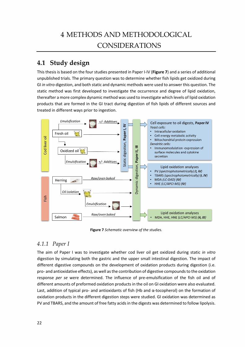
22
METHODS AND METHODOLOGICAL
CONSIDERATIONS
4.1 Study design This thesis is based on the four studies presented in Paper I-IV (Figure 7) and a series of additional
unpublished trials. The primary question was to determine whether fish lipids get oxidized during
GI in vitro digestion, and both static and dynamic methods were used to answer this question. The
static method was first developed to investigate the occurrence and degree of lipid oxidation,
thereafter a more complex dynamic method was used to investigate which levels of lipid oxidation
products that are formed in the GI tract during digestion of fish lipids of different sources and
treated in different ways prior to ingestion.
Figure 7 Schematic overview of the studies.
4.1.1 Paper I The aim of Paper I was to investigate whether cod liver oil get oxidized during static in vitro
digestion by simulating both the gastric and the upper small intestinal digestion. The impact of
different digestive compounds on the development of oxidation products during digestion (i.e.
pro- and antioxidative effects), as well as the contribution of digestive compounds to the oxidation
response per se were determined. The influence of pre-emulsification of the fish oil and of
different amounts of preformed oxidation products in the oil on GI oxidation were also evaluated.
Last, addition of typical pro- and antioxidants of fish (Hb and α-tocopherol) on the formation of
oxidation products in the different digestion steps were studied. GI oxidation was determined as
PV and TBARS, and the amount of free fatty acids in the digests was determined to follow lipolysis.
23
Analyses were done before digestion and at the end of the gastric and intestinal digestions,
respectively.
4.1.2 Paper II The aim of Paper II was to investigate what levels of the specific aldehydes MDA, HHE and HNE
that are formed in the gastric and intestinal lumen during digestion of cod liver oil using a dynamic
GI in vitro method (tiny-TIM). To analyze the specific aldehydes MDA, HHE and HNE, a LC/APCI-
MS method was developed and validated (see section 4.7.5) [74]. Cod liver oil with different
amounts of preformed oxidation products, as well as the impact of pre-emulsification, were
evaluated with respect to GI oxidation. Addition of cod Hb to the cod liver oil prior digestion was
tested to evaluate whether the strong pro-oxidative effect of Hb found during static digestion of
cod liver oil was also confirmed with a dynamic digestion method. The impact of EDTA as a
tentative antioxidant on GI oxidation was also examined. Analyses were here done in lumen
samples at repeated points during the gastric and the intestinal digestion steps.
4.1.3 Paper III The aim of Paper III was to examine what levels of MDA, HHE and HNE are formed in the gastric
and intestinal lumen after ingesting fish or fish oil using a dynamic GI in vitro method (tiny-TIM).
The impact of the fish muscle matrix, its lipid content, oven baking, species as well as oil
emulsification on GI oxidation were evaluated. Herring and salmon were chosen as representative
examples of two common food fishes. Fillets of both species were oven baked to study the
influence of cooking on GI oxidation. In order to evaluate the impact of the form in which the fish
lipids are supplied, herring oil was isolated from ground herring muscle, obtained from a batch of
high-fat herring. The amount of lipid oxidation products formed during digestion of herring mince
and crude as well as emulsified herring oil were then compared. Further, the determined
concentrations of MDA and HHE in intestinal lumen were compared with the concentrations of
the intestinal filtrate in order to evaluate the amount of aldehydes accessible for cellular uptake.
Hydroperoxides were also determined during the dynamic GI digestion of herring oil and raw
herring mince to compare the kinetics of primary oxidation products with the secondary aldehyde
products.
4.1.4 Paper IV The aim of Paper IV was to evaluate oxidative, proteomic and inflammatory responses on a cellular
level upon exposure to digests of non-oxidized and oxidized cod liver oil. Yeast, Saccharomyces
cerevisiae, was used as a model organism to study intracellular oxidation, cell energy metabolic
activity and mitochondrial protein expression. The immunomodulatory effects of the cod liver oil
digest were evaluated when maturing and stimulating human monocyte-derived dendritic cells
and were determined as expression of CD86 and HLA-DR and secretion of IL-6, IL-10, IL-12p40 and
IL-23. A second aim with this paper was to analyze the specific aldehydes MDA and HHE, in
addition to PV and TBARS, formed during GI digestion of cod liver oil. This required the
development and validation of new methods in the lab (see section 4.7.3 and 4.7.4).

24
4.2 Raw materials and additives
4.2.1 Cod liver oil Refined, but non-stabilized, cod liver oil was supplied by LYSI hf. (Reykjavík, Iceland). This
industrially manufactured oil was selected to represent a commercial fish oil, rich in LC n-3 PUFA,
but with the distinction that no extra antioxidants were added. The purpose was to acquire a fish
oil as neutral as possible as the addition of antioxidants would possibly disturb the evaluation of
the subsequent GI oxidation. It has been shown that fish oil supplements on the market often are
above recommended quality guide lines [10, 137]. Therefore, we wanted to produce cod liver oil
of different oxidative quality. Different levels of preformed oxidation products were obtained by
storing fresh oil in a closed Erlenmeyer flask in ambient light at room temperature for 6-13 days.
The resulting concentrations of lipid oxidation products can be found in Table 5 and the lipid
composition in Table 4.
4.2.2 Herring oil To evaluate the impact of the fish muscle matrix on GI oxidation, the herring oil was first isolated.
Centrifugation of minced raw herring fillets (see below) at 18 400×g for 30 min at room
temperature resulted in a floating layer of fish oil on top of the muscle tissue pellet and the
aqueous extract consisting of intra- and extracellular fluid. This is an easy and gentle method to
isolate mainly TAG from oily fish species. All herring lipids were not separated by this method; e.g.
membrane-bound phospholipids are thought to stay in the aqueous and protein fractions. In
addition to TAG, also lipid soluble antioxidants could go together with the oil fraction. The lipid
composition and the quality of the recovered herring oil are presented in Table 4 and 5,
respectively.
4.2.3 Emulsions Oil-in-water emulsions containing 20% (w/w) of fish oils were prepared according to the
procedure described by Sasaki et al. [138]. They reported that the mean particle diameter of
emulsions made of menhaden oil with this procedure ranged from 0.35 to 0.45 µM, and the mean
particle size was d32. Emulsions were always prepared fresh prior to the digestion experiment, but
as there were no access to an ultrasonicator when making dynamic digestions, the preparation of
those emulsions only included homogenization with a homogenizer, forming a coarse emulsion.
Emulsifier solutions consisted of 17 mM Brij 35 in 10 mM phosphate buffer, pH 7.0. Brij 35, also
referred to as polyoxyethylene glycol dodecyl ether or polyoxyethylene 23 lauryl ether, is an
uncharged, non-ionic detergent, with a hydrophilic polyoxyethylene chain and a hydrophobic
carbon chain. In contrast to e.g. tween 20 and 80, Brij 35 does not contain any fatty acid ester
moiety, which possibly could influence the GI oxidation result.
Figure 8 Chemical structure of Brij 35.
25
4.2.4 Herring mince Herring (Clupea harengus) is a small pelagic fish, caught world-wide. It has a high proportion of
dark muscle (around 20%) [139], which is rich in pro-oxidative heme-proteins. Herring is a fish
species with huge compositional variations depending on season, i.e. spawning vs non-spawning,
but also geographical place and feed may influence [13, 15, 140]. The amount and composition of
lipids, but also endogenous pools of e.g. antioxidants will therefore differ between different
catches. Herring thus represented a fish species which could be expected to be especially
susceptible for lipid oxidation. In Paper III, two batches of herring fillets were investigated. The
first batch was caught in the Baltic Sea (catch zone 27IIId) in June 2011 and contained 17.5% lipid,
while the second batch was caught in Skagerrak in February 2012 and had a lipid content of 4.2%.
Butterfly fillets from the above batches were bought as fresh as possible and received in the
laboratory two days post mortem where they were de-skinned before making mince of the muscle
tissue. Herring fillets which were supposed to be digested in the raw state were ground directly,
while the oven baked herring fillets were ground after cooking at 125°C to an inner temperature
of 55°C. One part of the mince made of the high-fat herring (1.6 kg) was used to isolate herring oil
(see above). All minces were kept at -80°C until use.
4.2.5 Salmon mince Salmon (Salmo salar) is a common food-fish species with high levels of carotenoids, such as
astaxanthin and canthaxanthin. Farmed Atlantic salmon was harvested by SalMar ASA (Kverva,
Norway) in February 2012, and one fillet of this lot was received in the laboratory three days after
slaughter. The fillet was deskinned and cut into 6x ~200 g pieces, whereof 3 pieces were oven
baked at 125°C (to an inner temperature of 55°C) before grinding to obtain a mince; the other 3
matching pieces were ground directly to raw mince.
4.2.6 Hemoglobin Heme-proteins, such as Hb, from fish are strong pro-oxidants and can induce lipid oxidation in e.g.
fish muscle [58, 141]. To evaluate the pro-oxidative effect of Hb during GI digestion, 11.5 µM cod
Hb (on emulsion basis) was added to emulsified cod liver oil prior to digestion. This concentration
was based on our previously determined Hb levels in herring fillets (unpublished data). Hb from
different fish species differ in its catalytic activity, possibly because of an adaptation to different
depths and water temperatures [58]. Therefore, we chose to keep the Hb origin similar to the oil
origin. Blood was obtained from freshly slaughtered farmed Atlantic cod, Gadus morhua.
Hemolysate was prepared from the blood as described by Richards and Hultin [142], and the Hb
concentration was determined against a bovine Hb standard after conversion to the carbon
monoxide form [143].
26
4.3 Gastrointestinal in vitro digestion method To investigate the extent of oxidation of fish lipids occurring during GI digestion, human studies
would ideally be made to include all physiological aspects. However, despite in vivo studies
possessing several benefits compared to in vitro studies, there are also some disadvantages that
makes in vitro methods preferable. A summary of the important differences between the two
methods is given in Table 1. For our research questions, in vitro methods were more suitable as
they e.g. enable screening, easy sampling and no ethical aspects.
Table 1 Summary of general advantages and disadvantages of using in vitro methods compared with in vivo trials.
Pros Cons + Possible to test any condition (pH, concentration, toxic compounds) + Easy sampling at various locations + No natural variation, reduces the required number of replicates + No ethical aspects + Low cost + Good for screening
- Too homogenous conditions (proper mixing, no phase separation of particle size in the stomach - No physiological feedback system - No absorption or removal of compounds in static models
4.3.1 Static in vitro digestion method Currently, no standardized method especially developed for evaluating GI oxidation exists. A
consensus static digestion method was, however, recently developed within the COST Action
FA1005 Infogest with the aim to facilitate comparison of static digestion results across researchers
[44]. At the start of this study, the Infogest consensus protocol by Minekus et al. [44] was not
initiated, and could thus not be used as a reference protocol. Unfortunately, a general protocol
suitable for various digestion questions is very difficult to establish, if the simplicity should still
remain. The consensus protocol is still not perfectly adapted for GI oxidation studies as e.g. no
lipase in the gastric phase is included. This is mainly due to the lack of a commercially available
human gastric lipase.
Instead, the static digestion protocol used in paper I and IV was a modification of the procedure
developed for determining in vitro bioaccessibility of carotenoids by Svelander et al. [144], which
originally was based on Garrett et al. [145] and then modified by Hedrén et al. [146]. As we
digested bulk oil, no starch or solid particles were included in the meal requiring addition of
amylase and mastication to simulate the initiation of digestion occurring in the mouth. Inclusion
of an oral phase is not considered necessary for liquid food according to the digestion method of
Minekus et al. [44]. To study the degree of lipid oxidation during digestion of fish lipids, certain
parameters were especially important and hence required a modification of the protocol by
Svelander et al. [144]. The major changes are listed below.
i) Predilution of the test meal in NaCl-solution with addition of high amounts of ascorbic
acid (originally to prevent oxidation of the carotenoids) was removed to keep the
environment free from pro- and antioxidants in our standard digestion protocol. The
original addition of pyrogallol and α-tocopherol to the simulated intestinal fluid was
excluded for the same reason. Selected pro- and antioxidants were then added
separately to the test meals in this study to investigate their influence on GI oxidation.

27
ii) In contrast to the studies of Svelander et al. [144], where they protected the
carotenoids as much as possible from exposure to oxygen by blanketing the samples
with nitrogen, we wanted to simulate more realistic oxygen levels during digestion. It
can be assumed that the oxygen level in the gastric phase is not a limiting factor for
lipid oxidation as air is swallowed during chewing and the food matrix itself contains
air and dissolved oxygen [63]. Air was therefore fully accessible in this step. However,
as the physiological oxygen concentration becomes lower in the small intestine, the
oxygen access was reduced in the head space of the samples during the intestinal
digestion step. For that reason samples were blanketed with nitrogen gas.
iii) Lipolysis may affect lipid oxidation, and it has been shown that gastric lipase
contribute with approximately 5-40% of total lipolysis [16]. A fungal lipase was
therefore added in the gastric phase.
Figure 9 Schematic overview of the static two-step digestion method simulating the gastric and the intestinal digestion.
4.3.1.1 Final static in vitro digestion method Our final digestion method is shown in Figure 9. To simulate the gastric phase, fish oil or fish mince
was mixed with gastric solution containing potassium, sodium, chloride, phosphate, magnesium
and calcium ions, and the digestive enzymes pepsin (porcine gastric mucosa) and lipase (Rhizopus
oryzae). The samples were incubated at pH 4 (adjusted with 0.1 M NaHCO3) for 30 min at 37°C on
an orbital shaker in darkness, followed by 30 min of incubation at pH 2 (adjusted with 1 M HCl).
To simulate intestinal digestion, the pH was raised to 6.9 (1 M NaHCO3) and a pancreatin/bile
extract solution of porcine origin was added. Samples were blanketed with nitrogen and incubated
for 120 min. An equal amount of water as the simulated gastric solution, was added to the start
sample to ensure similar extraction conditions.

28
Control samples containing fish oil or fish mince were made prior to each digestion experiment to
determine the required amount of hydrogen chloride or bicarbonate to obtain the desired pH for
each digestion phase. This procedure limited the unavoidable extra time between the gastric and
intestinal incubations. At the end of the digestion, the pH was occasionally checked in control
samples and no drift in pH was observed. Individual test tubes were made for each digestion step
to avoid possible sampling errors upon withdrawal of aliquots. Test tubes were frozen directly at
-80°C, and kept frozen until extraction of lipid oxidation products. As the activity of pepsin was
not considered important when digesting fish oil, no neutralization of gastric samples were made
prior to freezing and no protease inhibitor was added. PV and TBARS were determined in all
samples from static digestions (Paper I and IV), and MDA and HHE were also analyzed in Paper IV.
The used protocol simulates the conditions in the digestive tract during fed state, and was chosen
as both fish oil and fish mince were digested, the former is usually taken together with, or in close
connection to a meal. A fed state justify the gradual pH decrease applied during gastric digestion
as the pH in the stomach is raised immediately after ingesting a standard meal due to its buffering
capacity. A summary of 83 individual experiments determining the gastric pH during test meal
digestion in healthy subjects, shows that the pH in most cases increases to 5-7 [21]. The pH will
then slowly decrease, as hydrochloric acid is secreted. The bile extract concentration was ~4
mg/mL in the intestinal phase, which corresponds to postprandial in vivo levels.
The postprandial activity of human gastric lipase in gastric juice range between 10-120 U/mL in
healthy adults, using tributyrin (a short-chain non-physiological triglyceride) as substrate [16]. In
pure gastric juice the lipase activity was 130±62 U/mL [21]. Human gastric lipase was, however,
not commercially available, and no perfect alternatives exist having the same characteristics. As a
possible lipolytic effect in the stomach was considered important, a fungal lipase from Rhizopus
oryzae (same as arrhizus [21]) was used at 100 U/mL. This lipase was also used in the dynamic
digestion studies. It has a pH optimum of 7.5, but is stable between pH 3.5 and 8 [147]. Like human
gastric lipase, it is resistant to pepsin, but it is inhibited by bile salts at high concentrations [21,
148], which is acceptable as the pancreatic lipase is dominating during intestinal digestion where
bile salts are present. The fungal lipase has a reversed stereospecificity compared to gastric lipases
as the sn-1 position of TAG is preferred and not the sn-3 position [21]. Lipase from Aspergillus
niger could have been an alternative as it is active down to pH 2.5 and resistant to pepsin, like
human gastric lipase, but its stereospecificity on TAG is unknown [21].
4.3.1.2 Determination of oxygen in headspace during static digestion A 7.5-fold up-scaled digestion of cod liver oil was made to determine the actual oxygen
concentration in the head space of the digestion tubes during static in vitro digestion. Glass tubes
with a volume of 100 mL was used to ensure proper oxygen determination with a PBI Dansensor
CheckMate II as ~5 mL of gas was required for the measurement. The oxygen concentration was
determined both at start and at the end of each digestion phase to follow any change during
digestion.

29
4.3.2 Dynamic in vitro digestion method (tiny-TIM) By increasing the complexity of the in vitro digestion method, and thereby approaching more
physiological conditions, dynamic methods have been developed. We used the dynamic
computer-controlled system TIM, developed at TNO (Zeist, The Netherlands). The first model
(TIM-1), developed in the early 1990s, simulates four successive compartments (stomach,
duodenum, jejunum and ileum) and is described in detail by Minekus et al. [45]. This model has
been further developed and apart from the even more sophisticated or specialized models, such
as TIM-agc (advanced gastric compartment) and TIM-2 (including the gut), also a more simplified
version has been designed, tiny-TIM. A schematic presentation of tiny-TIM is seen in Figure 10.
Compared to TIM-1, a half-sized stomach was used, which is the standard size for non-
pharmaceutical studies. All test meals were accordingly down-scaled by a factor of two. Instead
of having three separate intestinal compartments, the small intestinal phase in tiny-TIM is
simulated by a single compartment without ileal efflux. Lipophilic products, incorporated into
micelles, are removed from the intestinal compartment as they pass through a filter with pore size
of 50 nm, while lipid droplets >50 nm remain in the intestinal lumen. When separation of the
different intestinal compartments is assumed to play a minor role for the research question, this
model is adequate. Fat digestion is primarily occurring in the upper small intestine (duodenum
and jejunum). One advantage with tiny-TIM over TIM-1 is that two separate digestions can be run
in parallel, thus enabling a doubled number of digestions within the same time period.
Figure 10 Schematic overview of the tiny-TIM system: A, gastric compartment; B, pyloric sphincter; C, chyme; D, small intestinal compartment; E, gastric secretion; F, intestinal secretion; G, semi-permeable membrane (<50 nm); H, intestinal filtrate; I, pH electrodes; J, pressure sensor.

30
4.3.2.1 Test meals and digestive fluids The test meals consisted of varying amount of fish lipids, depending on the research question. In
Paper II, bulk cod liver oil was compared with its corresponding emulsion, and each meal consisted
of 5 g cod liver oil (equals 25 g emulsion, 20% o/w). When investigating GI oxidation of different
fish minces in Paper III, 50 g mince was used. To investigate the matrix effect when digesting
herring lipids, 8.75 g herring oil or 44 g herring oil emulsion (equals 8.75 g oil) was used as this was
the lipid content in 50 g of high-fat herring mince. To avoid floating oil and ensure proper gastric
emptying when fish oil was digested, 20 g wheat flour bread was added as bulk meal. Bread was
also included in test meals of emulsion and mince to avoid background differences. All test meals
were made up to 150 g with water and adjusted to pH 6.5 prior to feeding.
Table 2 Composition of a 150 g portion (intake) of each test meal digested in the dynamic in vitro model.
Saliva (g)
Bread (g)
Water (g)
Fish mince/ oil/emulsion (g)
Cod liver oil (bread/no bread) 20 20/0 105/125 5 Cod liver oil emulsion (bread/no bread) 20 20/0 85/105 25 Herring/salmon mince (bread/no bread) 20 20/0 60/80 50 Herring oil 20 20 101.25 8.75 Herring oil emulsion 20 0 86 44 Blank (bread/no bread) 20 20/0 110/130 0
The enzymes and electrolytes used in the different simulated digestive fluids are listed here:
Saliva: Amylase (7 mg/meal) and the electrolytes NaCl, KCl and CaCl2.2H2O.
Gastric secretion: HCl, pepsin (porcine gastric mucosa), lipase (Rhizopus oryzae) and the electrolytes NaCl, KCl, CaCl2.2H2O and NaOAc. Secretion rate was 1mL/min.
Intestinal secretion: NaHCO3, bile (fresh porcine), pancreatin (Pancrex-Vet) and the electrolytes NaCl, KCl and CaCl2.2H2O. Secretion rate was 1mL/min.
Samples from the gastric and the intestinal lumen were withdrawn as snap-shot samples every 30
min, while intestinal filtrate samples were collected as pooled samples every 30 min, thus
representing an average for each 30 min interval. All samples were frozen directly after sampling
and stored at -80°C until analysis. The levels of MDA, HHE and HNE, and in selected test meals also
lipid hydroperoxides, were determined in samples from dynamic digestions.
Table 3 Summary of advantages and disadvantages of using dynamic in vitro digestion methods compared with static methods.
Pros Cons + pH gradient + Gradual secretion of digestive fluids + Realistic transit time + More realistic mixing + Removal of digestive products, possibly avoiding product inhibition + Can follow the concentration of a particular compound in different compartments over time
-Time consuming -Expensive -Sometimes very diluted samples, which may affect sample analysis

31
4.4 What type of answers can be obtained by the static and
the dynamic digestion models? In this study we wanted to investigate GI oxidation of fish lipids from several aspects. Static
digestions can give answers to fundamental questions, e.g. how physicochemical factors affect GI
oxidation and results from these experiments are expressed as µmol lipid oxidation product/kg
lipid. Further, an important aim was to explore which aldehyde and lipid hydroperoxide levels the
GI tract theoretically can be exposed to. This was investigated by following the concentration of
lipid oxidation products in the gastric and the intestinal compartment of the dynamic model over
time, and these results are presented as µmol oxidation product/L digesta. To also evaluate the
relative degree of oxidation of fish lipids under the dynamic condition, it is necessary to
understand the dilution behavior of the dynamic system. Without any formation or degradation,
the initial concentration of the test meal will change over time according to Figure 11, here
demonstrated by the coloring agent Patent Blue V. A linear decrease is observed in the gastric
phase up to 150 min, and approximately 50% of the initial intake concentration remains at 90 min
of digestion. At the same time, a peak concentration of 35% is reached in the intestinal lumen.
The concentration in the intestinal filtrate sample is slightly delayed compared to the intestinal
lumen, and peaks at 120 min of digestion.
Figure 11 Concentration of Patent Blue V marker in the gastric and the intestinal compartments and in the intestinal filtrate, expressed as per cent of intake, during dynamic in vitro digestion in tiny-TIM. The intestinal filtrate represents compounds passing a filter with a pore size of 50 nm. Error bars is (max-min)/2 of two digestions.
Based on these curves and the absolute oxidation concentration at each time point, it is possible
to calculate the relative change in oxidation over time (see equation below). Such data can be
used to compare results from the dynamic and the static digestions.

32
4.5 Choice of methods to follow lipid oxidation No universal method exists that completely covers all changes of lipids taking place during
oxidation. Therefore, it is necessary to choose a method that monitors relevant changes for a
particular question and specific food item. There are different general ways to get an overview of
the oxidation reaction, which can be categorized as i) oxygen consumption, ii) loss of initial
substrate, iii) formation of free radicals, iv) formation of primary oxidation products and v)
formation of secondary oxidation products, vi) formation of tertiary products. Depending on
substrate, matrix, number of possible measurements, etc. a selection has to be made for which
method that is most applicable.
As the lipid oxidation process requires oxygen, the oxygen consumption of the headspace above
the sample can be assumed to be a good indicator. The decrease in oxygen concentration in the
headspace can be determined e.g. by an Oxidograph or by gas chromatography [149]. This method
is useful for lipid products, but is not applicable for complex matrices containing e.g. proteins as
the protein oxidation process also absorbs oxygen. Due to oxygen consumption being a non-
specific method, and we specifically wanted to follow lipid oxidation, this method was rejected.
Instead, determination of lipid oxidation products was preferred. As the kinetics for primary and
secondary oxidation product formation were expected to differ during the oxidation process, it
was relevant to follow the concentration of compounds belonging to both these categories.
4.5.1 Choice of method to determine primary oxidation products Hydroperoxides are often determined to study primary lipid oxidation products and several
methods exist, which are relatively quick and easy. The redox property of hydroperoxides can be
used to oxidize reagents, e.g. iodide or ferrous ion. Another method to determine hydroperoxides
is iodometry, which is described in an official method by AOAC. Peroxides liberate iodine from the
iodide ion, and iodine are then titrated with sodium thiosulfate in the presence of starch. This
volumetric method requires large amounts of lipid sample and has some drawbacks with risk of
over- and underestimating the hydroperoxides in the sample. Methods built on chromatography
also exist to specifically determine different hydroperoxides, which can be relevant in certain
studies. Usually, chromatography methods are more time-consuming in sample preparation and
data processing than e.g. spectrophotometric methods.
Determination of conjugated dienes/trienes is yet another non-specific method to follow primary
oxidation products, as almost all (>90%) hydroperoxides contain conjugated diene structures.
Hydrogen abstraction from PUFA generates radicals, which after rearrangement form conjugated
dienes/trienes, due to the higher stability of the radical after electron delocalization. These
conjugated compounds absorb in the UV range and is easily measured by a spectrophotometer
[48]. High backgrounds are however seen.
The aim in this study was to get a general picture of the amount of primary oxidation products in
the samples during digestion. Therefore, the spectrophotometric methods were considered more
suitable than chromatographic methods. However, as small sample amounts were available and
we wanted to avoid the known background disturbance with conjugated dienes, the ferrous
oxidation method to determine lipid hydroperoxides was used in this study (see section 4.7.1).
33
4.5.2 Choice of method to determine secondary oxidation products As mentioned in the background, the class of secondary oxidation products consists of various
compounds. Volatiles (aldehydes, ketones, alcohols, short carboxylic acids, hydrocarbons etc.) can
be determined by sensorial or gas chromatography (GC) analysis of the headspace with/without
prior extraction. Dynamic headspace and solid phase microextraction (SPME) are methods to
concentrate volatiles prior to analysis. This equipment was not available. A classical quality
indication used by the oil industry to determine aldehydes is the anisidine test, generating the
anisidine value (AV). The reaction between the carbonyl bond of the aldehyde and anisidine amine
group leads to a Schiff base that absorbs at 350 nm [48]. This method also accounts for non-
volatile aldehydes. To follow lipid oxidation in both biological samples and foods, the aldehyde
MDA has traditionally been determined, mostly after its reaction with the chromophore TBA,
forming TBA-reactive substances (TBARS). This product can either be determined by
spectrophotometric or liquid chromatography (LC) methods. Nowadays, using chromatography
methods coupled to mass spectrometry (MS), a multitude of compounds can be determined
within one analysis [86]. The spectrophotometric method determining TBARS was chosen in order
to rapidly being able to analyze samples (see section 4.7.2), as no chromatographic method were
developed in the lab at the start of the project. More specific methods were developed later on.
The chosen methods for determination of lipid hydroperoxides and TBARS required lipid
extraction of the samples prior to analysis; this extraction is described below.
4.6 Lipid analysis
4.6.1 Extraction of lipids for total lipid determination, fatty acid
composition, lipolysis and lipid oxidation analysis Total lipid content of fish minces and fresh bile (Paper III) were determined gravimetrically after
extraction with chloroform and methanol as described by Undeland et al. [150] with minor
modifications. Solvent ratio of chloroform:methanol was according to the recommended ratios
given by Lee et al. [151]. Fish minces containing >6% lipids were extracted with a
chloroform:methanol ratio of 2:1, while minces with 2-6% lipids were extracted with 1:1 ratio, and
minces with <2% lipids with a 1:2 ratio. Two g of fish mince or bile was used and solvent volumes
were changed to 15 mL in the first homogenization step, and 5 mL was added in the second step.
To separate the one-phase system, 8 mL of NaCl solution (0.5%) was added. To prevent lipid
oxidation during preparation, 0.05% BHT was added to the solvent solutions and samples and
solvents were kept on ice.
According to Schmedes and Hølmer [152], a Bligh and Dyer extraction could simultaneously
partition lipid hydroperoxides into the chloroform phase and the non-protein bound MDA into the
methanol/water phase. The separation of hydroperoxides before addition of the TBA reagent
avoids falsely high TBARS values, which otherwise can be formed by hydroperoxide cleavage
during the heating step. Briefly, for oxidation product analyses (Paper I and IV), liquid samples
(digests or start samples consisting of oil and electrolyte) were vortexed with
chloroform:methanol (1:2, with 0.05% BHT) at a volume ratio of 4:15 (same proportions as
described in Schmedes and Hølmer [152]), and then additional 5 volumes of chloroform was
added. Aliquots of both phases were stored at -80°C prior to analysis.

34
4.6.2 Fatty acid composition (Paper II and III) Fatty acid composition of lipids extracted from fish minces, fish oil and bile, as described above,
was determined after methylation of the fatty acids to fatty acid methyl esters (FAME) according
to the principle of Lepage and Roy [153]. Extracted lipids, redissolved in toluene, were methylated
over night at 20°C after addition of acetylchlorid in methanol. C17:0 was added to the samples
before methylation as internal standard. Separation and quantification of FAME was done with
GC-MS. A methylated fatty acid mixture was used for identification of the different peaks.
4.6.3 Lipolysis (Paper I) Lipolysis was followed in digests of cod liver oil as the formation of free fatty acids of bulk oil e.g.
can contribute to emulsification and further lipolysis in the intestinal digestion. Formation of free
fatty acids during lipolysis of TAG may also increase the amount of lipid oxidation products formed
during digestion. To evaluate the degree of lipolysis during static in vitro digestion and relate this
feature with reported in vivo data, lipids were first extracted from the digests using the extraction
method described above for the lipid oxidation analyses, with slight modification. Non-digested
and intestinal samples were acidified to pH 1-2 to ensure that the fatty acids were protonated and
partitioned into the chloroform phase. Neutral and free fatty acids were separated and detected
according to the high performance liquid chromatography (HPLC) method described by Silversand
and Haux [154]. Lipolysis degree, expressed as per cent of maximal FFA formation, was calculated
according to the equation below. The calculation is based on the assumption that maximum two
FFA is formed from one TAG, that all fatty acids are randomly distributed on the glycerol molecule
and that the cod liver oil only consists of TAG.
4.7 Lipid oxidation analysis
4.7.1 Analysis of lipid hydroperoxides (Paper I, III and IV) A ferrous oxidation method was used in this study to determine lipid hydroperoxides, but with
two different complexation compounds. In Paper I and IV, the peroxide value was determined
under acidic conditions where thiocyanate formed a complex with ferric iron, a method described
by Undeland et al. [150]. A standard curve was made with cumene hydroperoxide.
In Paper III, the lipid hydroperoxide concentration in gastric and intestinal lumen samples were
determined with the xylenol orange reagent (FOX) using the PeroxiDetect� Kit. The reagent was
dissolved in methanol solution to avoid lipid oxidation during preparation. Prior to analysis, also
lumen samples were diluted in methanol:water (90:10) and BHT was added to the reagent mixture
to prevent oxidation. Tert-butyl hydroperoxide was used to make a standard curve. The reason to
change from the ferric thiocyanate method to the FOX assay in Paper III was that the chloroform
phase containing the extracted lipids had an interfering yellow color, which most likely originated
from the fresh bile used in the dynamic digestions. This interference was not observed in the static
digestions using bile extract.

35
4.7.2 Colorimetric analysis of free TBARS (Paper I and IV) Secondary oxidation products were determined as TBARS in Paper I and IV according to the
spectrophotometric method described by Schmedes and Hølmer [152]. MDA in the
methanol/water phase obtained after the chloroform/methanol extraction of samples (see
section 4.6.1) was allowed to react with the TBA reagent (dissolved in TCA), forming a red TBA2-
MDA complex [152]. 1,1,3,3-Tetraethoxypropane (TEP), which is a precursor of MDA, was
hydrolyzed by boiling in 0.1 M HCl to prepare a stock solution of MDA. The TBARS method was
chosen as it conveniently could be applied to the aqueous phase after lipid extraction. It
determines non-protein bound MDA due to the separation of proteins taking place during
extraction with chloroform/methanol and avoids thereby false high TBARS values as proteins also
can respond in the TBARS test.
The same method to determine TBARS could not be used for samples from the dynamic digestion
studies. After chloroform/methanol extraction of intestinal filtrate samples, the aqueous phase
obtained a strong yellow color with a peak at 420 nm. This color interfered with the absorbance
reading of TBA2-MDA at 532 nm because the baseline was affected in the range of 350-600 nm.
The yellow color, probably originating from the fresh bile, also remained after reaction with TBA
reagent and subsequent boiling and centrifugation. When looking at the spectra of the final
sample, two peaks between 420 and 532 were in fact seen. To overcome this problem a specific
method to determine the MDA using HPLC was developed (see below). In addition, the
spectrophotometric measurement of TBARS has some drawbacks due to its non-specificity. Apart
from MDA, TBA can also react with e.g. other lipid decomposition products (alkanals, alkenals and
alkadienals), browning reaction products, protein and sugar degradation products, amino acids
and nucleic acids [93, 155, 156]. Another criticism to the method is the high temperature (boiling
water) used in the derivatization step. Antioxidants are added to prevent lipid oxidation during
preparation, but it some oxidation might still occur.
4.7.3 Analysis of total MDA by HPLC-DAD (Paper IV) The problem with the non-specific reaction with TBA and spectrophotometric methods can be
overcome with a separation of the TBA2-MDA complex by chromatography prior to detection.
MDA can be detected without any derivatization [52], but to increase the sensitivity during
detection, derivatization agents are most frequently used. 2,4-Dinitrophenyl hydrazine (DNPH) is
commonly used to analyze MDA with HPLC, but also for studying protein oxidation as it reacts
with carbonyl groups [157]. Other examples of derivatization agents for MDA analysis is
diaminonaphtalene (DAN) [158], cyclohexanedione (CDH), FMOC-hydrazine [159] and
dansylhydrazine (DH) [160]. MDA can also be determined by GC. This method requires a
derivatization step prior analysis and a variety of agents are proposed, e.g. 2-hydroxypyrimidine
[161], 2-hydrazino-benzothiazole [162], and pentafluoro-phenylhydrazine [163].
In order to specifically measure MDA, the HPLC method described by Mateos et al. [157] with
prior derivatization with DNPH was selected. The decision to use DNPH as a derivative was based
on the comparative study by Mendes et al. [164], who concluded that DNPH was superior to TBA
as a derivatization agent also when HPLC was used to separate the MDA-derivative. During acid
conditions DNPH reacts with carbonyl groups yielding 2,4-dinitrophenylhydrazone derivatives,
which can be detected by UV/VIS at a wavelength between 300 and 380 nm depending on the
36
absorption maximum for the compound of interest. MDA-DNPH has an absorption maximum at
307 nm, while other small aliphatic aldehydes like formaldehyde, acetaldehyde, acetone, and
propionaldehyde have their maximum at 356 nm [165]. The 4-hydroxyalkenals HHE and HNE have
their maximum at 365 nm. In order to measure total MDA, protein bound MDA was released
during an alkaline hydrolysis step at 60°C for 30 min. MDA-DNPH derivatives were then separated
and detected by HPCL coupled to a photodiode array detector (DAD). A stock solution of MDA was
prepared by hydrolyzing TEP in 1% sulphuric acid for 2 h at 20°C [52]. The spectrum of the MDA-
DNPH standard was in accordance with the data given by Cordis et al. [165].
When the method was set up, small modifications of the original method was made. The very
strong oxidizing agent perchloric acid was replaced by trichloroacetic acid (TCA), and we observed
that the detectable concentration of MDA increased with increasing TCA concentration. A possible
drawback with the method for determining total MDA was that the hydrolysis step was made at
elevated temperature, which may induce lipid oxidation during sample preparation. However, no
difference in MDA concentration was seen with or without BHT addition.
4.7.4 Analysis of free HHE by LC/APCI-MS (Paper IV) A method for analyzing HHE was set-up to determine the amount before and after static digestion
of cod liver oil. Several methods were available, some based on GC-MS, which either required
headspace equipment or deuterated standards, but also HPLC-based methods were reported. For
example, Sakai et al. [166] and Grune et al. [167] presented HPLC methods to analyze the HHE
content in fish muscle and egg yolk, respectively. A combination of these HPLC methods and
sample preparations were used as a basis for our method development since a LC-MS equipment
was available and we wanted to add MS detection for better detection. The developed method
with low limit of detection (16 nM) is described in Larsson et al. [168]. Briefly, HCl was added to
the sample to precipitate the proteins, and HHE in the supernatant was allowed to form a
derivative with DNPH. The repeatability of the LC/APCI-MS method was 2-7% for six different HHE
concentrations in the range 0.05-2 µM (n=4). The RSD of digests, including sample preparation,
was 5% (n=6).
4.7.5 Combined analysis of free MDA, HHE and HNE (Paper II and III) The LC/APCI-MS method to determine HHE described above was further developed to determine
MDA, HHE and HNE in one single analysis as they all form derivatives with DHNP. A gradient
program separated the different aldehyde-DNPH derivatives, but small background disturbance
in the MS signal was seen for the HNE-DNPH ion coming from DNPH. Also the sample preparation
was slightly modified, in that BHT and EDTA was added to the sample to prevent possible oxidation
during sample preparation. However, no differences in determined aldehyde levels were seen
between digested samples with antioxidant addition compared to samples without antioxidants,
indicating that lipid oxidation during sample preparation is no major problem. The final method is
described in Tullberg et al. [74]. Linearity of standards was controlled for: MDA, R2=0.985 (0.025-
100 µM); HHE, R2=0.997 (0.01-10 µM) and HNE, R2=0.997 (0.005-10 µM). The determined relative
standard deviations (RSD) for digests analyzed in 6 replicates were: 2.6% (MDA), 7.6% (HHE) and
5.6% (HNE). Others, describing methods for total amounts of aldehydes, add TCA to the samples.
However, when we added TCA to aldehyde standards the peaks in the chromatograms
disappeared.

37
4.8 Exposure of yeast cells to cod liver oil digests to study
metabolic activity, oxidative stress and proteomic
response
4.8.1 Cultivation of yeast cells and digest exposure Yeast is one of the most well-studied eukaryotic cell models and despite the apparent difference
between yeast and human, surprisingly many basic cellular processes are conserved between the
species. The effect of cod liver oil digests of different quality on intracellular oxidative and
proteomic responses in yeast cells were investigated in Paper IV. Saccharomyces cerevisiae ZIM
2155 was cultivated according to the conditions described by Zakraj�ek et al. [169] They used yeast
in the stationary phase as a model to study the responses to environmental stressors. Also others
have made oxidative stress studies using yeast cells during their stationary phase [170, 171]. Cod
liver oil digests or control digests were added to yeast cell cultures at a concentration of 1.7 mg
lipids/mL (equivalent to 35 µL digest/mL) and incubated for 2 h before subsequent analyses.
4.8.2 Intracellular oxidation 2,7-Dichlorodihydrofluorescin (DCFH2) is extensively used to measure cellular ROS production
[172]. The non-fluorescent 2,7-dichlorodihydrofluorescin diacetate (DCFH2-DA) dye was added to
aliquots of resuspended pre-washed yeast cells to measure intracellular oxidation. After
penetration of the dye through the cell membrane, the dye is hydrolyzed by non-specific esterases
in the cytoplasm and can then oxidize to fluorescent 2,7-dichlorofluorescin (DCF) [173]. After 20
min of incubation, the rate of oxidation was monitored by a Safire II microplate reader at the
excitation and emission wavelengths of DCF; 488 nm and 520 nm, respectively. Results were
expressed as fluorescence intensity relative to control (buffer).
4.8.3 Cell energy metabolic activity Cell energy metabolic activity was determined by BacTiter-Glo� Microbial Cell Viability Assay,
which is based on the quantification of ATP. After 2 h of incubation of yeast cells with digests or
control buffer, the BacTiter-Glo� reagent was added and luminescence was recorded after 5 min
by a Safire II microplate reader. Results were expressed as luminescence intensity relative to
control (buffer).
4.8.4 Expression of mitochondrial proteins Mitochondrial proteins were extracted using Cytosol/Mitochondria Fractionation kits. Protein
concentration was determined by the method of Bradford [174]. Proteins were separated by 2-D
electrophoresis, with subsequent staining with the stable fluorescent stain SYPRO Ruby. Duplicate
gels were used to obtain an average gel sample using normalized spot volumes. Image analysis
revealed the expression changes, which were considered significant for spots which reproducibly
had minimum 1.5-fold change in expression compared to digested control. Protein identification
of statistically different spots were based on MS/MS spectra using the NCBInr database
(20130918) refined to taxonomy Saccharomyces cerevisiae. One drawback with this method is
that also pre-existing proteins are labelled, and not only newly synthesized proteins.
38
4.9 Exposure of human monocyte-derived dendritic cells
to cod liver oil digests to study immunomodulation
4.9.1 Dendritic cell model The monocyte-derived dendritic cell model was described by Jonsdottir et al. [175]. The dendritic
cell model has been developed for the purpose of screening possible pro- and anti-inflammatory
effects of natural products [175, 176]. Immature dendritic cells were obtained by isolating CD14+
monocytes from peripheral blood mononuclear cells (PBMCs) from healthy subjects [176]. Cells
were matured in the presence of IL-1β and tumor necrosis factor alpha (TNF-α) and stimulated
with lipopolysaccharides (LPS) for two days. At the same time, the dendritic cells were incubated
with freeze-dried digesta at 1, 10 and 100 µg /mL 0.002% DMSO (equivalent to 0.015, 0.015 and
0.15 µL wet digest/mL). DMSO without any digest was used as solvent control. The effect of
digested cod liver oil at various concentrations on the maturation and stimulation of dendritic cells
was evaluated by measuring expression of cell surface receptors (HLA-DR and CD86) and signaling
molecules (cytokines). Cell viability tests were performed to check the viability of the cells in the
presence of digests at the highest concentration (100 µg dw/mL). To study whether digests were
affected by the freeze drying, immunomodulatory responses in dendritic cells were repeated
using liquid digests (from another digestion) showing the same results as when freeze-dried
digests were used.
4.9.2 Expression of surface molecules Expression of HLA-DR and CD86, involved in the presentation of antigens to T cells and their
activation, were analyzed by flow cytometry. Dendritic cells were stained with monoclonal
antibodies against HLA-DR and CD86 and then collected and analyzed using FACScalibur and
CellQuest software. The expression of HLA-DR and CD86 was reported as percentage positive cells,
or mean fluorescence intensity (MFI), which evaluated the level of staining.
4.9.3 Secretion of cytokines Dendritic cells produce cytokines that could either be pro- or anti-inflammatory. The
concentration of the cytokines IL-6, IL-10, IL-12p40 and IL-23, was determined by the conventional
ELISA (enzyme-linked immunosorbent assay) method according to the manufacturer�s
instructions. Results were expressed as secretion index (SI), in order to normalize for large
individual differences in secretion. SI was calculated by dividing the cytokine concentration
secreted by dendritic cells exposed to cod liver oil digest with the concentration secreted after
exposure to a control (DMSO). The proportional index, calculated by dividing SIIL-12p40 with SIIL-10,
was used to evaluate the overall pro- or anti-inflammatory effect.
4.10 Statistical analysis Statistical analyses were made using SPSS Statistics version 19 (IBM Corporation, New York, USA)
and p ≤ 0.05 was considered significant. ANOVA was used to compare means between more than
two groups and Student�s t-test was used when only two groups were to be compared. Different
post hoc tests have been used in different papers and for different analyses, and a detailed
description is given in each paper.
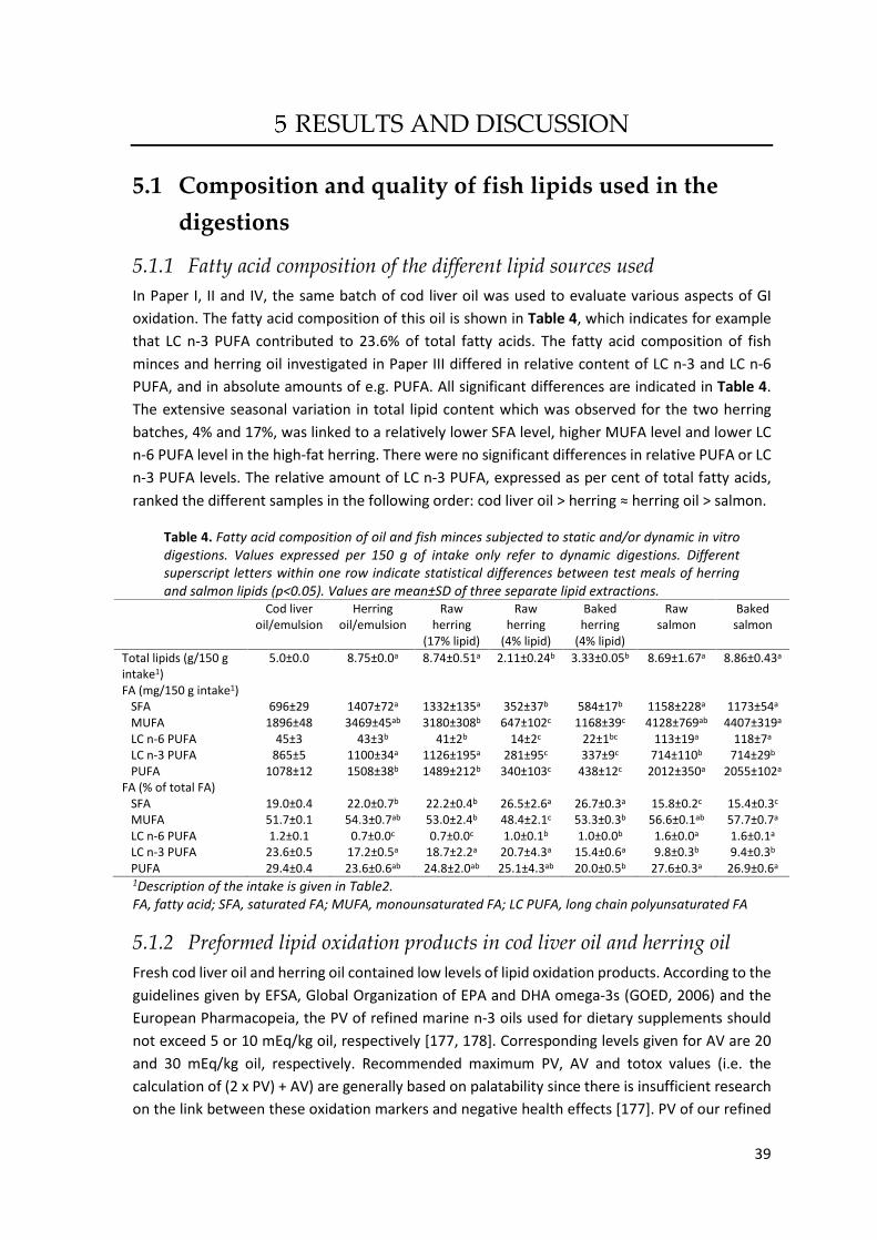
39
RESULTS AND DISCUSSION
5.1 Composition and quality of fish lipids used in the
digestions
5.1.1 Fatty acid composition of the different lipid sources used In Paper I, II and IV, the same batch of cod liver oil was used to evaluate various aspects of GI
oxidation. The fatty acid composition of this oil is shown in Table 4, which indicates for example
that LC n-3 PUFA contributed to 23.6% of total fatty acids. The fatty acid composition of fish
minces and herring oil investigated in Paper III differed in relative content of LC n-3 and LC n-6
PUFA, and in absolute amounts of e.g. PUFA. All significant differences are indicated in Table 4.
The extensive seasonal variation in total lipid content which was observed for the two herring
batches, 4% and 17%, was linked to a relatively lower SFA level, higher MUFA level and lower LC
n-6 PUFA level in the high-fat herring. There were no significant differences in relative PUFA or LC
n-3 PUFA levels. The relative amount of LC n-3 PUFA, expressed as per cent of total fatty acids,
ranked the different samples in the following order: cod liver oil > herring ≈ herring oil > salmon.
Table 4. Fatty acid composition of oil and fish minces subjected to static and/or dynamic in vitro digestions. Values expressed per 150 g of intake only refer to dynamic digestions. Different superscript letters within one row indicate statistical differences between test meals of herring and salmon lipids (p<0.05). Values are mean±SD of three separate lipid extractions.
Cod liver oil/emulsion
Herring oil/emulsion
Raw herring
(17% lipid)
Raw herring
(4% lipid)
Baked herring
(4% lipid)
Raw salmon
Baked salmon
Total lipids (g/150 g intake1)
5.0±0.0 8.75±0.0a 8.74±0.51a 2.11±0.24b 3.33±0.05b 8.69±1.67a 8.86±0.43a
FA (mg/150 g intake1) SFA 696±29 1407±72a 1332±135a 352±37b 584±17b 1158±228a 1173±54a
MUFA 1896±48 3469±45ab 3180±308b 647±102c 1168±39c 4128±769ab 4407±319a
LC n-6 PUFA 45±3 43±3b 41±2b 14±2c 22±1bc 113±19a 118±7a
LC n-3 PUFA 865±5 1100±34a 1126±195a 281±95c 337±9c 714±110b 714±29b
PUFA 1078±12 1508±38b 1489±212b 340±103c 438±12c 2012±350a 2055±102a
FA (% of total FA) SFA 19.0±0.4 22.0±0.7b 22.2±0.4b 26.5±2.6a 26.7±0.3a 15.8±0.2c 15.4±0.3c
MUFA 51.7±0.1 54.3±0.7ab 53.0±2.4b 48.4±2.1c 53.3±0.3b 56.6±0.1ab 57.7±0.7a
LC n-6 PUFA 1.2±0.1 0.7±0.0c 0.7±0.0c 1.0±0.1b 1.0±0.0b 1.6±0.0a 1.6±0.1a
LC n-3 PUFA 23.6±0.5 17.2±0.5a 18.7±2.2a 20.7±4.3a 15.4±0.6a 9.8±0.3b 9.4±0.3b
PUFA 29.4±0.4 23.6±0.6ab 24.8±2.0ab 25.1±4.3ab 20.0±0.5b 27.6±0.3a 26.9±0.6a
1Description of the intake is given in Table2. FA, fatty acid; SFA, saturated FA; MUFA, monounsaturated FA; LC PUFA, long chain polyunsaturated FA
5.1.2 Preformed lipid oxidation products in cod liver oil and herring oil Fresh cod liver oil and herring oil contained low levels of lipid oxidation products. According to the
guidelines given by EFSA, Global Organization of EPA and DHA omega-3s (GOED, 2006) and the
European Pharmacopeia, the PV of refined marine n-3 oils used for dietary supplements should
not exceed 5 or 10 mEq/kg oil, respectively [177, 178]. Corresponding levels given for AV are 20
and 30 mEq/kg oil, respectively. Recommended maximum PV, AV and totox values (i.e. the
calculation of (2 x PV) + AV) are generally based on palatability since there is insufficient research
on the link between these oxidation markers and negative health effects [177]. PV of our refined

40
fresh cod liver oil and the crude herring oil were 2.6 and 1.4 mEq/kg, respectively. According to
the manufacturer, the AV of fresh cod liver oil was 8.5 mEq/kg oil, which together with the PV
conclude that oxidation of the cod liver oil was below the recommended upper limits. Storage of
the cod liver oil for 6-13 days at 20°C generated PV from 25-68 mEq/kg (Table 5). In a study
screening different commercial fish oil supplements on the New Zeeland market, PV up to 33
mEq/L was reported, despite 12-24 months remaining of the best-before date [10]. Also secondary
oxidation products (AV) were above recommended levels in several supplements. A similar study,
but on the Norwegian market, determined following median values in mEq/kg (total range is given
in the parenthesis): PV 9.9 (0.8-39.8), AV 8.0 (2.1-34.0) and totox 29.0 (8.1-113.6), among 56
different supplements [11]. These studies implies that it is relevant to evaluate GI oxidation also
in oils that are oxidized, since they obviously appears on the market.
Table 5. Amount of preformed oxidation products in cod liver oil and herring oil used for subsequent digestion studies. The different batches of oxidized cod liver oils are indicated by A-D. A and B have the same denomination in Paper IV, while C and D corresponds to �medium ox� and �high ox�, respectively, in Paper II.
CO
Fresh CO
Ox A CO
Ox B CO
Ox C CO
Ox D Herring oil
Fresh
Used in paper I, II, IV I, IV I, IV II II III
PV (mmol/kg oil)1 1.3±0.1 23.0±0.3 33.7±0.8 12.4±0.6 20.0±1.8 0.7±0.1 Total MDA (µmol/kg oil) 15±2 337±14 551±35 na na na Free TBARS (µmol/kg oil) 4.1±1.3 23.2±3.3 94.2±4.3 na na 1.1±0.5 Free MDA (µmol/kg oil) 2.8±0.2 na 56±3 25±1 47±8 0.8±0.1 Free HHE (µmol/kg oil) 1.9±0.1 na 9.3±17 4.1±0.5 5.5±1.2 0.07±0.02 Free HNE (µmol/kg oil) 0.03±0.01 na 0.15±0.04 0.06±0.01 0.08±0.02 0.003±0.003
1Peroxide value expressed as mEq/kg oil is obtained by multiplying the number with 2. CO, cod liver oil; na, not analyzed
5.2 Factors of the in vitro digestion method influencing GI
oxidation
5.2.1 Influence of oxygen access during static digestion The influence of oxygen access on GI oxidation has to our knowledge not been reported by others.
Therefore, a pilot study was made to evaluate the effect of blanketing fresh and oxidized cod liver
oil with nitrogen prior to the gastric and the intestinal steps during static digestion. As seen in
Figure 12, oxidized oil showed a relative decline in both PV and TBARS in the gastric and the
intestinal phase compared to control samples digested without nitrogen blanketing. Also fresh
cod liver oil reached lower PV and TBARS levels after intestinal digestion compared to the control
when nitrogen blanketing was used.

41
Figure 12 Effect of blanketing cod liver oil digests with nitrogen prior to static gastric and intestinal in vitro digestion on the formation of A) PV and B) TBARS. Results are expressed in per cent relative cod liver oil digested without nitrogen blanketing.
Absolute amount of TBARS increased gradually during each digestion step when highly oxidized
cod liver oil was digested, in both control and nitrogen blanketed samples (Figure 13 A). In
contrast, the PV levels in control samples without nitrogen blanketing increased slightly during
digestion, while the PV declined after each digestion step when samples were blanketed with
nitrogen (Figure 13 B). These results indicate that the amount of dissolved oxygen in nitrogen
blanketed digests of highly oxidized oil was not enough for unrestricted production of new lipid
hydroperoxides, instead the net-effect was a decomposition of lipid hydroperoxides into
secondary oxidation products.
Figure 13 Effect of blanketing highly oxidized cod liver oil with nitrogen prior to static gastric and intestinal in vitro digestion on the formation of A) TBARS and B) PV.
Because the nitrogen blanketing had an impact on the determined lipid oxidation products, the
elaborated static digestion protocol used in Paper I and IV included a nitrogen blanketing step
prior to the intestinal digestion step to simulate assumed physiological conditions, i.e. with more
or less full oxygen access in the stomach, but with restricted oxygen access in the intestine. The
measured oxygen concentration in the headspace of the blanketed digesta during each digestion
step is seen in Figure 14. No significant differences in oxygen concentration were determined in
the head space after completed gastric or intestinal digestion compared to initial values of each
step. The oxygen level in the head space during intestinal digestion was 0.72±0.68% (~5 torr, n=4),
which is close to the determined levels in the mid small intestine of rats (11 Torr) [66].

42
Figure 14 Oxygen concentration in the head space of cod liver oil digests during gastric (G) and intestinal (I) digestion. G0=at start, G30=after 30 min of gastric digestion, G60= after 60 min of gastric digestion, I0=at start of intestinal digestion, I120=after 120 min of intestinal digestion (end of digestion). Values are mean±SD (n=4).
5.2.2 Influence of pH adjustments As described in the background, the low pH in the stomach could promote lipid oxidation. To
investigate the effect of subjecting cod liver oil to the stepwise pH changes occurring during static
digestion, one sample was subjected to the pH changes according to the digestion protocol, while
another sample only was incubated at 37°C with no pH adjustments. This sample consisted of oil
dissolved in electrolyte at pH ~4.5. No digestive compounds were added to any of these two
samples. No significant differences were found between the samples, neither after the gastric
incubation (60 min at 37°C), nor at the end of intestinal digestion (180 min of incubation at 37°C,
Paper I). In both samples, no oxidation was observed without digestive compounds added.
We also evaluated the possible influence of pH adjustments vs a constant pH using emulsified cod
liver oil in the presence of 11.5 µM Hb. Also here, no digestive compounds were added. In
accordance with the experiment using only bulk oil, no significant difference in TBARS were
obtained between the samples as a function of pH adjustments, however a significant increase in
both TBARS and PV was found after gastric and intestinal digestion in both samples (Paper I).
Similarly, Kenmogne-Domguia et al. [82] incubated a metMb-fortified (20 µM) protein-stabilized
emulsion at constant pH 6.5 for 180 min at 37°C and reported increased oxygen consumption and
MDA levels during this period. This shows that in the presence of a strong pro-oxidant, lipid
oxidation in an emulsion will proceed without any digestive compounds, but to a lower extent
than with digestive compounds present. To conclude, the effect of a low pH will differ depending
on the presence of strong pro-oxidants, and possibly whether the oil is pre-emulsified.
5.2.3 Influence of digestive compounds The addition of digestive compounds (pepsin, lipase, bile and pancreatin) had a large influence on
the formation of TBARS during static digestion of cod liver oil (Figure 15). Significantly higher levels
of TBARS and PV were also determined in Hb-enriched emulsified cod liver oil in the presence of
digestive compounds than without (Paper I).
Others have also evaluated the impact of digestive compounds on GI oxidation. Kenmogne-
Domguia et al. [9, 82] replaced the simulated gastric and intestinal fluids by equivalent volumes
of saline solutions at pH 2.5 and 6.5, respectively, when digesting protein-stabilized oil-in-water
emulsions in the presence of 20 µM metMb. They reported similar changes in oxygen
consumption, hydroperoxides and MDA of samples digested with or without digestive
compounds, indicating that the physicochemical environment had a higher impact than the
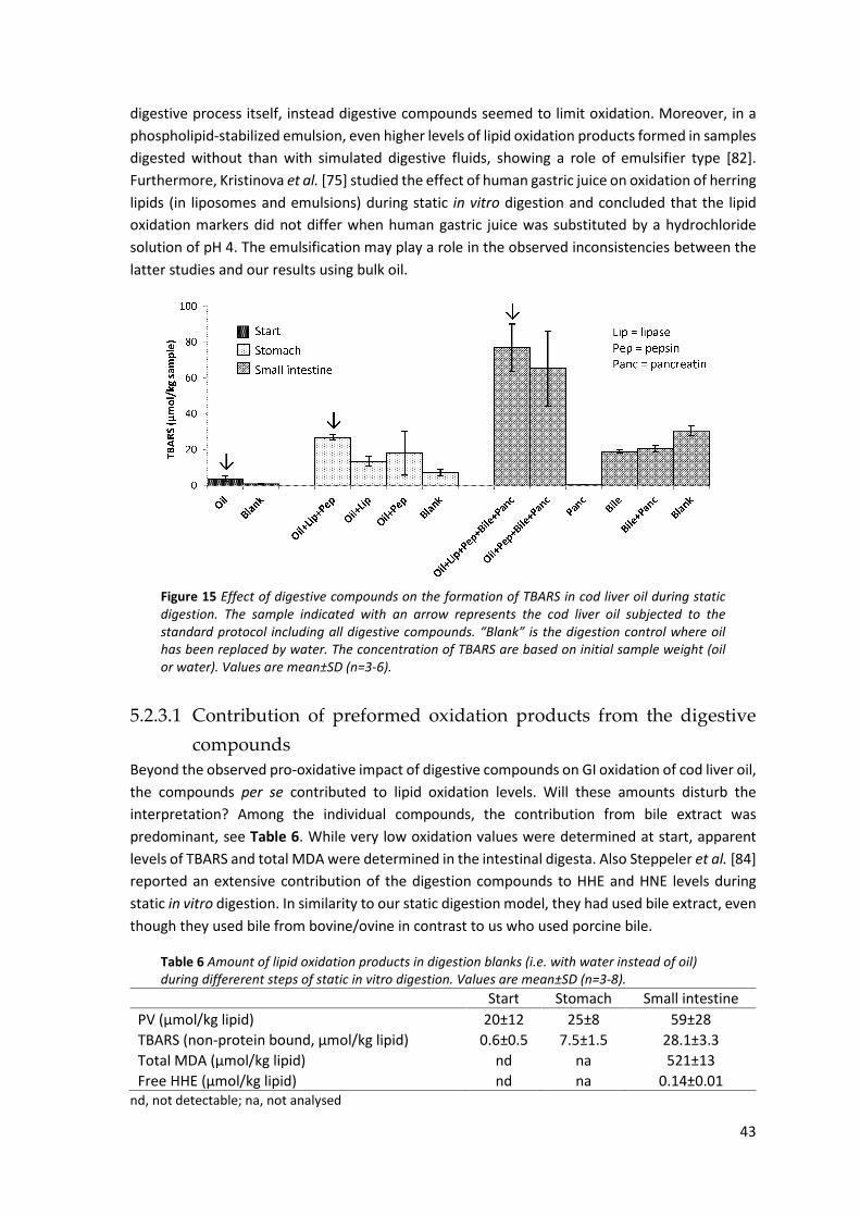
43
digestive process itself, instead digestive compounds seemed to limit oxidation. Moreover, in a
phospholipid-stabilized emulsion, even higher levels of lipid oxidation products formed in samples
digested without than with simulated digestive fluids, showing a role of emulsifier type [82].
Furthermore, Kristinova et al. [75] studied the effect of human gastric juice on oxidation of herring
lipids (in liposomes and emulsions) during static in vitro digestion and concluded that the lipid
oxidation markers did not differ when human gastric juice was substituted by a hydrochloride
solution of pH 4. The emulsification may play a role in the observed inconsistencies between the
latter studies and our results using bulk oil.
Figure 15 Effect of digestive compounds on the formation of TBARS in cod liver oil during static digestion. The sample indicated with an arrow represents the cod liver oil subjected to the standard protocol including all digestive compounds. �Blank� is the digestion control where oil has been replaced by water. The concentration of TBARS are based on initial sample weight (oil or water). Values are mean±SD (n=3-6).
5.2.3.1 Contribution of preformed oxidation products from the digestive
compounds Beyond the observed pro-oxidative impact of digestive compounds on GI oxidation of cod liver oil,
the compounds per se contributed to lipid oxidation levels. Will these amounts disturb the
interpretation? Among the individual compounds, the contribution from bile extract was
predominant, see Table 6. While very low oxidation values were determined at start, apparent
levels of TBARS and total MDA were determined in the intestinal digesta. Also Steppeler et al. [84]
reported an extensive contribution of the digestion compounds to HHE and HNE levels during
static in vitro digestion. In similarity to our static digestion model, they had used bile extract, even
though they used bile from bovine/ovine in contrast to us who used porcine bile.
Table 6 Amount of lipid oxidation products in digestion blanks (i.e. with water instead of oil) during differerent steps of static in vitro digestion. Values are mean±SD (n=3-8).
Start Stomach Small intestine PV (µmol/kg lipid) 20±12 25±8 59±28 TBARS (non-protein bound, µmol/kg lipid) 0.6±0.5 7.5±1.5 28.1±3.3 Total MDA (µmol/kg lipid) nd na 521±13 Free HHE (µmol/kg lipid) nd na 0.14±0.01
nd, not detectable; na, not analysed

44
In the dynamic digestions, fresh porcine bile (frozen directly after collection at the slaughterhouse)
was used. Also this bile contained preformed secondary oxidation products; MDA, HHE and HNE
levels were determined to 0.18 µM, 0.008 µM and 0.019 µM, respectively. When analyzing the
digests from the digestion blanks, relatively low concentrations of MDA, HHE and HNE were
determined, which implies that the preformed oxidation products of the digestive compounds did
not disturb the data interpretation from digestions of the fish lipid containing test meals. The
maximum aldehyde levels formed by digestion blanks at any time point were: 0.36 µM MDA, 0.006
µM HHE and 0.035 µM HNE. As marine lipids contain very low amounts of n-6 PUFA, and HNE is
uniquely formed from those fatty acids, the HNE concentration in samples from digested test
meals in most cases were in the same range as the digestion blanks. The average concentration of
MDA, HHE and HNE in the blanks from intestinal lumen was 0.29 µM, 0.005 µM and 0.014 µM,
respectively. The MDA level was similar to the MDA contribution of commercial porcine bile
extract to digesta taken in the intestinal phase of static digestions; 0.35 µM. The corresponding
concentration of TBARS in the bile extract was 0.95 µM. This difference is probably due to the
unspecific determination of MDA using a spectrophotometric method to measure the
concentration of the TBA2-MDA adduct, or induction of lipid oxidation during sample preparation.
5.2.3.2 Influence of pure bile acids and bile extract Because of the known difference in composition of bile acids between porcine and human bile, a
separate study was made with a mixture of pure bile acids mimicking the human composition
[179]. Individual bile acids have been shown to affect lipid oxidation differently, and especially
taurolithocholic acid could have a strong pro-oxidative effect [180, 181]. After digesting emulsified
cod liver oil with only electrolyte, with only pancreatin, with a mixture of pure bile acids (10 mM
in total) plus pancreatin or with porcine bile extract plus pancreatin, it was seen that the
concentration of TBARS increased in all samples. Significantly higher TBARS values were
determined in the samples digested with bile acids, and the highest level was reached with the
commercial bile extract (Figure 16). The difference in TBARS between emulsions digested with bile
extract and the mixture of pure bile acids could partly be explained by the TBARS contribution of
the bile extract per se. Interestingly, the level of TBARS in a separate bile extract control did not
increase during 120 min of simulated intestinal digestion, but remained similar to the non-
incubated bile extract control; contributing with 20 TBARS units in Figure 16. Apart from bile salts,
bile extract also contain phospholipids and cholesterol, which otherwise could have been
expected to oxidize further during the intestinal digestion. The pure mixture of undigested bile
acids did not respond in the TBARS measurement.
One explanation for the increase in TBARS of emulsion samples containing bile in any form is the
formation of micelles. Despite the use of an emulsion in this experiment, the lipid surface area will
largely increase during micelle formation in the presence of bile acids, facilitating the exposure of
pro- and antioxidants. It is suggested that bile acids promote lipid oxidation at lipid membranes
by an interaction of bile acids with iron ions or by increasing the permeability of lipid bilayers to
iron ions [181].

45
Figure 16 Effect of bile acids and bile extract on the formation of TBARS in cod liver oil emulsion oil during static digestion. Values are mean±SD (n=4).
5.2.3.3 Lipolysis in relation to GI oxidation It is assumed that FFA are more easily oxidized than TAG [182], and to verify whether lipolysis in
the GI tract could have contributed to the oxidation, the formation of FFA during GI digestion was
determined. No FFA was detected in the cod liver oil before digestion, but after 60 min of gastric
and 120 min of intestinal digestion, 29% and 68% of the fatty acids were hydrolyzed, respectively
(Figure 17 A). Control samples without cod liver oil contained very small amounts of FFA after the
intestinal digestion. These traces of FFA probably originated from the bile, or were formed during
hydrolysis of bile phospholipids during digestion. Similar pattern for the degree of lipolysis was
obtained when the amount of TAG was followed during digestion. After gastric and intestinal
digestion, 40% and 67% of the TAG were hydrolyzed, respectively (Figure 17 B).
Figure 17 Lipolysis degree expressed as A) FFA in per cent of maximal possible FFA formation (2 FFA from 1 TAG) and B) loss of TAG, TAG in per cent of initial TAG amount, during static in vitro digestion of cod liver oil. Values are mean±(max-min)/2 (n=2).
In human, a lipolysis degree of 5-40% has been reported in the stomach [16], indicating that our
in vitro results were in the upper range of that interval. In vivo, the postprandial concentration of
gastric lipase in the gastric content will change over time, as the meal will dilute the basal gastric
juice, but then gradually increase following gastric emptying and secretion of new gastric juice.
Recently, Sams et al. [21] reported that the human gastric lipase concentration is 15-20 µg/mL at
50% gastric emptying. Given the specific activity of 1.2 U/µg lipase, the human gastric lipase
activity would be ~20 U/mL. However, during our simulated gastric digestion, a fungal lipase
activity of 100 U/mL was used, which is higher than under the physiological conditions and hence

46
could contribute to the high lipolysis degree. Other parameters, such as pH, substrate and
different characteristics of the lipases will also influence the enzyme activity and affect the
lipolysis degree.
Based on our lipolysis data, it could be hypothesized that the formation of FFA contributes to the
observed GI oxidation. Despite lower average levels of TBARS and PV when cod liver oil was
digested according to the standard protocol with and without lipase in the gastric phase, however,
no significant differences were obtained as the standard deviation of samples without lipase was
large (Figure 15). The role of the fungal lipase in the gastric phase for the subsequent intestinal
oxidation and lipolysis is unknown as we don�t have a control without pancreatic lipase. Lorrain et
al. [76] reported a gastric lipolysis degree of 30-40% during static digestion of metMb enriched
emulsified sunflower oil using human gastric juice, which was similar to our data. They found that
the formation of conjugated dienes was not significantly affected by the presence of gastric juice,
as similar oxidation rate was obtained during digestion without gastric juice and concluded that
the released fatty acids were not more prone to oxidation than TAG.
5.2.4 Influence of ascorbic acid in simulated gastric juice Ascorbic acid may work either as a pro- or antioxidant depending on the concentration and on
presence of other compounds present in the oxidation system. It can work in synergy with e.g.
tocopherols as an antioxidant, but can also have pro-oxidative effects in the presence of transition
metal ions by reducing e.g. ferric ions, which can accelerate the lipid radical formation [183]. In
humans, ascorbic acid is found in gastric juice, but the concentration varies between individuals
and can also be affected by diseases. A concentration range of 3-463 µM among healthy people
(n=11), with a median of 87 µM has been reported [56].
The level of 85 µM ascorbic acid in gastric secretion was chosen. Since 5 mL of gastric residue was
added to each test meal prior to ingestion, this residue contributed to an additional ascorbic acid
concentration of 2.7 µM to the test meal. Thereafter, 85 µM ascorbic acid was secreted into the
gastric compartment at the flow rate of 1 mL/min, which made the ascorbic acid concentration in
the gastric lumen to gradually increase over time. Inclusion of ascorbic acid during the dynamic
digestion of medium oxidized cod liver oil (Ox C in Table 5) led to ~3-fold increased MDA levels of
both gastric and intestinal lumen compared to the control without ascorbic acid (Paper II). Also
raised HHE concentrations were obtained in intestinal lumen during digestion with ascorbic acid.
Hence, under these conditions the pro-oxidative effect of ascorbic acid was dominating. According
to the �polar paradox�, a water soluble antioxidant is not the preferred choice in oil-in-water
emulsions as it will not partition at the lipid-water interface [184, 185]. As we used oxidized cod
liver oil in this trial, the amount of endogenous α-tocopherol in the oil was most likely also reduced
(fresh oil contained 167 µg/g oil), which further limits the possible synergy effect of ascorbic acid
and α-tocopherol. Ascorbic acid may have reduced transition metal ions in the test meal and in
the secreted digestive fluids. The amount of iron and copper in the cod liver oil per se was 1.17
and 0.26 µg/g oil, respectively.
Oven baked herring mince was also subjected to dynamic digestion with and without ascorbic
acid-fortified gastric juice. The results then indicated rather similar MDA and HHE concentrations.
Herring mince also contain ascorbic acid; 4 and 35 µmol ascorbic acid/kg was found in herring

47
press juice and frozen herring fillets, respectively (own unpublished data) [186]. Ascorbic acid is
primary a pro-oxidant at low levels; as the metal reducing ability then dominates.
In conclusion, endogenous amounts of ascorbic acid can promote the extent of GI oxidation, but
the composition of the whole meal will influence the net-effect. By adding antioxidant-rich
components in the diet, the concentration where ascorbic acid acts as a pro-oxidant can be
crossed.
5.3 Effect of preformed lipid oxidation products on GI
oxidation The quality of e.g. fish oil supplements has been shown to vary extensively. We therefore wanted
to investigate the effect of preformed lipid oxidation products on subsequent GI oxidation. Cod
liver oil of different initial quality was subjected to both static and dynamic in vitro digestions. The
amount of preformed oxidation products of the different oils is given in Table 5; Ox A and Ox B
were subjected to static digestions, while Ox C and Ox D were subjected to dynamic digestions.
5.3.1 Influence of preformed oxidation products during static digestion The initial PV of the oil had no decisive role on the PV level obtained at the end of the gastric and
the intestinal phases (Figure 18). In none of the oils a build-up of PV during digestion was seen,
demonstrating that the break-down of hydroperoxides was faster than the formation. This is in
Figure 18 Effect of preformed lipid oxidation products in cod liver oil on the formation of A) PV, B) total MDA, C) non-protein bound TBARS and D) free HHE during static in vitro digestion. Blank (water instead of oil) is included to illustrate the contribution from the digestive compounds to each oxidation marker. Herring oil was included to compare cod liver oil with another oil source. Samples for the gastric step were not always prepared. Values are mean±SD (n=3-10). CO, cod liver oil; HO, herring oil.

48
accordance with the kinetics of primary oxidation products during the oxidation process. When
the oxidation starts, the amount of lipid hydroperoxides usually increases, but when the rate is
higher for reactions forming secondary oxidation products, such as TBARS and HHE, the net-effect
is a decrease in PV. Higher initial value of TBARS, total MDA and HHE, was on the other hand
followed by higher values during digestion. However, the relative changes after the gastric and
the intestinal digestion steps were not proportional to the initial amounts of aldehydes.
Figure 19 Formation of TBARS in different emulsion preparations made of fresh cod liver oil during static digestion. The initial TBARS level of each sample is indicated in the legend. The contribution of the digestion blank to the TBARS value has been subtracted. Values are mean±SD (n=2-7).
Also different preparations of emulsified cod liver oil, generating slightly different initial amounts
of oxidation products, were evaluated for GI oxidation (Figure 19). The relative increase in TBARS
during digestion was not affected by the small differences in initial TBARS concentration.
However, a rather good correlation of initial TBARS levels and the levels determined after gastric
digestion of emulsions was found; (R2=0.77, Figure 20). The corresponding correlation after the
intestinal phase was <0.5.
Figure 20 Correlation between initial TBARS level and TBARS levels in gastric digesta of cod liver oil emulsions after 60 min of static digestion.

49
5.3.2 Influence of preformed oxidation products during dynamic digestion The impact of preformed lipid oxidation products on GI oxidation during dynamic digestion was
evaluated in both non-emulsified and emulsified cod liver oil. Starting with bulk oil, different
amounts of preformed oxidation products generated different relative oxidative changes during
digestion (Figure 21). Formation of MDA was seen in all non-emulsified oils in the gastric phase,
but digests of fresh oil showed the highest relative increase. The same trend with a higher relative
MDA increase using fresh oil was obtained when these were emulsified. The formation of HHE
was more moderate than MDA. A temporary drop in the relative changes of MDA and HHE was
seen in some samples at 120 digestion, indicating that the dilution effect became predominant.
Between 150 and 180 min, the relative values increased for all samples. The uncertainties of these
relative values are high, due to the large dilution of the intake in combination with the small
remaining gastric digesta volume. Yet, the continued oxidation determined at the end of digestion
(180 min) showed that time is an important factor for lipid oxidation. The same pattern in relative
changes as in the gastric digestion was also seen during intestinal digestion.
Figure 21 Relative changes in MDA and HHE in gastric (A and B) and intestinal lumen (C and D) during dynamic in vitro digestion of cod liver oil. Oil medium ox, Ox C; Oil high ox, Ox D (Table 5).
To conclude, the relative changes indicate that there is not a strict proportional formation of MDA
and HHE based on the initial amount of oxidation products of the oil. However, because of the
relatively large differences in preformed oxidation products in the oils, the highest absolute
aldehyde concentrations in the gastric and intestinal lumen were generally obtained by the most
oxidized oil (Figure 22). Despite similar or different relative changes in aldehyde concentration, it
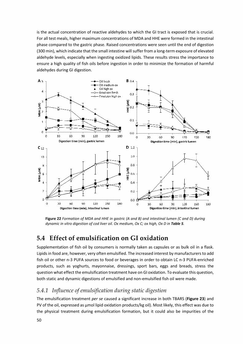
50
is the actual concentration of reactive aldehydes to which the GI tract is exposed that is crucial.
For all test meals, higher maximum concentrations of MDA and HHE were formed in the intestinal
phase compared to the gastric phase. Raised concentrations were seen until the end of digestion
(300 min), which indicate that the small intestine will suffer from a long-term exposure of elevated
aldehyde levels, especially when ingesting oxidized lipids. These results stress the importance to
ensure a high quality of fish oils before ingestion in order to minimize the formation of harmful
aldehydes during GI digestion.
Figure 22 Formation of MDA and HHE in gastric (A and B) and intestinal lumen (C and D) during dynamic in vitro digestion of cod liver oil. Ox medium, Ox C; ox high, Ox D in Table 5.
5.4 Effect of emulsification on GI oxidation Supplementation of fish oil by consumers is normally taken as capsules or as bulk oil in a flask.
Lipids in food are, however, very often emulsified. The increased interest by manufacturers to add
fish oil or other n-3 PUFA sources to food or beverages in order to obtain LC n-3 PUFA-enriched
products, such as yoghurts, mayonnaise, dressings, sport bars, eggs and breads, stress the
question what effect the emulsification treatment have on GI oxidation. To evaluate this question,
both static and dynamic digestions of emulsified and non-emulsified fish oil were made.
5.4.1 Influence of emulsification during static digestion The emulsification treatment per se caused a significant increase in both TBARS (Figure 23) and
PV of the oil, expressed as µmol lipid oxidation products/kg oil). Most likely, this effect was due to
the physical treatment during emulsification formation, but it could also be impurities of the

51
emulsifier. The amount of TBARS differed significantly (p<0.001) also between the different
emulsion preparations (n=6, Figure 19). During subsequent GI digestion of the emulsions, TBARS
values increased after each digestion phase, but only small changes in PV were found. The same
was noted when fresh bulk cod liver oil was digested. However, the relative increase in TBARS
after the gastric and the intestinal phases was larger for bulk oil (6.2 and 14.8, respectively)
compared to the emulsions (4.5±1.5 and 8.6±3.0, respectively). During these conditions and with
this emulsifier, emulsification did not have an impact on GI oxidation.
Figure 23 Effect of emulsification of fresh cod liver oil on the formation of TBARS during subsequent static digestion. TBARS values are only statistically higher in emulsions at start. Digestion blank is subtracted from the values.
5.4.2 Influence of emulsification during dynamic digestion Increased amounts of lipid oxidation products were also determined in emulsions of fresh cod
liver oil made prior to the dynamic digestions compared to bulk oil (data not shown). A 3-fold
increase was found for MDA and HNE, while HHE levels only increased slightly (30%, data from 3
different preparations of fresh cod liver oil). These differences were evened out during the meal
preparation, and similar levels of oxidation products were found in test meals of non-emulsified
and emulsified cod liver oil (Figure 24). However, the initial MDA and HHE levels of emulsion made
of oxidized cod liver oil (�Ox D� according to Table 5) were lower compared to non-emulsified
oxidized oil after the emulsification treatment. Emulsified cod liver oils (both fresh and oxidized
oil) evolved lower MDA levels during gastric digestion compared to non-emulsified oil, but in the
intestinal phase the concentrations were equal. In contrast, the emulsification had a lowering
effect of the oxidized oil on the amount of HHE in the gastric phase, but no apparent effect on
fresh oil. However, both emulsions generated HHE levels that were 2- to 3-fold higher in intestinal
digesta compared to corresponding bulk oil.
In addition to the refined cod liver oil, also crude herring oil was studied. Opposite to the cod liver
oil, emulsified herring oil generated 4-fold higher MDA and HHE concentrations in gastric lumen
at 90 min of digestion compared to non-emulsified herring oil. The difference between emulsified
and non-emulsified herring oil remained in the intestinal lumen. The observed discrepancy of the
emulsification effect in the gastric phase between the different oil sources is difficult to explain. It
should be mentioned though, that the herring oil was digested in the presence of bread and
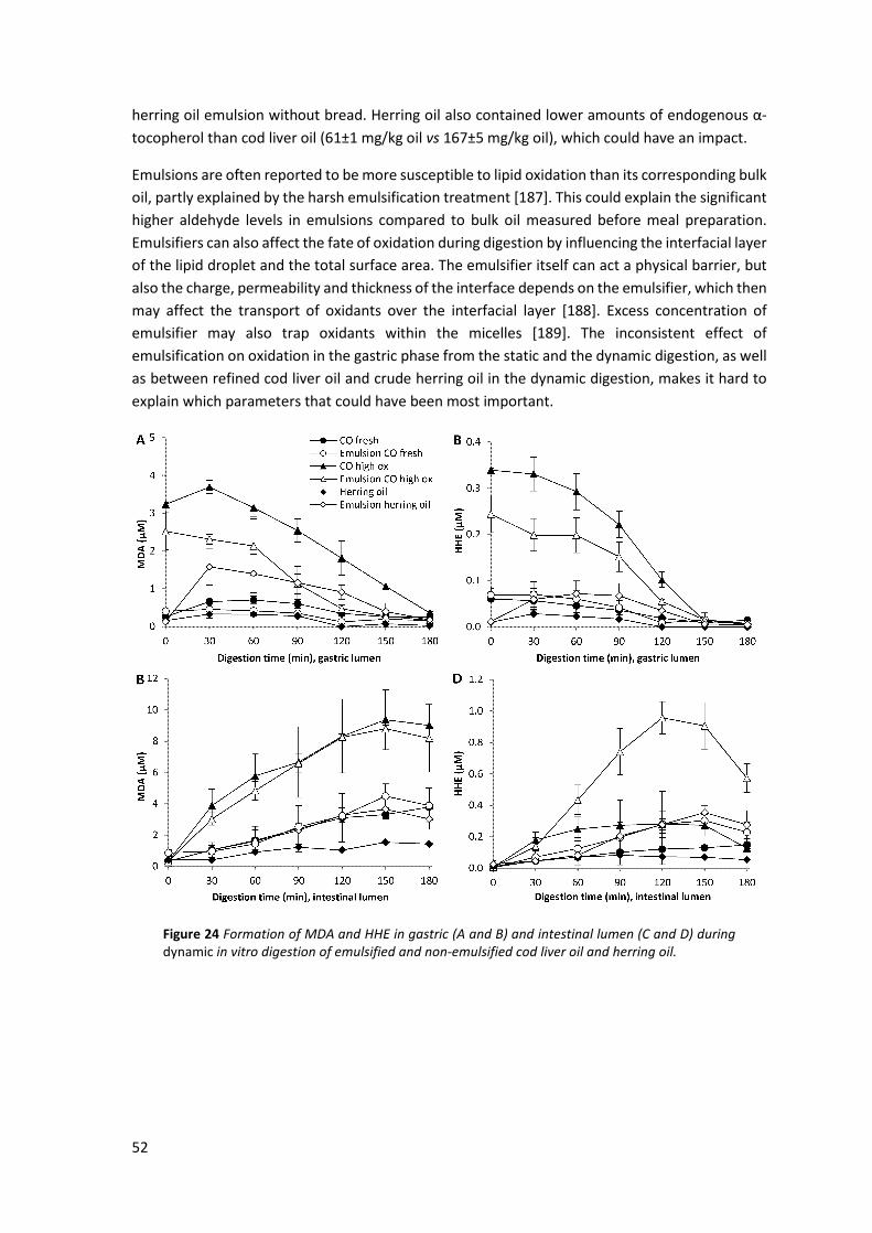
52
herring oil emulsion without bread. Herring oil also contained lower amounts of endogenous α-
tocopherol than cod liver oil (61±1 mg/kg oil vs 167±5 mg/kg oil), which could have an impact.
Emulsions are often reported to be more susceptible to lipid oxidation than its corresponding bulk
oil, partly explained by the harsh emulsification treatment [187]. This could explain the significant
higher aldehyde levels in emulsions compared to bulk oil measured before meal preparation.
Emulsifiers can also affect the fate of oxidation during digestion by influencing the interfacial layer
of the lipid droplet and the total surface area. The emulsifier itself can act a physical barrier, but
also the charge, permeability and thickness of the interface depends on the emulsifier, which then
may affect the transport of oxidants over the interfacial layer [188]. Excess concentration of
emulsifier may also trap oxidants within the micelles [189]. The inconsistent effect of
emulsification on oxidation in the gastric phase from the static and the dynamic digestion, as well
as between refined cod liver oil and crude herring oil in the dynamic digestion, makes it hard to
explain which parameters that could have been most important.
Figure 24 Formation of MDA and HHE in gastric (A and B) and intestinal lumen (C and D) during dynamic in vitro digestion of emulsified and non-emulsified cod liver oil and herring oil.
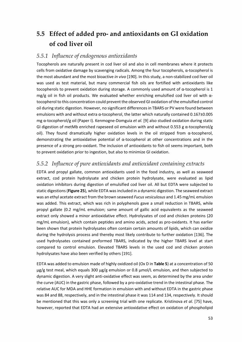
53
5.5 Effect of added pro- and antioxidants on GI oxidation
of cod liver oil
5.5.1 Influence of endogenous antioxidants Tocopherols are naturally present in cod liver oil and also in cell membranes where it protects
cells from oxidative damage by scavenging radicals. Among the four tocopherols, α-tocopherol is
the most abundant and the most bioactive in vivo [190]. In this study, a non-stabilized cod liver oil
was used as test material, but many commercial fish oils are fortified with antioxidants like
tocopherols to prevent oxidation during storage. A commonly used amount of α-tocopherol is 1
mg/g oil in fish oil products. We evaluated whether enriching emulsified cod liver oil with α-
tocopherol to this concentration could prevent the observed GI oxidation of the emulsified control
oil during static digestion. However, no significant differences in TBARS or PV were found between
emulsions with and without extra α-tocopherol, the latter which naturally contained 0.167±0.005
mg α-tocopherol/g oil (Paper I). Kenmogne-Domguia et al. [9] also studied oxidation during static
GI digestion of metMb enriched rapeseed oil emulsion with and without 0.553 g α-tocopherol/g
oil). They found dramatically higher oxidation levels in the oil stripped from α-tocopherol,
demonstrating the antioxidative potential of α-tocopherol at other concentrations and in the
presence of a strong pro-oxidant. The inclusion of antioxidants to fish oil seems important, both
to prevent oxidation prior to ingestion, but also to minimize GI oxidation.
5.5.2 Influence of pure antioxidants and antioxidant containing extracts EDTA and propyl gallate, common antioxidants used in the food industry, as well as seaweed
extract, cod protein hydrolysate and chicken protein hydrolysate, were evaluated as lipid
oxidation inhibitors during digestion of emulsified cod liver oil. All but EDTA were subjected to
static digestions (Figure 25), while EDTA was included in a dynamic digestion. The seaweed extract
was an ethyl acetate extract from the brown seaweed Fucus vesiculosus and 1.45 mg/mL emulsion
was added. This extract, which was rich in polyphenols gave a small reduction in TBARS, while
propyl gallate (0.2 mg/mL emulsion; same amount of gallic acid equivalents as the seaweed
extract only showed a minor antioxidative effect. Hydrolysates of cod and chicken proteins (20
mg/mL emulsion), which contain peptides and amino acids, acted as pro-oxidants. It has earlier
been shown that protein hydrolysates often contain certain amounts of lipids, which can oxidize
during the hydrolysis process and thereby most likely contribute to further oxidation [136]. The
used hydrolysates contained preformed TBARS, indicated by the higher TBARS level at start
compared to control emulsion. Elevated TBARS levels in the used cod and chicken protein
hydrolysates have also been verified by others [191].
EDTA was added to emulsion made of highly oxidized oil (Ox D in Table 5) at a concentration of 50
µg/g test meal, which equals 300 µg/g emulsion or 0.8 µmol/L emulsion, and then subjected to
dynamic digestion. A very slight anti-oxidative effect was seem, as determined by the area under
the curve (AUC) in the gastric phase, followed by a pro-oxidative trend in the intestinal phase. The
relative AUC for MDA and HHE formation in emulsion with and without EDTA in the gastric phase
was 84 and 88, respectively, and in the intestinal phase it was 114 and 134, respectively. It should
be mentioned that this was only a screening trial with one replicate. Kristinova et al. [75] have,
however, reported that EDTA had an extensive antioxidative effect on oxidation of phospholipid

54
stabilized herring oil emulsion during gastric conditions without enzymes, and thereby concluded
that endogenous iron ions mediated the lipid oxidation. EDTA is a strong metal chelator and our
own results could imply that the rather low amount of iron ions in the oil (1.17 µg/g oil) was not
the main contributor to lipid oxidation in this system. The highly oxidized oil probably contained
plenty of lipid radicals to keep up the oxidation process. It is possible that EDTA would have an
antioxidative effect if it was added to fresh oil where iron ions may play a larger role. None of the
selected antioxidants could efficiently reduce GI oxidation at the tested concentration. It may be
that e.g. polyphenols present in red wine and grape seed extract could prevent GI oxidation,
because others have reported antioxidative effects from those during in vitro digestions [63, 75,
90].
Figure 25 Effect of potential antioxidants on TBARS formation in cod liver oil emulsion during static in vitro digestion. Values are mean±SD (n=3).
5.5.3 Influence of Hb The influence of 11.5 µM cod Hb added to emulsified fresh cod liver oil was evaluated during both
static and dynamic digestion conditions. Hb was also added to cod mince and then subjected to
static digestion. For emulsified cod liver oil, an immediate increase in all determined oxidation
markers (PV, TBARS, MDA, HHE and HNE) was seen even prior to the digestion, demonstrating the
strong pro-oxidative effect of fish Hb. Both PV and TBARS levels increased during the gastric
digestion, but at the end of the digestion, only TBARS continued to increase, while PV decreased
(Paper I). The increase in TBARS concentration after the gastric and the intestinal step compared
to the initial concentration was 5-fold and 6-fold, respectively, which corresponds to a final TBARS
concentration of 2270 µmol/kg lipid versus 370 µmol/kg lipid before digestion. The TBARS
concentration in the emulsion without Hb was 6 µmol/kg lipid.
The results from the dynamic digestion of emulsified cod liver oil with addition of 11.5 µM cod Hb
showed an apparent pro-oxidative effect of Hb (Figure 26). A peak was obtained in MDA (25 µM)
and HHE (0.5 µM) at 90 min of gastric digestion. Digests with Hb formed ~5.5 times higher MDA
and HHE levels when it peaked in the intestinal phase at 150 min of digestion compared to digest
of emulsion alone. Three times higher HHE levels were generated in the intestinal digesta of
emulsion with Hb compared to in the gastric digesta, while almost similar MDA levels were formed
in the gastric and intestinal digests.
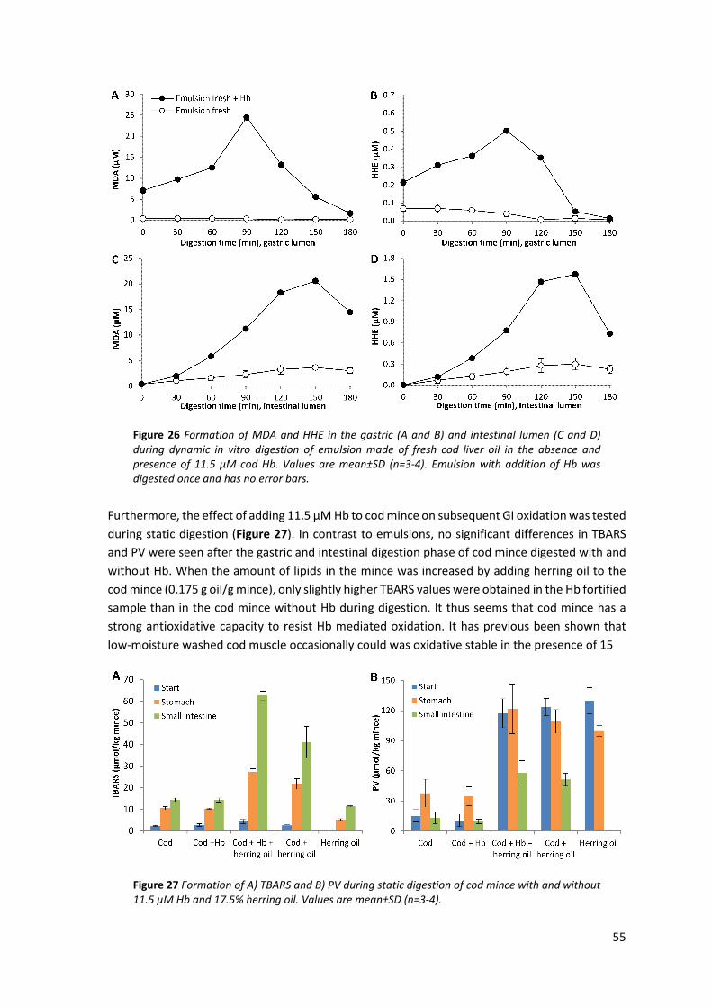
55
Figure 26 Formation of MDA and HHE in the gastric (A and B) and intestinal lumen (C and D) during dynamic in vitro digestion of emulsion made of fresh cod liver oil in the absence and presence of 11.5 µM cod Hb. Values are mean±SD (n=3-4). Emulsion with addition of Hb was digested once and has no error bars.
Furthermore, the effect of adding 11.5 µM Hb to cod mince on subsequent GI oxidation was tested
during static digestion (Figure 27). In contrast to emulsions, no significant differences in TBARS
and PV were seen after the gastric and intestinal digestion phase of cod mince digested with and
without Hb. When the amount of lipids in the mince was increased by adding herring oil to the
cod mince (0.175 g oil/g mince), only slightly higher TBARS values were obtained in the Hb fortified
sample than in the cod mince without Hb during digestion. It thus seems that cod mince has a
strong antioxidative capacity to resist Hb mediated oxidation. It has previous been shown that
low-moisture washed cod muscle occasionally could was oxidative stable in the presence of 15
Figure 27 Formation of A) TBARS and B) PV during static digestion of cod mince with and without 11.5 µM Hb and 17.5% herring oil. Values are mean±SD (n=3-4).

56
µM Hb during ice storage [192]. Different amounts of α-tocopherol and different degrees of
disruption in the myofibrillar structure were then suggested explanations. The PV of the cod mince
in our case was low, and perhaps this was a contributing factor for the low response since e.g.
metHb is known to react with hydroperoxides, resulting in the formation of radicals that can
propagate the oxidation process.
Many other studies on GI oxidation have added Hb to initiate lipid oxidation, and have then
evaluated the effect of e.g. various antioxidants. Hb is, indeed, a strong initiator and examples of
its different actions at low pH have been described earlier in this thesis (section 3.7.1.1). Our data
confirms that it is important to include efficient antioxidants to a meal when heme-proteins are
present.
5.6 Effect of fish muscle matrix on GI oxidation To evaluate the impact of the fish muscle matrix on GI oxidation, herring oil was isolated from
herring mince with a lipid content of 17% and then subjected to both static and dynamic GI
digestions along with the whole mince. Herring oil was studied either as emulsified or crude oil.
All test meals contained the amount of total lipids provided by 50 g of herring mince, i.e. 8.75 g.
Raw herring mince formed higher concentrations of MDA and HHE compared to meals with
herring oil during gastric digestion (Figure 28). Based on maximum MDA and HHE levels, the
different herring lipid meals were ranked: herring mince >> herring oil emulsion > herring oil.
Figure 28 Formation of MDA and HHE in gastric (A and B) and intestinal lumen (C and D) during dynamic in vitro digestion of raw herring mince, herring oil emulsion and herring oil.

57
Figure 29 Relative change in concentration of MDA (A) and HHE (B) during dynamic digestion of raw herring mince, herring oil emulsion and herring oil.
Despite that herring mince formed the highest absolute levels of MDA and HHE, the relative
change in concentration (based on initial concentration at the intake point) increased the most in
emulsified herring oil (Figure 29). It should be mentioned though that the aldehyde levels in the
herring mince test meal before digestion were 13 µM MDA and 0.29 µM HHE versus 0.14 µM MDA
and 0.012 µM HHE in the emulsified herring oil test meal. Herring oil contained similar
concentrations as emulsified herring oil (0.16 µM MDA and 0.01 µM HHE) at the intake point, but
still the emulsified oil had a higher relative increase. It is likely that the increased surface area for
emulsified herring lipid droplets facilitated gastric oxidation and once the lipid oxidation is
initiated, propagation can continue during the remaining digestion time.
Also during the intestinal digestion (Figure 28 C and D), herring mince evolved extensively higher
MDA levels than the oil samples, and again emulsified oil formed higher amounts than bulk oil.
However, HHE data did not follow the same pattern as MDA. At 30 and 60 min of intestinal
digestion, the herring lipid test meals showed inseparable HHE concentrations, despite their
different concentrations developed in the gastric lumen (Figure 28 B and D). Later during the
digestion, between 90 and 180 min, the HHE levels in the intestinal lumen from herring mince and
emulsified herring oil increased remarkably, with a peak at 150 min. The higher HHE concentration
with herring oils in intestinal lumen compared to that in gastric lumen, especially with the
emulsified oil, shows a continued intestinal oxidation. The intestinal tract can therefore also be
exposed to high levels of harmful aldehydes.
Also static in vitro digestions were made using the same raw herring mince (17% lipid) and herring
oil as was used in the dynamic digestions (Figure 30). Extensively higher TBARS levels were
generated during static digestion of herring mince compared to herring oil. However, it was also
confirmed by TBARS analyses that herring mince contained higher initial levels of oxidation
products compared to herring oil; 235 vs 1 µmol TBARS/kg lipid, respectively. The generated
TBARS concentration after gastric digestion of herring mince (1730 µmol/kg lipid; 89 µmol/L
digesta) was ~75 times higher than the corresponding value after digesting herring oil (22 µmol/kg
lipid; 1.2 µmol/L digesta). The relative increment of TBARS after the gastric and the intestinal step
compared to the start values were 7- and 13-fold in herring mince versus 21- and 32-fold in herring
oil (Figure 31). The large difference in both absolute and relative TBARS concentration observed
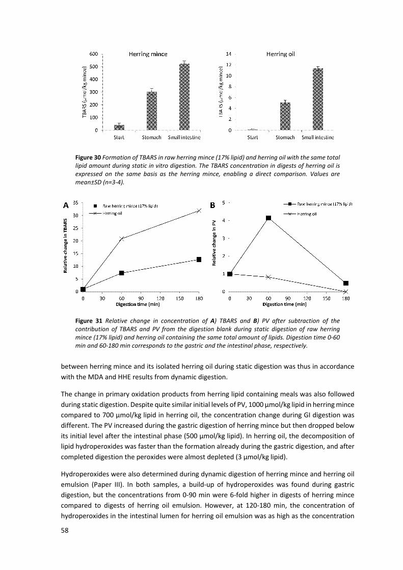
58
Figure 30 Formation of TBARS in raw herring mince (17% lipid) and herring oil with the same total lipid amount during static in vitro digestion. The TBARS concentration in digests of herring oil is expressed on the same basis as the herring mince, enabling a direct comparison. Values are mean±SD (n=3-4).
Figure 31 Relative change in concentration of A) TBARS and B) PV after subtraction of the contribution of TBARS and PV from the digestion blank during static digestion of raw herring mince (17% lipid) and herring oil containing the same total amount of lipids. Digestion time 0-60 min and 60-180 min corresponds to the gastric and the intestinal phase, respectively.
between herring mince and its isolated herring oil during static digestion was thus in accordance
with the MDA and HHE results from dynamic digestion.
The change in primary oxidation products from herring lipid containing meals was also followed
during static digestion. Despite quite similar initial levels of PV, 1000 µmol/kg lipid in herring mince
compared to 700 µmol/kg lipid in herring oil, the concentration change during GI digestion was
different. The PV increased during the gastric digestion of herring mince but then dropped below
its initial level after the intestinal phase (500 µmol/kg lipid). In herring oil, the decomposition of
lipid hydroperoxides was faster than the formation already during the gastric digestion, and after
completed digestion the peroxides were almost depleted (3 µmol/kg lipid).
Hydroperoxides were also determined during dynamic digestion of herring mince and herring oil
emulsion (Paper III). In both samples, a build-up of hydroperoxides was found during gastric
digestion, but the concentrations from 0-90 min were 6-fold higher in digests of herring mince
compared to digests of herring oil emulsion. However, at 120-180 min, the concentration of
hydroperoxides in the intestinal lumen for herring oil emulsion was as high as the concentration

59
in lumen for herring mince. As emulsified herring oil was neither subjected to static digestion, nor
subjected to hydroperoxides analyses, we cannot make a direct comparison of the effect of static
and dynamic digestions on the formation of primary oxidation products between these two
samples.
To conclude, herring mince generally generated higher concentrations of secondary lipid oxidation
products during static and dynamic digestions compared to samples without the muscle matrix.
The herring mince contained higher amounts of preformed lipid oxidation products and also has
a known content of heme-proteins. Under the intestinal conditions of the dynamic digestion,
emulsified herring oil formed equally high levels of HHE and hydroperoxides as the herring mince,
and in the gastric phase, it also had the highest relative increase in MDA and HHE of all three
samples. This could indicate that aldehydes of the fish mince to a larger extent form adducts with
proteins and DNA during digestion, which are not measurable with the used method.
5.7 Comparison of herring and salmon muscle regarding
GI oxidation To compare the fate of two common food fish species regarding GI oxidation, herring and salmon
was selected. The lipid content of herring varies extensively during season and therefore two
batches of herring were studied where the lipid content was 4% and 17%. The salmon contained
17% lipid, which enabled a comparison with the high fat herring regarding the role of endogenous
pro- and antioxidants. Herring (17% lipid) formed significantly higher values of MDA and HHE
during digestion compared to salmon (Figure 32). In the gastric phase, five times higher MDA
concentration was generated by herring compared to salmon (23.9 µM vs 4.6 µM) and in the
intestinal phase the relative difference was further enlarged; 22.5 µM vs 2.1 µM.
Similar results were determined for HHE; herring (17% lipid) formed 4-fold and 30-fold higher HHE
concentrations in the gastric and in the intestinal phase, respectively, compared to salmon. Also
herring (4% lipid) evolved higher levels of MDA (~3-fold) in both the gastric and the intestinal
lumen compared to salmon.
Interestingly, the MDA and HHE levels with herring (4% lipid) in the intestinal lumen were much
lower than expected when taking into account the released amount from the gastric lumen. It is
possible that proteolysis, caused by the digestion, generated more exposed amino groups that
could form covalent adducts with MDA and HHE, which will affect the detectable lipid oxidation
products. The fact that herring (17% lipid) gave rise to significantly higher HHE and MDA levels
than herring (4% lipid) in the intestinal lumen could be a molar effect. HHE derives uniquely from
n-3 PUFA, while MDA can be produced from PUFA containing minimum 3 double bonds [93].
Herring (17% lipid) contained e.g. four times more LC n-3 PUFA than herring (4% lipid), which
hence could generate higher quantities of HHE during GI digestion. Endogenous pro- and
antioxidants of the fish mince could also play a role in the observed discrepancy in aldehyde levels
between low-fat and high-fat herring in the intestinal phase. Depending on the amount of lipids
available for oxidation, and partitioning of pro-and antioxidants in the oil phase vs aqueous phase,
the overall effect of pro- and antioxidants will differ.

60
Figure 32 Formation of MDA and HHE in gastric (A and B) and intestinal lumen (C and D) during dynamic in vitro digestion of raw mince of herring and salmon.
The apparent species difference could be related to several factors. Firstly, the presence and
concentration of endogenous pro- and antioxidants differ between the species. Herring contains
high levels of Hb, which exerts several pro-oxidative effects as discussed earlier. Salmon has lower
heme-protein levels, and in addition salmon contains the antioxidants astaxanthine and
canthaxanthine with conjugated double bonds able to scavenge radicals. To conclude, salmon may
have a favorable composition of antioxidants preventing GI oxidation. Secondly, although the
relative proportion of total PUFA was not significantly different between the three samples (Table
4), the dominating fatty acids in herring had a higher degree of unsaturation, which may
contribute to the more pronounced lipid oxidation observed in herring compared to salmon
during digestion (data not shown). Herring (17% lipid) also contains significantly larger quantities
of LC n-3 PUFA than salmon, which partly could explain the reported higher HHE values of herring.
However, the fact that herring (4% lipid) contained significantly less LC n-3 PUFA than salmon but
still generated higher HHE concentrations, indicating that the influence from other parameters on
GI oxidation dominated, e.g. the abundance of heme proteins in herring.
Thirdly, it should be noted that the initial amount of MDA and HHE of the minces also differed
significantly, which could influence the degree of GI oxidation. Herring (17% lipid) contained 39
µmol MDA/kg mince and 0.87 µmol HHE/kg mince at start, while raw salmon contained only 2.2
µmol MDA/kg mince and 0.072 µmol HHE/kg mince. Corresponding starting values of raw herring
(4% lipid) were in between herring (17% lipid) and salmon (14 µmol MDA/kg mince and 0.22 µmol
HHE/kg mince) and it also yielded in between-levels of MDA and HHE during digestion. Despite
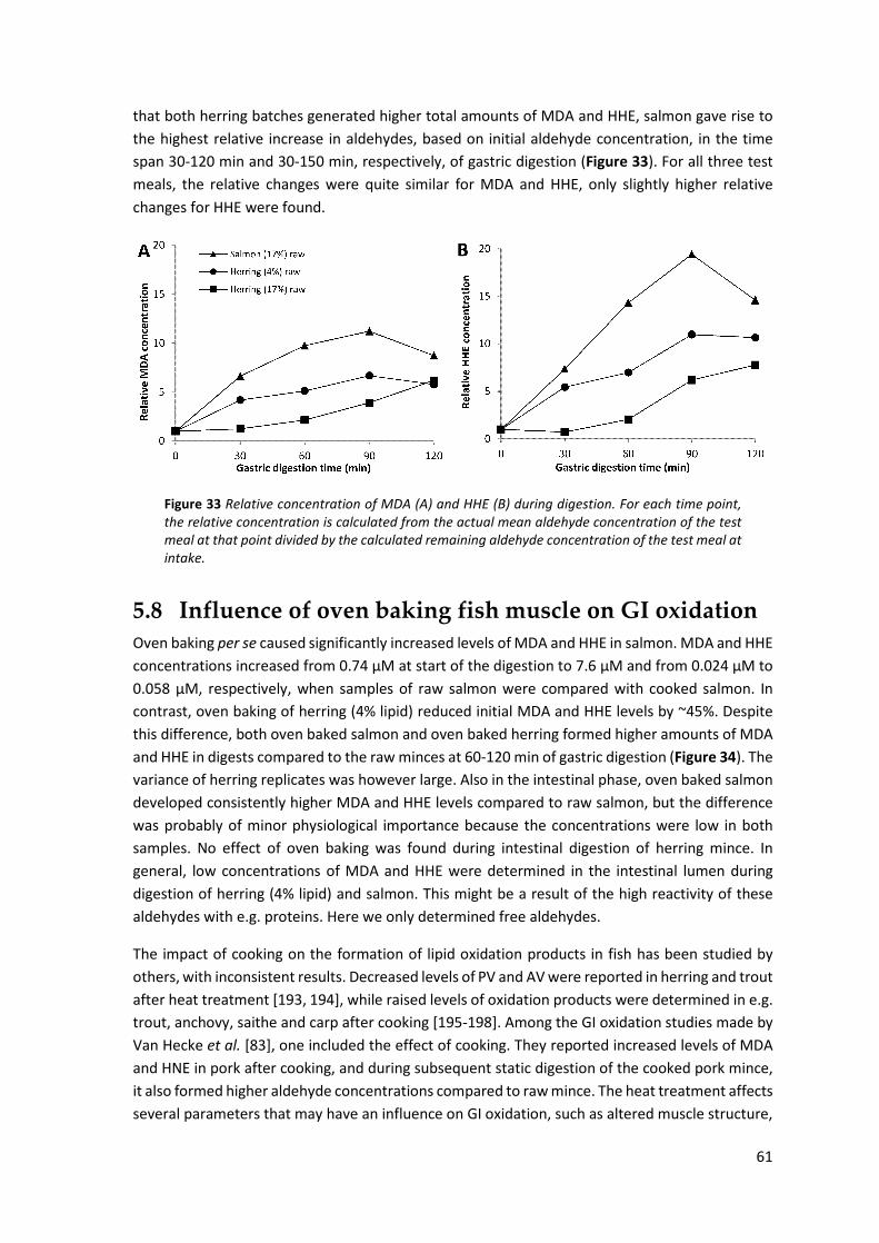
61
that both herring batches generated higher total amounts of MDA and HHE, salmon gave rise to
the highest relative increase in aldehydes, based on initial aldehyde concentration, in the time
span 30-120 min and 30-150 min, respectively, of gastric digestion (Figure 33). For all three test
meals, the relative changes were quite similar for MDA and HHE, only slightly higher relative
changes for HHE were found.
Figure 33 Relative concentration of MDA (A) and HHE (B) during digestion. For each time point, the relative concentration is calculated from the actual mean aldehyde concentration of the test meal at that point divided by the calculated remaining aldehyde concentration of the test meal at intake.
5.8 Influence of oven baking fish muscle on GI oxidation Oven baking per se caused significantly increased levels of MDA and HHE in salmon. MDA and HHE
concentrations increased from 0.74 µM at start of the digestion to 7.6 µM and from 0.024 µM to
0.058 µM, respectively, when samples of raw salmon were compared with cooked salmon. In
contrast, oven baking of herring (4% lipid) reduced initial MDA and HHE levels by ~45%. Despite
this difference, both oven baked salmon and oven baked herring formed higher amounts of MDA
and HHE in digests compared to the raw minces at 60-120 min of gastric digestion (Figure 34). The
variance of herring replicates was however large. Also in the intestinal phase, oven baked salmon
developed consistently higher MDA and HHE levels compared to raw salmon, but the difference
was probably of minor physiological importance because the concentrations were low in both
samples. No effect of oven baking was found during intestinal digestion of herring mince. In
general, low concentrations of MDA and HHE were determined in the intestinal lumen during
digestion of herring (4% lipid) and salmon. This might be a result of the high reactivity of these
aldehydes with e.g. proteins. Here we only determined free aldehydes.
The impact of cooking on the formation of lipid oxidation products in fish has been studied by
others, with inconsistent results. Decreased levels of PV and AV were reported in herring and trout
after heat treatment [193, 194], while raised levels of oxidation products were determined in e.g.
trout, anchovy, saithe and carp after cooking [195-198]. Among the GI oxidation studies made by
Van Hecke et al. [83], one included the effect of cooking. They reported increased levels of MDA
and HNE in pork after cooking, and during subsequent static digestion of the cooked pork mince,
it also formed higher aldehyde concentrations compared to raw mince. The heat treatment affects
several parameters that may have an influence on GI oxidation, such as altered muscle structure,

62
Figure 34 Formation of MDA and HHE in gastric (A and B) and intestinal lumen (C and D) during dynamic in vitro digestion of raw and oven baked fish mince of herring and salmon. Lipid content of the fish mince is indicated in the legend.
decomposition of heme-proteins under the liberation of heme/iron and loss of antioxidants. The
activity of GPx, an enzyme able to reduce hydrogen peroxide and lipid hydroperoxides in cells,
was for example reduced by 20-60% in duck and chicken meat heated to 60°C, which is similar to
the inner temperature of the oven baked fish fillets used in our study (55°C) [199]. All these
parameters might have contributed to the higher aldehyde levels in digests of cooked fish in the
gastric phase. Hypothesizing that denaturation of proteins in cooked fish mince generates more
exposed peptide bonds, and thus an assumed higher proteolysis degree, it can be expected that
the formation of protein-aldehyde adducts in the intestinal phase is promoted in digests from
cooked fish, which affects their concentration of detectable aldehydes.

63
5.9 What levels of oxidation products are reached during
dynamic digestion and do they possess any health
risks? One of the major aims with this study was to evaluate what levels of oxidation products that could
be reached in the GI tract after ingesting fish lipids. Most studies on GI oxidation have used static
methods, which will not supply such data as the continuous secretion of fluids and removal of
dissolved low molecular weight digestion products and water from the system is not simulated.
Dynamic in vitro methods are a better alternative, even if they can only simulate passive
absorption, and the impact of various cells types in the GI tract is not included.
5.9.1 Levels of MDA General for most test meals, and especially for fish, was the more or less pronounced bell-shaped
curves for MDA and HHE, indicating a formation of aldehydes despite the dilution effect of the
dynamic system. The highest concentration of MDA determined in gastric digesta from the
different fish lipid containing meals during dynamic digestion was around 25 µM. In fact, several
test meals reached this level; raw herring mince (17% lipid), oven baked herring mince (4% lipid,
+/- ascorbic acid in the gastric juice) and cod liver oil emulsion fortified with 11.5 µM Hb. For all
samples this peak occurred at 90 min of digestion. Since the initial MDA concentrations of these
test meals varied between 2-13 µM, the relative increase was in the range 4-28. Thus not only the
initial meal quality is decisive for the formation of lipid oxidation products during subsequent
digestion. In the intestinal lumen, only raw herring mince (17% lipid) and cod liver oil emulsion
with Hb generated MDA levels of similar magnitude; 22.5 µM and 20.5 µM, respectively.
Maximum levels were generally formed at 180 min of intestinal digestion.
According to in vivo results presented by Gobert et al. [91], the relative increase in TBARS during
gastric digestion of a beef meal in minipigs was about 5-fold between 15 min and 240 min. They
found that the concentration, expressed on a lipid basis, peaked at 240 min of digestion, but since
the lipid dilution was of the same order as the raise in TBARS, it can be concluded that the stomach
was exposed to a constant level of TBARS in their study. This may indicate that the lack of an
oxidative defense system in our in vitro model enables elevated aldehyde levels.
Maximum levels of MDA formed during gastric and intestinal digestion of fresh cod liver oil were
0.7 µM and 3.8 µM, respectively. Corresponding levels in gastric and intestinal digests of oxidized
cod liver oil (Ox D) were 3.7 µM and 9.4 µM MDA, respectively. Common for nearly all fish oil
samples, the maximum MDA concentration was determined in the intestinal lumen. In contrast,
the minces from herring and salmon generally generated higher MDA concentrations in the gastric
phase, and the concentrations of MDA in the intestinal lumen of fish minces were lower than
expected when taking into account the amounts of MDA released from the gastric compartment.
This was in accordance with the HHE data and might be a result of the high reactivity of these
aldehydes and the fact that we only determine free aldehydes.
MDA is mostly bound to proteins in foods as a N-2-propenal derivative, which after enzymatic
hydrolysis by digestive enzymes releases N-ε-(2-propenal)lysine [200]. Nevertheless, lysine-MDA
retains its ability to modify proteins and DNA [201]. Indeed, it has been shown that human
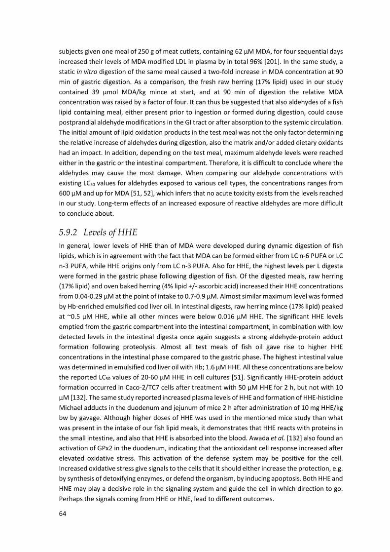
64
subjects given one meal of 250 g of meat cutlets, containing 62 µM MDA, for four sequential days
increased their levels of MDA modified LDL in plasma by in total 96% [201]. In the same study, a
static in vitro digestion of the same meal caused a two-fold increase in MDA concentration at 90
min of gastric digestion. As a comparison, the fresh raw herring (17% lipid) used in our study
contained 39 µmol MDA/kg mince at start, and at 90 min of digestion the relative MDA
concentration was raised by a factor of four. It can thus be suggested that also aldehydes of a fish
lipid containing meal, either present prior to ingestion or formed during digestion, could cause
postprandial aldehyde modifications in the GI tract or after absorption to the systemic circulation.
The initial amount of lipid oxidation products in the test meal was not the only factor determining
the relative increase of aldehydes during digestion, also the matrix and/or added dietary oxidants
had an impact. In addition, depending on the test meal, maximum aldehyde levels were reached
either in the gastric or the intestinal compartment. Therefore, it is difficult to conclude where the
aldehydes may cause the most damage. When comparing our aldehyde concentrations with
existing LC50 values for aldehydes exposed to various cell types, the concentrations ranges from
600 µM and up for MDA [51, 52], which infers that no acute toxicity exists from the levels reached
in our study. Long-term effects of an increased exposure of reactive aldehydes are more difficult
to conclude about.
5.9.2 Levels of HHE In general, lower levels of HHE than of MDA were developed during dynamic digestion of fish
lipids, which is in agreement with the fact that MDA can be formed either from LC n-6 PUFA or LC
n-3 PUFA, while HHE origins only from LC n-3 PUFA. Also for HHE, the highest levels per L digesta
were formed in the gastric phase following digestion of fish. Of the digested meals, raw herring
(17% lipid) and oven baked herring (4% lipid +/- ascorbic acid) increased their HHE concentrations
from 0.04-0.29 µM at the point of intake to 0.7-0.9 µM. Almost similar maximum level was formed
by Hb-enriched emulsified cod liver oil. In intestinal digests, raw herring mince (17% lipid) peaked
at ~0.5 µM HHE, while all other minces were below 0.016 µM HHE. The significant HHE levels
emptied from the gastric compartment into the intestinal compartment, in combination with low
detected levels in the intestinal digesta once again suggests a strong aldehyde-protein adduct
formation following proteolysis. Almost all test meals of fish oil gave rise to higher HHE
concentrations in the intestinal phase compared to the gastric phase. The highest intestinal value
was determined in emulsified cod liver oil with Hb; 1.6 µM HHE. All these concentrations are below
the reported LC50 values of 20-60 µM HHE in cell cultures [51]. Significantly HHE-protein adduct
formation occurred in Caco-2/TC7 cells after treatment with 50 µM HHE for 2 h, but not with 10
µM [132]. The same study reported increased plasma levels of HHE and formation of HHE-histidine
Michael adducts in the duodenum and jejunum of mice 2 h after administration of 10 mg HHE/kg
bw by gavage. Although higher doses of HHE was used in the mentioned mice study than what
was present in the intake of our fish lipid meals, it demonstrates that HHE reacts with proteins in
the small intestine, and also that HHE is absorbed into the blood. Awada et al. [132] also found an
activation of GPx2 in the duodenum, indicating that the antioxidant cell response increased after
elevated oxidative stress. This activation of the defense system may be positive for the cell.
Increased oxidative stress give signals to the cells that it should either increase the protection, e.g.
by synthesis of detoxifying enzymes, or defend the organism, by inducing apoptosis. Both HHE and
HNE may play a decisive role in the signaling system and guide the cell in which direction to go.
Perhaps the signals coming from HHE or HNE, lead to different outcomes.
65
5.9.3 Levels of HNE Low levels of HNE were determined in digests from all test meals evaluated for GI oxidation. This
was in conformity with the small amounts of LC n-6 PUFA found in fish lipids, 0.7-1.6% of total
fatty acids, compared to 9.4-23.6% of total fatty acids for LC n-3 PUFA. Moreover, the differences
between different test meals were sometimes difficult to interpret because of interfering high
values in the gastric phase when digesting blanks with bread (yielding 0.2 g vegetable oil per
intake). The background from all digestion blanks in the intestinal lumen were constantly around
0.014 µM. Of all analyzed test meals, the highest HNE concentration, 0.068 µM, was formed at
150 min in the intestinal lumen with emulsified oxidized cod liver oil. In agreement with MDA and
HHE data, the concentration of HNE in the intestinal lumen of fish containing test meals was lower
than in the gastric lumen, pointing towards increased protein-aldehyde interactions. Van Hecke
et al. [71] and Steppeler et al. [84] followed GI oxidation during static in vitro digestion of beef,
chicken, pork and salmon. They reported elevated TBARS levels in all minces after digestion, while
changes in HNE concentrations were not unequivocal. Steppeler et al. [84] found similar levels of
HNE in blank digesta as in digesta of muscle mince, which is in accordance with our HNE results
when digesting fish mince.
Our detectable HNE levels during dynamic in vitro digestion of various fish lipids are below the
cytotoxic values of 5-500 µM HNE in different cell types reported by Esterbauer et al. [52] and
Pillon et al. [51]. The formation of HNE after ingesting fish lipids seems thus not to be a major
concern. The formation of other lipid oxidation products from fish lipids may however contribute
to the oxidation of other dietary lipids present in the same meal or cause oxidative damages of
lipids and proteins in vivo.
5.9.4 Levels of lipid hydroperoxides The levels of lipid hydroperoxides were followed during dynamic in vitro digestion of raw herring
mince (17% lipid) and emulsified herring oil. Herring mince contained higher concentrations of
peroxides at start (160 µM in intake) compared to emulsified herring oil (15 µM in intake), and
these concentrations remained quite constant during the gastric digestion. However, in the
intestinal phase levels increased up to 220 µM and 270 µM for mince and emulsion, respectively.
Estimated average daily intake of lipid hydroperoxides in England is 1.5 mmol in human [202]. It
has been controversial whether lipid hydroperoxides are absorbed and transported into the
circulation. Few studies have determined lipid hydroperoxides in the lymph, chylomicrons or
plasma after administration of a diet containing lipid hydroperoxides [54, 203, 204]. In contrast,
others have only determined the degradation products of lipid hydroperoxides in the blood after
absorption. This could be related to the concentration in combination with the status of the cell�s
defense system, e.g. GSH and GPx [203, 205]. Wingler et al. [205] showed that low GPx in the GI
tract could suffice to prevent absorption and transport of lipid hydroperoxides in Caco-2 cells; a
concentration of 100 µM 13-HPODE (a specific lipid hydroperoxide) was not transported through
the cell membrane of CaCo-2 cells, while 500 µM was. The levels of hydroperoxides generated in
our study were in between these levels. To conclude, all lipid oxidation products will cause a raised
oxidative stress in the GI tract. Depending on the presence of dietary antioxidants together with
the ability of the endogenous defense to cope with all reactive compounds, this may cause more
or less harm.
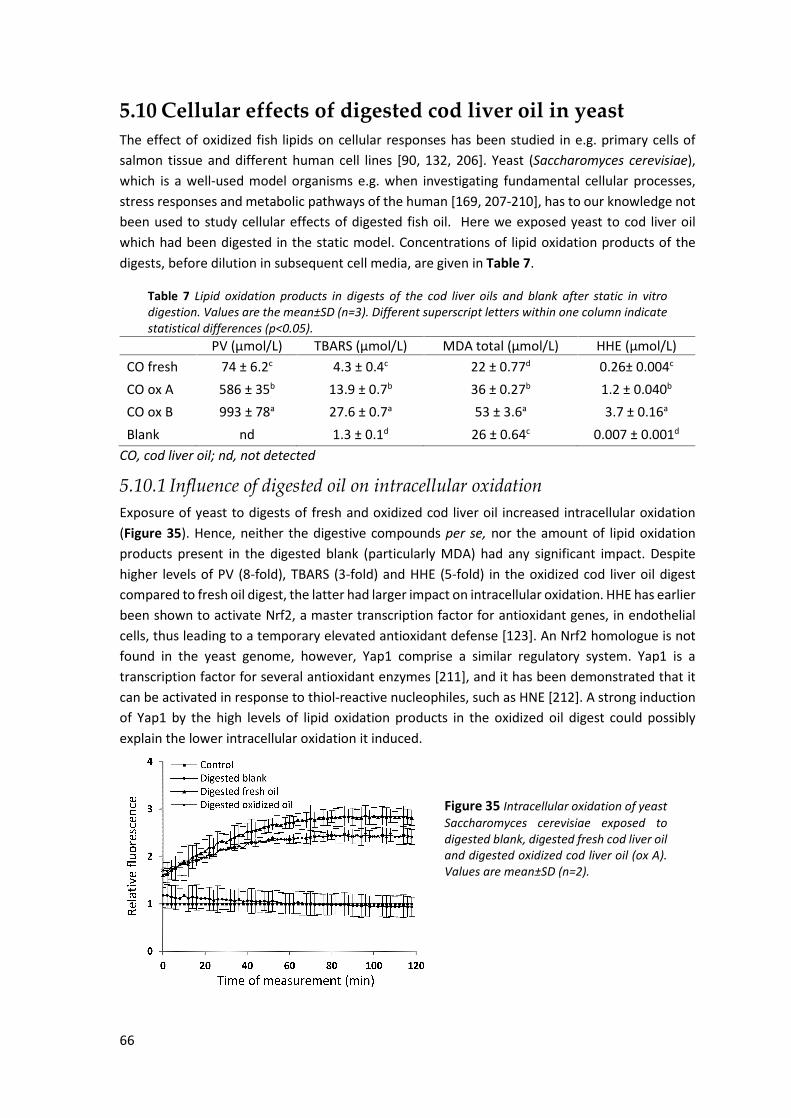
66
5.10 Cellular effects of digested cod liver oil in yeast The effect of oxidized fish lipids on cellular responses has been studied in e.g. primary cells of
salmon tissue and different human cell lines [90, 132, 206]. Yeast (Saccharomyces cerevisiae),
which is a well-used model organisms e.g. when investigating fundamental cellular processes,
stress responses and metabolic pathways of the human [169, 207-210], has to our knowledge not
been used to study cellular effects of digested fish oil. Here we exposed yeast to cod liver oil
which had been digested in the static model. Concentrations of lipid oxidation products of the
digests, before dilution in subsequent cell media, are given in Table 7.
Table 7 Lipid oxidation products in digests of the cod liver oils and blank after static in vitro digestion. Values are the mean±SD (n=3). Different superscript letters within one column indicate statistical differences (p<0.05).
PV (µmol/L) TBARS (µmol/L) MDA total (µmol/L) HHE (µmol/L)
CO fresh 74 ± 6.2c 4.3 ± 0.4c 22 ± 0.77d 0.26± 0.004c
CO ox A 586 ± 35b 13.9 ± 0.7b 36 ± 0.27b 1.2 ± 0.040b
CO ox B 993 ± 78a 27.6 ± 0.7a 53 ± 3.6a 3.7 ± 0.16a
Blank nd 1.3 ± 0.1d 26 ± 0.64c 0.007 ± 0.001d
CO, cod liver oil; nd, not detected
5.10.1 Influence of digested oil on intracellular oxidation Exposure of yeast to digests of fresh and oxidized cod liver oil increased intracellular oxidation
(Figure 35). Hence, neither the digestive compounds per se, nor the amount of lipid oxidation
products present in the digested blank (particularly MDA) had any significant impact. Despite
higher levels of PV (8-fold), TBARS (3-fold) and HHE (5-fold) in the oxidized cod liver oil digest
compared to fresh oil digest, the latter had larger impact on intracellular oxidation. HHE has earlier
been shown to activate Nrf2, a master transcription factor for antioxidant genes, in endothelial
cells, thus leading to a temporary elevated antioxidant defense [123]. An Nrf2 homologue is not
found in the yeast genome, however, Yap1 comprise a similar regulatory system. Yap1 is a
transcription factor for several antioxidant enzymes [211], and it has been demonstrated that it
can be activated in response to thiol-reactive nucleophiles, such as HNE [212]. A strong induction
of Yap1 by the high levels of lipid oxidation products in the oxidized oil digest could possibly
explain the lower intracellular oxidation it induced.
Figure 35 Intracellular oxidation of yeast Saccharomyces cerevisiae exposed to digested blank, digested fresh cod liver oil and digested oxidized cod liver oil (ox A). Values are mean±SD (n=2).

67
5.10.2 Influence of digested oil on cell energy metabolic activity The cell energy metabolic activity of the yeast increased significantly after exposure to all three
digests compared to the control (buffer; Figure 36). No difference in cell energy metabolic activity
was seen between exposures to digests of fresh or oxidized cod liver oil, while exposure to digest
without oil caused significantly lower activity. Because the different levels of lipid oxidation
products had no influence, the raised activity is possibly mainly a result of β-oxidation of fatty
acids in peroxisomes. The peroxisome biogenesis is induced when yeast cells are cultivated in a
fatty acid containing medium [213]. Produced acetyl-CoA is then transferred from peroxisomes to
mitochondria and consequently ATP is formed [214, 215]. The metabolism of e.g. fatty acids
generates hydrogen peroxide, but ROS can also form in the respiration process in mitochondria.
Increased metabolic activity is therefore related to the increased response in the intracellular
oxidation assay. To cope with the risk caused by elevated ROS levels, protecting enzymes are
expressed by the cell. For example, induced expressions of peroxisomal catalase and Gpx1 were
seen when yeast was grown in medium containing fatty acids [213, 216].
Figure 36 Cell energy metabolic activity of yeast Saccharomyces cerevisiae exposed to digests. DB, blank; DCO fresh, digested fresh cod liver oil and DCO ox, digested oxidized cod liver oil (ox A). Values are mean±SD (n=2). Values followed by different letters (a-c) are statistically different.
5.10.3 Influence of digested oil on expression of mitochondrial proteins Expression of mitochondrial proteins in yeast cells exposed to oil digests was compared with the
expression obtained after exposure to blank digest. Six proteins were statistically affected; four
proteins were up-regulated and two were down-regulated. Four of these proteins were
successfully identified; acetyl-CoA hydrolase (Ach1), dihydrolipolyllysine-residue
succinyltransferase component of α-ketoglutarate dehydrogenase complex (Kgd2), 2-
methylcitrate dehydratase (Pdh1) and peroxyredoxin (Hyr1 Gpx3). Among these, only Gpx3 was
up-regulated.
Table 8 Relative fold changes of mitochondrial proteins after 2 h exposure of yeast cells to digests of cod liver oil and blank.
Blank Fresh oil Oxidized oil1 Control (buffer) Protein 1 -1.30 -1.57 1.22 Ach1 1 -1.32 -2.26 1.24 Kgd2 1 -1.66 -1.86 -1.10 Pdh1 1 1.53 1.58 -1.09 Gpx3 1 -1.09 -1.47 1.43 Not id 1 2.11 2.29 1.05 Not id
1 Cod liver oil Ox B according to Table 5 Not id, not identified

68
All down-regulated proteins were linked to energy metabolic functions. Firstly, as demonstrated
by Eisenberg et al. [217], deletion of the gene encoding Ach1 led to accumulation of acetate, which
in turn activated enzymes responsible for its conversion to acetyl-CoA. Down-regulation of Ach1
could be related to increased cell energy metabolic activity in the cells exposed to oil digests.
Secondly, the α-ketoglutarate dehydrogenase complex is involved in the regulation of the citric
acid cycle, hence controlling mitochondrial metabolism. Thirdly, Phd1 is part of the methylcitrate
cycle, which mediates the conversion of propionyl-CoA to pyruvate [218, 219]. In contrast to the
down-regulated proteins, Gpx3 is involved in oxidative stress responses. Gpx3, together with the
two other glutathione peroxidases expressed by S. cerevisiae, Gpx1 and Gpx2, are structural
homologues of mammalian GPx [213].
More intense protein expression changes were seen after exposure to digests of oxidized oil
compared to digests of fresh oil, which could be related to the higher amounts of lipid oxidation
products in the former (Table 8). The down-regulation of proteins could indicate that MDA, HHE
and/or ROS caused oxidative damage of proteins and DNA. As mentioned earlier, MDA and HHE,
but also other reactive aldehydes formed during the oxidation process, reacts readily with amino
and thiol groups of proteins generating Michael and Schiff adducts [101, 120]. Both HNE and ROS
have been reported to inactivate α-ketoglutarate dehydrogenase by oxidation [220]. In addition,
metabolic enzymes containing iron-sulfur in their prosthetic group are sensitive to ROS [221], and
Pdh1 contain a 2Fe-2S cluster, making it susceptible to oxidative damage. Almost identical fold
changes of Gpx3 were seen in yeast cells after exposure to both fresh and oxidized oil digests. It
could thus not be determined whether the elevated Gpx3 levels were connected to the oil per se
or increased levels of oxidation products compared to the digested blank. But the results are in
line with the observed increased intracellular oxidation, which should activate the defense
mechanisms of the cell.
Genome responses in rats after an oxidized diet compared to a control diet have been reported
by others. Awada et al. [132] showed increased expression of Gpx2 by the oxidized diet. Sulzle et
al. [222] showed that various genes were upregulated after consumption of a diet with oxidized
oil compared to a control diet. The up-regulated genes were related to cytochrome P450A, β-
oxidation, lipid metabolism and protein metabolism. Also Chao et al. [223] reported up-regulation
of cytochrome P450A in rats fed an oxidized soyabean-oil diet. An elevated expression of
cytochrome P450A, which may be caused by the activation of PPARα, will increase the oxidative
stress [5]. However, investigation of healthy rat heart mitochondria showed a large occurrence of
modifications in enzymes of lipid metabolism and in proteins of the citric acid cycle and respiratory
chain [2]. Such modifications involved Michael adducts of e.g. HHE and HNE with cysteine and
histidine residues. Biological effects of undigested marine oil containing a varying degree of
oxidation products were evaluated in primary cells of salmon tissue and in human Caco-2 cells and
monocytes [206]. When the oxidation level of the oil increased, the activity of superoxide
dismutase (SOD) increased to a certain level and then decreased again. Oxidized marine oil also
promoted lipid oxidation of cell membranes and increased expression of genes involved in stress
and inflammation responses. Our results using digested oils are thus in line with their results.

69
5.11 Inflammatory responses of digested cod liver oil in
dendritic cells The progress from the immature to the mature state of dendritic cells is accompanied by an
upregulation of the cell surface receptors CD86 and HLA-DR [224]. Results from the expression of
CD86 (Figure 37) and HLA-DR (data not shown) showed that significantly lower percentage of
dendritic cells matured and stimulated in the presence of digested fresh oil (100 µg digest dw/mL)
compared to cells matured with digested blank. Also in the presence of digested oxidized oil, a
tendency of fewer cells expressing CD86 was seen. Undigested oils (1-100 µg/mL) were also
tested, but without any influence on the dendritic cell maturation and stimulation.
Figure 37 Expression of CD86 by dendritic cells cultured with DMSO as a control and digests of fresh cod liver oil, oxidized cod liver oil and blank. The concentration of digesta was 1, 10 or 100 µg dw/mL cell suspension. MC, monocytes; imDC, immature dendritic cells; DCO, digested cod liver oil; DCO ox, digested oxidized cod liver oil; DB, digested blank. Values are mean±SD (n=5).
Additionally, the effect of digested cod liver oil on the monocyte-derived dendritic secretion of
key cytokines was investigated. IL-10 is one of the major anti-inflammatory cytokines and inhibits
the synthesis of other cytokines. On the other hand, IL-12p40 affects the differentiation of naïve
T cells into Th1 cells, and IL-6 and IL-23 towards Th17 cells, thus, these three cytokines act pro-
inflammatory. Secretion of IL-12p40 and IL-10 was analyzed in order to determine whether the
digests induced a Th1-like response (high IL-12 and low IL-10) or a Th2-like response (low IL-12
and high IL-10) [176]. Induction of Th2 could be indications of anti-inflammatory effects [225].
Again, the highest response was seen in dendritic cells exposed to digested fresh cod liver oil (100
µg digest dw/mL), which caused significantly lower secretion levels of all analyzed cytokines
compared to digested blank (Figure 38, IL-6 and IL-23 are not shown). The oxidized cod liver oil
digest led to lower secretion index of IL-10 and IL-12p40 compared to the digested fresh oil. When
the IL-12p40/IL-10 proportional index was calculated, both fresh and oxidized oil digests
generated significantly higher proportion values compared to control (DMSO), and fresh oil digest
also compared to digested blank. In the cytokine analyses, also the undigested oils contributed to
a trend for lower IL-10 and subsequently higher IL-12p40/IL-10 proportional index, however the
differences were not significant. In conclusion, these data suggest that cod liver oil per se has a
slight pro-inflammatory effect, but including lipid oxidation products and digestive compounds in
the cod liver oil digests further stimulates the dendritic cells to obtain a pro-inflammatory
phenotype.
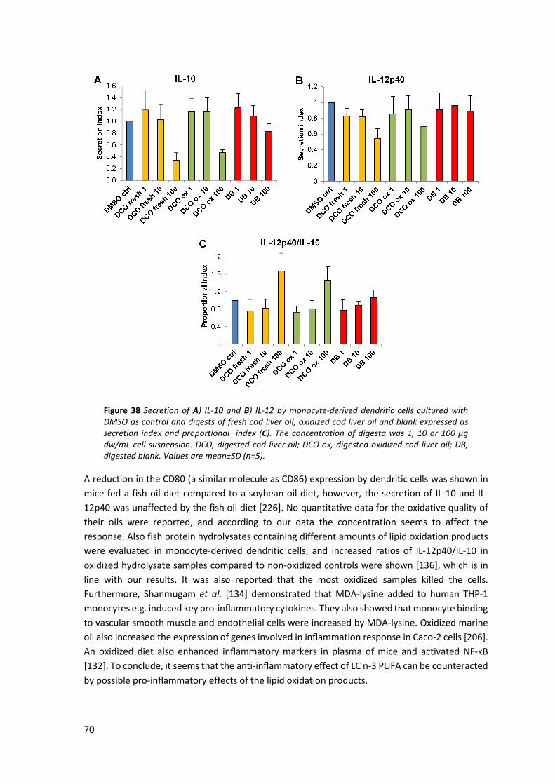
70
Figure 38 Secretion of A) IL-10 and B) IL-12 by monocyte-derived dendritic cells cultured with DMSO as control and digests of fresh cod liver oil, oxidized cod liver oil and blank expressed as secretion index and proportional index (C). The concentration of digesta was 1, 10 or 100 µg dw/mL cell suspension. DCO, digested cod liver oil; DCO ox, digested oxidized cod liver oil; DB, digested blank. Values are mean±SD (n=5).
A reduction in the CD80 (a similar molecule as CD86) expression by dendritic cells was shown in
mice fed a fish oil diet compared to a soybean oil diet, however, the secretion of IL-10 and IL-
12p40 was unaffected by the fish oil diet [226]. No quantitative data for the oxidative quality of
their oils were reported, and according to our data the concentration seems to affect the
response. Also fish protein hydrolysates containing different amounts of lipid oxidation products
were evaluated in monocyte-derived dendritic cells, and increased ratios of IL-12p40/IL-10 in
oxidized hydrolysate samples compared to non-oxidized controls were shown [136], which is in
line with our results. It was also reported that the most oxidized samples killed the cells.
Furthermore, Shanmugam et al. [134] demonstrated that MDA-lysine added to human THP-1
monocytes e.g. induced key pro-inflammatory cytokines. They also showed that monocyte binding
to vascular smooth muscle and endothelial cells were increased by MDA-lysine. Oxidized marine
oil also increased the expression of genes involved in inflammation response in Caco-2 cells [206].
An oxidized diet also enhanced inflammatory markers in plasma of mice and activated NF-κB
[132]. To conclude, it seems that the anti-inflammatory effect of LC n-3 PUFA can be counteracted
by possible pro-inflammatory effects of the lipid oxidation products.
71
CONCLUSIONS
During static in vitro digestion of cod liver oil, gradually higher TBARS levels were formed during the gastric and the intestinal steps. Addition of digestive compounds promoted lipid oxidation, and especially bile acids played an important role.
Cod liver oils with elevated levels of preformed oxidation products generated higher
concentrations of lipid oxidation products during digestion compared to oils of better quality. However, the initial amount of oxidation products was not directly proportional to the levels reached during subsequent GI digestion.
Emulsification of cod liver oil had inconsistent impact on GI oxidation during dynamic
digestion. Emulsified oils generated lower MDA concentrations during gastric digestion, but in the intestinal phase they formed equally high MDA levels as non-emulsified oil. Emulsification on the other hand stimulated HHE formation in the intestinal phase, generating 3-fold higher levels. TBARS levels were unaffected by emulsification during static digestion.
Cod Hb (11.5 µmol/L emulsion) had a strong and immediate pro-oxidant effect on emulsified cod liver oil during digestion. Equally high MDA concentrations (25 µM) were obtained in the gastric and the intestinal phase during dynamic digestion in the presence of Hb. Hb-enriched emulsion also yielded the highest HHE level (1.6 µM) of all test meals in this study.
Extensively higher levels of lipid oxidation products were formed during dynamic digestion of raw herring mince compared to its corresponding isolated oil (emulsified or non-emulsified). A suggested explanation was the large endogenous amounts of heme-proteins in the herring muscle. Emulsification of the herring oil prior to ingestion also generated higher MDA and HHE levels during digestion compared to non-emulsified oil, which could be due to the increased surface area of the lipid droplets in the emulsion.
Raw mince of herring and salmon generated different amounts of lipid oxidation products
during dynamic digestion; higher levels of MDA and HHE were formed by herring minces containing 4 and 17% lipids compared to salmon mince with 17% lipids. The amount of preformed lipid oxidation products, however, was higher in the herring minces. Pro-oxidative Hb in the herring and antioxidative astaxanthine in the salmon are likely explanations.
Oven baking per se had a pro-oxidative effect on salmon, and higher MDA and HHE levels were also formed during subsequent digestion of cooked salmon compared to raw salmon. Higher maximum concentrations of MDA was also generated by the oven baked herring in the gastric phase compared to raw herring.
Maximum levels of MDA, HHE and HNE determined in the gastric phase during dynamic digestion of all fish lipid containing test meals were 27 µM, 0.85 µM and 0.06 µM, respectively. With most fish meals, lower aldehyde concentrations were determined in the intestinal phase than in the gastric phase, with the levels being even lower than the contribution from the gastric compartment. This might be a result of the high reactivity of these aldehydes in combination with the increased proteolysis, enabling raised aldehyde-protein adduct formation. In contrast, oil containing test meals generally gave higher
72
aldehyde concentrations in the intestinal phase. Maximum MDA, HHE and HNE levels determined in the intestinal phase were 23 µM, 1.6 µM, 0.07 µM, respectively.
Exposure of yeast to digests of fresh and oxidized cod liver oil, containing varying amounts of lipid oxidation products, caused increased intracellular oxidation and cell energy metabolic activity. At the proteome level, a down-regulation of proteins, which are directly or indirectly involved in energy metabolism, and an up-regulation of the antioxidant enzyme peroxyredoxin was observed. The results suggested that oil digests caused a high oxidant load for the yeast, leading to increased oxidative stress and damage of cellular components.
Exposure of monocyte-derived dendritic cells to digests of fresh and oxidized cod liver oil indicated a reduced maturation and a raised proportional index of the cytokines IL-12p40 and IL-10, which suggested a pro-inflammatory effect compared to digested blank.

73
FUTURE PERSPECTIVES
The work presented in this thesis showed that the amounts of lipid oxidation products increased
during in vitro digestion of fish lipids. However, based on the initial amount of oxidation products
there was not a constant relative formation of aldehydes during digestion. Furthermore, the
highest aldehyde levels were not consistently formed in the same digestive phase. These topics
could be investigated in more detail, and in different matrices, in order to find what food related
factors that influence GI oxidation the most. The outcome should answer the question in which
step it is most important to limit oxidation in order to minimize total oxidant load in the GI tract;
prior to ingestion, in the stomach or in the intestine? This is important knowledge both in the
design of LC n-3 enriched products and for dietary recommendations. Proper efforts could then
be directed to add antioxidants that acts efficiently in the most critical step(s). Hb was shown to
strongly increase lipid oxidation during digestion of emulsified cod liver oil, and the high aldehyde
levels determined in herring mince was also partly ascribed heme protein mediated oxidation.
Therefore, it seems especially important to find ways to inhibit the pro-oxidative effect of Hb
during digestion.
Suggested methodological changes for future GI digestion experiments includes e.g. a change of
static digestion method to the consensus digestion method for static in vitro studies that was
developed within the COST Action Infogest after the initiation of our studies. The inclusion of a
gastric lipase and fresh bovine bile, which has a similar bile acid composition to human would
hopefully generate even more realistic data on GI oxidation. These changes will facilitate
comparison of results among different researcher�s data. More studies using human gastric and
intestinal juices would also be encouraged, because extensively higher aldehyde levels were
formed during digestion of fresh cod liver oil using human juices compared to simulated fluids
following the consensus protocol for static in vitro digestion [74].
Another method related issue is the choice of oxidation marker. Since we determined non-protein
bound aldehydes during dynamic digestion, it is important to test our hypothesis that the lower
levels of aldehydes found in the intestinal phase of fish containing test meals are due to increased
protein-aldehyde adduct formation. This could be done with monoclonal antibodies against
specific aldehyde-adducts, such as HHE-adducts or other reliable methods where both free and
bound aldehydes are determined. It can be admitted that in vitro data always needs to be verified
by in vivo studies and it would be highly interesting to correlate our findings with a study on GI
oxidation in ileostomy patients. Further, potential damage of the epithelium caused by lipid
oxidation products could be studied on biopsies.
In the yeast cell model, we could not separate the effects of lipid oxidation products from effects
of cod liver oil digests as an energy source. In a follow-up study, a stripped stable oil with minimal
amounts of preformed oxidation products could be included, as well as glucose, as a control for
the contribution of an energy source. Effect on cells during non-starvation condition should also
be evaluated. Changes in the total proteome, and a focus on newly synthesized proteins could
also give more information about the instant effects of digested oil. Another step forward is to
use primary cells of human epithelium.
74
ACKNOWLEDGEMENTS
Many people have helped me and supported me during my years as a PhD student; you all have
my gratitude. I would especially like to thank:
Ingrid Undeland, my supervisor, and Marie Alminger, my co-supervisor, who have guided me
through this trip. Ingrid, thanks for always having your door open and for giving your opinion on
things. Thanks for all these years, and I appreciate your principles. Marie, thanks for the company
in Holland; it was a pity the police had closed our cosy hotel during my second visit�
Thomas Andlid, Bo Ekstrand and Ann-Sofie Sandberg for your constructive feedback on this thesis.
A special thanks to Thomas for introducing me to Food Science many years ago.
Mattias Fredrikson for being my mentor and for attending various LiFT activities.
Rob Havenaar, for your expertise and helpfulness. I also would like to thank the other people at
TNO, Tom, Hans, Casper, Jaan, Big-Rob, Wendy, Mark, Susann, Mans, Evelijn, who helped me in
one way or another, e.g. by delivering samples and printing stickers, and for laughing at my jokes.
Nisse and Annette for always helping out in the lab and for coming with good suggestions on all
kinds of questions. Nisse, without you the LC-MS method would probably not exist. Mi-Lo, Helena
and Anna for helping me with all administrative issues and for arranging nice Food Science events.
Malin, Cecilia S, Cristina, Graziele and Cecilia M for being really nice roommates. Malin and Cecilia
S, it has been a pleasure to share room with you and thanks for all research related discussions
and life related social chats. Cecilia S, I miss you and all the laughs we shared.
All co-workers at Food and Nutrition Science, both present and former. It has been many people
over the years, and you all deserves my gratitude for contributing to a nice atmosphere. Special
thanks to Hanna, Cecilia T and Lillie for practical contribution to this work. Hanna, you were the
best master thesis student I could wish for, in all aspects, and I am grateful that you are still
around. Hanna and Cecilia T, I appreciate all collaborations; work is much more fun when you do
it together. Thanks for also being my back-up, e.g. when I had to run from the lab to pick up the
children before all vials were in the autosampler�
Lars, Alva, Erik and Isak, my greatest gratitude goes to you; you are the most important for me.
Lars, thanks for always being there for me, and for helping me with computer questions and with
photo editing late evenings when the children had fell asleep. I am also thankful to Håkan and
Kerstin, who have assisted Lars with the children when I have been abroad or been working
weekends. I am also grateful to mum, dad, Anna, Lena, Anders and all friends for your encouraging
words and your contribution to a meaningful life.
This work was financially supported by the Swedish Research Council for Environment, Agricultural
Sciences and Spatial Planning (FORMAS, Project 222-2007-1007) and Nordic Innovation Centre
(SAFEFOODERA, Project 08202). Foundation Olle Engkvist, Byggmästare and LiFT have supported
the research with equipment and covered my travel expenses during my trips to the Netherlands,
respectively.
75
REFERENCES
1. Maehre, H.K., I.J. Jensen, E.O. Elvevoll, and K.E. Eilertsen, ω-3 fatty acids and cardiovascular diseases: Effects, mechanisms and dietary relevance. International Journal of Molecular Sciences, 2015. 16(9): p. 22636-22661.
2. Domingues, R.M., P. Domingues, T. Melo, D. Pérez-Sala, A. Reis, and C.M. Spickett, Lipoxidation adducts with peptides and proteins: Deleterious modifications or signaling mechanisms? Journal of Proteomics, 2013. 92: p. 110-131.
3. Liao, F., A. Andalibi, J.H. Qiao, H. Allayee, A.M. Fogelman, and A.J. Lusis, Genetic evidence for a common pathway mediating oxidative stress, inflammatory gene induction, and aortic fatty streak formation in mice. Journal of Clinical Investigation, 1994. 94(2): p. 877-884.
4. Penumetcha, M., N. Khan-Merchant, and S. Parthasarathy, Enhanced solubilization and intestinal absorption of cholesterol by oxidized linoleic acid. Journal of Lipid Research, 2002. 43(6): p. 895-903.
5. Turner, R., C.H. McLean, and K.M. Silvers, Are the health benefits of fish oils limited by products of oxidation? Nutrition Research Reviews, 2006. 19(1): p. 53-62.
6. Halliwell, B., K. Zhao, and M. Whiteman, The gastrointestinal tract: A major site of antioxidant action? Free Radical Research, 2000. 33(6): p. 819-830.
7. Kanner, J. and T. Lapidot, The stomach as a bioreactor: Dietary lipid peroxidation in the gastric fluid and the effects of plant-derived antioxidants. Free Radical Biology and Medicine, 2001. 31(11): p. 1388-1395.
8. Gorelik, S., M. Ligumsky, R. Kohen, and J. Kanner, The stomach as a "bioreactor": When red meat meets red wine. Journal of Agricultural and Food Chemistry, 2008. 56(13): p. 5002-5007.
9. Kenmogne-Domguia, H.B., A. Meynier, C. Boulanger, and C. Genot, Lipid oxidation in food emulsions under gastrointestinal-simulated conditions: The key role of endogenous tocopherols and initiator. Food Digestion, 2012. 3(1-3): p. 46-52.
10. Albert, B.B., J.G. Derraik, D. Cameron-Smith, P.L. Hofman, S. Tumanov, S.G. Villas-Boas, M.L. Garg, and W.S. Cutfield, Fish oil supplements in New Zealand are highly oxidised and do not meet label content of n-3 PUFA. Sci Rep, 2015. 5: p. 7928.
11. Thorkildsen, T., I. Ottestad, and G. Vogt, Oxidation status in omega-3 oils in the Norwegian health care market, in 26th LipidForum Symposium. 2011: Ålesund, Norway. p. 52.
12. Ratnayake, W.M.N. and C. Galli, Fat and fatty acid terminology, methods of analysis and fat digestion and metabolism: A background review paper. Annals of Nutrition and Metabolism, 2009. 55(1-3): p. 8-43.
13. Jensen, K.N., C. Jacobsen, and H.H. Nielsen, Fatty acid composition of herring (Clupea harengus L.): Influence of time and place of catch on n-3 PUFA content. Journal of the Science of Food and Agriculture, 2007. 87(4): p. 710-718.
14. Ackman, R.G., Nutritional composition of fats in seafoods. Progress in Food and Nutrition Science, 1989. 13(3-4): p. 161-289.
15. Larsson, K., A. Almgren, and I. Undeland, Hemoglobin-mediated lipid oxidation and compositional characteristics of washed fish mince model systems made from cod (Gadus morhua), herring (Clupea harengus), and salmon (Salmo salar) muscle. Journal of Agricultural and Food Chemistry, 2007. 55(22): p. 9027-9035.
16. Armand, M., Lipases and lipolysis in the human digestive tract: Where do we stand? Current Opinion in Clinical Nutrition and Metabolic Care, 2007. 10(2): p. 156-164.
17. Bakker, J. and D.J. Mela, Effect of emulsion structure on flavor release and taste perception. 1996. p. 36-47. 18. Vingerhoeds, M.H., T.B.J. Blijdenstein, F.D. Zoet, and G.A. van Aken, Emulsion flocculation induced by saliva
and mucin. Food Hydrocolloids, 2005. 19(5): p. 915-922. 19. McClements, D.J., E.A. Decker, and Y. Park, Controlling lipid bioavailability through physicochemical and
structural approaches. Critical Reviews in Food Science and Nutrition, 2009. 49(1): p. 48-67. 20. Kalantzi, L., K. Goumas, V. Kalioras, B. Abrahamsson, J. Dressman, and C. Reppas, Characterization of the
Human Upper Gastrointestinal Contents Under Conditions Simulating Bioavailability/Bioequivalence Studies. Pharmaceutical Research, 2006. 23(1): p. 165-176.
21. Sams, L., J. Paume, J. Giallo, and F. Carriere, Relevant pH and lipase for in vitro models of gastric digestion. Food & Function, 2016.
22. Armand, M., B. Pasquier, M. André, P. Borel, M. Senft, J. Peyrot, J. Salducci, H. Portugal, V. Jaussan, and D. Lairon, Digestion and absorption of 2 fat emulsions with different droplet sizes in the human digestive tract. American Journal of Clinical Nutrition, 1999. 70(6): p. 1096-1106.
23. Bauer, E., S. Jakob, and R. Mosenthin, Principles of physiology of lipid digestion. Asian-Australasian Journal of Animal Sciences, 2005. 18(2): p. 282-295.
24. Pafumi, Y., D. Lairon, P.L. De La Porte, C. Juhel, J. Storch, M. Hamosh, and M. Armand, Mechanisms of inhibition of triacylglycerol hydrolysis by human gastric lipase. Journal of Biological Chemistry, 2002. 277(31): p. 28070-28079.

76
25. Bodmer, M.W., S. Angal, G.T. Yarranton, T.J.R. Harris, A. Lyons, D.J. King, G. Pieroni, C. Riviere, R. Verger, and P.A. Lowe, Molecular cloning of a human gastric lipase and expression of the enzyme in yeast. Biochimica et Biophysica Acta (BBA) - Gene Structure and Expression, 1987. 909(3): p. 237-244.
26. Moreau, H., R. Laugier, Y. Gargouri, F. Ferrato, and R. Verger, Human preduodenal lipase is entirely of gastric fundic origin. Gastroenterology, 1988. 95(5): p. 1221-1226.
27. Mukherjee, M., Human digestive and metabolic lipases--a brief review. Journal of Molecular Catalysis B: Enzymatic, 2003. 22(5-6): p. 369-376.
28. McClements, D.J., E.A. Decker, and Y. Park, Controlling Lipid Bioavailability through Physicochemical and Structural Approaches. Critical Reviews in Food Science and Nutrition, 2009. 49(1): p. 48 - 67.
29. Armand, M., M. Hamosh, J.S. DiPalma, J. Gallagher, S.B. Benjamin, J.R. Philpott, D. Lairon, and P. Hamosh, Dietary fat modulates gastric lipase activity in healthy humans. American Journal of Clinical Nutrition, 1995. 62(1): p. 74-80.
30. Favé, G., T.C. Coste, and M. Armand, Physicochemical properties of lipids: new strategies to manage fatty acid bioavailability. Cellular and molecular biology (Noisy-le-Grand, France), 2004. 50(7): p. 815-831.
31. Mu, H. and C.E. Høy, The digestion of dietary triacylglycerols. Progress in Lipid Research, 2004. 43(2): p. 105-133.
32. Carriere, F., J.A. Barrowman, R. Verger, and R. Laugier, Secretion and contribution to lipolysis of gastric and pancreatic lipases during a test meal in humans. Gastroenterology, 1993. 105(3): p. 876-888.
33. Lindqvist, A. and C. Erlanson-Albertsson, Fat digestion and its role in appetite regulation and energy balance - The importance of enterostatin and tetrahydrolipstatin. Current Medicinal Chemistry - Central Nervous System Agents, 2003. 3(3): p. 157-175.
34. Lowe, M.E., The triglyceride lipases of the pancreas. J. Lipid Res., 2002. 43(12): p. 2007-2016. 35. Roger, E., F. Lagarce, E. Garcion, and J.P. Benoit, Biopharmaceutical parameters to consider in order to alter
the fate of nanocarriers after oral delivery. Nanomedicine, 2010. 5(2): p. 287-306. 36. Kamp, F., W. Guo, R. Souto, P.F. Pilch, B.E. Corkey, and J.A. Hamilton, Rapid Flip-flop of Oleic Acid across the
Plasma Membrane of Adipocytes. Journal of Biological Chemistry, 2003. 278(10): p. 7988-7995. 37. Mansbach, C.M., II and F. Gorelick, Development and Physiological Regulation of Intestinal Lipid Absorption.
II. Dietary lipid absorption, complex lipid synthesis, and the intracellular packaging and secretion of chylomicrons. Am J Physiol Gastrointest Liver Physiol, 2007. 293(4): p. G645-650.
38. Hussain, M.M., Intestinal lipid absorption and lipoprotein formation. Current Opinion in Lipidology, 2014. 25(3): p. 200-206.
39. Nassir, F., B. Wilson, X. Han, R.W. Gross, and N.A. Abumrad, CD36 is important for fatty acid and cholesterol uptake by the proximal but not distal intestine. Journal of Biological Chemistry, 2007. 282(27): p. 19493-19501.
40. Chinetti, G., S. Griglio, M. Antonucci, I.P. Torra, P. Delerive, Z. Majd, J.C. Fruchart, J. Chapman, J. Najib, and B. Staels, Activation of proliferator-activated receptors α and γ induces apoptosis of human monocyte-derived macrophages. Journal of Biological Chemistry, 1998. 273(40): p. 25573-25580.
41. Tyagi, S., P. Gupta, A.S. Saini, C. Kaushal, and S. Sharma, The peroxisome proliferator-activated receptor: A family of nuclear receptors role in various diseases. Journal of Advanced Pharmaceutical Technology & Research, 2011. 2(4): p. 236-240.
42. Calder, P.C., Marine omega-3 fatty acids and inflammatory processes: Effects, mechanisms and clinical relevance. Biochimica et Biophysica Acta - Molecular and Cell Biology of Lipids, 2015. 1851(4): p. 469-484.
43. Hur, S.J., B.O. Lim, E.A. Decker, and D.J. McClements, In vitro human digestion models for food applications. Food Chemistry, 2011. 125(1): p. 1-12.
44. Minekus, M., M. Alminger, P. Alvito, S. Ballance, T. Bohn, C. Bourlieu, F. Carrière, R. Boutrou, M. Corredig, D. Dupont, C. Dufour, L. Egger, M. Golding, S. Karakaya, B. Kirkhus, S. Le Feunteun, U. Lesmes, A. MacIerzanka, A. MacKie, S. Marze, D.J. McClements, O. Ménard, I. Recio, C.N. Santos, R.P. Singh, G.E. Vegarud, M.S.J. Wickham, W. Weitschies, and A. Brodkorb, A standardised static in vitro digestion method suitable for food-an international consensus. Food and Function, 2014. 5(6): p. 1113-1124.
45. Minekus, M., P. Marteau, R. Havenaar, and J. Huis in't Veld, A multi compartmental dynamic computer-controlled model simulating the stomach and small intestine. Alternatives to laboratory animals (ATLA), 1995. 23: p. 197-209.
46. Minekus, M., M. Smeets-Peeters, R. Havenaar, A. Bernalier, G. Fonty, S. Marol-Bonnin, M. Alric, P. Marteau, and J.H.J. Huis In't Veld, A computer-controlled system to simulate conditions of the large intestine with peristaltic mixing, water absorption and absorption of fermentation products. Applied Microbiology and Biotechnology, 1999. 53(1): p. 108-114.
47. Molly, K., M. Vande Woestyne, and W. Verstraete, Development of a 5-step multi-chamber reactor as a simulation of the human intestinal microbial ecosystem. Applied Microbiology and Biotechnology, 1993. 39(2): p. 254-258.
48. Laguerre, M., J. Lecomte, and P. Villeneuve, Evaluation of the ability of antioxidants to counteract lipid oxidation: Existing methods, new trends and challenges. Progress in Lipid Research, 2007. 46(5): p. 244-282.
49. Apel, K. and H. Hirt, Reactive oxygen species: Metabolism, oxidative stress, and signal transduction, in Annual Review of Plant Biology. 2004. p. 373-399.
77
50. Guillén, M.D. and E. Goicoechea, Toxic oxygenated α,β-unsaturated aldehydes and their study in foods: A review. Critical Reviews in Food Science and Nutrition, 2008. 48(2): p. 119-136.
51. Pillon, N. and C. Soulage, Lipid Peroxidation by-Products and the Metabolic Syndrome, Lipid Peroxidation, A. Catala, Editor. 2012, InTech.
52. Esterbauer, H., R.J. Schaur, and H. Zollner, Chemistry and biochemistry of 4-hydroxynonenal, malonaldehyde and related aldehydes. Free Radical Biology and Medicine, 1991. 11(1): p. 81-128.
53. Fernández, J., J.A. Pérez-Álvarez, and J.A. Fernández-López, Thiobarbituric acid test for monitoring lipid oxidation in meat. Food Chemistry, 1997. 59(3): p. 345-353.
54. Ursini, F., A. Zamburlini, G. Cazzolato, M. Maiorino, G.B. Bon, and A. Sevanian, Postprandial plasma lipid hydroperoxides: A possible link between diet and atherosclerosis. Free Radical Biology and Medicine, 1998. 25(2): p. 250-252.
55. Bull, A.W., N.D. Nigro, W.A. Golembieski, J.D. Crissman, and L.J. Marnett, In vivo stimulation of DNA synthesis and induction of ornithine decarboxylase in rat colon by fatty acid hydroperoxides, autoxidation products of unsaturated fatty acids. Cancer Research, 1984. 44(11): p. 4924-4928.
56. Waring, A.J., I.M. Drake, C.J. Schorah, K.L.M. White, D.A.F. Lynch, A.T.R. Axon, and M.F. Dixon, Ascorbic acid and total vitamin C concentrations in plasma, gastric juice, and gastrointestinal mucosa: Effects of gastritis and oral supplementation. Gut, 1996. 38(2): p. 171-176.
57. Alayash, A.I., R.P. Patel, and R.E. Cashon, Redox reactions of hemoglobin and myoglobin: Biological and toxicological implications. Antioxidants and Redox Signaling, 2001. 3(2): p. 313-327.
58. Undeland, I., H.G. Kristinsson, and H.O. Hultin, Hemoglobin-mediated oxidation of washed minced cod muscle phospholipids: Effect of pH and hemoglobin source. Journal of Agricultural and Food Chemistry, 2004. 52(14): p. 4444-4451.
59. Aranda, R.t., H. Cai, C.E. Worley, E.J. Levin, R. Li, J.S. Olson, G.N. Phillips, Jr., and M.P. Richards, Structural analysis of fish versus mammalian hemoglobins: effect of the heme pocket environment on autooxidation and hemin loss. Proteins, 2009. 75(1): p. 217-30.
60. Reeder, B.J. and M.T. Wilson, Mechanism of reaction of myoglobin with the lipid hydroperoxide hydroperoxyoctadecadienoic acid. Biochemical Journal, 1998. 330(3): p. 1317-1323.
61. Carlsen, C.U. and L.H. Skibsted, Myoglobin Species with Enhanced Prooxidative Activity Is Formed during Mild Proteolysis by Pepsin. Journal of Agricultural and Food Chemistry, 2004. 52(6): p. 1675-1681.
62. Maestre, R., M. Pazos, and I. Medina, Involvement of methemoglobin (MetHb) formation and hemin loss in the pro-oxidant activity of fish hemoglobins. Journal of Agricultural and Food Chemistry, 2009. 57(15): p. 7013-7021.
63. Gorelik, S., T. Lapidot, I. Shaham, R. Granit, M. Ligumsky, R. Kohen, and J. Kanner, Lipid peroxidation and coupled vitamin oxidation in simulated and human gastric fluid inhibited by dietary polyphenols: Health implications. Journal of Agricultural and Food Chemistry, 2005. 53(9): p. 3397-3402.
64. Iwao, T., A. Toyonaga, H. Shigemori, M. Ikegami, and K. Tanikawa, Omeprazole administration does not impair gastrointestinal mucosal perfusion, oxygenation, and hexosamine generation. Gastrointestinal Endoscopy, 1995. 41(5): p. 497-501.
65. Hillman, K., A.L. Whyte, and C.S. Stewart, Dissolved oxygen in the porcine gastrointestinal tract. Letters in Applied Microbiology, 1993. 16(6): p. 299-302.
66. He, G., R.A. Shankar, M. Chzhan, A. Samouilov, P. Kuppusamy, and J.L. Zweier, Noninvasive measurement of anatomic structure and intraluminal oxygenation in the gastrointestinal tract of living mice with spatial and spectral EPR imaging. Proceedings of the National Academy of Sciences of the United States of America, 1999. 96(8): p. 4586-4591.
67. Andersson, K. and H. Lingnert, Influence of oxygen concentration on the flavour and chemical stability of cream powder. LWT - Food Science and Technology, 1998. 31(3): p. 245-251.
68. Gorelik, S., R. Kohen, M. Ligumsky, and J. Kanner, Saliva plays a dual role in oxidation process in stomach medium. Archives of Biochemistry and Biophysics, 2007. 458(2): p. 236-243.
69. Peri, L., D. Pietraforte, G. Scorza, A. Napolitano, V. Fogliano, and M. Minetti, Apples increase nitric oxide production by human saliva at the acidic pH of the stomach: A new biological function for polyphenols with a catechol group? Free Radical Biology and Medicine, 2005. 39(5): p. 668-681.
70. Tirosh, O., A. Shpaizer, and J. Kanner, Lipid Peroxidation in a Stomach Medium Is Affected by Dietary Oils (Olive/Fish) and Antioxidants: The Mediterranean versus Western Diet. Journal of Agricultural and Food Chemistry, 2015. 63(31): p. 7016-7023.
71. Van Hecke, T., J. Vanden Bussche, L. Vanhaecke, E. Vossen, J. Van Camp, and S. De Smet, Nitrite Curing of Chicken, Pork, and Beef Inhibits Oxidation but Does Not Affect N-Nitroso Compound (NOC)-Specific DNA Adduct Formation during in Vitro Digestion. Journal of Agricultural and Food Chemistry, 2014. 62(8): p. 1980-1988.
72. Hoshino, C., Y. Tagawa, S. Wada, J.H. Oh, D.K. Park, A. Nagao, and J. Terao, Antioxidant Activity of Quercetin against Metmyoglobin-induced Oxidation of Fish Oil-bile Salt Emulsion. Bioscience, Biotechnology and Biochemistry, 1997. 61(10): p. 1634-1640.
78
73. Lorrain, B., O. Dangles, C. Genot, and C. Dufour, Chemical modeling of heme-induced lipid oxidation in gastric conditions and inhibition by dietary polyphenols. Journal of Agricultural and Food Chemistry, 2010. 58(1): p. 676-683.
74. Tullberg, C., K. Larsson, N.G. Carlsson, I. Comi, N. Scheers, G. Vegarud, and I. Undeland, Formation of reactive aldehydes (MDA, HHE, HNE) during the digestion of cod liver oil: comparison of human and porcine in vitro digestion models. Food Funct, 2016. 7(3): p. 1401-12.
75. Kristinova, V., I. Storrø, and T. Rustad, Influence of human gastric juice on oxidation of marine lipids -in vitro study. Food Chemistry, 2013. 141(4): p. 3859-3871.
76. Lorrain, B., O. Dangles, M. Loonis, M. Armand, and C. Dufour, Dietary iron-initiated lipid oxidation and its inhibition by polyphenols in gastric conditions. Journal of Agricultural and Food Chemistry, 2012. 60(36): p. 9074-9081.
77. Lapidot, T., R. Granit, and J. Kanner, Lipid peroxidation by "free" iron ions and myoglobin as affected by dietary antioxidants in simulated gastric fluids. Journal of Agricultural and Food Chemistry, 2005. 53(9): p. 3383-3390.
78. Tagliazucchi, D., V. Elena, and C. Angela, Effect of Dietary Melanoidins on Lipid Peroxidation during Simulated Gastric Digestion: Their Possible Role in the Prevention of Oxidative Damage. Journal of Agricultural and Food Chemistry, 2010. 58(4): p. 2513-2519.
79. Tesoriere, L., D. Butera, C. Gentile, and M.A. Livrea, Bioactive Components of Caper (Capparis spinosa L.) from Sicily and Antioxidant Effects in a Red Meat Simulated Gastric Digestion. Journal of Agricultural and Food Chemistry, 2007. 55(21): p. 8465-8471.
80. Kuffa, M., T.J. Priesbe, C.G. Krueger, J.D. Reed, and M.P. Richards, Ability of dietary antioxidants to affect lipid oxidation of cooked turkey meat in a simulated stomach and blood lipids after a meal. Journal of Functional Foods, 2009. 1(2): p. 208-216.
81. Lapidot, T., R. Granit, and J. Kanner, Lipid hydroperoxidase activity of myoglobin and phenolic antioxidants in simulated gastric fluid. Journal of Agricultural and Food Chemistry, 2005. 53(9): p. 3391-3396.
82. Kenmogne-Domguia, H.B., S. Moisan, M. Viau, C. Genot, and A. Meynier, The initial characteristics of marine oil emulsions and the composition of the media inflect lipid oxidation during in vitro gastrointestinal digestion. Food Chemistry, 2014. 152: p. 146-154.
83. Van Hecke, T., E. Vossen, L.Y. Hemeryck, J. Vanden Bussche, L. Vanhaecke, and S. De Smet, Increased oxidative and nitrosative reactions during digestion could contribute to the association between well-done red meat consumption and colorectal cancer. Food Chemistry, 2015. 187: p. 29-36.
84. Steppeler, C., J.-E. Haugen, R. Rødbotten, and B. Kirkhus, Formation of Malondialdehyde, 4-Hydroxynonenal, and 4-Hydroxyhexenal during in Vitro Digestion of Cooked Beef, Pork, Chicken, and Salmon. Journal of Agricultural and Food Chemistry, 2015.
85. Goicoechea, E., K. Van Twillert, M. Duits, E.D.F.A. Brandon, P.R. Kootstra, M.H. Blokland, and M.D. Guillén, Use of an in vitro digestion model to study the bioaccessibility of 4-hydroxy-2-nonenal and related aldehydes present in oxidized oils rich in omega-6 acyl groups. Journal of Agricultural and Food Chemistry, 2008. 56(18): p. 8475-8483.
86. Goicoechea, E., E.F.A. Brandon, M.H. Blokland, and M.D. Guillen, Fate in digestion in vitro of several food components, including some toxic compounds coming from omega-3 and omega-6 lipids. Food and Chemical Toxicology, 2011. 49(1): p. 115-124.
87. Hur, S.J., S.Y. Lee, and S.J. Lee, Effect of biopolymer encapsulation on the digestibility of lipid and cholesterol oxidation products in beef during in vitro human digestion. Food Chemistry, 2015. 166: p. 254-260.
88. Hur, S.J., S.Y. Lee, S.S. Moon, and S.J. Lee, In vitro effects of cooking methods on digestibility of lipids and formation of cholesterol oxidation products in pork. Korean Journal for Food Science of Animal Resources, 2014. 34(3): p. 280-286.
89. Tarvainen, M., A. Phuphusit, J.P. Suomela, A. Kuksis, and H. Kallio, Effects of antioxidants on rapeseed oil oxidation in an artificial digestion model analyzed by UHPLC-ESI-MS. Journal of Agricultural and Food Chemistry, 2012. 60(14): p. 3564-3579.
90. Maestre, R., J.D. Douglass, S. Kodukula, I. Medina, and J. Storch, Alterations in the intestinal assimilation of oxidized PUFAs are ameliorated by a polyphenol-rich grape seed extract in an in vitro model and Caco-2 Cells1-3. Journal of Nutrition, 2013. 143(3): p. 295-301.
91. Gobert, M., D. Rémond, M. Loonis, C. Buffière, V. Santé-Lhoutellier, and C. Dufour, Fruits, vegetables and their polyphenols protect dietary lipids from oxidation during gastric digestion. Food and Function, 2014. 5(9): p. 2166-2174.
92. Gorelik, S., M. Ligumsky, R. Kohen, and J. Kanner, A novel function of red wine polyphenols in humans: prevention of absorption of cytotoxic lipid peroxidation products. The FASEB Journal, 2008. 22(1): p. 41-46.
93. Esterbauer, H., Cytotoxicity and genotoxicity of lipid-oxidation products. The American Journal of Clinical Nutrition, 1993. 57(5): p. 779S-785S.
94. Kanazawa, K., E. Kanazawa, and M. Natake, Uptake of secondary autoxidation products of linoleic acid by the rat. Lipids, 1985. 20(7): p. 412-419.
95. Kaneda, T. and T. Miyazawa, Lipid peroxides and nutrition. World review of nutrition and dietetics, 1987. 50: p. 186-214.

79
96. Negishi, H., K. Fujimoto, and T. Kaneda, Effect of autoxidized methyl linoleate on glutathione peroxidase. Journal of Nutritional Science and Vitaminology, 1980. 26(3): p. 309-317.
97. Suomela, J.-P., M. Ahotupa, and H. Kallio, Triacylglycerol oxidation in pig lipoproteins after a diet rich in oxidized sunflower seed oil. Lipids, 2005. 40(5): p. 437-444.
98. Pillon, N.J., L. Soulère, R.E. Vella, M. Croze, B.R. Caré, H.A. Soula, A. Doutheau, M. Lagarde, and C.O. Soulage, Quantitative structure-activity relationship for 4-hydroxy-2-alkenal induced cytotoxicity in L6 muscle cells. Chemico-Biological Interactions, 2010. 188(1): p. 171-180.
99. Cighetti, G., S. Debiasi, R. Paroni, and P. Allevi, Free and total malondialdehyde assessment in biological matrices by gas chromatography-mass spectrometry: What is needed for an accurate detection. Analytical Biochemistry, 1999. 266(2): p. 222-229.
100. Schaur, R.J., Basic aspects of the biochemical reactivity of 4-hydroxynonenal. Molecular Aspects of Medicine, 2003. 24(4-5): p. 149-159.
101. Kanner, J., Dietary advanced lipid oxidation endproducts are risk factors to human health. Molecular Nutrition & Food Research, 2007. 51(9): p. 1094-1101.
102. Lopachin, R.M. and T. Gavin, Molecular mechanisms of aldehyde toxicity: A chemical perspective. Chemical Research in Toxicology, 2014. 27(7): p. 1081-1091.
103. Choudhary, S., W. Zhang, F. Zhou, G.A. Campbell, L.L. Chan, E.B. Thompson, and N.H. Ansari, Cellular lipid peroxidation end-products induce apoptosis in human lens epithelial cells. Free Radical Biology and Medicine, 2002. 32(4): p. 360-369.
104. Vaillancourt, F., H. Fahmi, Q. Shi, P. Lavigne, P. Ranger, J.C. Fernandes, and M. Benderdour, 4-Hydroxynonenal induces apoptosis in human osteoarthritic chondrocytes: The protective role of glutathione-S-transferase. Arthritis Research and Therapy, 2008. 10(5).
105. Jacobs, A.T. and L.J. Marnett, Systems analysis of protein modification and cellular responses induced by electrophile stress. Accounts of Chemical Research, 2010. 43(5): p. 673-683.
106. Reed, T., M. Perluigi, R. Sultana, W.M. Pierce, J.B. Klein, D.M. Turner, R. Coccia, W.R. Markesbery, and D.A. Butterfield, Redox proteomic identification of 4-Hydroxy-2-nonenal-modified brain proteins in amnestic mild cognitive impairment: Insight into the role of lipid peroxidation in the progression and pathogenesis of Alzheimer's disease. Neurobiology of Disease, 2008. 30(1): p. 107-120.
107. Smith, M.A., L.M. Sayre, V.E. Anderson, P.L.R. Harris, M.F. Beal, N. Kowall, and G. Perry, Cytochemical demonstration of oxidative damage in Alzheimer disease by immunochemical enhancement of the carbonyl reaction with 2,4- dinitrophenylhydrazine. Journal of Histochemistry and Cytochemistry, 1998. 46(6): p. 731-735.
108. Kanner, J., S. Gorelik, S. Roman, and R. Kohen, Protection by polyphenols of postprandial human plasma and low-density lipoprotein modification: The stomach as a bioreactor. Journal of Agricultural and Food Chemistry, 2012. 60(36): p. 8790-8796.
109. Negre-Salvayre, A., C. Coatrieux, C. Ingueneau, and R. Salvayre, Advanced lipid peroxidation end products in oxidative damage to proteins. Potential role in diseases and therapeutic prospects for the inhibitors. British Journal of Pharmacology, 2008. 153(1): p. 6-20.
110. Pillon, N.J., R.E. Vella, L. Soulère, M. Becchi, M. Lagarde, and C.O. Soulage, Structural and functional changes in human insulin induced by the lipid peroxidation byproducts 4-hydroxy-2-nonenal and 4-hydroxy-2-hexenal. Chemical Research in Toxicology, 2011. 24(5): p. 752-762.
111. Winter, C.K., H.J. Segall, and W.F. Haddon, Formation of cyclic adducts of deoxyguanosine with the aldehydes trans-4-hydroxy-2-hexenal and trans-4-hydroxy-2-nonenal in vitro. Cancer Research, 1986. 46(11): p. 5682-5686.
112. Marnett, L.J., Lipid peroxidation - DNA damage by malondialdehyde. Mutation Research - Fundamental and Molecular Mechanisms of Mutagenesis, 1999. 424(1-2): p. 83-95.
113. Brambilla, G., L. Sciabà, P. Faggin, A. Maura, U.M. Marinari, M. Ferro, and H. Esterbauer, Cytotoxicity, DNA fragmentation and sister-chromatid exchange in Chinese hamster ovary cells exposed to the lipid peroxidation product 4-hydroxynonenal and homologous aldehydes. Mutation Research/Genetic Toxicology, 1986. 171(2-3): p. 169-176.
114. Demir, E., F. Turna, B. Kaya, A. Creus, and R. Marcos, Mutagenic/recombinogenic effects of four lipid peroxidation products in Drosophila. Food and Chemical Toxicology, 2013. 53: p. 221-227.
115. Kanazawa, A., T. Sawa, T. Akaike, and H. Maeda, Dietary lipid peroxidation products and DNA damage in colon carcinogenesis. European Journal of Lipid Science and Technology, 2002. 104(7): p. 439-447.
116. Loguercio, C. and M. Di Pierro, The role of glutathione in the gastrointestinal tract: A review. Italian Journal of Gastroenterology and Hepatology, 1999. 31(5): p. 401-407.
117. Brigelius-Flohé, R., Tissue-specific functions of individual glutathione peroxidases. Free Radical Biology and Medicine, 1999. 27(9-10): p. 951-965.
118. Kanazawa, K. and H. Ashida, Dietary hydroperoxides of linoleic acid decompose to aldehydes in stomach before being absorbed into the body. Biochimica et Biophysica Acta - Lipids and Lipid Metabolism, 1998. 1393(2-3): p. 349-361.
119. Kanazawa, K. and H. Ashida, Catabolic fate of dietary trilinoleoylglycerol hydroperoxides in rat gastrointestines. Biochimica et Biophysica Acta - Lipids and Lipid Metabolism, 1998. 1393(2-3): p. 336-348.

80
120. Long, E.K. and M.J. Picklo, Trans-4-hydroxy-2-hexenal, a product of n-3 fatty acid peroxidation: Make some room HNE. Free Radical Biology and Medicine, 2010. 49(1): p. 1-8.
121. Amer, M.A., M.H. Ghattas, D.M. Abo-Elmatty, and S.H. Abou-El-Ela, Influence of glutathione S-transferase polymorphisms on type-2 diabetes mellitus risk. Genetics and molecular research : GMR, 2011. 10(4): p. 3722-3730.
122. Singh, S.P., M. Niemczyk, D. Saini, Y.C. Awasthi, L. Zimniak, and P. Zimniak, Role of the electrophilic lipid peroxidation product 4-hydroxynonenal in the development and maintenance of obesity in mice. Biochemistry, 2008. 47(12): p. 3900-3911.
123. Ishikado, A., K. Morino, Y. Nishio, F. Nakagawa, A. Mukose, Y. Sono, N. Yoshioka, K. Kondo, O. Sekine, T. Yoshizaki, S. Ugi, T. Uzu, H. Kawai, T. Makino, T. Okamura, M. Yamamoto, A. Kashiwagi, and H. Maegawa, 4-Hydroxy Hexenal Derived from Docosahexaenoic Acid Protects Endothelial Cells via Nrf2 Activation. PLoS ONE, 2013. 8(7).
124. Skendera, B., A.H. Vaculova, and J. Hofmanova, Docosahexaenoic fatty acid (DHA) in the regulation of colon cell growth and cell death: A review. Biomedical Papers, 2012. 156(SUPPL. 3): p. 186-199.
125. Merendino, N., L. Costantini, L. Manzi, R. Molinari, D. D'Eliseo, and F. Velotti, Dietary ω-3 polyunsaturated fatty acid DHA: A potential adjuvant in the treatment of cancer. BioMed Research International, 2013. 2013.
126. Liu, J. and D.W.L. Ma, The role of n-3 polyunsaturated fatty acids in the prevention and treatment of breast cancer. Nutrients, 2014. 6(11): p. 5184-5223.
127. Geelen, A., J.M. Schouten, C. Kamphuis, B.E. Stam, J. Burema, J.M.S. Renkema, E.J. Bakker, P. Van't Veer, and E. Kampman, Fish consumption, n-3 fatty acids, and colorectal cancer: A meta-analysis of prospective cohort studies. American Journal of Epidemiology, 2007. 166(10): p. 1116-1125.
128. Gerber, M., Omega-3 fatty acids and cancers: A systematic update review of epidemiological studies. British Journal of Nutrition, 2012. 107(SUPPL. 2): p. S228-S239.
129. MacLean, C.H., S.J. Newberry, W.A. Mojica, P. Khanna, A.M. Issa, M.J. Suttorp, Y.W. Lim, S.B. Traina, L. Hilton, R. Garland, and S.C. Morton, Effects of omega-3 fatty acids on cancer risk: A systematic review. Journal of the American Medical Association, 2006. 295(4): p. 403-415.
130. Turk, H.F. and R.S. Chapkin, Membrane lipid raft organization is uniquely modified by n-3 polyunsaturated fatty acids. Prostaglandins Leukotrienes and Essential Fatty Acids, 2013. 88(1): p. 43-47.
131. Siow, R.C., T. Ishii, and G.E. Mann, Modulation of antioxidant gene expression by 4-hydroxynonenal: atheroprotective role of the Nrf2/ARE transcription pathway. Redox Rep, 2007. 12(1): p. 11-5.
132. Awada, M., C.O. Soulage, A. Meynier, C. Debard, P. Plaisancié, B. Benoit, G. Picard, E. Loizon, M.-A. Chauvin, M. Estienne, N. Peretti, M. Guichardant, M. Lagarde, C. Genot, and M.-C. Michalski, Dietary oxidized n-3 PUFA induce oxidative stress and inflammation: role of intestinal absorption of 4-HHE and reactivity in intestinal cells. Journal of Lipid Research, 2012. 53(10): p. 2069-2080.
133. Dolcet, X., D. Llobet, J. Pallares, and X. Matias-Guiu, NF-kB in development and progression of human cancer. Virchows Archiv, 2005. 446(5): p. 475-482.
134. Shanmugam, N., J.L. Figarola, Y. Li, P.M. Swiderski, S. Rahbar, and R. Natarajan, Proinflammatory effects of advanced lipoxidation end products in monocytes. Diabetes, 2008. 57(4): p. 879-888.
135. Verhoeckx, K., P. Cotter, I. López-Expósito, C. Kleiveland, T. Lea, A. Mackie, T. Requena, D. Swiatecka, and H. Wichers, The impact of food bioactives on health: In vitro and Ex Vivo models. The Impact of Food Bioactives on Health: In Vitro and Ex Vivo Models. 2015. 1-327.
136. Halldorsdottir, S.M., H. Sveinsdottir, J. Freysdottir, and H.G. Kristinsson, Oxidative processes during enzymatic hydrolysis of cod protein and their influence on antioxidant and immunomodulating ability. Food Chemistry, 2014. 142: p. 201-209.
137. Vogt, G., T. Thorkildsen, I. Ottestad, J.E. Haugen, and A. Nilsson, Oxidation status in omega-3 oils in the Norwegian health care market, in 26th LipidForum Symposium. 2011: Ålesund, Norway.
138. Sasaki, K., J. Alamed, J. Weiss, P. Villeneuve, L.J. López Giraldo, J. Lecomte, M.C. Figueroa-Espinoza, and E.A. Decker, Relationship between the physical properties of chlorogenic acid esters and their ability to inhibit lipid oxidation in oil-in-water emulsions. Food Chemistry, 2010. 118(3): p. 830-835.
139. Undeland, I., B. Ekstrand, and H. Lingnert, Lipid oxidation in herring (Clupea harengus) light muscle, dark muscle, and skin, stored separately or as intact fillets. JAOCS, Journal of the American Oil Chemists' Society, 1998. 75(5): p. 581-590.
140. Huynh, M.D., D.D. Kitts, C. Hu, and A.W. Trites, Comparison of fatty acid profiles of spawning and non-spawning Pacific herring, Clupea harengus pallasi. Comparative Biochemistry and Physiology - B Biochemistry and Molecular Biology, 2007. 146(4): p. 504-511.
141. Larsson, K.J. and I.K. Undeland, Effect of caffeic acid on haemoglobin-mediated lipid and protein oxidation in washed cod mince during ice and frozen storage. Journal of the Science of Food and Agriculture, 2010. 90(14): p. 2531-2540.
142. Richards, M.P. and H.O. Hultin, Contributions of blood and blood components to lipid oxidation in fish muscle. Journal of Agricultural and Food Chemistry, 2002. 50(3): p. 555-564.
143. Brown, W.D., Chromatography of Myoglobin on Diethylaminoethyl Cellulose Columns. Journal of Biological Chemistry, 1961. 236(8): p. 2238-2240.

81
144. Svelander, C.A., E.A. Tibäck, L.M. Ahrné, M.I.B.C. Langton, U.S.O. Svanberg, and M.A.G. Alminger, Processing of tomato: Impact on in vitro bioaccessibility of lycopene and textural properties. Journal of the Science of Food and Agriculture, 2010. 90(10): p. 1665-1672.
145. Garrett, D.A., M.L. Failla, and R.J. Sarama, Development of an in vitro digestion method to assess carotenoid bioavailability from meals. Journal of Agricultural and Food Chemistry, 1999. 47(10): p. 4301-4309.
146. Hedrén, E., V. Diaz, and U. Svanberg, Estimation of carotenoid accessibility from carrots determined by an in vitro digestion method. European Journal of Clinical Nutrition, 2002. 56(5): p. 425-430.
147. Kermanshahi, H., D.D. Maenz, and H.L. Classen, Stability of Porcine and Microbial Lipases to Conditions that Approximate the Proventriculus of Young Birds. Poultry Science, 1998. 77(11): p. 1665-1670.
148. Hiol, A., M.D. Jonzo, N. Rugani, D. Druet, L. Sarda, and L.C. Comeau, Purification and characterization of an extracellular lipase from a thermophilic Rhizopus oryzae strain isolated from palm fruit. Enzyme and Microbial Technology, 2000. 26(5-6): p. 421-430.
149. Villiere, A., M. Viau, I. Bronnec, N. Moreau, and C. Genot, Oxidative Stability of Bovine Serum Albumin- and Sodium Caseinate-Stabilized Emulsions Depends on Metal Availability. Journal of Agricultural and Food Chemistry, 2005. 53(5): p. 1514-1520.
150. Undeland, I., H.O. Hultin, and M.P. Richards, Added triacylglycerols do not hasten hemoglobin-mediated lipid oxidation in washed minced cod muscle. Journal of Agricultural and Food Chemistry, 2002. 50(23): p. 6847-6853.
151. Lee, C.M., B. Trevino, and M. Chaiyawat, A Simple and Rapid Solvent Extraction Method for Determining Total Lipids in Fish Tissue. Journal of AOAC International, 1996. 79(2): p. 487-492.
152. Schmedes, A. and G. Hølmer, A new thiobarbituric acid (TBA) method for determining free malondialdehyde (MDA) and hydroperoxides selectively as a measure of lipid peroxidation. Journal of the American Oil Chemists Society, 1989. 66(6): p. 813-817.
153. Lepage, G. and C.C. Roy, Direct transesterification of all classes of lipids in a one-step reaction. Journal of Lipid Research, 1986. 27(1): p. 114-120.
154. Silversand, C. and C. Haux, Improved high-performance liquid chromatographic method for the separation and quantification of lipid classes: Application to fish lipids. Journal of Chromatography B: Biomedical Applications, 1997. 703(1-2): p. 7-14.
155. Janero, D.R., Malondialdehyde and thiobarbituric acid-reactivity as diagnostic indices of lipid peroxidation and peroxidative tissue injury. Free Radical Biology and Medicine, 1990. 9(6): p. 515-540.
156. Pilz, J., I. Meineke, and C.H. Gleiter, Measurement of free and bound malondialdehyde in plasma by high-performance liquid chromatography as the 2,4-dinitrophenylhydrazine derivative. Journal of Chromatography B: Biomedical Sciences and Applications, 2000. 742(2): p. 315-325.
157. Mateos, R., E. Lecumberri, S. Ramos, L. Goya, and L. Bravo, Determination of malondialdehyde (MDA) by high-performance liquid chromatography in serum and liver as a biomarker for oxidative stress: Application to a rat model for hypercholesterolemia and evaluation of the effect of diets rich in phenolic antioxidants from fruits. Journal of Chromatography B: Analytical Technologies in the Biomedical and Life Sciences, 2005. 827(1): p. 76-82.
158. Steghens, J.P., A.L. Van Kappel, I. Denis, and C. Collombel, Diaminonaphtalene, a new highly specific regent for HPLC-UV measurement of total and free malondialdehyde in human plasma or serum. Free Radical Biology and Medicine, 2001. 31(2): p. 242-249.
159. Mao, J., H. Zhang, J. Luo, L. Li, R. Zhao, R. Zhang, and G. Liu, New method for HPLC separation and fluorescence detection of malonaldehyde in normal human plasma. Journal of Chromatography B: Analytical Technologies in the Biomedical and Life Sciences, 2006. 832(1): p. 103-108.
160. Lord, H.L., J. Rosenfeld, V. Volovich, D. Kumbhare, and B. Parkinson, Determination of malondialdehyde in human plasma by fully automated solid phase analytical derivatization. Journal of Chromatography B: Analytical Technologies in the Biomedical and Life Sciences, 2009. 877(13): p. 1292-1298.
161. Hamberg, M., W.G. Niehaus Jr, and B. Samuelsson, Preparation, isolation, and characterization of a derivative of malonaldehyde. Analytical Biochemistry, 1968. 22(1): p. 145-153.
162. Beljean-Leymarie, M. and E. Bruna, Subpicogram determination of malondialdehyde by gas-liquid chromatography with nitrogen phosphorus detector. I. Standardization. Analytical Biochemistry, 1988. 173(1): p. 174-184.
163. Tomita, M., T. Okuyama, Y. Hatta, and S. Kawai, Determination of free malonaldehyde by gas chromatography with an electron-capture detector. Journal of Chromatography B: Biomedical Sciences and Applications, 1990. 526(C): p. 174-179.
164. Mendes, R., C. Cardoso, and C. Pestana, Measurement of malondialdehyde in fish: A comparison study between HPLC methods and the traditional spectrophotometric test. Food Chemistry, 2009. 112(4): p. 1038-1045.
165. Cordis, G.A., D.K. Das, and W. Riedel, High-performance liquid chromatographic peak identification of 2,4- dinitrophenylhydrazine derivatives of lipid peroxidation aldehydes by photodiode array detection. Journal of Chromatography A, 1998. 798(1-2): p. 117-123.
82
166. Sakai, T., K. Sugamoto, and N. Eto, Cytotoxicity of 4-hydroxy-2E-hexenal, a lipid peroxidation-derived aldehyde, and changes of its content in frozen yellowtail meat. Journal of the Food Hygienic Society of Japan, 2000. 41(6): p. 368-370.
167. Grune, T., K. Krämer, P.P. Hoppe, and W. Siems, Enrichment of eggs with n-3 polyunsaturated fatty acids: Effects of vitamin E supplementation. Lipids, 2001. 36(8): p. 833-838.
168. Larsson, K., K. Istenic, T. Wulff, R. Jonsdottir, H. Kristinsson, J. Freysdottir, I. Undeland, and P. Jamnik, Effect of in vitro digested cod liver oil of different quality on oxidative, proteomic and inflammatory responses in the yeast Saccharomyces cerevisiae and human monocyte-derived dendritic cells. J Sci Food Agric, 2015. 95(15): p. 3096-106.
169. Zakraj�ek, T., P. Raspor, and P. Jamnik, Saccharomyces cerevisiae in the stationary phase as a model organism - characterization at cellular and proteome level. Journal of Proteomics, 2011. 74(12): p. 2837-2845.
170. Cyrne, L., L. Martins, L. Fernandes, and H.S. Marinho, Regulation of antioxidant enzymes gene expression in the yeast Saccharomyces cerevisiae during stationary phase. Free Radical Biology and Medicine, 2003. 34(3): p. 385-393.
171. Longo, V.D., L.L. Liou, J.S. Valentine, and E.B. Gralla, Mitochondrial superoxide decreases yeast survival in stationary phase. Archives of Biochemistry and Biophysics, 1999. 365(1): p. 131-142.
172. Jakubowski, W. and G. Bartosz, 2,7-dichlorofluorescin oxidation and reactive oxygen species: What does it measure? Cell Biology International, 2000. 24(10): p. 757-760.
173. Jakubowski, W. and G. Bartosz, Estimation of oxidative stress in Saccharomyces cerevisiae with fluorescent probes. International Journal of Biochemistry and Cell Biology, 1997. 29(11): p. 1297-1301.
174. Bradford, M.M., A rapid and sensitive method for the quantitation of microgram quantities of protein utilizing the principle of protein-dye binding. Analytical Biochemistry, 1976. 72(1-2): p. 248-254.
175. Jonsdottir, G., S. Omarsdottir, A. Vikingsson, I. Hardardottir, and J. Freysdottir, Aqueous extracts from Menyanthes trifoliate and Achillea millefolium affect maturation of human dendritic cells and their activation of allogeneic CD4+ T cells in vitro. Journal of Ethnopharmacology, 2011. 136(1): p. 88-93.
176. Omarsdottir, S., E.S. Olafsdottir, and J. Freysdottir, Immunomodulating effects of lichen-derived polysaccharides on monocyte-derived dendritic cells. International Immunopharmacology, 2006. 6(11): p. 1642-1650.
177. EFSA, EFSA BIOHAZ Panel (EFSA Panel on Biological Hazards) Scientific Opinion on Fish Oil for Human Consumption. Food Hygiene, including Rancidity. . EFSA Journal, 2010. 8(10): p. 1874.
178. Pharmacopoeia, E., European Pharmacopoeia, C.o. Europe, Editor. 2008: Strasbourg, France. 179. Mel'nikov, S.M., J.W.M. Seijen Ten Hoorn, and A.P.A.M. Eijkelenboom, Effect of phytosterols and phytostanols
on the solubilization of cholesterol by dietary mixed micelles: An in vitro study. Chemistry and Physics of Lipids, 2004. 127(2): p. 121-141.
180. Fuentes-Broto, L., E. Martínez-Ballarín, J. Miana-Mena, C. Berzosa, E. Piedrafita, I. Cebrián, R.J. Reiter, and J.J. García, Lipid and protein oxidation in hepatic homogenates and cell membranes exposed to bile acids. Free Radical Research, 2009. 43(11): p. 1080-1089.
181. Sreejayan, N. and C. Von Ritter, Effect of bile acids on lipid peroxidation: The role of iron. Free Radical Biology and Medicine, 1998. 25(1): p. 50-56.
182. Shewfelt, R.L., Fish muscle lipolysis. A review. Journal of Food Biochemistry, 1981. 5: p. 79-100. 183. Kojo, S., Vitamin C: Basic metabolism and its function as an index of oxidative stress. Current Medicinal
Chemistry, 2004. 11(8): p. 1041-1064. 184. Decker, E.A., K. Warner, M.P. Richards, and F. Shahidi, Measuring antioxidant effectiveness in food. Journal of
Agricultural and Food Chemistry, 2005. 53(10): p. 4303-4310. 185. Laguerre, M., C. Bayrasy, A. Panya, J. Weiss, D.J. McClements, J. Lecomte, E.A. Decker, and P. Villeneuve, What
Makes Good Antioxidants in Lipid-Based Systems? The Next Theories Beyond the Polar Paradox. Critical Reviews in Food Science and Nutrition, 2015. 55(2): p. 183-201.
186. Undeland, I. and H. Lingnert, Lipid Oxidation in Fillets of Herring (Clupea harengus) during Frozen Storage. Influence of Prefreezing Storage. Journal of Agricultural and Food Chemistry, 1999. 47(5): p. 2075-2081.
187. Jacobsen, C., Oxidative stability and shelf life of food emulsions, in Oxidative stability and shelf life of foods containing oils and fats, C. Jacobsen, Editor. 2016, AOCS press. p. 287-312.
188. Waraho, T., D.J. McClements, and E.A. Decker, Mechanisms of lipid oxidation in food dispersions. Trends in Food Science & Technology, 2011. 22(1): p. 3-13.
189. Berton-Carabin, C.C., M.H. Ropers, and C. Genot, Lipid Oxidation in Oil-in-Water Emulsions: Involvement of the Interfacial Layer. Comprehensive Reviews in Food Science and Food Safety, 2014. 13(5): p. 945-977.
190. van Acker, S.A.B.E., L.M.H. Koymans, and A. Bast, Molecular pharmacology of vitamin E: Structural aspects of antioxidant activity. Free Radical Biology and Medicine, 1993. 15(3): p. 311-328.
191. Jónsdóttir, R., M. Geirsdóttir, P.Y. Hamaguchi, P. Jamnik, H.G. Kristinsson, and I. Undeland, The ability of in vitro antioxidant assays to predict the efficiency of a cod protein hydrolysate and brown seaweed extract to prevent oxidation in marine food model systems. Journal of the Science of Food and Agriculture, 2016. 96(6): p. 2125-2135.

83
192. Sannaveerappa, T., A.S. Sandberg, and I. Undeland, Evaluation of occasional nonresponse of a washed cod mince model to hemoglobin (Hb)-mediated oxidation. Journal of Agricultural and Food Chemistry, 2007. 55(11): p. 4429-4435.
193. Domiszewski, Z., Effect of heating fatty fish: Baltic herring (Clupea harengus membras), European sprat (Sprattus sprattus) and rainbow trout (Oncorhynchus mykiss) on lipid oxidation and contents of eicosapentaenoic and docosahexaenoic acids. International Journal of Food Science and Technology, 2013. 48(4): p. 786-793.
194. Regulska-Ilow, B. and R. Ilow, Comparison of the effects of microwave cooking and conventional cooking methods on the composition of fatty acids and fat quality indicators in herring. Nahrung - Food, 2002. 46(6): p. 383-388.
195. Karlsdottir, M.G., K. Sveinsdottir, H.G. Kristinsson, D. Villot, B.D. Craft, and S. Arason, Effect of thermal treatment and frozen storage on lipid decomposition of light and dark muscles of saithe (Pollachius virens). Food Chemistry, 2014. 164: p. 476-484.
196. Turhan, S., N.S. Ustun, and H. Temiz, Lipid Quality of Anchovy (Engraulis Encrasicholus) Fillets Affected by Different Cooking Methods. International Journal of Food Properties, 2011. 14(6): p. 1358-1365.
197. Naseri, M., M. Rezaei, S. Moieni, H. Hosseni, and S. Eskandari, Effect of different precooking methods on chemical composition and lipid damage of silver carp (Hypophthalmichthys molitrix) muscle. International Journal of Food Science and Technology, 2010. 45(10): p. 1973-1979.
198. Tokur, B., The effect of different cooking methods on proximate composition and lipid quality of rainbow trout (Oncorhynchus mykiss). International Journal of Food Science and Technology, 2007. 42(7): p. 874-879.
199. Hoac, T., C. Daun, U. Trafikowska, J. Zackrisson, and B. Åkesson, Influence of heat treatment on lipid oxidation and glutathione peroxidase activity in chicken and duck meat. Innovative Food Science and Emerging Technologies, 2006. 7(1-2): p. 88-93.
200. Piche, L.A., P.D. Cole, M. Hadley, R. Van den Bergh, and H.H. Draper, Identification of N-ε-(2-propenal)lysine as the main form of malondialdehyde in food digesta. Carcinogenesis, 1988. 9(3): p. 473-477.
201. Gorelik, S., J. Kanner, D. Schurr, and R. Kohen, A rational approach to prevent postprandial modification of LDL by dietary polyphenols. Journal of Functional Foods, 2013. 5(1): p. 163-169.
202. Wolff, S.P. and J. Nourooz-Zadeh, Hypothesis: UK consumption of dietary lipid hydroperoxides - A possible contributory factor to atherosclerosis [1]. Atherosclerosis, 1996. 119(2): p. 261-263.
203. Aw, T.Y., M.W. Williams, and L. Gray, Absorption and lymphatic transport of peroxidized lipids by rat small intestine in vivo: Role of mucosal GSH. American Journal of Physiology - Gastrointestinal and Liver Physiology, 1992. 262(1 25-1): p. G99-G106.
204. Staprans, I., J.H. Rapp, X.M. Pan, and K.R. Feingold, Oxidized lipids in the diet are incorporated by the liver into very low density lipoprotein in rats. Journal of Lipid Research, 1996. 37(2): p. 420-430.
205. Wingler, K., C. Muller, K. Schmehl, S. Florian, and R. Brigelius-Flohe, Gastrointestinal glutathione peroxidase prevents transport of lipid hydroperoxides in CaCo-2 cells. Gastroenterology, 2000. 119(2): p. 420-430.
206. Grimmer, S., M. Todorcevic, M. Lilac, G. Vogt, and B. Ruyter, Potential health effects of oxidised marine oil enriched with omega-3, in 26th LipidForum Symposium. 2011: Ålesund, Norway. p. 54.
207. Jamieson, D.J., Oxidative stress responses of the yeast Saccharomyces cerevisiae. Yeast, 1998. 14(16): p. 1511-1527.
208. Sigler, K., J. Chaloupka, J. Brozmanová, N. Stadler, and M. Höfer, Oxidative stress in microorganisms - I: Microbial vs. higher cells - Damage and defenses in relation to cell aging and death. Folia Microbiologica, 1999. 44(6): p. 587-624.
209. Menacho-Márquez, M. and J.R. Murguía, Yeast on drugs: Saccharomyces cerevisiae as a tool for anticancer drug research. Clinical & Translational Oncology, 2007. 9(4): p. 221-228.
210. Ma, D., Applications of yeast in drug discovery. 2001. p. 117-162. 211. Temple, M.D., G.G. Perrone, and I.W. Dawes, Complex cellular responses to reactive oxygen species. Trends
in Cell Biology, 2005. 15(6): p. 319-326. 212. Azevedo, D., F. Tacnet, A. Delaunay, C. Rodrigues-Pousada, and M.B. Toledano, Two redox centers within Yap1
for H2O2 and thiol-reactive chemicals signaling. Free Radical Biology and Medicine, 2003. 35(8): p. 889-900. 213. Ohdate, T. and Y. Inoue, Involvement of glutathione peroxidase 1 in growth and peroxisome formation in
Saccharomyces cerevisiae in oleic acid medium. Biochimica et Biophysica Acta - Molecular and Cell Biology of Lipids, 2012. 1821(9): p. 1295-1305.
214. Poirier, Y., V.D. Antonenkov, T. Glumoff, and J.K. Hiltunen, Peroxisomal β-oxidation-A metabolic pathway with multiple functions. Biochimica et Biophysica Acta - Molecular Cell Research, 2006. 1763(12): p. 1413-1426.
215. Brown, L.A. and A. Baker, Peroxisome biogenesis and the role of protein import. Journal of Cellular and Molecular Medicine, 2003. 7(4): p. 388-400.
216. Hiltunen, J.K., A.M. Mursula, H. Rottensteiner, R.K. Wierenga, A.J. Kastaniotis, and A. Gurvitz, The biochemistry of peroxisomal β-oxidation in the yeast Saccharomyces cerevisiae. FEMS Microbiology Reviews, 2003. 27(1): p. 35-64.
217. Eisenberg, T., S. Schroeder, A. Andryushkova, T. Pendl, V. Küttner, A. Bhukel, G. Mariño, F. Pietrocola, A. Harger, A. Zimmermann, T. Moustafa, A. Sprenger, E. Jany, S. Büttner, D. Carmona-Gutierrez, C. Ruckenstuhl, J. Ring, W. Reichelt, K. Schimmel, T. Leeb, C. Moser, S. Schatz, L.-P. Kamolz, C. Magnes, F. Sinner, S. Sedej, K.-
84
U. Fröhlich, G. Juhasz, Thomas R. Pieber, J. Dengjel, S.J. Sigrist, G. Kroemer, and F. Madeo, Nucleocytosolic Depletion of the Energy Metabolite Acetyl-Coenzyme A Stimulates Autophagy and Prolongs Lifespan. Cell Metabolism, 2014. 19(3): p. 431-444.
218. Brämer, C.O. and A. Steinbüchel, The methylcitric acid pathway in Ralstonia eutropha: New genes identified involved in propionate metabolism. Microbiology, 2001. 147(8): p. 2203-2214.
219. Upton, A.M. and J.D. McKinney, Role of the methylcitrate cycle in propionate metabolism and detoxification in Mycobacterium smegmatis. Microbiology, 2007. 153(12): p. 3973-3982.
220. Humphries, K.M. and L.I. Szweda, Selective inactivation of α-ketoglutarate dehydrogenase and pyruvate dehydrogenase: Reaction of lipoic acid with 4-hydroxy-2-nonenal. Biochemistry, 1998. 37(45): p. 15835-15841.
221. Cantu, D., J. Schaack, and M. Patel, Oxidative inactivation of mitochondrial aconitase results in iron and H2O2-mediated neurotoxicity in rat primary mesencephalic cultures. PLoS ONE, 2009. 4(9).
222. Sülzle, A., F. Hirche, and K. Eder, Thermally oxidized dietary fat upregulates the expression of target genes of PPARα in rat liver. Journal of Nutrition, 2004. 134(6): p. 1375-1383.
223. Chao, P.M., S.C. Hsu, F.J. Lin, Y.J. Li, and C.J. Huang, The up-regulation of hepatic acyl-CoA oxidase and cytochrome P 450 4A1 mRNA expression by dietary oxidized frying oil is comparable between male and female rats. Lipids, 2004. 39(3): p. 233-238.
224. De Jong, E.C., H.H. Smits, and M.L. Kapsenberg, Dendritic cell-mediated T cell polarization. Springer Seminars in Immunopathology, 2005. 26(3): p. 289-307.
225. Huang, J.L., C. Reis e Sousa, Y. Itoh, J. Inman, and D.E. Scott, IL-12 induction by a Th1-inducing adjuvant in vivo: Dendritic cell subsets and regulation by IL-10. Journal of Immunology, 2001. 167(3): p. 1423-1430.
226. Teague, H., B.D. Rockett, M. Harris, D.A. Brown, and S.R. Shaikh, Dendritic cell activation, phagocytosis and CD69 expression on cognate T cells are suppressed by n-3 long-chain polyunsaturated fatty acids. Immunology, 2013. 139(3): p. 386-394.