15 v 15 Nationwide vs colleges 2 25 Minute Halves 15 & 10 second shot counters Neutral Zones Lines
Editors: Tasman, William; Jaeger, Edward A.
Title: Duane's Ophthalmology, 2008 Edition
Copyright ©2008 Lippincott Williams & Wilkins
> Table of Contents > Duane's Clinical Ophthalmology > Volume 2 > Neuro-Ophthalmology > Chapter 11 - Nystagmus and Saccadic
Intrusions and Oscillations
Chapter 11
Nystagmus and Saccadic Intrusions and Oscillations
Louis F. Dell'Osso
Robert B. Daroff
Every truth passes through three stages before it is recognized. In the first it is ridiculed, in
the second it is opposed, in the third it is regarded as self-evident.
Schopenauer
NYSTAGMUSNystagmus (derived from the Greek word, νµσταγµνσ, meaning drowsiness, is derived from νµσταζειν, meaning “to
nod in one's sleep”), the rhythmic to-and-fro oscillation of the eyes, has been regarded as enigmatic. In fact, the
distinguished neuro-ophthalmologist Wilbrand once advised “never write on nystagmus, it will lead you nowhere.”1
Although technologic advances have permitted quantitative insights into nystagmus analysis, the clinician should not
be daunted. Many useful, often diagnostic, observations can be made by physical examination alone. Figures 1 and 2
are examples of one convenient method of diagramming nystagmus. Also, nystagmus can be further described when
the globes are inspected under slit-lamp magnification or when the fundus is viewed. However, due to the
complexity of nystagmus waveforms and the possibility of combinations of different types of nystagmus, only ocular
motility recordings can guarantee diagnosis that is both accurate and repeatable.
Fig. 1 Simple diagrammatic method for depicting nystagmus. Two arrowheads indicate that velocity of nystagmus phases are equal (i.e.,pendular). Single arrowhead indicates jerk nystagmus and points in direction of fast phase. Heavy lines indicate more intense nystagmus.A. Pendular nystagmus in primary position and up or down, converting to jerk on lateral gaze. B. First-degree jerk nystagmus present only
Ovid: Duane's Ophthalmology http://ovidsp.tx.ovid.com.ezproxy.fiu.edu/spb/ovidweb.cgi
1 of 67 12/30/2008 7:23 AM

on left lateral gaze. C. Second-degree jerk nystagmus beating leftward in primary position and increasing on left gaze. D. Third-degreeleftward jerk nystagmus.
Fig. 2 Nystagmus diagrams can be detailed and complex if one uses these symbols.
This chapter is a coalescence of the traditional neuro-ophthalmologic approach to nystagmus diagnosis and the
impact of the newer capabilities of electronic eye movement recording and mathematical “biomodeling,”
specifically, top-down, behavioral models capable of simulating human ocular motor responses to known target
inputs in the presence of nystagmus and saccadic disorders.
Eye movement recordings have allowed definition of 49 types of nystagmus (Table 1) and new insights into their
pathophysiology. For precise analysis, special recording techniques are necessary, such as infrared, magnetic
search-coil, or high-speed digital video recording systems, which can faithfully reproduce the eye-movement
trajectories and provide accurate information on eye position without drift or noise. For quantitative purposes, all
systems should record by way of direct current, with a bandwidth of 100 Hz. The eyes should be recorded separately
in horizontal, vertical, and (if possible) torsional directions, with the tracing analogs written on rectilinear graph
paper. Recording should be performed during fixation of visible targets and sometimes in the dark with eyes open
(see Chapter 9). For detailed quantitative analysis, the data should be digitized at 200 Hz or higher.
TABLE 1. Forty-nine Types of Nystagmus*
Acquired Gaze-evoked Pursuit-defect1
“fixation” Deviational Pursuit-system
Anticipatory Gaze-paretic Infantile
Induced “Neurasthenic” Pseudospontaneous
Ovid: Duane's Ophthalmology http://ovidsp.tx.ovid.com.ezproxy.fiu.edu/spb/ovidweb.cgi
2 of 67 12/30/2008 7:23 AM

Arthrokinetic “Seducible” Induced
Induced “Setting-in” Rebound
Somatosensory Horizontal Reflex
Associated Induced Baer's
Induced Provoked See-saw
Stransky's Infantile Somatosensory
Audiokinetic Congenital Induced
Induced “Fixation” Spontaneous
Bartels' Hereditary Stepping around
Induced Pursuit-system Apparent/real
Bruns' Intermittent vertical Induced
Centripetal Jerk Somatosensory
Cervical Lateral medullary Torsional
Neck torsion Lid Rotary
Vertebral-basilar artery insufficiency Miner's1 Uniocular
Circular/Elliptic/Oblique Occupational Upbeat
Alternating windmill Muscle-paretic Vertical
Circumduction Myasthenic Vestibular
Diagonal Nucleus of the optic tract A(po)geotropic/geotropic
Elliptic Optokinetic Alternating current
Gyratory Induced Bechterew's
Ovid: Duane's Ophthalmology http://ovidsp.tx.ovid.com.ezproxy.fiu.edu/spb/ovidweb.cgi
3 of 67 12/30/2008 7:23 AM

Oblique “Kinetic” Caloric/caloric-after
Radiary “Optic” Compensatory
Convergence Optomotor Electrical/faradic/galvanic
Convergence-evoked Panoramic Head-shaking
Dissociated “Railway” Induced
Disjunctive Sigma L-
Downbeat “Train” Labyrinthine
Drug-induced Optokinetic after- Perverted
Barbiturate Induced Pneumatic/compression
Bow tie Post-optokinetic Positional/alcohol
Induced Reverse post-optokinetic Positioning
Epileptic Pendular Pseudocaloric
Ictal Talantropia Rotational/perrotary
Fusion maldevelopment Periodic/Aperiodic alternating Secondary phase
Latent/manifest latent Alternans
Monocular “fixation” Physiologic
Unimacular End-point
Flash-induced Fatigue
Flicker-induced Pursuit after-
Induced Induced
*Synonyms and other terms are indented under either the preferred or the more inclusive designation; some nystagmus types may be acquired or congenital; quoted
terms are erroneous or nonspecific.1May not exist.
Ovid: Duane's Ophthalmology http://ovidsp.tx.ovid.com.ezproxy.fiu.edu/spb/ovidweb.cgi
4 of 67 12/30/2008 7:23 AM

Nystagmus has traditionally been divided into two types on the basis of the clinical impression of the waveform.
Thus, if the eyes appeared to oscillate with equal speed in either direction, it was called “pendular” nystagmus; if
movement in one direction was faster than in the other, it was called “jerk” nystagmus. True pendular nystagmus is
sinusoidal, whereas jerk nystagmus has a slow phase away from the object of regard, followed by a fast (saccadic)
phase toward the target. The direction of the fast component, by convention, defines the nystagmus direction. Only
accurate recordings can often assess these criteria. Nystagmus should be described not only by its waveform and
direction but also by its amplitude and frequency, the product of which is intensity. The examiner should also note
the positions of gaze in which the nystagmus occurs and whether the intensity changes with gaze direction. Jerk
nystagmus is usually accentuated in amplitude on gaze in the direction of the fast component, a characteristic
referred to as Alexander's law.2
The field of gaze in which nystagmus intensity is minimal is termed the null zone. The neutral zone is that eye
position in which a reversal of direction of jerk nystagmus occurs and in which no nystagmus, any of several
bidirectional waveforms, or pendular nystagmus is present. The null and neutral zones usually, but not always,
overlap. Gaze-angle nulls usually result in head postures that allow use of the null to fixate targets that are directly
in front of the patient.3
Based on quantitative eye-movement recordings, we identified three underlying mechanistic defects in the slow eye
movement (SEM) subsystem (see Chapter 9) that produce nystagmus.
High gain instability. In some persons, because of abnormally high gain in the SEM subsystem, a runaway
(increasing velocity) movement or a pendular oscillation is evoked. In this chapter, the term high gain can also
imply excessive delay for the gain present (i.e., the control loop may have a normal gain, but an increased
delay). Control theory suggests how particular changes in gain can result in either a pendular or a jerk
nystagmus. Pendular nystagmus can be “congenital” (see section on Infantile Nystagmus Syndrome) or acquired,
whereas horizontal jerk nystagmus with slow phases of increasing velocity usually is associated with congenital
nystagmus; however, the latter may result from an Arnold-Chiari malformation.4 Vertical nystagmus with an
exponential slow phase of increasing velocity may be secondary to acquired cerebellar disease.5
1.
Visual-vestibular tone imbalance. The nystagmus of tone imbalance of the visual-vestibular subsystem results
from the imposition of asymmetric input on an inherently normal horizontal gaze generator. This asymmetric
input occurs if one vestibular apparatus (labyrinths, nerve, and brain stem nuclei) functions abnormally, if both
sides are asymmetrically defective, or if there is a central imbalance of the optokinetic subsystem. The
nystagmus recording always shows a linear (straight line) slow phase, reflecting a persistent tone to drive the
eyes toward the side of the relatively damaged vestibular apparatus. The slow-phase amplitude is reduced by
fixation and enhanced by darkness, Frenzel (high-plus) lenses, or closing the eyes. Fixation inhibition may be
related to an opposing smooth-pursuit force and requires the integrity of the cerebellar flocculus.
2.
Integrator leak. Nystagmus caused by a “leaky integrator” occurs only in an eccentric gaze position; thus, it is
gaze evoked. The eyes are unable to maintain the eccentric position and drift back to the primary position with a
decreasing velocity, reflecting a passive movement resisted by the viscous forces of orbital soft tissues. The
defect may reside in the brain stem “neural integrator” or its connections, (such as in the cerebellum), which
mediate eye deviation. This form of gaze-evoked nystagmus is called “gaze-paretic” nystagmus (see Chapter 9,
Fig. 8 for an illustration of the gaze-paretic waveform).
3.
One means of classification of nystagmus is based on whether it is a gaze-evoked or gaze-modulated type; the
former category requires that there be no primary-position nystagmus. The nystagmus exhibited in two benign
syndromes (infantile nystagmus and binocular maldevelopment nystagmus), physiologic types (vestibular), and
symptomatic types (vestibular) fall in the gaze-modulated category. Some physiologic types (end-point) and
symptomatic types (gaze-paretic) are gaze evoked. Although these concepts of a control mechanism represent
useful approaches toward a more meaningful classification of nystagmus, they are far from inclusive. For practical
reasons, an empirical nystagmus classification is presented that will aid the clinician in bedside and office
evaluation, without the use of sophisticated recording instrumentation. This classification continues to change as
our understanding of nystagmus advances. It must be emphasized, however, that such clinical diagnoses should be
considered speculative and that definitive diagnosis is often only possible by means of accurate ocular motor
Ovid: Duane's Ophthalmology http://ovidsp.tx.ovid.com.ezproxy.fiu.edu/spb/ovidweb.cgi
5 of 67 12/30/2008 7:23 AM

recordings.
The localizing significance of nystagmus is often a mere indication of dysfunction somewhere in the posterior fossa
(i.e., vestibular end-organ, brain stem, or cerebellum). However, certain nystagmus patterns are quite specific and
permit reasonably accurate neuroanatomic diagnosis. When possible, the specific and nonspecific forms are
separated on the basis of clinical appearance and associated signs and symptoms.
NYSTAGMUS IN INFANCYThere are several types of benign nystagmus usually seen in infancy. The characteristic types of nystagmus in the
Infantile Nystagmus Syndrome (INS, fka “congenital” nystagmus—CN) are the most common. Others are the
nystagmus of the Fusion Maldevelopment Nystagmus Syndrome (FMNS, fka latent/manifest latent nystagmus—LMLN)
and the pendular nystagmus of the Spasmus Nutans Syndrome (SNS).6 We have adopted the nomenclature
recommended by the Classification of Eye Movement Abnormalities and Strabismus (CEMAS) Working Group in an
attempt to eliminate the confusing and misleading terminology of some of the classical names found in the
literature. The new terminology differentiates between a syndrome that includes nystagmus (often several different
types of nystagmus) and a specific type of nystagmus. For example, the nystagmus seen in the INS may be any
combination of two or three mechanistically different types of nystagmus (see previous) resulting in 12 to 14
specific waveforms. Using this terminology facilitates more accurate descriptions of each type of nystagmus when
required (e.g., pendular, pursuit-system nystagmus) while still allowing for the inclusion of several types found in
each syndrome by simply appending the word nystagmus to the syndrome (e.g., INS nystagmus). Similarly, if the
shorthand, “IN” is used, it too must be understood to be a general description encompassing all of the specific types
of nystagmus possible in the INS; the same applies to “FMN.”
Infantile Nystagmus SyndromeThe nystagmus of the INS is usually present at birth or noted in early infancy at the time of development of visual
fixation, and it persists throughout life. The syndrome consists of one or more types of nystagmus with characteristic
waveforms, head turns, tilts, or oscillations. Rarely, the nystagmus becomes manifest later in life,7 so the term
congenital should be thought of as a congenital predisposition for this particular type of ocular motor instability,
rather than taken literally. This syndrome may accompany primary visual defects, which led to the assumption that
the nystagmus is secondary to poor vision and that both “sensory defect” and “motor defect” types existed. In fact,
eye-movement recordings demonstrated that the specific types of nystagmus found in the INS had the same
waveforms and underlying mechanism, regardless of the coincidental, perhaps facilitating, existence of a sensory
deficit. The nystagmus itself is the direct result of an ocular motor control instability that may develop with or
without an accompanying sensory deficit. Thus, for those cases in which a sensory deficit exists, it can only be a
subordinate factor in the development of the nystagmus, perhaps interfering with the normal calibration of one or
more of the ocular motor subsystems, thereby precipitating instability. The common association of “pendular”
nystagmus with a sensory defect and “jerk” nystagmus with a primary motor abnormality was both simplistic and
erroneous. Ocular motor studies of infants with INS showed no difference in waveforms associated with the presence
or absence of sensory deficits; the infants exhibited the same waveforms that have been recorded in children and
adults.8,9,10 Specifically, the development of foveation periods in INS waveforms begins early in infancy as acuity and
fixation develop. This is clearly seen in infrared recordings of infants when they are attending to a visual task. INS
may appear spontaneously or be familial. Hereditary INS may be sex linked, recessive, or dominant; the dominant
form has been linked with chromosome 6p12.11
The relationship of the visual defect to the nystagmus possibly represents simple genetic association. Although the
visual problem is not causal, it may contribute to the intensity of the nystagmus. At least one form of INS nystagmus
represents a high-gain instability in a SEM subsystem,12 and fixation attempt (the effort to see) is its main driving
force. Poor vision will increase fixation effort and increase the intensity of the nystagmus. Moreover, a subclinical
motor instability may become manifest by this exaggerated visual effort. Although the exact anatomical location of
the source of the instability present in INS nystagmus is unknown, we hypothesize that the various pendular
waveforms (and some jerk waveforms) are due to a gain/delay problem in an internal (brain stem) feedback loop in
the pursuit subsystem.12 That is, the pendular nystagmus waveforms of INS stem from “pursuit-system” nystagmus,
Ovid: Duane's Ophthalmology http://ovidsp.tx.ovid.com.ezproxy.fiu.edu/spb/ovidweb.cgi
6 of 67 12/30/2008 7:23 AM

modified by the saccadic system's attempts to foveate the target and the fixation subsystem's attempt to extend
foveation; this hypothesis is embodied in an ocular motor system model (see Chapter 9).13,14,15,16 The much greater
frequency of horizontal–torsional nystagmus, compared with vertical or diagonal nystagmus, probably reflects
inherent differences in the stability of the respective pursuit subsystems (i.e., the horizontal is more unstable than
the vertical). Although there is no torsional smooth pursuit system per se, the torsional component of the nystagmus
reflects instability in torsional control.17 Another factor in support of the hypothesis of pursuit-system nystagmus is
that no oscillopsia is perceived from oscillations in pursuit velocity, not in normals and not in those with INS. Thus,
no additional adaptation mechanism need be proposed to account for the absence of oscillopsia in INS; it is
suppressed by the same mechanism by which normals suppress it during pursuit. Initially, we proposed that excessive
positive feedback around the common neural integrator might be responsible for the accelerating slow phases of INS
nystagmus.18 We subsequently demonstrated that the common neural integrator is not the site of the INS
instability.19 However, several models have been proposed that attempt to explain the genesis of some INS
waveforms, based on that premise.20,21,22 Although each can generate limited, specific INS waveforms, the models
exhibit behaviors inconsistent with data from individuals with INS and do not simulate the known broad range of
human ocular motor responses (both normal and during nystagmus) to common stimuli (i.e., they are demonstrations
of putative mechanisms to generate waveforms rather than models capable of simulating ocular motor system
behavior). Because IN appears to be activated and intensified by fixation attempt, the deficit may also be linked to
the fixation subsystem (see Chapter 9). The coexistence of a high-frequency pendular oscillation with a
low-frequency jerk nystagmus (resulting in a dual-jerk waveform) in some INS subjects and also in FMNS, suggests
that the high-frequency pendular oscillation is due to an instability at a different site. Recent evidence points to the
nucleus of the optic tract (NOT) as the site of this oscillation shared by patients with either the INS or FMNS.23
Goldstein suggested that IN is caused by oscillations at two frequencies whose interactions may approximate some of
the known INS waveforms.24 However, such interactions do not produce the absolutely motionless (i.e., “flat”)
periods of extended foveation (300–400 msec) recorded in many patients.
Distinguishing the lower frequency pendular nystagmus from jerk nystagmus may be difficult clinically, particularly in
the INS. Certain forms of jerk nystagmus are invariably mislabeled as pendular, or the direction is misidentified.
Even with oculographic recordings, the direction of the fast phase may be misinterpreted unless velocity tracings are
obtained.25 In the absence of oculography, clinicians should describe the nystagmus carefully or use diagrammatic
methods (see Figs. 1, 2, and 3). Monocular visual deprivation induced, in some monkeys, a diagonal nystagmus whose
horizontal component initially looked like FMNS slow phases (see later discussion) and then developed to resemble
INS slow phases. This deprivation took place from birth to 25 days and was followed by monocular deprivation of the
other eye.26 The role of the NOT in FMN in monkeys has been more clearly defined recently.27,28
Fig. 3 A typical downbeat nystagmus that is maximum in frequency and amplitude on eccentric and downward gaze. The nystagmus isabsent during up and left gaze and up and right gaze; it is minimal during straight up or straight down gaze. In primary position thedown-beating nystagmus is moderate in amplitude and slow in frequency. The frequency but not the amplitude increases on gaze right and
Ovid: Duane's Ophthalmology http://ovidsp.tx.ovid.com.ezproxy.fiu.edu/spb/ovidweb.cgi
7 of 67 12/30/2008 7:23 AM

left. On oblique downward gaze both amplitude and frequency increase, and on down and left gaze the eyes have a mixed patterncombining vertical and rotary components.
The INS nystagmus usually damps significantly with convergence. Although the exact mechanism responsible for this
damping in unknown, there was speculation that it might result from co-contraction of antagonist muscles of each
eye during convergence. However, recent work by Miller found no co-contraction.29 We hypothesized that damping
during convergence might result from an effective increase in the stiffness of the ocular motor plant brought about
by the increased innervation to the two medial recti. (i.e., co-contraction of antagonist muscles of the two eyes,
rather than of each eye). The Orbit 1.8 simulation (J. M. Miller, personal communication), predicted that the 8 g
primary-position tension in the medial rectus increased to 13 g at 20° adduction (40° of convergence) and to 18 g at
30° adduction (60° of convergence), 75% and 125% increases, respectively. Because convergence results in a change
in the muscle pulley system,30,31 the latter may be the mechanism by which the stiffness is increased. The
observations of convergence-induced damping of other types of nystagmus support this peripheral mechanism in
preference to one relying on an inherent property of the nystagmus. As previously mentioned, the intensity of INS
nystagmus is related to the fixation attempt, which probably explains why it sometimes persists with eyes open in
darkness (when the subject will probably attempt to “see”) and damps behind closed lids (when the subject will,
unless instructed to the contrary, reduce any attempt to “see”).25 The defining criterion is fixation attempt, not
retinal illumination or lid position. Therefore, reports of the presence or absence of nystagmus with lid closure or
darkness that lack a description of the instructions to the subject, provide little useful information.
The recognition of INS is of extreme importance, particularly in the adult patient, and may obviate unnecessary
neurodiagnostic procedures; its characteristics are listed in Table 2. INS is almost always binocular and never shows
more than minor amplitude dissociation between the two eyes. Clinically, the nystagmus usually appears uniplanar.
Like vestibular end-organ nystagmus, horizontal nystagmus remains horizontal when the eyes are deviated vertically
and does not convert to vertical nystagmus. Using new, sensitive techniques for recording torsional eye movements,
we found small but significant torsional components in the nystagmus of subjects previously thought to have purely
horizontal INS.17 Because the prominent horizontal movement masks the usually smaller torsional component, the
latter appears to be a common characteristic of “horizontal” INS. In most patients, rightward movements were
accompanied by clockwise torsion and leftward movements by counterclockwise torsion.32 We discuss later the
superimposition of a latent component on an ongoing IN.
TABLE 2. Characteristics of Infantile Nystagmus
Binocular with similar amplitude in both eyes
Usually horizontal and torsional (vertical rare)
Pendular or increasing velocity slow phases
Distinctive waveforms with foveation periods and braking saccades
Asymmetric aperiodic alternation possible (Baclofen ineffective)
Provoked or increased by fixation attempt
Ovid: Duane's Ophthalmology http://ovidsp.tx.ovid.com.ezproxy.fiu.edu/spb/ovidweb.cgi
8 of 67 12/30/2008 7:23 AM
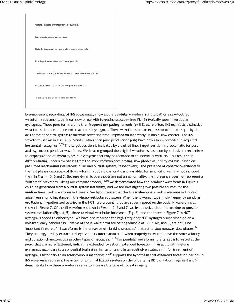
Abolished in sleep or inattention to visual tasks
Gaze-modulated, not gaze-evoked
Diminished (damped) by gaze-angle or convergence nulls
Superimposition of latent component possible
“Inversion” of the optokinetic reflex (actually, reversal of the IN)
Associated head oscillation (not compensatory) or turn
No oscillopsia except under rare conditions
Eye-movement recordings of INS occasionally show a pure pendular waveform (sinusoidal) or a saw-toothed
waveform (equiamplitude linear slow phase with foveating saccade) (see Fig. 8) typically seen in vestibular
nystagmus. These pure forms are neither frequent nor pathognomonic for INS. More often, INS manifests distinctive
waveforms that are not present in acquired nystagmus. These waveforms are an expression of the attempts by the
ocular motor control system to increase foveation time, imposed on inherently unstable slow control. The INS
waveforms shown in Figs. 4, 5, 6 and 7 (other than pure pendular or jerk) have never been recorded in acquired
horizontal nystagmus.8,33 The target position is indicated by a dashed line; target position is problematic for pure
and asymmetric pendular waveforms. We have regrouped the original waveforms based on hypothesized mechanisms
to emphasize the different types of nystagmus that may be recorded in an individual with INS. This resulted in
differentiating linear slow phases from the more common accelerating slow phases of jerk nystagmus, based on
presumed mechanisms (visual-vestibular and pursuit-system, respectively). The presence of dynamic overshoots in
the fast phases (saccades) of IN waveforms is both idiosyncratic and variable; for simplicity, we have not included
them in Figs. 4, 5, 6 and 7. Because dynamic overshoots are not an abnormality, their presence does not represent a
“different” waveform. Using our computer model,14,16 we demonstrated how the pendular waveforms in Figure 4
could be generated from a pursuit-system instability, and we are investigating two possible sources for the
unidirectional jerk waveforms in Figure 5. We hypothesize that the linear-slow-phase jerk waveforms in Figure 6
arise from a tonic imbalance in the visual-vestibular subsystem. When the low-amplitude, high-frequency pendular
oscillations, hypothesized to arise in the NOT, are present, they are superimposed on the basic IN waveforms as
shown in Figure 7. Of the 15 waveforms shown in Figs. 4, 5, 6 and 7, we hypothesize that nine are due to pursuit-
system oscillation (Figs. 4, 5), three to visual-vestibular imbalance (Fig. 6), and the three in Figure 7 to NOT
nystagmus added to either type. We have also recorded the high-frequency NOT nystagmus superimposed on a
low-frequency pendular IN. Twelve of these waveforms are pathognomonic of IN; P, AP, and JL are not. One
important feature of IN waveforms is the presence of “braking saccades” that act to stop runaway slow phases.34
They are triggered by extraretinal eye-velocity information and, when properly measured, have the same velocity
and duration characteristics as other types of saccades.35,36 For pendular waveforms, the target is foveated at the
peaks that are more flattened, indicating extended foveation. Extended foveation in an adult with lifelong
nystagmus secondary to a congenital brain stem hamartoma and in an adult given gabapentin for treatment of
nystagmus secondary to an arteriovenous malformation37 supports the hypothesis that extended foveation periods in
INS waveforms represent the action of a normal fixation system on the underlying INS oscillation. Figures 8 and 9
demonstrate how these waveforms serve to increase the time of foveal imaging.
Ovid: Duane's Ophthalmology http://ovidsp.tx.ovid.com.ezproxy.fiu.edu/spb/ovidweb.cgi
9 of 67 12/30/2008 7:23 AM

Fig. 4 Variations of pendular nystagmus due to pursuit-system instability: pure (P), asymmetric (AP), pendular with foveating saccades(PFS), pseudopendular (PP), and pseudopendular with foveating saccades (PPFS). Note that although foveating saccades vary in amplitude,
all achieve foveation (i.e., they all return the eyes to same point, the target). Foveation takes place at the peaks that are more flattened;here shown as the leftmost peaks. In this and Figures 5, 6, 7 and Figure 16, dashed lines indicate target position (see text). In all figures,upward deflections indicate rightward, upward, or clockwise eye movement (all eye movements are from the patient's perspective).
Ovid: Duane's Ophthalmology http://ovidsp.tx.ovid.com.ezproxy.fiu.edu/spb/ovidweb.cgi
10 of 67 12/30/2008 7:23 AM

Fig. 5 Unidirectional types of jerk nystagmus, mechanism unknown, including two with saccadic foveation (pure jerk and jerk withextended foveation) and two with slow eye movement (SEM) foveation (pseudocycloid and pseudojerk). The increasing velocity slowphases are common in IN. Note small and variable saccadic amplitude in pseudocycloid waveform and further reduction in pseudojerkwaveform.
Fig. 6 Unidirectional (jerk, J) and bidirectional (triangular, T and bidirectional jerk, BDJ) nystagmus with linear slow phases hypothesized tobe due to visual-vestibular imbalance. All saccades are in a corrective direction (i.e., toward target).
Fig. 7 Dual jerk nystagmus hypothesized to be comprised of NOT nystagmus superimposed on IN waveforms shown in Figures 5 and 6. Theresulting waveforms show sinusoidal modulation of the underlying slow eye movement off target. Depicted are dual jerk right withaccelerating slow phases (DJRA), dual jerk right with extended foveation (DJREF), and dual jerk right with linear slow phases (DJRL).
Ovid: Duane's Ophthalmology http://ovidsp.tx.ovid.com.ezproxy.fiu.edu/spb/ovidweb.cgi
11 of 67 12/30/2008 7:23 AM

Fig. 8 Foveation “strategy” employed during pendular (P) and jerk left (JL) nystagmus. The target is only briefly foveated at points 0, 2, andso forth. t, time scale.
Ovid: Duane's Ophthalmology http://ovidsp.tx.ovid.com.ezproxy.fiu.edu/spb/ovidweb.cgi
12 of 67 12/30/2008 7:23 AM
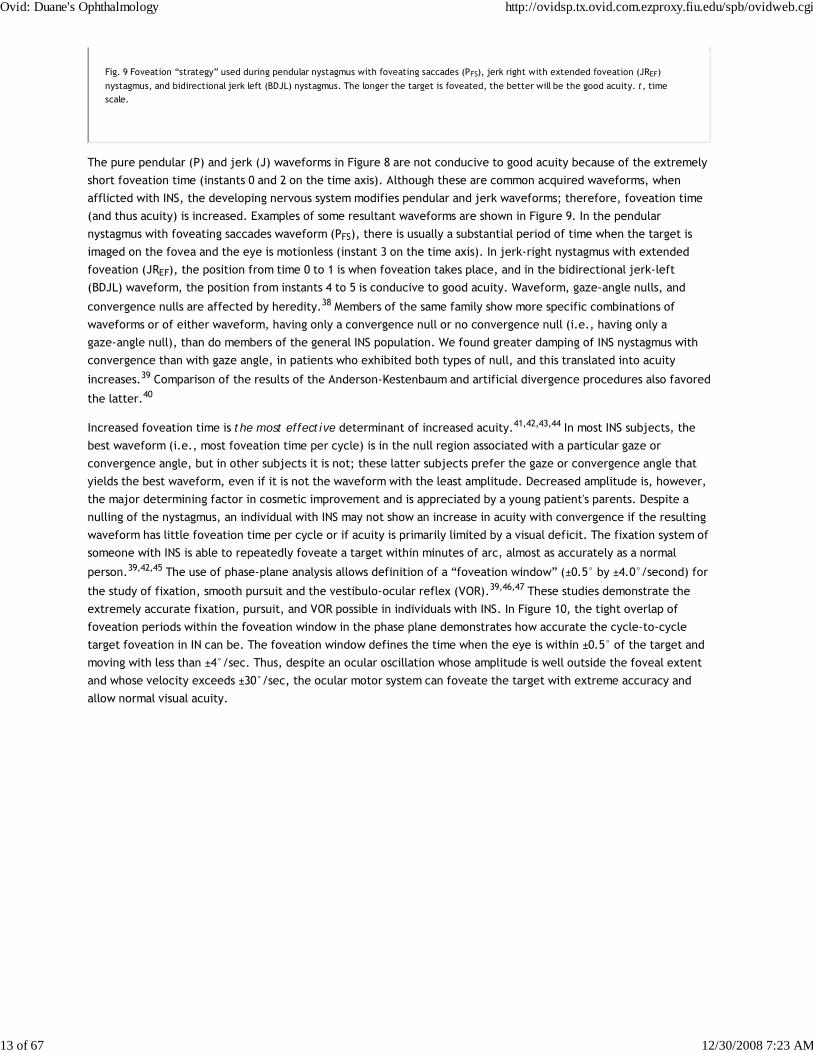
Fig. 9 Foveation “strategy” used during pendular nystagmus with foveating saccades (PFS), jerk right with extended foveation (JREF)
nystagmus, and bidirectional jerk left (BDJL) nystagmus. The longer the target is foveated, the better will be the good acuity. t, timescale.
The pure pendular (P) and jerk (J) waveforms in Figure 8 are not conducive to good acuity because of the extremely
short foveation time (instants 0 and 2 on the time axis). Although these are common acquired waveforms, when
afflicted with INS, the developing nervous system modifies pendular and jerk waveforms; therefore, foveation time
(and thus acuity) is increased. Examples of some resultant waveforms are shown in Figure 9. In the pendular
nystagmus with foveating saccades waveform (PFS), there is usually a substantial period of time when the target is
imaged on the fovea and the eye is motionless (instant 3 on the time axis). In jerk-right nystagmus with extended
foveation (JREF), the position from time 0 to 1 is when foveation takes place, and in the bidirectional jerk-left
(BDJL) waveform, the position from instants 4 to 5 is conducive to good acuity. Waveform, gaze-angle nulls, and
convergence nulls are affected by heredity.38 Members of the same family show more specific combinations of
waveforms or of either waveform, having only a convergence null or no convergence null (i.e., having only a
gaze-angle null), than do members of the general INS population. We found greater damping of INS nystagmus with
convergence than with gaze angle, in patients who exhibited both types of null, and this translated into acuity
increases.39 Comparison of the results of the Anderson-Kestenbaum and artificial divergence procedures also favored
the latter.40
Increased foveation time is the most effective determinant of increased acuity.41,42,43,44 In most INS subjects, the
best waveform (i.e., most foveation time per cycle) is in the null region associated with a particular gaze or
convergence angle, but in other subjects it is not; these latter subjects prefer the gaze or convergence angle that
yields the best waveform, even if it is not the waveform with the least amplitude. Decreased amplitude is, however,
the major determining factor in cosmetic improvement and is appreciated by a young patient's parents. Despite a
nulling of the nystagmus, an individual with INS may not show an increase in acuity with convergence if the resulting
waveform has little foveation time per cycle or if acuity is primarily limited by a visual deficit. The fixation system of
someone with INS is able to repeatedly foveate a target within minutes of arc, almost as accurately as a normal
person.39,42,45 The use of phase-plane analysis allows definition of a “foveation window” (±0.5° by ±4.0°/second) for
the study of fixation, smooth pursuit and the vestibulo-ocular reflex (VOR).39,46,47 These studies demonstrate the
extremely accurate fixation, pursuit, and VOR possible in individuals with INS. In Figure 10, the tight overlap of
foveation periods within the foveation window in the phase plane demonstrates how accurate the cycle-to-cycle
target foveation in IN can be. The foveation window defines the time when the eye is within ±0.5° of the target and
moving with less than ±4°/sec. Thus, despite an ocular oscillation whose amplitude is well outside the foveal extent
and whose velocity exceeds ±30°/sec, the ocular motor system can foveate the target with extreme accuracy and
allow normal visual acuity.
Ovid: Duane's Ophthalmology http://ovidsp.tx.ovid.com.ezproxy.fiu.edu/spb/ovidweb.cgi
13 of 67 12/30/2008 7:23 AM
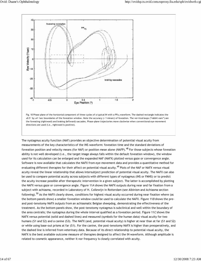
Fig. 10 Phase plane of the horizontal component of three cycles of a typical IN with a PPFS waveform. The dashed rectangle indicates the
±0.5° by ±4°/sec boundaries of the foveation window. Note the accuracy (< 1 minarc) of foveation. The ver-tical loops (“rabbit ears”) arethe foveating (rightward) and braking (leftward) saccades. Phase-plane trajectories move clockwise when conventional eye-movementdirections are used (i.e., rightward is positive).
The nystagmus acuity function (NAF) provides an objective determination of potential visual acuity from
measurements of the key characteristics of the INS waveform: foveation time and the standard deviations of
foveation position and velocity means (for NAF) or position mean alone (NAFP).48 For those subjects whose foveation
ability is not well developed (i.e., the target image always falls within the default foveation window), the window
used for its calculation can be enlarged and the expanded NAF (NAFX) plotted versus gaze or convergence angle.
Software is now available that calculates the NAFX from eye-movement data and provides a quantitative method for
evaluating different therapies for their affect on potential visual acuity.49 Plots of the NAF or NAFX versus visual
acuity reveal the linear relationship that allows intersubject prediction of potential visual acuity. The NAFX can also
be used to compare potential acuity across subjects with different types of nystagmus (INS or FMNS) or to predict
the acuity increase possible after therapeutic intervention in a given subject. The latter is accomplished by plotting
the NAFX versus gaze or convergence angle. Figure 11A shows the NAFX outputs during near and far fixation from a
subject with achiasma, recorded in Laboratory of H. Collewijn in Rotterdam (see Albinism and Achiasma section
following).50 As the NAFX clearly shows, conditions for highest visual acuity occurred during near fixation where (as
the bottom panels show) a smaller foveation window could be used to calculate the NAFX. Figure 11B shows the pre-
and post-tenotomy NAFX outputs from an achiasmatic Belgian sheepdog, demonstrating the effectiveness of the
treatment. As the bottom panels show, the post-tenotomy nystagmus is subclinical and well within the boundary of
the area centralis; the nystagmus during the whole interval qualified as a foveation period. Figure 11C shows the
NAFX versus potential (solid and dashed lines) and measured (symbols for the human data) visual acuity for two
humans (S1 and S2) and a canine (S3). The NAFX (and, potential visual acuity) is higher at near than at far (S1 and S2)
or while using base-out prisms at far (S1). For the canine, the post-tenotomy NAFX is higher than preoperatively, and
the dashed line is inferred from veterinary data. Because of its direct relationship to potential visual acuity, the
NAFX is the best available outcome measure of therapies designed to affect the IN waveform. Although amplitude is
related to cosmetic appearance, neither it nor frequency is closely correlated with acuity.
Ovid: Duane's Ophthalmology http://ovidsp.tx.ovid.com.ezproxy.fiu.edu/spb/ovidweb.cgi
14 of 67 12/30/2008 7:23 AM
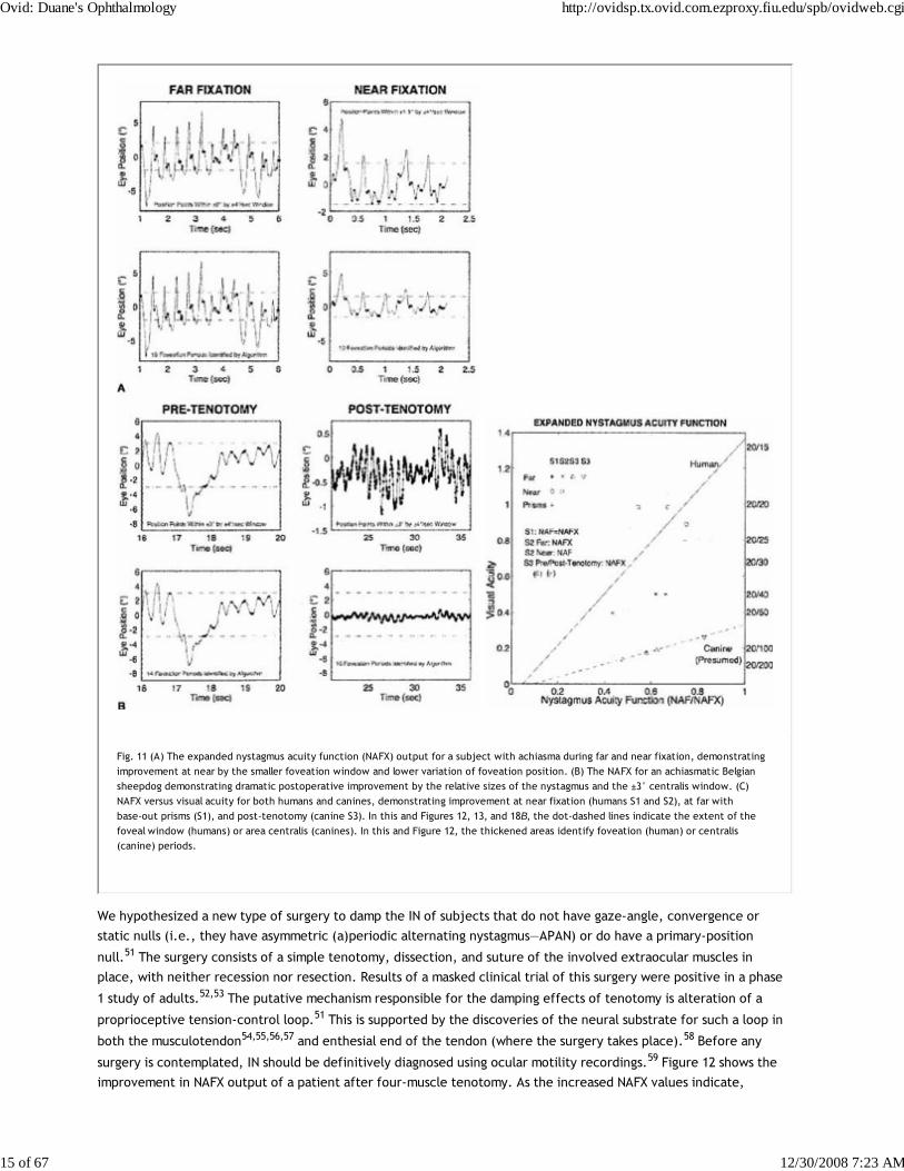
Fig. 11 (A) The expanded nystagmus acuity function (NAFX) output for a subject with achiasma during far and near fixation, demonstratingimprovement at near by the smaller foveation window and lower variation of foveation position. (B) The NAFX for an achiasmatic Belgiansheepdog demonstrating dramatic postoperative improvement by the relative sizes of the nystagmus and the ±3° centralis window. (C)NAFX versus visual acuity for both humans and canines, demonstrating improvement at near fixation (humans S1 and S2), at far withbase-out prisms (S1), and post-tenotomy (canine S3). In this and Figures 12, 13, and 18B, the dot-dashed lines indicate the extent of thefoveal window (humans) or area centralis (canines). In this and Figure 12, the thickened areas identify foveation (human) or centralis(canine) periods.
We hypothesized a new type of surgery to damp the IN of subjects that do not have gaze-angle, convergence or
static nulls (i.e., they have asymmetric (a)periodic alternating nystagmus—APAN) or do have a primary-position
null.51 The surgery consists of a simple tenotomy, dissection, and suture of the involved extraocular muscles in
place, with neither recession nor resection. Results of a masked clinical trial of this surgery were positive in a phase
1 study of adults.52,53 The putative mechanism responsible for the damping effects of tenotomy is alteration of a
proprioceptive tension-control loop.51 This is supported by the discoveries of the neural substrate for such a loop in
both the musculotendon54,55,56,57 and enthesial end of the tendon (where the surgery takes place).58 Before any
surgery is contemplated, IN should be definitively diagnosed using ocular motility recordings.59 Figure 12 shows the
improvement in NAFX output of a patient after four-muscle tenotomy. As the increased NAFX values indicate,
Ovid: Duane's Ophthalmology http://ovidsp.tx.ovid.com.ezproxy.fiu.edu/spb/ovidweb.cgi
15 of 67 12/30/2008 7:23 AM

tenotomy damped the IN and improved the waveform. Both the foveation time per cycle and the mean foveation
position improved and the post-tenotomy foveation window was smaller in both position and velocity. The various
therapies available for INS, based on the presence or absence of gaze and convergence nulls, is summarized in Table
3. Note that for patients with both convergence and gaze-angle nulls, exploitation of the former (surgically or with
vergence prisms) usually damps the nystagmus and increases acuity most; it is necessary to add -1.00 S (OU) to
vergence prisms for prepresbyopic patients. Preliminary studies (at all gaze angles) of the broad damping effects of
convergence on IN (see Fig. 14 and discussion of convergence following) suggest that composite prisms (unequal
base-out) are not necessary in these cases; vergence prisms (equal base-out) will achieve the same damping.60 As
indicated in Table 3, regardless of the presence of nulls, afferent stimulation can be used in all patients who exhibit
nystagmus damping with active stimulation (see later discussion).
Fig. 12 Comparison of NAFX outputs in a patient who underwent a four-muscle tenotomy procedure. Both the foveation time per cycle andaccuracy of foveations (smaller foveation window) improved, resulting in the 67% higher NAFX value.
Ovid: Duane's Ophthalmology http://ovidsp.tx.ovid.com.ezproxy.fiu.edu/spb/ovidweb.cgi
16 of 67 12/30/2008 7:23 AM

Fig. 14 NAFX versus gaze angle and fitted polynomial curves for both far and near fixation with best potential visual acuities indicated. Notethe dramatic increase with convergence and the broader range of gaze angles with higher potential acuities.
TABLE 3. Therapies for Infantile Nystagmus
If the IN nulls ONLY with lateral gaze:
Resection and recession (four-muscle)
Version prisms
Afferent stimulation (passive or active)
If the IN nulls ONLY with convergence:
Bimedial recession1 (artificial divergence) plus bilateral tenotomy
7D BO vergence prisms with (1.00 S1 (OU)
Ovid: Duane's Ophthalmology http://ovidsp.tx.ovid.com.ezproxy.fiu.edu/spb/ovidweb.cgi
17 of 67 12/30/2008 7:23 AM

Afferent stimulation (passive or active)
If the IN nulls with BOTH lateral gaze and convergence:
Bimedial recession1 plus bilateral tenotomy or combined with resection and recession
7D BO vergence prisms with (1.00 S1 (OU)
Afferent stimulation (passive or active)
If the IN nulls with NEITHER lateral gaze nor convergence or is asymmetric aperiodic alternating IN:
Four-muscle tenotomy, dissection and suture
Maximal recession (Four-muscle)*
Afferent stimulation (passive or active)
1Damps IN only for nonstrabismic, binocular patients.
*This surgery should be replaced by the four-muscle tenotomy procedure that does not carry the risk of inducing diplopia.
The so-called inversion of the optokinetic reflex seems to occur only with INS.61 When optokinetic stimuli are
presented to a patient with INS, a peculiar phenomenon may occur: the resulting nystagmus may be opposite in
direction from what would be anticipated if the evoked optokinetic nystagmus (OKN) simply summated with the
ongoing nystagmus. For example, in the presence of left-beating INS nystagmus, the response to right-going
optokinetic targets (a leftward fast phase) should add to the left-beating INS nystagmus to produce enhancement of
the nystagmus intensity. In “inversion,” the nystagmus may either damp or be converted to right-beating nystagmus.
If right-going targets are presented at a gaze angle at which the nystagmus is either absent or pendular, a right-
beating nystagmus may result. Inversion of the optokinetic reflex is present in 67% of INS patients. The observation
of optokinetic inversion establishes the nystagmus as IN. The phenomenon is, in reality, merely a reversal of the INS
nystagmus direction due to a null shift; it is not a true inversion of the optokinetic response (see discussion of
reversed pursuit later). The basic function of the optokinetic system is to stabilize slowly moving retinal images, but
the rapidly moving retina of an INS patient may interfere with this function. The optokinetic response appears
suppressed in some patients; however, the perceived circularvection is in the proper direction, and OKN dynamics
appear to be normal in individuals with INS.
The head oscillations that often accompany INS increase with visual intent and have traditionally been regarded as
compensatory. For compensation to be achieved, head movements would have to be equal in amplitude and opposite
in direction to the eye movements. For such a mechanism to work, the VOR would have to be totally inhibited (gain
reduced to 0). Accurate objective observations of the head movements in patients with INS do not support that
hypothesis.62 Rather, the head oscillation is merely an extension of the motor instability, and the VOR functions
normally to cancel the effects of head oscillation during the periods of target foveation normally present in the INS
waveform.47 The head tremor in INS can be distinguished from that in acquired disease; it is easily suppressed
voluntarily in the former but not in the latter.
Point out the head tremor to the patient. If it stops, the nystagmus is IN; if it persists, both
Ovid: Duane's Ophthalmology http://ovidsp.tx.ovid.com.ezproxy.fiu.edu/spb/ovidweb.cgi
18 of 67 12/30/2008 7:23 AM
are acquired.
Individuals with INS usually do not experience an illusory oscillatory movement of their environment
(oscillopsia).63,64 This lack of oscillopsia in INS, and also in FMNS, suggests that both oscillations occur within an
efference copy feedback loop that serves to nullify the effects of retinal-image oscillation induced by either of
these instabilities.65 Like most ocular oscillations (myoclonus being the exception), INS nystagmus disappears in
sleep. In two patients with INS plus an acquired nystagmus, their acquired oscillopsia seemed to be related to an
inability to maintain repeatable periods of good foveation in a particular plane.66,67 However, that inability was an
epiphenomenon caused by the addition of a transitory acquired nystagmus to the ever-present INS nystagmus.65
Oscillopsia suppression in INS and other types of nystagmus appears to be accomplished by efference copy of the
nystagmus signal.65,68,69,70,71,72 Oscillopsia may occur in some patients with very poor foveation stability73 or may
occur in later life secondary to afferent deficits.74 The thresholds for motion detection in INS differ from normal and
may also play a role in oscillopsia suppression.75,76 We reported oscillopsia of a migraine aura in an individual with
INS,77 as well as vertical oscillopsia secondary to a decompensated phoria; the latter event led to the discovery of
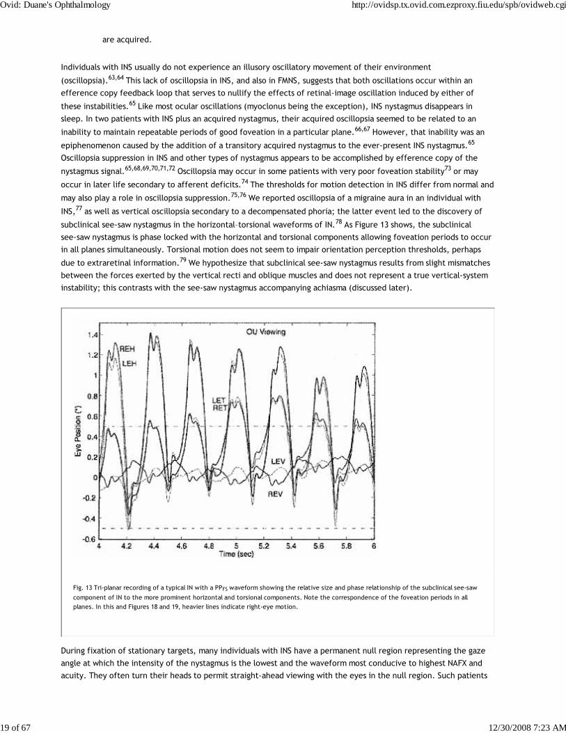
subclinical see-saw nystagmus in the horizontal–torsional waveforms of IN.78 As Figure 13 shows, the subclinical
see-saw nystagmus is phase locked with the horizontal and torsional components allowing foveation periods to occur
in all planes simultaneously. Torsional motion does not seem to impair orientation perception thresholds, perhaps
due to extraretinal information.79 We hypothesize that subclinical see-saw nystagmus results from slight mismatches
between the forces exerted by the vertical recti and oblique muscles and does not represent a true vertical-system
instability; this contrasts with the see-saw nystagmus accompanying achiasma (discussed later).
Fig. 13 Tri-planar recording of a typical IN with a PPFS waveform showing the relative size and phase relationship of the subclinical see-saw
component of IN to the more prominent horizontal and torsional components. Note the correspondence of the foveation periods in allplanes. In this and Figures 18 and 19, heavier lines indicate right-eye motion.
During fixation of stationary targets, many individuals with INS have a permanent null region representing the gaze
angle at which the intensity of the nystagmus is the lowest and the waveform most conducive to highest NAFX and
acuity. They often turn their heads to permit straight-ahead viewing with the eyes in the null region. Such patients
Ovid: Duane's Ophthalmology http://ovidsp.tx.ovid.com.ezproxy.fiu.edu/spb/ovidweb.cgi
19 of 67 12/30/2008 7:23 AM

benefit from appropriate version prism spectacles that alleviate the necessity for the head turn and the resulting
increased fixation attempt.25,41 However, as Figure 14 shows, if the IN damps with convergence, the higher NAFX
values will persist over a broader range of gaze angles than during fixation on a far target. This allows higher acuity
over most useful gaze angles and demonstrates the advantage of either base-out prisms or the bimedial rectus
recession procedure over therapies aimed at moving a gaze-angle null to primary position.
Some INS patients may exhibit a superimposed latent component that induces null shifts toward an eye that is
covered (Fig. 15).80 Demonstration of such a shift and maintenance of any of the INS waveforms establish the
nystagmus as belonging to INS rather than the FMNS (see later discussion). Rarely, a null shift is toward the viewing
eye.25
Fig. 15 Depiction of shifts of neutral zone or null (N) in congenital nystagmus. Tracing demonstrates an idealized nystagmus pattern withboth eyes open (OU). Neutral zone extends over several degrees on either side of 0°. When gaze is directed laterally, nystagmus ofincreasing amplitude develops with fast phase in direction of gaze. Occlusion of right eye (RE) shifts zone to the right; at 0° there isleft-beating nystagmus. Occlusion of left eye (LE) shifts zone to the left; at 0° there is right-beating nystagmus.
Some studies of INS and smooth pursuit have led to confusion between the reversal of INS direction that may occur
during pursuit and reversed pursuit. This confusion is similar to that discussed earlier for the optokinetic response.
Accurate eye-movement recordings show that neither the optokinetic nor the pursuit subsystem responds in a
reversed manner, as should be obvious both by the absence of any symptoms of such a grave deficit and the normal
abilities of individuals with INS in sports. Also, their perceptions of both the direction and magnitude of movements
in the periphery and on the fovea are normal. Just as the INS waveform is distorted by SEM (creating periods of
extended foveation) during fixation of a stationary target, the pursuit system is able to generate pursuit movements
with a direction and velocity that match those of a moving target during these same periods of the INS
waveform.46,68,81 This ensures extended foveation of the moving target and results in accurate smooth pursuit during
the periods when the target image is on the fovea. Pursuit during foveation is all that is necessary for good acuity;
the same conditions are met during smooth pursuit as are met during fixation of a stationary target. During smooth
pursuit (or during optokinetic or VOR stimuli) the gaze angle at which the INS null region occurs shifts in the
direction opposite to the pursuit (optokinetic grating or VOR-induced eye motion).46,47 The amount of null shift is a
function of the pursuit or VOR velocity. This measurable shift in the INS null angle causes the INS nystagmus reversal
that has been mistakenly equated with “reversed” responses of both the optokinetic and pursuit subsystems.
In many individuals with INS, afferent stimulation of the ophthalmic division of the trigeminal nerve or of the neck
may damp the nystagmus and improve the waveform, allowing increased visual acuity.48,82 Neck or forehead
vibration prolonged foveation periods, yielding higher values of the NAF and improved visual acuity in 9 of 13
patients with INS.48 This noninvasive and benign therapy (active afferent stimulation) may prove useful in both INS
Ovid: Duane's Ophthalmology http://ovidsp.tx.ovid.com.ezproxy.fiu.edu/spb/ovidweb.cgi
20 of 67 12/30/2008 7:23 AM
and acquired nystagmus. The use of soft contact lenses to improve the acuity of individuals with INS takes advantage
of the damping effect on nystagmus of (passive) afferent stimulation.83,84,85,86
Soft contact lenses are not contraindicated in INS and can provide better acuity than
spectacles in patients whose nystagmus damps with afferent stimulation. Plano soft contact
lenses can be used if no refractive correction is required.
Both surgical and nonsurgical treatments for INS have been reviewed elsewhere.3,87 Relatives of individuals with INS
may have saccadic instabilities,88 and carriers of blue-cone monochromatism may have vertical (upbeat and
downbeat) nystagmus and FMNS.89
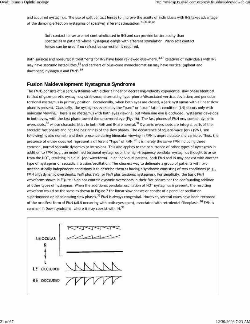
Fusion Maldevelopment Nystagmus SyndromeThe FMNS consists of: a jerk nystagmus with either a linear or decreasing-velocity exponential slow phase identical
to that of gaze-paretic nystagmus; strabismus; alternating hyperphoria/dissociated vertical deviation; and pendular
torsional nystagmus in primary position. Occasionally, when both eyes are closed, a jerk nystagmus with a linear slow
phase is present. Classically, the nystagmus evoked by the “pure” or “true” latent condition (LN) occurs only with
uniocular viewing. There is no nystagmus with both eyes viewing, but when one eye is occluded, nystagmus develops
in both eyes, with the fast phase toward the uncovered eye (Fig. 16). The fast phases of FMN may contain dynamic
overshoots,90 whose characteristics in both FMN and IN are normal.91 Dynamic overshoots are integral parts of the
saccadic fast phases and not the beginnings of the slow phases. The occurrence of square-wave jerks (SWJ, see
following) is also normal, and their presence during binocular viewing in FMN is unpredictable and variable. Thus, the
presence of either does not represent a different “type” of FMN;92 it is merely the same FMN including these
common, normal saccadic dynamics or intrusions. This also applies to the occurrence of other types of nystagmus in
addition to FMN (e.g., an undefined torsional nystagmus or the high-frequency pendular nystagmus thought to arise
from the NOT, resulting in a dual-jerk waveform). In an individual patient, both FMN and IN may coexist with another
type of nystagmus or saccadic intrusion/oscillation. The clearest way to delineate a group of patients with two
mechanistically independent conditions is to describe them as having a syndrome consisting of two conditions (e.g.,
FMN with dynamic overshoots, FMN plus SWJ, or FMN plus torsional nystagmus). For simplicity, the basic FMN
waveforms shown in Figure 16 do not contain dynamic overshoots in their fast phases nor the confounding addition
of other types of nystagmus. When the additional pendular oscillation of NOT nystagmus is present, the resulting
waveform would be the same as shown in Figure 7 for linear slow phases or consist of a pendular oscillation
superimposed on decelerating slow phases.38 FMN is always congenital. However, several cases have been recorded
of the manifest form of FMN (MLN occurring with both eyes open), associated with retrolental fibroplasia.90 FMN is
common in Down syndrome, where it may coexist with IN.93
Ovid: Duane's Ophthalmology http://ovidsp.tx.ovid.com.ezproxy.fiu.edu/spb/ovidweb.cgi
21 of 67 12/30/2008 7:23 AM
Fig. 16 Fusion maldevelopment nystagmus (FMN). With both eyes open there is either low-amplitude nystagmus (when only one eye isfixating) or, rarely, no nystagmus (when both eyes are fixating). Closure of either eye results in jerk nystagmus with fast phases toward theviewing (unoccluded) eye. When both eyes are open, the nystagmus fast phases are toward the fixating eye. Slow phases may be eitherlinear (usually when both eyes are open) or decreasing-velocity exponentials (usually upon occlusion of one eye), unlike those of IN. Notethat the fast phases may be foveating (for low-amplitude FMN with linear slow phases) or defoveating (for the higher amplitude FMN withdecreasing velocity slow phases).
Early theories postulated that a unilateral retinal stimulus was the necessary condition for FMN, but this concept was
discounted by observations of FMN in monocular fixation with a blind eye or with an acoustic stimulus in complete
darkness. Similarly, the hypothesis that the FMNS is caused by nasal-temporal asymmetries in the optokinetic reflex
is not supported by evidence that subjects with FMNS are able to use retinal slip information to adapt motion-
detection sensitivities94 and are able to pursue symmetrically.95 Also, because nasal-temporal asymmetries exist in
individuals with strabismus but not FMN,94 this cannot be the primary causal factor in the genesis of the nystagmus.
Asymmetries in the monocular optokinetic response of monkeys deprived of binocular input early in life may result
from, rather than cause, their nystagmus. In normal monkeys, each nucleus of the optic tract (NOT) is driven
binocularly; in these monkeys, they are driven by the contralateral eye.96 Although the resulting imbalance may
provide the tonic signal that produces the FMNS slow phases (inactivation of the NOT with muscimol abolishes the
nystagmus), the cause of the imbalance appears to lie in higher centers. The spontaneous reversal of FMN in the dark
has led to the speculation that eye dominance is predetermined.97,98 Shallo-Hoffmann et al. identified an alternating
vertical component to FMN,99 and Brodsky linked the genesis of FMN and dissociated vertical divergence to the dorsal
light reflex present in many animals.100
We relate the FMNS to the cortical switching that must occur in the calculation of egocentric direction when going
from binocular to monocular viewing.90 Under binocular conditions, the gaze angle of each eye is summed with the
other and divided by two to obtain the egocentric direction, referenced to the “cyclopean eye.” However, with
monocular viewing, egocentric direction depends only on the viewing eye, and the cortical operation of summing
and dividing by two must be altered to process unchanged information from the viewing eye. The shift in egocentric
direction toward the nonviewing eye causes the slow drift of the eyes in that direction. Both eyes are then
corrected by a saccade in the direction of the viewing eye, which brings the eyes to the target (or, in darkness, to
the intended gaze angle). This contention is supported by unilateral strabismus surgery causing central effects on
egocentric localization.101 Thus, FMNS nystagmus may be generated by this inability to properly alter the cortical
mathematical operation normally used to define egocentric direction (i.e., this deficit in higher centers may result
in a tonic imbalance in the visual-vestibular subsystem, producing the linear slow phases of FMN).
The shift to monocular egocentric localization can also produce a mode whereby the saccadic system generates
defoveating saccades that momentarily carry the fixating eye past the target in a temporal direction, followed by a
decelerating-velocity nasal drift back toward the target.102 This results from generating a pulse, but not a step, of
innervation to drive the fast phases of the FMNS nystagmus. Presumably, the common neural integrator is kept from
integrating these defoveating pulses by the signal representing the correct eye position vis-à-vis the target. These
hypotheses have been combined in an ocular motor system model (see Chapter 9) capable of simulating responses of
an individual with FMNS.15,103,104
FMNS nystagmus occurs in patients with strabismus who, although viewing with both eyes open, are fixing
monocularly. The slow phases are either linear or decelerating, and the fast phases are always in the direction of the
viewing eye.90 The nystagmus of patients with strabismus, alternating fixation, and FMNS nystagmus with both eyes
open has fast phases always in the direction of the fixating eye. Such patients may be easily misdiagnosed as having
INS, because the nystagmus is present with both eyes open. Recordings are required to document the decelerating
or linear slow-phase waveforms characteristic of FMNS from the accelerating slow phases predominant in the INS.
Strabismus is a necessary (but not sufficient) condition for FMN.105 That is, all individuals with FMN have strabismus,
consisting of a phoria under cover and a tropia with both eyes open, if nystagmus is present under these respective
conditions. Conversely, FMNS is not significantly associated with early-onset strabismus.106 Rarely, on occlusion of a
Ovid: Duane's Ophthalmology http://ovidsp.tx.ovid.com.ezproxy.fiu.edu/spb/ovidweb.cgi
22 of 67 12/30/2008 7:23 AM
preferred eye, during which fixation with an amblyopic eye is forced, both eyes drift in the direction of the covered
eye without corrections by fast phases; this is called latent deviation. Early surgical correction of infantile
strabismus may convert the nystagmus of FMNS present with both eyes open (the manifest condition) to nystagmus
present only upon occlusion of one eye (the latent condition),107 thereby supporting a previous hypothesis.105
Because the good acuity of INS patients is related to the long, postsaccadic foveation periods of many waveforms, it
was difficult to explain the equally good acuity of FMNS patients, given the absence of such periods. However,
accurate studies of FMNS foveation revealed a dual strategy.102 During the low-amplitude, linear-slow-phase FMNS
waveform, the saccadic fast phases foveate the target, and the low-velocity slow phases take the eye away from the
target with little effect on acuity. During the higher amplitude, decelerating slow-phase FMNS waveform, the
saccadic fast phases defoveate the target, allowing foveation during the low-velocity, tail ends of the slow phases
(see Fig. 16); this ensures the best acuity possible.
Although most patients have nystagmus from either the INS or the FMNS, some have both; three unambiguous patient
groups have been identified: INS, FMNS, and INS + FMNS.38,108,109 The three groups exhibit different clinical signs and
relations to strabismus; most INS patients do not have strabismus, but all FMNS patients do. Thus, INS and FMNS are
specific, easily differentiated syndromes and do not, as has been suggested,110 represent a unitary disorder with a
broad spectrum of expression. Because no acquired, time-independent, primary-position jerk nystagmus reverses
direction with alternate eye cover, a simple reverse-cover test can be a powerful clinical tool.
To distinguish between benign, infantile, primary-position, jerk nystagmus, and that which is
acquired and symptomatic, first verify that there is no periodic alternation in direction and
then perform a reverse-cover test. If the cover test causes a reversal in the nystagmus
direction consistent with FMNS, the nystagmus is benign (FMNS or INS with a latent
component). If not, attempt to rule out INS (by history, clinical signs [see Table 2], and
waveforms).
Nystagmus Blockage SyndromeThe nystagmus blockage syndrome (NBS) is both a poorly understood and an overdiagnosed phenomenon related to
INS. As the name suggests, the nystagmus of these patients diminishes or disappears with the act of willed esotropia
while fixating a distant target. This should not be confused with the damping of INS nystagmus during convergence
on a near target. There are two mechanisms by which blockage of the ongoing nystagmus can be accomplished.111
During the willed esotropia, some INS nystagmus merely damps or stops, in much the same way as with true
convergence. In the second type of NBS, the INS waveform converts to a FMNS waveform with the onset of the
strabismus. Normally, the substitution of the FMNS slow phases for the INS waveforms that allow for better foveation
would not be advantageous. However, in these few patients, the small FMNS amplitude results in better acuity than
the larger INS amplitude. NBS is often misdiagnosed in FMNS patients with a strong Alexander's law variation of their
nystagmus, which causes them to fixate with their adducting eye.111
Spasmus Nutans SyndromeSpasmus nutans syndrome (SNS) is a rare constellation of ocular oscillation, head nodding, and torticollis that begins
in infancy (usually between 4 and 18 months of age) and disappears clinically in childhood (usually before 3 years of
age). The nystagmus is generally bilateral (but can differ in each eye and may even be strictly monocular), and it
oscillates in horizontal, torsional, or vertical directions. Hoyt reported an instance of SNS presenting with monocular
nystagmus in monozygous twins.112
The nystagmus tends to be asymmetric in the two eyes, to vary in different directions of gaze, and to be rapid and of
small amplitude. The head nodding is inconstant and irregular and can be horizontal, vertical, or both. The average
duration of SNS is 12 to 24 months; rarely, it lasts a number of years. Studies of quantitative head- and
eye-movement recordings indicate that the head movement may, using the normal VOR, actually serve to abolish the
eye movements.113 In some patients, it may be only compensatory with suppression of the VOR. Compare this to INS,
Ovid: Duane's Ophthalmology http://ovidsp.tx.ovid.com.ezproxy.fiu.edu/spb/ovidweb.cgi
23 of 67 12/30/2008 7:23 AM
where the head oscillation is an extension of the nystagmus and the VOR is normal (see preceding discussion).
The pendular oscillation of SNS is characterized by a variable phase difference between the oscillations of each
eye.114 These phase differences can appear from minute to minute and during the child's development. The
dissociated nystagmus is usually of a higher frequency than INS nystagmus, and the result can be disjugate,
conjugate, or uniocular. We hypothesize that SNS reflects a yoking abnormality, perhaps due to delayed
development. Recordings show that SNS nystagmus may not disappear completely but may recede to a subclinical
level; neither INS nor FMNS nystagmus disappears with age.
Acquired
SECONDARY TO VISUAL LOSS.Nystagmus occurring in early childhood consequent to progressive bilateral visual loss should not be classified as INS
unless INS waveforms are documented. The conceptual problems in the classification were discussed earlier. Usually,
nystagmus secondary to visual loss cannot be distinguished from INS in a patient with coexisting primary visual
abnormalities.
The nystagmus associated with rod monochromacy (complete congenital achromatopsia) is said to be distinguishable
from other forms of nystagmus of infancy on the basis of slow buildup of the slow component velocity of OKN. This
occurs during monocular stimulation with directional asymmetry of OKN when the temporal-to-nasal direction is
compared with the nasal-to-temporal direction.115 Patients with blindness from birth and nystagmus may have an
impaired VOR and an inability to initiate saccades voluntarily, despite the presence of quick phases of nystagmus.116
Adults with “eye movements of the blind” may exhibit features similar to those of patients with cerebellar disease116
or may exhibit jerk nystagmus, see-saw nystagmus, or a combination of oscillations. Cats reared from birth in
stroboscopic illumination develop low-amplitude nystagmus; this is believed to be an animal model for nystagmus
secondary to visual loss.117
Monocular visual loss may produce monocular nystagmus, usually vertical, at any age from birth through adult life.
That the nystagmus is monocular and usually vertical makes it distinguishable from INS, but it may mimic the
nystagmus of spasmus nutans, particularly if there is associated head nodding.
SPASMUS NUTANS.The nystagmus of the benign spasmus nutans syndrome may sometimes be mimicked by tumors of the optic nerve,
chiasm, or third ventricle,118 although both the true SNS nystagmus and that secondary to tumor have been called
spasmus nutans, we prefer to limit that term to the benign condition. Any child with nystagmus resembling SNS
nystagmus should have brain imaging if tumor cannot be ruled out by other clinical signs. Retinal disease may mimic
the clinical signs of the SNS,119 as may opsoclonus-myoclonus.120
ACQUIRED PENDULAR NYSTAGMUS (ADULTS)Acquired pendular nystagmus may reflect brain stem or cerebellar dysfunction, or both. It occurs in patients with
vascular or demyelinating disease. In the latter, it has been regarded as a sign of cerebellar nuclear lesions. The
nystagmus is multivectorial (i.e., horizontal, vertical, diagonal, elliptic, or circular) and usually is associated with a
head tremor. Marked dissociation between the two eyes often exists and may not correlate with differences in visual
acuity from coexisting optic neuropathy.121 Despite the dissociation, the oscillations of the two eyes in a patient
with MS are phase-locked, even though they may differ in their frequencies.122 Das et al. postulated a neural-net
model for acquired pendular nystagmus in MS; the model duplicated the resetting effect of saccades on the
oscillation.123 Acquired pendular nystagmus also occurs in an autosomal peroxisomal disorder.124 Gabapentin is
effective in treating some forms of acquired pendular nystagmus.124,125,126 Averbuch-Heller et al. published a
comprehensive review of the pathogenesis of acquired pendular nystagmus in 1995.127
Rarely, acquired pendular nystagmus in the adult becomes manifest with acquired amblyopia, as mentioned earlier.
Ovid: Duane's Ophthalmology http://ovidsp.tx.ovid.com.ezproxy.fiu.edu/spb/ovidweb.cgi
24 of 67 12/30/2008 7:23 AM

Scopolamine may be an effective treatment,128 but botulinum toxin is of limited efficacy in treating acquired
pendular nystagmus.129 A review of current therapeutic approaches to various types of nystagmus and saccadic
oscillations, based on known physiology and pharmacology, points out the need for more precise, double-blind
studies.130
Miner's nystagmus is a rarity limited presumably to mine workers in the United Kingdom. This historical anachronism
was described as a small-amplitude, horizontal, and vertical nystagmus that is often more pronounced in upward
gaze. The pathogenesis of this putative dysfunction is uncertain, but functional contamination with voluntary
“nystagmus” is suspected; a secondary gain setting is usually present.
Except for the possible dissociation between the two eyes, acquired pendular nystagmus may be similar to a
pendular INS waveform; both can have associated head tremor and characteristically damp with eyelid closure.
Studies into the pathogenesis of acquired pendular nystagmus have ruled out delayed visual feedback and increased
gain in the visually enhanced VOR as causal factors.127
ACQUIRED HORIZONTAL JERK NYSTAGMUS
VestibularWe generally delimit vestibular nystagmus as being consequent to dysfunction of the vestibular end-organ, nerve, or
nuclear complex within the brain stem. It is a horizontal-torsional or purely horizontal, primary-position jerk
nystagmus with a linear slow phase. The nystagmus intensity increases with gaze toward the fast phase (obeying
Alexander's law); it decreases and, with central lesions, may reverse directions on gaze toward the direction of the
slow phase. The symptom of vertigo usually coexists. As might be expected, acute lesions of the cerebellar flocculus
(the vestibulocerebellum) can produce a similar nystagmus (see Chapter 10). For practical clinical purposes, the
responsible lesion in vestibular nystagmus is located in either the end-organ, nerve, or brain stem. Such localization
requires an appreciation of the manifestations of end-organ dysfunction. In normal subjects, some degree of
nystagmus and vertigo develops when the labyrinth (end-organ) is stimulated with warm or cold water applied to the
tympanic membrane. The direction of the resulting nystagmus, in terms of the fast (jerk) phase, can be remembered
by the mnemonic “COWS” (Cold, Opposite; Warm, Same). Cold water in the left ear (or warm water in the right)
induces a right-beating nystagmus; cold water in the right ear (or warm water in the left) induces a left-beating
nystagmus. In addition, the subject experiences vertigo and, with eye closure, past-points with an outstretched arm
and falls in a consistent direction on Romberg testing. The apparent direction of the vertiginous movement, whether
of the environment or self, is always in the direction of the fast phase of the nystagmus. The past-pointing and
Romberg fall are always in the direction of the slow phase. For example, with cold water placed in the external canal
of the left ear, the subject develops a right-beating jerk nystagmus and experiences environmental or bodily
movement to the right (paradoxically appearing to move continuously in one direction).131 With the eyes closed, the
patient's attempts at pointing an outstretched finger at a target in front of him result in past-pointing to the left; on
standing there is a tendency to fall to the left (in the direction of the slow phase of the nystagmus). This Romberg
fall can be directionally altered by head turning: with the head turned to the left, the slow phase is directed toward
the rear and the fall is backward; with the head turned to the right, the fall is forward.132
These manifestations of cold-water irrigation mimic the effects of a destructive lesion of the vestibular end-organ;
warm-water irrigation mimics an irritative lesion. Clinically, most diseases of the end-organ create destructive
effects. Irritative phenomena occur but are transient, often subclinical, and usually of interest only to the
electronystagmographer. During an attack of Ménière's disease, there may be ipsilateral (jerk toward the affected
side) nystagmus. Perhaps the most common cause of ipsilateral nystagmus secondary to end-organ disease is recovery
nystagmus.133 Here, spontaneous nystagmus that occurs after a unilateral labyrinthine lesion may transiently reverse
direction as some function is restored in the damaged end-organ. This probably reflects the compensatory “central
rebalancing” of the vestibular nuclei. This compensation can also change a primary-position vestibular nystagmus (of
peripheral or central etiology) to a paroxysmal positional nystagmus.134
A patient with unidirectional jerk nystagmus, vertigo in the direction of the fast-phase component, and
past-pointing and Romberg fall in the direction of the slow component is suffering acute dysfunction of the
Ovid: Duane's Ophthalmology http://ovidsp.tx.ovid.com.ezproxy.fiu.edu/spb/ovidweb.cgi
25 of 67 12/30/2008 7:23 AM
vestibular end-organ on the side of the nystagmus slow phase. When the pattern of direction for the nystagmus,
vertigo, past-pointing, and Romberg fall is not as just described but varies in some aspect, the symptom complex
represents a central vestibular abnormality. Thus, in central vestibular disease, the vertigo may be in the direction
of the slow phase of the nystagmus, and the past-pointing or Romberg fall may be toward the fast phase.
Other factors distinguish peripheral from central vestibular nystagmus. Pure vertical or pure torsional nystagmus is
never peripheral and always represents central dysfunction. Similarly, pure horizontal nystagmus without a torsional
component is suggestive of central disease.131 Nystagmus that is reduced in intensity by visual fixation is peripheral,
whereas nystagmus due to central lesions is usually not reduced, and may even be enhanced, by fixation. Peripheral
vestibular nystagmus is best visualized clinically behind Frenzel lenses (+20 diopters), which eliminate the inhibiting
effects of visual fixation and magnify the eyes.135 A marked bidirectionality to the nystagmus (left-beating on left
gaze and a similarly severe right-beating nystagmus on right gaze) is almost always central. Nystagmus may
accompany episodic attacks of ataxia.136 Evidence has been presented supporting a specific chromosomal
abnormality in some cases137 and brain stem lesions in others.138 Table 4 presents the differential features of
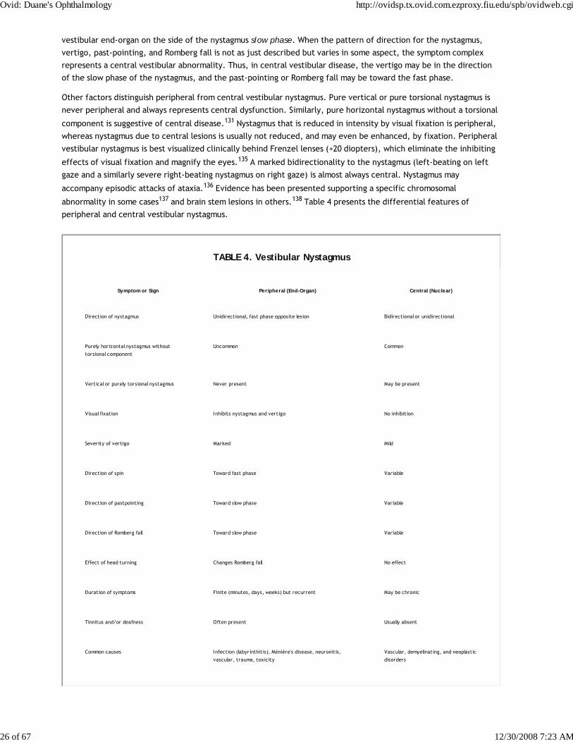
peripheral and central vestibular nystagmus.
TABLE 4. Vestibular Nystagmus
Symptom or Sign Peripheral (End-Organ) Central (Nuc lear)
Direction of nystagmus Unidirectional, fast phase opposite lesion Bidirectional or unidirectional
Purely horizontal nystagmus without
torsional component
Uncommon Common
Vertical or purely torsional nystagmus Never present May be present
Visual fixation Inhibits nystagmus and vertigo No inhibition
Severity of vertigo Marked Mild
Direction of spin Toward fast phase Variable
Direction of pastpointing Toward slow phase Variable
Direction of Romberg fall Toward slow phase Variable
Effect of head turning Changes Romberg fall No effect
Duration of symptoms Finite (minutes, days, weeks) but recurrent May be chronic
Tinnitus and/or deafness Often present Usually absent
Common causes Infection (labyrinthitis), Ménière's disease, neuronitis,
vascular, trauma, toxicity
Vascular, demyelinating, and neoplastic
disorders
Ovid: Duane's Ophthalmology http://ovidsp.tx.ovid.com.ezproxy.fiu.edu/spb/ovidweb.cgi
26 of 67 12/30/2008 7:23 AM
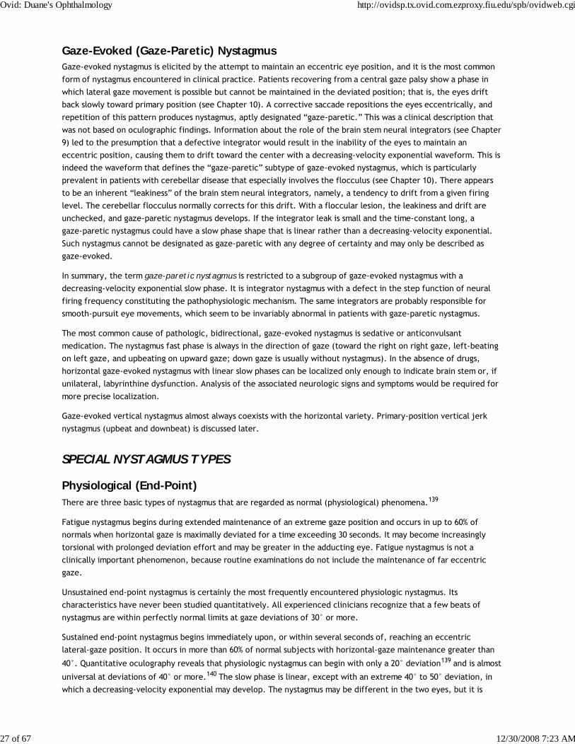
Gaze-Evoked (Gaze-Paretic) NystagmusGaze-evoked nystagmus is elicited by the attempt to maintain an eccentric eye position, and it is the most common
form of nystagmus encountered in clinical practice. Patients recovering from a central gaze palsy show a phase in
which lateral gaze movement is possible but cannot be maintained in the deviated position; that is, the eyes drift
back slowly toward primary position (see Chapter 10). A corrective saccade repositions the eyes eccentrically, and
repetition of this pattern produces nystagmus, aptly designated “gaze-paretic.” This was a clinical description that
was not based on oculographic findings. Information about the role of the brain stem neural integrators (see Chapter
9) led to the presumption that a defective integrator would result in the inability of the eyes to maintain an
eccentric position, causing them to drift toward the center with a decreasing-velocity exponential waveform. This is
indeed the waveform that defines the “gaze-paretic” subtype of gaze-evoked nystagmus, which is particularly
prevalent in patients with cerebellar disease that especially involves the flocculus (see Chapter 10). There appears
to be an inherent “leakiness” of the brain stem neural integrators, namely, a tendency to drift from a given firing
level. The cerebellar flocculus normally corrects for this drift. With a floccular lesion, the leakiness and drift are
unchecked, and gaze-paretic nystagmus develops. If the integrator leak is small and the time-constant long, a
gaze-paretic nystagmus could have a slow phase shape that is linear rather than a decreasing-velocity exponential.
Such nystagmus cannot be designated as gaze-paretic with any degree of certainty and may only be described as
gaze-evoked.
In summary, the term gaze-paretic nystagmus is restricted to a subgroup of gaze-evoked nystagmus with a
decreasing-velocity exponential slow phase. It is integrator nystagmus with a defect in the step function of neural
firing frequency constituting the pathophysiologic mechanism. The same integrators are probably responsible for
smooth-pursuit eye movements, which seem to be invariably abnormal in patients with gaze-paretic nystagmus.
The most common cause of pathologic, bidirectional, gaze-evoked nystagmus is sedative or anticonvulsant
medication. The nystagmus fast phase is always in the direction of gaze (toward the right on right gaze, left-beating
on left gaze, and upbeating on upward gaze; down gaze is usually without nystagmus). In the absence of drugs,
horizontal gaze-evoked nystagmus with linear slow phases can be localized only enough to indicate brain stem or, if
unilateral, labyrinthine dysfunction. Analysis of the associated neurologic signs and symptoms would be required for
more precise localization.
Gaze-evoked vertical nystagmus almost always coexists with the horizontal variety. Primary-position vertical jerk
nystagmus (upbeat and downbeat) is discussed later.
SPECIAL NYSTAGMUS TYPES
Physiological (End-Point)There are three basic types of nystagmus that are regarded as normal (physiological) phenomena.139
Fatigue nystagmus begins during extended maintenance of an extreme gaze position and occurs in up to 60% of
normals when horizontal gaze is maximally deviated for a time exceeding 30 seconds. It may become increasingly
torsional with prolonged deviation effort and may be greater in the adducting eye. Fatigue nystagmus is not a
clinically important phenomenon, because routine examinations do not include the maintenance of far eccentric
gaze.
Unsustained end-point nystagmus is certainly the most frequently encountered physiologic nystagmus. Its
characteristics have never been studied quantitatively. All experienced clinicians recognize that a few beats of
nystagmus are within perfectly normal limits at gaze deviations of 30° or more.
Sustained end-point nystagmus begins immediately upon, or within several seconds of, reaching an eccentric
lateral-gaze position. It occurs in more than 60% of normal subjects with horizontal-gaze maintenance greater than
40°. Quantitative oculography reveals that physiologic nystagmus can begin with only a 20° deviation139 and is almost
universal at deviations of 40° or more.140 The slow phase is linear, except with an extreme 40° to 50° deviation, in
which a decreasing-velocity exponential may develop. The nystagmus may be different in the two eyes, but it is
Ovid: Duane's Ophthalmology http://ovidsp.tx.ovid.com.ezproxy.fiu.edu/spb/ovidweb.cgi
27 of 67 12/30/2008 7:23 AM

symmetric in the two lateral directions. The amplitude of physiologic nystagmus does not exceed 3°.139 Thus, small-
amplitude gaze-evoked nystagmus may be a normal phenomenon, provided the slow phase, with gaze angles up to
40°, is linear. The onset of end-point nystagmus is related to slow drift velocity,141 and the reduction of drift
velocity during fixation (probably by the fixation subsystem) inhibits the nystagmus. Gaze-evoked nystagmus is by
necessity “pathologic” if any of three features are present: (1) asymmetry in the two directions, (2) amplitude of 4°
or more, or (3) exponential slow phase within a gaze angle of less than 40°.
DissociatedNystagmus in which the two eyes show a significant asymmetry in either amplitude or direction is designated
“dissociated.” The most common type of dissociation is in internuclear ophthalmoplegias where it is most marked in
the abducting eye (see Chapter 10). Abduction nystagmus, which is sometimes designated by the confusing term
ataxic nystagmus, is not really a nystagmus. This saccadic oscillation is secondary to lesions of the medial longitudinal
fasciculus and is discussed under Saccadic Pulses/Pulse Trains later in this chapter and in Chapter 10.
The pendular nystagmus in patients with multiple sclerosis is usually dissociated. There are a variety of nystagmus
dissociations with lesions of the posterior fossa (e.g., asymmetric vertical nystagmus greater in one eye on looking
up and in the other eye on looking down).
TorsionalTorsional nystagmus describes a torsional movement of the globe about its anteroposterior axis; the term rotary
nystagmus is used interchangeably. Most nystagmus consequent to vestibular end-organ dysfunction has a torsional
component admixed with a major horizontal or vertical nystagmus. A purely torsional nystagmus never occurs with
vestibular end-organ disease. When of small amplitude, torsional nystagmus may reflect a medullary lesion. Larger-
amplitude torsional nystagmus may be congenital, but when it is acquired it often indicates diencephalic (thalamic)
involvement, in which case it is the underlying pattern in see-saw nystagmus. Torsional nystagmus can be classified
into two groups.142 In one the nystagmus is in primary position, and in the other it is gaze evoked. The etiologies of
both are demyelinating, vascular, or neoplastic.
For consistency, nystagmus in the torsional plane should be defined with respect to the subject, not the observer.
When a subject's nystagmus beats to the right, it is called jerk-right nystagmus; if it beats toward the forehead, it is
upbeat (even if the subject is in the head-hanging position). Therefore, when the nystagmus fast phases bring the
top of the eye toward the subject's right shoulder, it is clockwise nystagmus. Clinical descriptions of torsional
nystagmus, made from the observer's point of view, deviate from the accepted convention that applies to both
horizontal and vertical movements. Maintaining the subject-based directions reduces confusion, eases understanding
of anatomic substrates, and anchors the subject's perception of oscillopsia to the direction of his nystagmus. Just as
leftward horizontal slow phases cause rightward, perceived world motion, clockwise torsional slow phases cause
counterclockwise, perceived world motion (see elliptic nystagmus, discussed later).
See-SawSee-saw nystagmus is characterized by a conjugate, pendular, torsional oscillation with a superimposed disjunctive
vertical vector. The intorting eye rises, and the opposite, extorting eye falls. Repetition of this sequence in the
alternate direction provides the see-saw effect. The torsional movements predominate in all fields of gaze, but the
see-saw feature may be restricted to the primary position or, more commonly, to downward or lateral gaze. See-saw
nystagmus can be of the jerk type (with one phase being slow and the other fast) with unilateral meso-diencephalic
lesions.143 Most patients with acquired pendular see-saw nystagmus have bitemporal hemianopias consequent to
large parasellar tumors expanding within the third ventricle. It is occasionally evoked transiently after blinks or
saccades.144 Upper brain stem vascular disease and severe head trauma are the next most common etiologies.
Posttraumatic see-saw may be temporarily abolished by ingestion of alcohol.145 Rarely, the nystagmus is associated
with septo-optic dysplasia, an Arnold-Chiari malformation,146 multiple sclerosis,147 or loss of vision alone.148 See-saw
nystagmus probably reflects diencephalic (thalamic) dysfunction possibly of a pathway or pathways from the zona
incerta to the interstitial nucleus of Cajal. A study of visual–vestibular interaction concluded that see-saw nystagmus
Ovid: Duane's Ophthalmology http://ovidsp.tx.ovid.com.ezproxy.fiu.edu/spb/ovidweb.cgi
28 of 67 12/30/2008 7:23 AM

resulted from a loss of retinal error signals secondary to disruption of chiasmal crossing fibers.149
Congenital see-saw nystagmus manifests either in constant vertical disjugacies without a significant torsional
component or in conjugate torsional nystagmus with a vertical component said to be opposite that of the acquired
variety; that is, the intorting eye falls while the extorting eye rises. However, all cases of congenital see-saw
nystagmus that have been recorded follow the rule that the rising eye intorts while the falling eye extorts.
Congenital see-saw nystagmus is also seen in canine and human achiasma (see Albinism and Achiasma section later in
the chapter). An unusual instance of congenital see-saw nystagmus in two mentally retarded adult siblings,
associated with retinitis pigmentosa, also had the typical vertical/torsional relationship associated with acquired
see-saw.150
The ocular tilt reaction, described in Chapter 10, is actually one-half of a see-saw cycle.
Convergence/Convergence-EvokedThe act of convergence usually damps nystagmus, particularly the congenital type.151 Convergence can also damp151
or evoke152 lid nystagmus and may damp or enhance downbeat nystagmus.153 Upbeat nystagmus may change to
downbeat with convergence.154
Repetitive divergence is a term describing a slow divergence movement followed by a rapid convergence to the
primary position. The movements occur at irregular intervals, distinguishing this from nystagmus.155 In the single
reported instance of this phenomenon in a patient with hepatic encephalopathy, an entire cycle lasted from 4 to 10
seconds, and the interval between cycles was 1 to 15 seconds.
Conjugate nystagmus evoked by convergence (convergence-evoked nystagmus) must be distinguished from
convergence nystagmus. Convergence-retraction nystagmus as a manifestation of the dorsal midbrain syndrome is
discussed in Chapter 10; here, the initiating convergence movements are saccadic156 and, thus not a true nystagmus.
Fast divergent movements, followed by a slow convergence associated with epileptic electroencephalographic
activity, occurred in a neonate with an intraventricular hemorrhage.157
With the exception of pure convergence nystagmus in infants with spasmus nutans, true pendular convergence
nystagmus is uncommon. We observed it in a patient with presumed progressive supranuclear palsy who had paralysis
of all volitional eye movements. In retrospect, however, the patient probably had central nervous system Whipple's
disease, perhaps the most common cause of pendular convergence nystagmus (see Chapter 10). We studied three
patients with convergence nystagmus with phase shifts of about 180° in both the horizontal and torsional planes with
conjugate nystagmus in the vertical plane.158 Convergence increased the nystagmus in two of the patients. The
waveforms were either sinusiodal or complex sums of sinusoids, and in one patient they were cycloidal. Unlike the
pseudocycloid waveform of INS, there were no initiating saccades to these cycloidal movements. We hypothesized
that the low-frequency convergence nystagmus resulted from a visually mediated vergence instability, whereas
high-frequency forms might have arisen from instability of brain stem pathways associated with vergence.
Nystagmus evoked by convergence is unusual and may be either conjugate or disjugate, congenital or acquired.159 In
two cases reported, no definite clinical correlation could be made with a specific lesion. The neuropathologic
examination revealed no morphologic explanation for nystagmus in the patient with congenital convergence-evoked
nystagmus; the patient with the acquired form had demyelinating disease with a spastic paraparesis and no cranial
nerve abnormality other than the ocular motor findings.159 Horizontal pendular nystagmus rarely is evoked by
accommodative vergence.160 This is to be distinguished from psychogenic flutter (the so-called voluntary nystagmus
discussed later), which is often best induced when the eyes are slightly converged.
Periodic AlternatingPeriodic alternating nystagmus (PAN) is an extraordinary ocular motor phenomenon in which a persisting horizontal
jerk nystagmus periodically changes directions. PAN may be congenital or acquired. The congenital variety (i.e., INS
nystagmus), which may be associated with albinism,161,162 has slow-phase waveforms of both linear and increasing
velocity, usually lacks the well-defined stereotyped periodicity seen in acquired PAN (i.e., it is asymmetric
Ovid: Duane's Ophthalmology http://ovidsp.tx.ovid.com.ezproxy.fiu.edu/spb/ovidweb.cgi
29 of 67 12/30/2008 7:23 AM
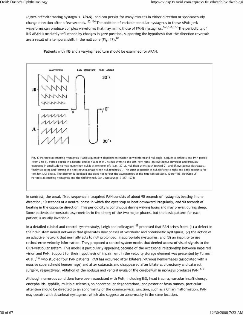
(a)periodic alternating nystagmus—APAN), and can persist for many minutes in either direction or spontaneously
change direction after a few seconds.163,164 The addition of variable pendular nystagmus to these APAN jerk
waveforms can produce complex waveforms that may mimic those of FMNS nystagmus.165,166,167 The periodicity of
INS APAN is markedly influenced by changes in gaze position, supporting the hypothesis that the direction reversals
are a result of a temporal shift in the null zone (Fig. 17).80
Patients with INS and a varying head turn should be examined for APAN.
Fig. 17 Periodic alternating nystagmus (PAN) sequence is depicted in relation to waveform and null angle. Sequence reflects one PAN period(from 0 to T). Period begins in a neutral phase; null is at 0°. As null shifts to the left, jerk right (JR) nystagmus develops and graduallyincreases in amplitude to maximum when null is at extreme left (e.g., 30°L). Null then shifts back toward 0°, and JR nystagmus decreases,finally stopping and forming the next neutral phase when null reaches 0°. The same sequence of null shifting to right and back accounts forjerk left (JL) phase. The diagram is idealized and does not reflect the asymmetries of the true clinical state. (Daroff RB, Dell'Osso LF:Periodic alternating nystagmus and the shifting null. Can J Otolaryngol 3:367, 1974)
In contrast, the usual, fixed sequence in acquired PAN consists of about 90 seconds of nystagmus beating in one
direction, 10 seconds of a neutral phase in which the eyes stop or beat downward irregularly, and 90 seconds of
beating in the opposite direction. This periodicity is continuous during waking hours and may prevail during sleep.
Some patients demonstrate asymmetries in the timing of the two major phases, but the basic pattern for each
patient is usually invariable.
In a detailed clinical and control-system study, Leigh and colleagues168 proposed that PAN arises from: (1) a defect in
the brain stem neural networks that generates slow phases of vestibular and optokinetic nystagmus, (2) the action of
an adaptive network that normally acts to null prolonged, inappropriate nystagmus, and (3) an inability to use
retinal-error velocity information. They proposed a control system model that denied access of visual signals to the
OKN-vestibular system. This model is particularly appealing because of the occasional relationship between impaired
vision and PAN. Support for their hypothesis of impairment in the velocity storage element was presented by Furman
et al.,169 who studied four PAN patients. PAN has occurred after bilateral vitreous hemorrhages (associated with a
massive subarachnoid hemorrhage) and after cataracts and disappeared after bilateral vitrectomy and cataract
surgery, respectively. Ablation of the nodulus and ventral uvula of the cerebellum in monkeys produces PAN.170
Although numerous conditions have been associated with PAN, including INS, head trauma, vascular insufficiency,
encephalitis, syphilis, multiple sclerosis, spinocerebellar degenerations, and posterior fossa tumors, particular
attention should be directed to an abnormality of the craniocervical junction, such as a Chiari malformation. PAN
may coexist with downbeat nystagmus, which also suggests an abnormality in the same location.
Ovid: Duane's Ophthalmology http://ovidsp.tx.ovid.com.ezproxy.fiu.edu/spb/ovidweb.cgi
30 of 67 12/30/2008 7:23 AM
Campbell described PAN secondary to phenytoin intoxication in a patient with alcoholic cerebellar degeneration.171
The antispasticity drug baclofen abolishes acquired PAN but has no effect on the INS variety.172 The drug abolished
experimentally created PAN in the monkey,170 as well as a single case of aperiodic alternating nystagmus in a patient
with vertebrobasilar insufficiency.173
PAN may be associated with a periodic alternating skew deviation.174 Periodic alternating gaze deviations, with and
without associated alternating nystagmus or alternating head turning, are rare, related phenomena.175
DownbeatDownbeat nystagmus is defined as nystagmus in primary gaze position, with the fast phase beating in a downward
direction. Patients with brain stem disease or drug intoxications usually lack gaze-evoked downward nystagmus
despite nystagmus in all other fields of gaze. Thus, nystagmus beating downward in the primary position is a striking
phenomenon and is highly suggestive of a craniocervical junction lesion, such as a Chiari malformation.176,177
Downbeat nystagmus is usually of sufficient amplitude in primary position to cause oscillopsia, and, contrary to
Alexander's law,2 it is not maximum at the extreme of downward gaze. Rather, it is usually of maximal intensity when
the eyes are deviated laterally and slightly below the horizontal;177 the nystagmus may be intermittent.
The other major cause of downbeat nystagmus is spinocerebellar degeneration. A variety of miscellaneous conditions
may also be associated with downbeat nystagmus,153,177 including anticonvulsants, alcohol and lithium intoxication,
magnesium deficiency, B12 deficiency, brain stem encephalitis, alcoholic cerebellar degeneration, and brain stem or
cerebellar infarcts.178 Rarely, downbeat nystagmus is secondary to upper brain stem or supratentorial lesions, or as a
form of INS nystagmus. Schmidt's179 article is an excellent clinical review of downbeat nystagmus.
Downbeat nystagmus was regarded as the prototype of nystagmus secondary to a “pursuit defect,” but studies have
cast serious doubt as to whether a unidirectional pursuit defect is a cause of any form of nystagmus.176 The
slow-phase waveform of downbeat nystagmus can vary from a linear, to an increasing-velocity, to a decreasing-
velocity exponential in the same patient, presumably reflecting short-term gain changes by cerebellar compensation
for leaky brain stem neural integrators.5 One patient with a pseudocycloid waveform damped with convergence,
prompting treatment with base-out prism spectacles.153 This has subsequently proved superior to retinal image
stabilization as a means of reducing oscillopsia.180 Downbeat is usually increased in intensity with head-hanging176 as
well as linear acceleration, and occasionally convergence. The mechanism of downbeat is postulated to be disruption
of central vestibular pathways,176 possibly involving specific connections from the otoliths or an asymmetry of the
vertical semicircular canal gains.181
UpbeatPrimary-position nystagmus with the fast phase beating upward may reflect drug or other intoxications182 but usually
indicates structural disease, usually of the brain stem.183,184,185 The location of the lesions in patients with upbeat
nystagmus after meningitis or Wernicke's encephalopathy is uncertain. Büttner et al. discussed the localizing value of
upbeat and other types of nystagmus.186 With convergence, upbeat may enhance184 or convert to downbeat.154 The
slow-phase waveform is usually linear but may be an increasing-velocity exponential.
Upbeat may be enhanced184 or suppressed187 by head tilt. Therefore, it is conceptualized hypothetically as
secondary to a disruption of central otolithic pathways; the effects of convergence on the nystagmus are presumed
to reflect vergence effects on these pathways. Tobacco smoking causes upbeat nystagmus in normal human subjects
in darkness, but the nystagmus is suppressed by visual fixation.188 The mechanism is presumed to be the excitatory
effects of nicotine on central vestibular pathways.
ReboundRebound nystagmus is either the diminution and direction change of gaze-evoked horizontal nystagmus during
sustained ocular deviation or a horizontal gaze-evoked nystagmus that, on refixation to primary position, transiently
beats in the opposite direction. The sign is often present in patients with cerebellar disease.189 Rebound nystagmus
Ovid: Duane's Ophthalmology http://ovidsp.tx.ovid.com.ezproxy.fiu.edu/spb/ovidweb.cgi
31 of 67 12/30/2008 7:23 AM
is conceptualized as a smooth eye movement bias generated to oppose gaze-evoked centripetal drift of the eyes.
Normal subjects may demonstrate rebound nystagmus after prolonged far lateral gaze if the lights are shut off the
moment the eyes are returned to primary position and may even occur in normals during fixation in a fully lit
room.140 Rebound and centripetal nystagmus may occur in Creutzfeldt-Jakob disease.190
Circular, Elliptic, and ObliqueCircular nystagmus, sometimes confusingly mislabeled as “rotary,” is a form of pendular nystagmus in which the
globe oscillates continuously in a fine, rapid, circular path. Unlike torsional nystagmus, in which the 12 o'clock
meridian of the limbus torts laterally, this point maintains its position in circular nystagmus. The nystagmus
represents the summation of simultaneous, equal-amplitude, horizontal, and vertical pendular oscillations that are
90° out of phase. Elliptic nystagmus is produced when the horizontal and vertical oscillations are 90° out of phase
but are of unequal amplitude. Analysis of the vertical and horizontal pendular nystagmus movements, which
summate to form circular and elliptic nystagmus, indicates that a true circular pattern is rarely sustained. More
often, the nystagmus varies between elliptic and circular. Unlike all other types of nystagmus, the oscillopsia
induced by elliptical nystagmus is in the same direction as the slow phases.191 Thus, if the subject's eye moves in a
clockwise, circular-elliptic manner (from the subject's point of view), the direction of perceived world motion will
also be clockwise. Acquired circular-elliptic nystagmus occurs in multiple sclerosis, is often dissociated in the two
eyes, and almost always coexists with truncal or extremity ataxia. Drug treatment with isoniazid may be effective.191
This type of nystagmus is often part of the INS and is identical in the two eyes in patients otherwise free of
neurologic signs.
Oblique or diagonal nystagmus results when simultaneous pendular, horizontal, and vertical vectors are either in
phase or 180° out of phase. The angle of the diagonal vector depends on the relative amplitudes of the horizontal
and vertical components. This type is more often acquired than infantile and has the same significance as acquired
circular-elliptic nystagmus.
CervicalThe literature on cervical (cord) nystagmus contains numerous examples of spontaneous or positional nystagmus
allegedly secondary to lesions of the cervical spinal cord or roots.192,193,194 Although this form of positional
nystagmus occurs only rarely, it is claimed to support the “legitimacy” of dizziness in patients who have sustained
“whiplash” injuries and are involved in litigation. For that reason, the entire concept of cervical nystagmus has
become highly suspect.193,194 We have never seen a patient with convincing cervical nystagmus.
Muscle-Paretic (Myasthenic)A paretic eye muscle, from whatever cause, can fatigue quickly during contraction, and muscle-paretic nystagmus
can be observed. This is often evident as gaze-evoked nystagmus in myasthenia gravis, in which there is usually
asymmetry between the two eyes.195 Another form of oscillation in myasthenia is nystagmus of the abducting eye
(may be saccadic pulse trains, discussed below), coexisting with paresis of adduction; this mimics an internuclear
ophthalmoplegia. Here the oscillation is not due to lateral rectus paresis, but rather to excessive innervation by
increased central gain, the result of paresis of the contralateral yoke medial rectus. Cessation of both muscle-
paretic and contralateral yoke nystagmus in myasthenia usually follows administration of anticholinesterase
medication.
LidLid nystagmus is a rhythmic, upward jerking of the upper eyelids that usually consists of the normally coordinated
movements of the lids and eyes during vertical ocular nystagmus. Several types of pathologic lid nystagmus are
recognized. Type I, with no specific localizing value coexists synchronously with vertical ocular nystagmus, but the
amplitude of the lid movements exceeds significantly that of the eyes. Type II is evoked by lateral gaze and is
characterized by rapid phasic twitches of the lids that are synchronous with the fast phases of horizontal ocular
nystagmus. This second type may be a sign of the lateral medullary syndrome and can be inhibited by near-effort.151
Ovid: Duane's Ophthalmology http://ovidsp.tx.ovid.com.ezproxy.fiu.edu/spb/ovidweb.cgi
32 of 67 12/30/2008 7:23 AM
Type III lid nystagmus is provoked by ocular convergence.152,196 In a patient with this type of nystagmus studied
pathologically, there was a large area of subacute demyelinization in the rostral medulla. Lid nystagmus, differing
from Types I–III, may occur with midbrain lesions.197
EpilepticNystagmus associated with epileptic activity includes retraction, pendular, torsional, and divergent-convergent
forms.157 At times, the pupils may constrict and dilate synchronously with the nystagmus.198 The usual form of
epileptic nystagmus is horizontal jerk with the fast phase contralateral to a posterior parietal focus, but a vertical
nystagmus may also be present.199 The eyes may tonically deviate toward or away from the side of the epileptic
focus.200 The decreasing-velocity slow phases suggest that a gaze-holding failure201 causes the slow phases, and the
relationship of the ictal activity suggests activation of the saccadic system (see Chapter 10).199
INDUCED NYSTAGMUS
CaloricWe previously described the characteristics of caloric-induced vestibular nystagmus. With unilateral irrigation, the
nystagmus is horizontal, torsional, or oblique, depending on the position of the head. Bilateral simultaneous caloric
stimulation produces vertical nystagmus; the direction of the fast (jerk) phase can be remembered by the mnemonic
CUWD (pronounced “cud” and designating Cold, Up; Warm, Down).
Traditionally, caloric nystagmus was regarded as being evoked entirely by thermal convection of the endolymph.
However, the fact that such nystagmus exists in the zero-gravity conditions of outer space indicates the necessity of
an alternative mechanism,202 which,203 in addition to the convection mechanism, is a direct temperature effect on
the canal's sensory apparatus that is independent of head orientation. A review of the technique of quantitative
bithermal caloric testing for evaluating vestibular function is provided elsewhere. Caloric and head rotation (doll's)
are useful tests in the evaluation of comatose patients.204
RotationalRotating or accelerating head movements induce motion of endolymph in the semicircular canals, with a resultant
jerk nystagmus. If the axis of rotation passes through the upright head, as happens with the Bárány chair, the
nystagmus fast phase is in the same direction as head (or chair) rotation. After cessation of the rotation, the
postrotary nystagmus is in the opposite direction. Rotational nystagmus is used in evaluating the ocular motor system
in infants (see Chapter 3, Fig. 2), and to evaluate vestibulo-ocular gain and other aspects (visual suppression of the
VOR) of the vestibular system.
PositionalIn patients complaining of vertigo related to shifts in position of the head or body, positional nystagmus should be
considered. The test is performed by observing for nystagmus produced when the patient rapidly reclines from a
sitting to a supine position, with the head either turned alternately to one side and then the other, or
hyperextended (hanging). Two types of nystagmus, peripheral and central, can be differentiated (Table 5).
TABLE 5. Positional Nystagmus
Features Peripheral Central
Latency 3–40 sec None; nystagmus begins immediately
Ovid: Duane's Ophthalmology http://ovidsp.tx.ovid.com.ezproxy.fiu.edu/spb/ovidweb.cgi
33 of 67 12/30/2008 7:23 AM
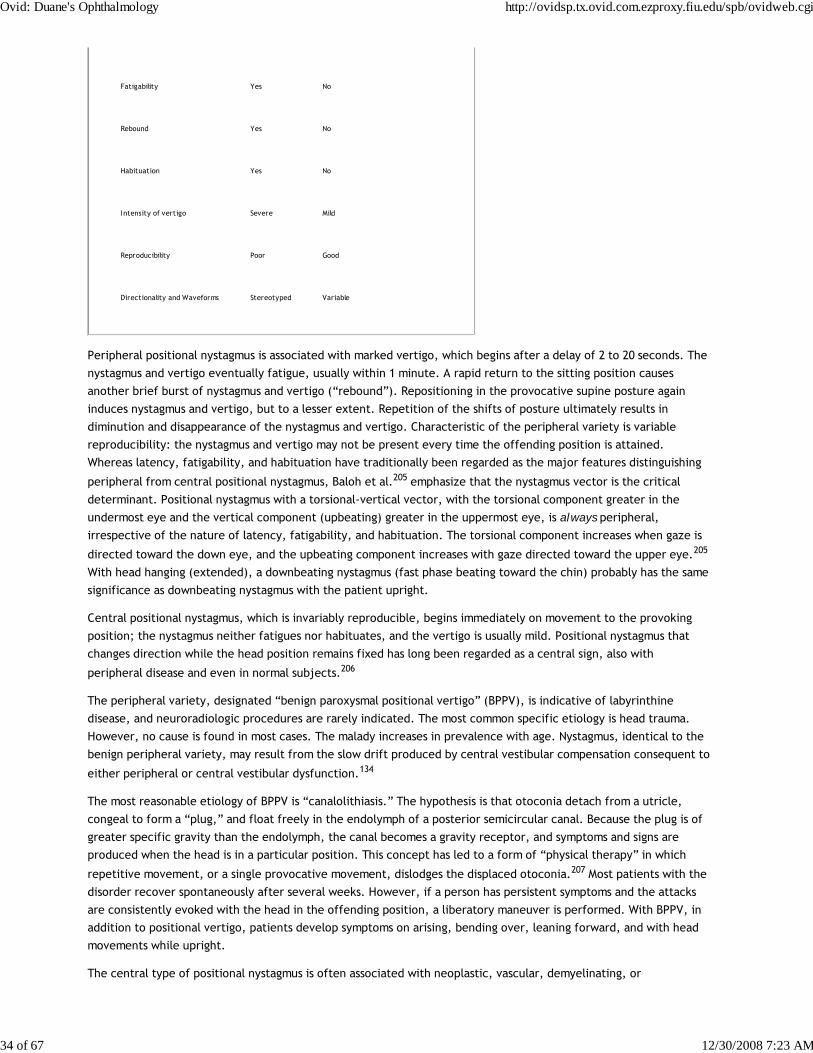
Fatigability Yes No
Rebound Yes No
Habituation Yes No
Intensity of vertigo Severe Mild
Reproducibility Poor Good
Directionality and Waveforms Stereotyped Variable
Peripheral positional nystagmus is associated with marked vertigo, which begins after a delay of 2 to 20 seconds. The
nystagmus and vertigo eventually fatigue, usually within 1 minute. A rapid return to the sitting position causes
another brief burst of nystagmus and vertigo (“rebound”). Repositioning in the provocative supine posture again
induces nystagmus and vertigo, but to a lesser extent. Repetition of the shifts of posture ultimately results in
diminution and disappearance of the nystagmus and vertigo. Characteristic of the peripheral variety is variable
reproducibility: the nystagmus and vertigo may not be present every time the offending position is attained.
Whereas latency, fatigability, and habituation have traditionally been regarded as the major features distinguishing
peripheral from central positional nystagmus, Baloh et al.205 emphasize that the nystagmus vector is the critical
determinant. Positional nystagmus with a torsional-vertical vector, with the torsional component greater in the
undermost eye and the vertical component (upbeating) greater in the uppermost eye, is always peripheral,
irrespective of the nature of latency, fatigability, and habituation. The torsional component increases when gaze is
directed toward the down eye, and the upbeating component increases with gaze directed toward the upper eye.205
With head hanging (extended), a downbeating nystagmus (fast phase beating toward the chin) probably has the same
significance as downbeating nystagmus with the patient upright.
Central positional nystagmus, which is invariably reproducible, begins immediately on movement to the provoking
position; the nystagmus neither fatigues nor habituates, and the vertigo is usually mild. Positional nystagmus that
changes direction while the head position remains fixed has long been regarded as a central sign, also with
peripheral disease and even in normal subjects.206
The peripheral variety, designated “benign paroxysmal positional vertigo” (BPPV), is indicative of labyrinthine
disease, and neuroradiologic procedures are rarely indicated. The most common specific etiology is head trauma.
However, no cause is found in most cases. The malady increases in prevalence with age. Nystagmus, identical to the
benign peripheral variety, may result from the slow drift produced by central vestibular compensation consequent to
either peripheral or central vestibular dysfunction.134
The most reasonable etiology of BPPV is “canalolithiasis.” The hypothesis is that otoconia detach from a utricle,
congeal to form a “plug,” and float freely in the endolymph of a posterior semicircular canal. Because the plug is of
greater specific gravity than the endolymph, the canal becomes a gravity receptor, and symptoms and signs are
produced when the head is in a particular position. This concept has led to a form of “physical therapy” in which
repetitive movement, or a single provocative movement, dislodges the displaced otoconia.207 Most patients with the
disorder recover spontaneously after several weeks. However, if a person has persistent symptoms and the attacks
are consistently evoked with the head in the offending position, a liberatory maneuver is performed. With BPPV, in
addition to positional vertigo, patients develop symptoms on arising, bending over, leaning forward, and with head
movements while upright.
The central type of positional nystagmus is often associated with neoplastic, vascular, demyelinating, or
Ovid: Duane's Ophthalmology http://ovidsp.tx.ovid.com.ezproxy.fiu.edu/spb/ovidweb.cgi
34 of 67 12/30/2008 7:23 AM

degenerative disorders involving the brain stem or cerebellum.208 Intermediate forms of positional nystagmus, such
as those that fatigue and habituate but have no noticeable latency, should raise suspicions of central disease. The
difference between central paroxysmal positioning nystagmus and that of BPPV is in the direction; the latter beats in
the direction aligned with the affected canal plane and any other direction indicates a central origin.209
Testing for positional nystagmus is fairly standardized and does not require electronystagmography. We prefer
self-illuminated Frenzel glasses (highly convex lenses, +20 D, which blur vision), but testing can also be performed in
a completely darkened room, with the examiner periodically observing the eyes with a dim light. Routine testing for
positional nystagmus in patients without the specific symptom of positional vertigo does not generally yield useful
results.
The BPPV just mentioned is due to dysfunction of a posterior semicircular canal. Some patients have
horizontal-canal BPPV210 or anterior-canal BPPV,211 which have different liberatory maneuvers.212 The term
positional nystagmus is often used interchangeably with positional vertigo in designating a syndrome, and
positional and positioning are used interchangeably to describe nystagmus.
OptokineticThe localizing value of OKN with cerebral hemispheric lesions is discussed in Chapter 10. OKN is used for several
other important functions in clinical neuro-ophthalmology. It provides evidence of at least gross levels of visual
function in infants or patients with functional visual loss. As mentioned in Chapter 10, OKN in the downward
direction is used to induce convergence-retraction “nystagmus,” and horizontal OKN is used to demonstrate the
adduction insufficiency in internuclear ophthalmoparesis. “Inversion” of OKN is diagnostic of INS. OKN can also be
used to diagnose oculomotor nerve misdirection.
Drug/Toxin-InducedDrug-induced nystagmus is a common sequela of barbiturate, tranquilizer, phenothiazine, and anticonvulsant
therapy.213 The nystagmus is generally gaze evoked and is usually horizontal or horizontal-torsional in direction.
Vertical nystagmus is often present on upward gaze and only rarely on downward gaze. At times the nystagmus may
be dissociated in the two eyes despite the lack of structural disease to account for the asymmetry. Although
primary-position nystagmus is usually indicative of severe drug intoxication, it may persist for 10 hours after the oral
ingestion of only 100 mg of secobarbital.134 In addition, this amount of secobarbital can produce positional
nystagmus. As mentioned, lithium can produce downbeat nystagmus,214,215 tobacco can induce upbeat nystagmus,188
and severe alcohol intoxication can produce downbeat nystagmus.216 Toluene (glue-sniffing) can induce pendular
nystagmus with both horizontal and vertical components.217
Unfortunately, that alcohol can produce horizontal gaze-evoked nystagmus has led to a “roadside sobriety” test
conducted by law-enforcement officers.218 Nystagmus as an indicator of alcohol intoxication is fraught with
extraordinary pitfalls: many normal individuals have physiologic end-point nystagmus; small doses of tranquilizers
that wouldn't interfere with driving ability can also produce nystagmus; nystagmus may be congenital or consequent
to structural neurologic disease; and often a neuro-ophthalmologist or sophisticated oculographer is required to
determine whether nystagmus is pathologic. Such judgments are difficult for experts to make under the best
conditions and impossible to make accurately under roadside conditions. It is unreasonable to have cursorily trained
law officers using the test, no matter how intelligent, perceptive, and well meaning they might be. As noted,
meticulous history taking and drug-screening blood studies are often essential in evaluating patients with nystagmus.
SPECIAL ANATOMIC CATEGORIES
Acoustic NeuromaSchwannomas of the eighth nerve grow so slowly that adaptive mechanisms often obscure clinical vestibular
manifestations. Vestibular nystagmus beating contralateral to the lesion may be present, particularly if fixation is
eliminated. As the tumor expands to compress the brain stem, a slow, gaze-evoked ipsilateral nystagmus is often
Ovid: Duane's Ophthalmology http://ovidsp.tx.ovid.com.ezproxy.fiu.edu/spb/ovidweb.cgi
35 of 67 12/30/2008 7:23 AM

added. The combination of a small-amplitude, rapid primary-position jerk nystagmus beating contralateral to the
lesion and a slower, larger-amplitude, gaze-evoked (Bruns') nystagmus ipsilateral to the lesion also occurs with other
extra-axial masses, including cerebellar tumors, compressing the brain stem. Rarely, Bruns' nystagmus is inverted.219
Lateral Medullary SyndromeThe lateral medullary syndrome (Wallenberg) is a distinctive constellation of signs. The nystagmus in this syndrome
tends to be stereotyped. With the eyes open there is horizontal-torsional jerk nystagmus beating contralateral to
the lesion; when recorded with the eyes closed, the nystagmus beats ipsilateral to the lesion. Other rare
manifestations, confined to single cases, are gaze-evoked eyelid and ocular nystagmus inhibited by the near
reflex151 and horizontal gaze-evoked monocular downbeat nystagmus.
An extraordinarily dramatic eye-movement abnormality, “saccadic lateropulsion,” may occur with lateral medullary
infarction. Eye movements as well as body and limb movements are biased toward the side of the lesion (ipsipulsion).
The ocular motor abnormality is most striking during shifts of fixation; all ipsilateral saccades are too large
(hypermetric), whereas those to the opposite side are too small (hypometric). Spontaneous drifts to the side of the
lesion are gaze dependent and may reflect disruptions of the gaze-holding mechanism.220 There is also reduced
capability to adjust saccadic gain in response to the dysmetria,221 significant tilt in the subjective vertical, with
ipsilateral excyclotropia,222 and torsional nystagmus and torsipulsion.223,224 Upward or downward refixations veer
ipsilaterally along an oblique rather than a vertical line. Lateropulsion away from the side of the lesion
(contrapulsion) has occurred with a unilateral disorder of the rostral cerebellum.225 Both types of lateropulsion are
regarded as saccadic instabilities (Table 6). The gaze deviation may reflect increased inhibition of the ipsilateral
vestibular nucleus and ipsipulsion may reflect decreased excitation of contralateral premotor areas in the pontine
paramedian reticular formation.226
TABLE 6. Saccadic Intrusions and Oscillations*
Bobbing/dipping Saccadic lateropulsion
inverse bobbing ipsipulsion
reverse bobbing contrapulsion
Convergence-retraction “nystagmus” Saccadic pulses/pulse trains
“nystagmus” retractoris abduction “nystagmus”
Double saccadic pulses (single/multiple) ataxic “nystagmus”
saccadic intrusions/oscillations saccadic intrusions/oscillations
Dynamic overshoot stepless saccades
“quiver” Square-wave jerks/oscillations
Dysmetria Gegenrucke
Ovid: Duane's Ophthalmology http://ovidsp.tx.ovid.com.ezproxy.fiu.edu/spb/ovidweb.cgi
36 of 67 12/30/2008 7:23 AM

Flutter hopping “nystagmus”
Flutter dysmetria “lightening eye movements”
Macro saccadic oscillations myoclonus
Myoclonus saccadic intrusions/oscillations
laryngeal “nystagmus” Zickzakbewegungen
“lightning eye movements” Square-wave pulses (bursts/single)
pharyngeal “nystagmus” “macro square-wave jerks”
Opsoclonus Kippdeviationen/“Kippnystagmus”
“dancing eyes” “pendular macro-oscillations”
“lightning eye movements” saccadic “nystagmus”
saccadomania saccadic oscillations/intrusions
Psychogenic flutter Staircase saccadic intrusions
hysterical flutter Superior oblique myokymia
hysterical “nystagmus”
“ocular fibrillation”
“ocular shuddering”
psychological “nystagmus”
voluntary flutter
voluntary “nystagmus”
*Synonyms and other terms are indented under either the preferred or the more inclusive designation; quoted terms are erroneous or misleading.
ALBINISM AND A(HEMI)(HYPO)CHIASMAOcular albinism is associated with anomalous visual projections that result in a variety of eye movement
Ovid: Duane's Ophthalmology http://ovidsp.tx.ovid.com.ezproxy.fiu.edu/spb/ovidweb.cgi
37 of 67 12/30/2008 7:23 AM

disturbances, with considerable intersubject variability. These individuals may have pendular or jerk nystagmus,
absent OKN, “inverted” pursuit, or “defective” pursuit (see section on INS) when targets are projected onto the
temporal half-retina.227 Periodic alternating nystagmus may also occur.161,162 Albinism may also exist in the absence
of any nystagmus.228,229
Achiasma is a very rare condition, first recognized in dogs (ca. 1974) and then in humans (ca. 1992). It is associated
with the combination of INS and see-saw nystagmus; we first diagnosed the see-saw nystagmus from videotape (in
canines in 1991 and in a human in 1993) and subsequently studied it using eye-movement
recordings.50,230,231,232,233,234,235, In achiasma, all retinal fibers remain ipsilateral, passing to the ipsilateral lateral
geniculates and visual cortexes. Thus, each visual cortex has a representation of the entire visual field, but
stereopsis is impossible; there is no bitemporal hemianopia. Figure 18 shows both the horizontal INS nystagmus and
the vertical component of the see-saw nystagmus in an achiasmatic canine (Fig. 18A) and an achiasmatic human (Fig.
18B). The horizontal and vertical waveforms of the canines' nystagmus and the human's vertical nystagmus were
pendular, whereas the human's horizontal waveforms were pendular with foveating saccades (PFS) or pseudopendular
with foveating saccades (PPFS). Because the achiasmatic human subject was not always able to achieve
well-developed foveation, as shown by her inability to repeatedly foveate the target with her fixating right eye, a
larger than normal foveation window for the NAFX would be required to assess the best potential visual acuity for
this segment. Unlike INS, which may or may not be associated with a particular sensory deficit, the see-saw
nystagmus appears to be directly associated with achiasma in both species. In achiasma, there is no lesion, indeed no
chiasm, and disruption of thalamic inputs to the nucleus of Cajal, postulated as the cause of acquired see-saw
nystagmus, cannot be causal. In hemichiasma, abnormalities at the chiasm prevent decussation of the retinal fibers
from one eye, the other decussating normally.236 Whereas achiasma appears to be sufficient for see-saw nystagmus,
hemichiasma is not. Ocular motility and retinal studies of humans with achiasma and hypochiasma (a maldeveloped
chiasm) revealed INS, see-saw nystagmus, strabismus, reduced vision, optic nerve dysplasia.237 Because of the rarity
of congenital see-saw nystagmus and its identification in both canines and humans with achiasma and in one of two
canines with hemichiasma, we regard its presence in infants as a diagnostic sign of possible achiasma or
hemichiasma.238 The presence of see-saw nystagmus in an infant is a strong indication for imaging of the optic chiasm
to rule out achiasma or structural abnormalities (hypochiasma) conducive to hemichiasma.
Ovid: Duane's Ophthalmology http://ovidsp.tx.ovid.com.ezproxy.fiu.edu/spb/ovidweb.cgi
38 of 67 12/30/2008 7:23 AM

Fig. 18 The horizontal IN and vertical component of see-saw nystagmus in (A) an achiasmatic Belgian sheepdog, during binocular (OU)viewing and (B) an achiasmatic human, during right-eye fixation. Note that the left eye in (B) was approximately 15° esotropic. Whereindicated, traces were shifted for clarity. REH—right eye horizontal, LEH—left eye horizontal, REV—right eye vertical, and LEV—left eyevertical. In (B), REV and LEV were shifted -15° for clarity.
Ovid: Duane's Ophthalmology http://ovidsp.tx.ovid.com.ezproxy.fiu.edu/spb/ovidweb.cgi
39 of 67 12/30/2008 7:23 AM
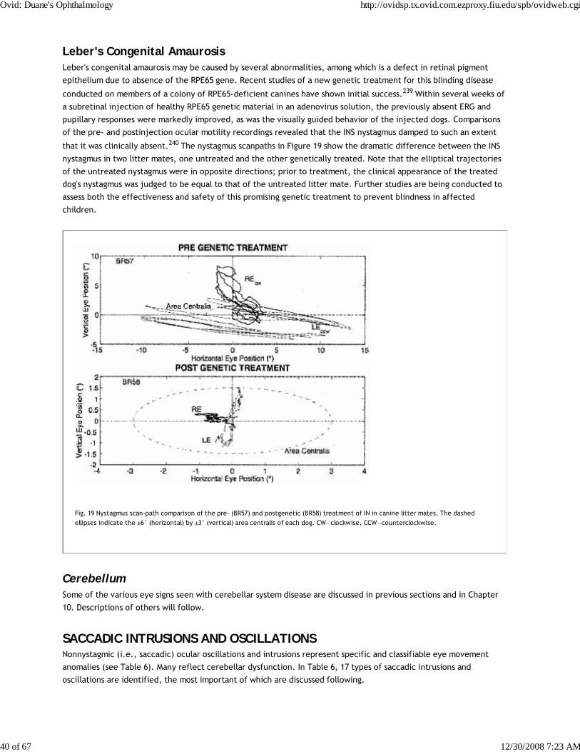
Leber's Congenital AmaurosisLeber's congenital amaurosis may be caused by several abnormalities, among which is a defect in retinal pigment
epithelium due to absence of the RPE65 gene. Recent studies of a new genetic treatment for this blinding disease
conducted on members of a colony of RPE65-deficient canines have shown initial success.239 Within several weeks of
a subretinal injection of healthy RPE65 genetic material in an adenovirus solution, the previously absent ERG and
pupillary responses were markedly improved, as was the visually guided behavior of the injected dogs. Comparisons
of the pre- and postinjection ocular motility recordings revealed that the INS nystagmus damped to such an extent
that it was clinically absent.240 The nystagmus scanpaths in Figure 19 show the dramatic difference between the INS
nystagmus in two litter mates, one untreated and the other genetically treated. Note that the elliptical trajectories
of the untreated nystagmus were in opposite directions; prior to treatment, the clinical appearance of the treated
dog's nystagmus was judged to be equal to that of the untreated litter mate. Further studies are being conducted to
assess both the effectiveness and safety of this promising genetic treatment to prevent blindness in affected
children.
Fig. 19 Nystagmus scan-path comparison of the pre- (BR57) and postgenetic (BR58) treatment of IN in canine litter mates. The dashedellipses indicate the ±6° (horizontal) by ±3° (vertical) area centralis of each dog. CW—clockwise, CCW—counterclockwise.
CerebellumSome of the various eye signs seen with cerebellar system disease are discussed in previous sections and in Chapter
10. Descriptions of others will follow.
SACCADIC INTRUSIONS AND OSCILLATIONSNonnystagmic (i.e., saccadic) ocular oscillations and intrusions represent specific and classifiable eye movement
anomalies (see Table 6). Many reflect cerebellar dysfunction. In Table 6, 17 types of saccadic intrusions and
oscillations are identified, the most important of which are discussed following.
Ovid: Duane's Ophthalmology http://ovidsp.tx.ovid.com.ezproxy.fiu.edu/spb/ovidweb.cgi
40 of 67 12/30/2008 7:23 AM
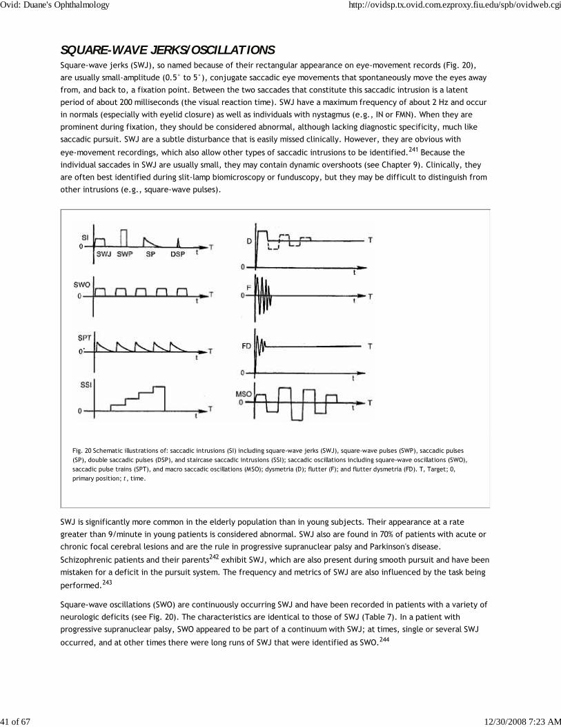
SQUARE-WAVE JERKS/OSCILLATIONSSquare-wave jerks (SWJ), so named because of their rectangular appearance on eye-movement records (Fig. 20),
are usually small-amplitude (0.5° to 5°), conjugate saccadic eye movements that spontaneously move the eyes away
from, and back to, a fixation point. Between the two saccades that constitute this saccadic intrusion is a latent
period of about 200 milliseconds (the visual reaction time). SWJ have a maximum frequency of about 2 Hz and occur
in normals (especially with eyelid closure) as well as individuals with nystagmus (e.g., IN or FMN). When they are
prominent during fixation, they should be considered abnormal, although lacking diagnostic specificity, much like
saccadic pursuit. SWJ are a subtle disturbance that is easily missed clinically. However, they are obvious with
eye-movement recordings, which also allow other types of saccadic intrusions to be identified.241 Because the
individual saccades in SWJ are usually small, they may contain dynamic overshoots (see Chapter 9). Clinically, they
are often best identified during slit-lamp biomicroscopy or funduscopy, but they may be difficult to distinguish from
other intrusions (e.g., square-wave pulses).
Fig. 20 Schematic illustrations of: saccadic intrusions (SI) including square-wave jerks (SWJ), square-wave pulses (SWP), saccadic pulses(SP), double saccadic pulses (DSP), and staircase saccadic intrusions (SSI); saccadic oscillations including square-wave oscillations (SWO),saccadic pulse trains (SPT), and macro saccadic oscillations (MSO); dysmetria (D); flutter (F); and flutter dysmetria (FD). T, Target; 0,primary position; t, time.
SWJ is significantly more common in the elderly population than in young subjects. Their appearance at a rate
greater than 9/minute in young patients is considered abnormal. SWJ also are found in 70% of patients with acute or
chronic focal cerebral lesions and are the rule in progressive supranuclear palsy and Parkinson's disease.
Schizophrenic patients and their parents242 exhibit SWJ, which are also present during smooth pursuit and have been
mistaken for a deficit in the pursuit system. The frequency and metrics of SWJ are also influenced by the task being
performed.243
Square-wave oscillations (SWO) are continuously occurring SWJ and have been recorded in patients with a variety of
neurologic deficits (see Fig. 20). The characteristics are identical to those of SWJ (Table 7). In a patient with
progressive supranuclear palsy, SWO appeared to be part of a continuum with SWJ; at times, single or several SWJ
occurred, and at other times there were long runs of SWJ that were identified as SWO.244
Ovid: Duane's Ophthalmology http://ovidsp.tx.ovid.com.ezproxy.fiu.edu/spb/ovidweb.cgi
41 of 67 12/30/2008 7:23 AM

TABLE 7. Characteristics of Saccadic Instabilities
Square-Wave Jerks Square-Wave Osc illations Square-WavePulses* Macro Saccadic Osc illations
Amplitude 0.5°–5°1 0.5°–5°1 4°–30° 1°–30°
Constant Constant Variable Increasing then decreasing
Time course Sporadic/bursts Bursts Bursts/sporatic Bursts
Latency 200 msec 200 msec 50–150 msec 200 msec
Foveation Yes Yes Yes No
Presence in darkness Yes Yes Yes No
*Previously designated macro square-wave jerks.1Occasionally up to 10°.
SQUARE-WAVE PULSESSquare-wave pulses (SWP), originally called “macro square-wave jerks,” are usually larger in amplitude than SWJ,
are related to fixation, and have a frequency of about 2 Hz.245 They generally occur in bursts but may appear as a
single saccadic intrusion. Both eyes suddenly and conjugately move off target with a saccade, and after a latent
period of only about 80 milliseconds, a nonvisually evoked reflex saccade brings them back on target (see Fig. 20).
SWP are not merely large SWJ; the characteristics of both are summarized in Table 7. SWP usually occur in patients
with marked extremity ataxia suggestive of cerebellar outflow disease, especially when the patient has
demyelinating lesions.245 A unique variety of SWP, present with binocular fixation at distance but stopping when
either eye was closed, prompted the designation “inverse latent SWP.”246
STAIRCCASE SACCADIC INTRUSIONSWe identified a unique type of saccadic intrusion in a patient with cerebellar atrophy, named, staircase saccadic
intrusions because of its appearance in eye-movement recordings.247 Fixation would be interrupted by a series of
saccades in one direction or the other with normal intersaccadic intervals (see Fig. 20). The individual saccades
could be of equal amplitude of could vary. Staircase saccadic intrusions were also present during smooth pursuit. In
normals, such “staircase” eye movements can be generated by feeding back the eye-movement signal and allowing it
to move the target in the same direction; this produces a constant retinal error signal that drives the saccadic
system. We modelled staircase saccadic intrusions using a behavioral OMS model15 by interfering with the retinal
error signal and creating a constant reconstructed error signal.
MACRO SACCADIC OSCILLATIONSMacro saccadic oscillations (MSO) increase and then damp in amplitude, bypassing the fixation angle with each
saccade (see Fig. 20).248 Unlike SWJ, SWO, and SWP, MSO are not present in darkness and have a longer latency for
the return saccade than do SWP. MSO may occur among a constellation of saccadic intrusions and oscillations
including SWJ, saccadic pulses (SP), double saccadic pulses (DSP), flutter, and flutter dysmetria (FD)241 (see
Ovid: Duane's Ophthalmology http://ovidsp.tx.ovid.com.ezproxy.fiu.edu/spb/ovidweb.cgi
42 of 67 12/30/2008 7:23 AM

following for descriptions of these latter eye signs).
A comparison of the features of the four saccadic instabilities described earlier (SWJ, SWO, SWP, and MSO) appears
in Table 7.
SACCADIC PULSES/PULSE TRAINSSaccadic pulses, originally called “stepless saccades,” are intrusions on fixation caused by a spurious pulse of
innervation, provided by the burst cells without the usual accompanying step. The resultant eye movement is a
saccade off-target followed immediately by a glissadic drift back to the target (see Fig. 20). The glissadic drift in SP
represents failure of the neural integrator to produce a step of innervation from the burst producing the SP. This
difference from SWJ suggests dysfunction in the pause cell/burst cell circuitry for SP and a more central dysfunction
for SWJ.
Saccadic pulse trains (SPT) are continuous runs of SP and, as Figure 20 shows, may be easily confused with
nystagmus. Even on good eye-movement records, SPT cannot be distinguished from jerk nystagmus with decreasing-
velocity slow phases, unless both eye position and target position are known. The initiation of an SP is a saccade
off-target, whereas jerk nystagmus is initiated by the slow phase off-target, with the saccadic fast phase bringing
the eye back to the target. The so-called abduction “nystagmus” of internuclear ophthalmoplegia is a SPT.249 We
recorded several patients with congenital achromatopsia thought to have INS, but their oscillations did not contain
any of the known INS waveforms and were most consistent with SPT. The waveforms mimicked those of FMNS, but
there were no evident effects of monocular fixation.
DOUBLE SACCADIC PULSESDouble saccadic pulses (DSP) are intrusions on fixation, consisting of two back-to-back saccades without latency
between them (see Fig. 20).241 Small DSP are common in normals and also occur occasionally with INS. Multiple DSP
(mDSP) are runs of DSP.241 There is a continuum between the saccadic intrusion DSP, mDSP, and the saccadic
oscillation, flutter.244 Vertical DSP has occurred superimposed on the pendular nystagmus of oculopalatal tremor.250
DYSMETRIAStrictly speaking, dysmetria refers to any inaccurate saccadic eye movement. With small saccades, normal subjects
may undershoot or overshoot; with large saccades, small undershooting is the rule. Consistent overshooting during
small refixations (10° or less), or more than occasional overshooting during refixations greater than 30°, is abnormal
and indicative of cerebellar dysfunction (see Fig. 20).251 The term ocular dysmetria is used to denote pathologic
hypermetria. In experimental animals with cerebellar lesions, dysmetria may be unequal in the two eyes.252 The
components of the oscillation are usually flat at the peaks, indicating an intersaccadic latency,251 but they may also
be triangular or sinusoidal in appearance; the latter are probably cycles of flutter dysmetria (FD) (see following).
Quantitative studies can differentiate normal from pathologic saccadic dysmetria,253 especially for saccades of 20° or
more. Saccadic dysmetria with preserved smooth pursuit may result from midline cerebellar lesions.254 This occurs
despite the known involvement of the cerebellar vermis in the control of smooth pursuit. A patient with a dorsal
cerebellar vermis angioma had dysmetria of all types of saccades (visually guided and visually, vestibularly, and
cervically remembered);255 the observation that final eye position was normal in the memory-guided saccades,
suggests that this area is involved in the neural integration of the saccadic pulse for such saccades.
FLUTTEROcular flutter was originally defined clinically as any brief, intermittent, binocular, horizontal ocular oscillation
occurring spontaneously during straight-ahead fixation. It differs from the oscillation of dysmetria, which always
follows a saccadic refixation. In eye-movement recordings, flutter is triangular or sinusoidal in appearance,
consisting of several back-to-back saccades (see Fig. 20).
Flutter represents a disturbance of the pause cells in the pontine paramedian reticular formation subserving
Ovid: Duane's Ophthalmology http://ovidsp.tx.ovid.com.ezproxy.fiu.edu/spb/ovidweb.cgi
43 of 67 12/30/2008 7:23 AM
horizontal eye movements.256 Inappropriate inhibition of the pause cells leads to the burst cell activity that
produces the flutter. Rarely, a blink will initiate a large-amplitude ocular flutter in patients with neurologic disease
and short bursts of low-amplitude flutter in normal subjects.257 Ashe et al. described microflutter (“microsaccadic”
flutter) in five patients with no identifiable neurological disorders,258 attributed to malfunction of the omnipause
neurons. There are also reports of flutter with the AIDS-related complex259 and with opsoclonus in patients with
anti-Ri antibodies.260
Flutter and opsoclonus represent a continuum of ocular motor instability. Patients recovering from opsoclonus often
develop a picture of flutter in which opsoclonus emerges, especially during upward gaze. As with opsoclonus, flutter
may be a manifestation of a paraneoplastic syndrome,261 and it may remit spontaneously even before the neoplasm is
diagnosed.262
FLUTTER DYSMETRIAFlutter dysmetria (FD) is the occurrence of flutter immediately after a saccade (Fig. 20).256 Although it superficially
resembles dysmetria, eye-movement recordings reveal that FD is an oscillation about the intended fixation angle and
consists of back-to-back saccades with no intersaccadic latencies. This contrasts with dysmetria in which the
saccadic oscillation has normal intersaccadic latencies. FD is seen in a setting of cerebellar disease.
OPSOCLONUSOpsoclonus is a bizarre ocular motor oscillation consisting of rapid, involuntary, chaotic, repetitive, unpredictable,
conjugate saccadic eye movements in all directions (and planes) and persisting during sleep.263 The movements are
usually continuous except during recovery and in mild forms, during which brief paroxysms interrupt stable fixation.
In this instance, the continuum between opsoclonus and ocular flutter, mentioned earlier, is readily apparent. Some
paroxysms have characteristic oblique or half-circle vectors,264 whereas others are entirely horizontal and
indistinguishable from flutter.
Opsoclonus, usually associated with extremity myoclonus and ataxia, occurs in a number of clinical settings. In
children, opsoclonus and generalized limb myoclonus may continue enigmatically for years, except when suppressed
by adrenocorticotropic hormone (ACTH) therapy, which seems to be more effective than corticosteroids.
Opsoclonus and acute cerebellar ataxia may represent the sole manifestations of occult neuroblastoma, and the eye
movements usually, but not always, remit after tumor removal. This variety of opsoclonus may also respond to ACTH.
Hattori et al. reported opsoclonus only on eye closure in two brothers with hereditary cerebellar ataxia.265
Rarely, opsoclonus is a self-limiting phenomenon that occurs in otherwise normal neonates. The so-called ocular tics
seen in children usually occur with other types of tics, may be imitated on request, and may be associated with
stress.266 They appear to be bursts of opsoclonus when recorded.267 In adults, opsoclonus can be secondary to a
postinfectious syndrome (which has an excellent prognosis for a complete recovery); brain stem encephalitis;
toxicity due to amitriptyline, lithium, haloperidol, chlordecone, thallium, toluene, chlorophenothane, phenytoin or
diazepam; hyperosmolar nonketotic coma; and degenerative diseases.268,269 It also occurs as a nonmetastatic,
paraneoplastic complication of visceral carcinoma; some of these patients have anticerebellar antibodies in the
serum and cerebrospinal fluid,270,271 and their manifestations may improve with steroid260,272 or
immunoadsorption273 therapy. Brain stem pause cell dysfunction is the presumed etiology, but these cells are normal
pathologically in this syndrome.274 Radiologic and pathologic correlation in a patient suggests involvement of both
cerebellum and brain stem.275 Electrophysiologic data suggest brain stem and cerebellar circuits are involved in
idiopathic opsoclonus-myoclonus.276 Extensive reviews of the literature are available.130,277,278
MYOCLONUSOcular myoclonus is a pendular oscillation that conforms to our definition of nystagmus and is indeed regarded as
nystagmus by European writers. However, the eye movement is usually classified separately as “myoclonus” because
of associated rhythmic movements of nonocular muscles in synchrony with the eyes. The soft palate is most
commonly involved, but the tongue, facial muscles, pharynx, larynx, and diaphragm may also participate.
Ovid: Duane's Ophthalmology http://ovidsp.tx.ovid.com.ezproxy.fiu.edu/spb/ovidweb.cgi
44 of 67 12/30/2008 7:23 AM
The term myoclonus is used, often confusingly, to describe several movement disorders. It may refer to
spontaneous, episodic, single, or multiple jerks of the extremities that constitute a form of seizure particularly
prevalent in infants. The same movement provoked by a loud noise is termed startle myoclonus and occurs in adults
with specific types of cerebral dysfunction, such as anoxic encephalopathy or Creutzfeldt-Jakob disease. Similar
movements occur in normal subjects before falling asleep and represent a physiologic phenomenon, probably of
spinal origin. Myoclonus also describes the arrhythmic, asymmetric, sudden, brief, involuntary jerks of one or more
muscles of the extremities, often reflecting a metabolic derangement or degenerative central nervous system
disease with cerebellar involvement.
Ocular myoclonus is characterized by continuous, rhythmic, to-and-fro pendular oscillation, usually in the vertical
plane, with a rate of 1.5 to 5 beats per second. Only the coexisting movements of other structures, such as the
palate, distinguish “ocular myoclonus” from pendular nystagmus. At times the eye movements have an oblique and
rotary vector.279 Actually, isolated palatal myoclonus (symptomatic or essential)280 is more common than the
oculopalatal variety and may be a benign, self-limited process that variably responds to anticonvulsants.
Oculopalatal myoclonus has a rather specific pathologic correlate—hypertrophy of the inferior olivary nucleus in the
medulla. The myoclonic triangle involves three structures: the red nucleus in the midbrain, the ipsilateral inferior
olive in the medulla, and the contralateral dentate nucleus of the cerebellum. The connecting pathways are the
central tegmental tract, the inferior cerebellar peduncle, and the superior cerebellar peduncle. Pathologic
involvement of the central tegmental tract produces hypertrophy of the ipsilateral inferior olive after a latency of
two to 49 months, with a mean at about 10.5 months.281 Damage to a dentate nucleus results in contralateral olivary
hypertrophy after a similar latency. Oculopalatal myoclonus develops as a consequence of the olivary hypertrophy
and, therefore, is not a manifestation of acute lesions. The mechanism for the hypertrophy is believed to be
denervation supersensitivity281 related to transneuronal degeneration.282 Once established, myoclonus ordinarily
persists, even during sleep, as a chronic sign until the death of the patient.283 Occasionally, isolated palatal
myoclonus disappears during sleep.284 A single case of ocular myoclonus responded dramatically to valproic acid285
and another to chronic one-eye patching.286 Trihexyphenidyl seems to be the most effective therapy.287
SUPERIOR OBLIQUE MYOKYMIASuperior oblique myokymia is an intermittent, small-amplitude, monocular, torsional eye movement (“microtremor”)
evoking oscillopsia, which appears spontaneously in otherwise healthy adults and only rarely, perhaps even
coincidentally, with other neurologic disease.288 The oscillation is rapid in rate (12 to 15 per second) and reflects
phasic contraction of the superior oblique muscle.289 The movement is detected most readily during ophthalmoscopy
or by use of the slit lamp. Eye-movement recordings reveal either slow, sustained tonic intorsion and depression, or
a phasic intorsion with superimposed high-frequency oscillations in both the vertical and torsional planes; each
intorsion is followed by a decreasing-velocity return.290,291 The spectrum of the oscillations includes low-amplitude
components up to 50 Hz and high-amplitude components from 1 to 6 Hz.
Recognition of the entity and reassurance of the patient are essential. Rosenberg and Glaser292 have presented an
extensive longitudinal study of patients and emphasize that the disorder may have varying clinical manifestations and
that spontaneous remissions and relapses occur. Treatment with carbamazepine provides short-term benefit to most
patients.292 There is disagreement as to the appropriateness of the term myokymia,293 but the usual benignity of the
condition can no longer remain unquestioned.290 The source of the oscillation is uncertain.294,295 A review of the
history and results of treatment is available.296
BOBBING/DIPPINGOcular bobbing is a distinctive spontaneous vertical eye-movement disturbance, readily distinguished from downbeat
nystagmus and ocular myoclonus. Bobbing refers to fast downward jerks of both eyes followed by a slow drift to
midposition. The downward jerks may be disjugate in the two eyes, and often the eyes remain deviated for several
seconds before returning to midposition. Bobbing usually occurs in comatose patients with extensive destruction of
the pons, but extrapontine compressions, obstructive hydrocephalus, metabolic encephalopathy, and encephalitis
occasionally are causative.297
Ovid: Duane's Ophthalmology http://ovidsp.tx.ovid.com.ezproxy.fiu.edu/spb/ovidweb.cgi
45 of 67 12/30/2008 7:23 AM
Bobbing can be divided into three types. Typical bobbing involves both eyes and appears in patients with paralysis of
horizontal conjugate eye movements. A monocular type reflects coexisting contralateral third-nerve paresis. The
third category, atypical bobbing, includes downward bobbing with convergence movements, asymmetric bobbing
without an associated oculomotor palsy, and bobbing with intact spontaneous or reflex horizontal eye movements;
the latter variety suggests diffuse encephalopathy, hydrocephalus, or organophosphate poisoning, rather than
severe intrinsic pontine disease. The pathophysiology of all forms of ocular bobbing is uncertain, but imaginative
hypotheses abound. In two cases, a pontine lesion plus an oculomotor lesion resulted in uniocular bobbing,298 but in
another, no explanation could be found.299
In addition to these three types of bobbing (typical, monocular, and atypical), we described a phenomenon
designated “reverse bobbing,” in which the eyes jerked upward with a fast movement and then slowly returned to
the horizontal; the patients were deeply comatose as a result of metabolic encephalopathy. Reverse bobbing may
coexist with typical bobbing with lesions of the dorsal median portion of the pontine tegmentum.300
There are two reports of comatose patients who demonstrated a slow downward eye movement, followed, after a
variable delay, by a quick saccade up to midposition. This disorder was called “inverse bobbing” in one report301 and
“ocular dipping” in the other.302 The latter term (dipping) seems to have achieved favor.303,304 The upward jerking
of the eyes is occasionally associated with contraction of the orbicularis oculi.305 The phenomenon is regarded as
mechanistically similar to the sustained down-gaze deviation seen occasionally in comatose patients and has
occurred in a patient with a pinealoblastoma.306 A depressed level of consciousness is not a prerequisite for its
appearance. Some patients in coma may demonstrate all three types of spontaneous vertical movements: ocular
bobbing, ocular dipping, and reverse bobbing.307
PSYCHOGENIC FLUTTER (VOLUNTARY “NYSTAGMUS”)Psychogenic flutter consists of bursts of an extremely rapid, conjugate, horizontal oscillation that appears pendular
but actually consists of back-to-back saccades.306 As shown in Table 6, voluntary “nystagmus” is actually flutter.256 It
may be used as a party trick or as a conscious attempt to feign illness. The oscillation is readily identified by the
extreme rapidity (approximately 20 Hz, with a range of 8 to 23 Hz) and brevity of each burst (maximum duration
usually less than 30 seconds). Most subjects do not sustain the oscillation for more than 10 seconds and manifest
facial distortions with eyelid closure to “rest” their eyes in preparation for another outburst. The ability to perform
this stunt may be hereditary and is present in about 5% of the population.308 Rarely, psychogenic flutter is in the
vertical plane309 or is multidirectional, mimicking opsoclonus.310
REFERENCES
1. Wartenberg R: Diagnostic tests in neurology. A selection for office use. Chicago: Yearbook, 1953:42
2. Doslak MJ, Dell'Osso LF, Daroff RB: Alexander's law: A model and resulting study. Ann Otol Rhinol Laryngol
91:316, 1982
3. Hertle RW, Zhu X: Oculographic and clinical characterization of thirty-seven children with anomalous head
postures, nystagmus, and strabismus: The basis of a clinical algorithm. J AAPOS 4(1):25, 2000
4. Barton JJS, Sharpe JA: Oscillopsia and horizontal nystagmus with accelerating slow phases following lumbar
puncture in the Arnold-Chiari malformation. Ann Neurol 33:418, 1993
5. Abel LA, Traccis S, Dell'Osso LF, et al: Variable waveforms in downbeat nystagmus imply short-term gain
changes. Ann Neurol 13:616, 1983
Ovid: Duane's Ophthalmology http://ovidsp.tx.ovid.com.ezproxy.fiu.edu/spb/ovidweb.cgi
46 of 67 12/30/2008 7:23 AM
6. CEMAS Working Group: A national eye institute sponsored workshop and publication on the classification of
eye movement abnormalities and strabismus (CEMAS). In The National Eye Institute Publications
(http://www.nei.nih.gov). National Institutes of Health, National Eye Institute, Bethesda, MD, 2001
7. Gresty MA, Bronstein AM, Page NG, et al: Congenital-type nystagmus emerging in later life Neurology 41:653,
1991
8. Dell'Osso LF, Daroff RB: Congenital nystagmus waveforms and foveation strategy. Doc Ophthalmol 39:155,
1975
9. Hertle RW, Dell'Osso LF: Clinical and ocular motor analysis of congenital nystagmus in infancy. J AAPOS
3(2):70, 1999
10. Hertle RW, Maldanado VK, Maybodi M, et al: Clinical and ocular motor analysis of the infantile nystagmus
syndrome in the first 6 months of life. Br J Ophthalmol 86(6):670, 2002
11. Kerrison JB, Koenekoop RK, Arnould VJ, et al: Clinical features of autosomal dominant congenital
nystagmus linked to chrosome 6p12. Am J Ophthalmol 125:64, 1998
12. Dell'Osso LF, Gauthier G, Liberman G, et al: Eye movement recordings as a diagnostic tool in a case of
congenital nystagmus. Am J Optom Arch Am Acad Optom 49:3, 1972
13. Jacobs JB: An ocular motor system model that simulates congenital nystagmus, including braking and
foveating saccades. (Ph.D. Dissertation). In: Biomedical Engineering. Case Western Reserve University,
Cleveland, 2001:1
14. Dell'Osso LF, Jacobs JB: A robust, normal ocular motor system model with congenital nystagmus (CN)
including braking and foveating saccades. In Sharpe JA (ed): Neuro-Ophthalmology at the beginning of the new
millennium. Englewood: Medimond Medical Publications, 2000:107
15. Dell'Osso LF: Nystagmus basics. Normal models that simulate dysfunction. In Hung GK, Ciuffreda KJ (eds):
Models of the visual system. New York: Kluwer Academic / Plenum Publishers, 2002:711
16. Dell'Osso LF: A hypothetical fixation system capable of extending foveation in congenital nystagmus. J
Neuro-Ophthalmol 22:185, 2002
17. Averbuch-Heller L, Dell'Osso LF, Leigh RJ, et al: The torsional component of “horizontal” congenital
nystagmus. J Neuro-Ophthalmol 22:22, 2002
18. Dell'Osso LF, Daroff RB: Clinical disorders of ocular movement. In Zuber BL (ed): Models of Oculomotor
Behavior and Control. West Palm Beach: CRC Press Inc, 1981:233
19. Dell'Osso LF, Weissman BM, Leigh RJ, et al: Hereditary congenital nystagmus and gaze-holding failure: The
Ovid: Duane's Ophthalmology http://ovidsp.tx.ovid.com.ezproxy.fiu.edu/spb/ovidweb.cgi
47 of 67 12/30/2008 7:23 AM

role of the neural integrator. Neurology 43:1741, 1993
20. Optican LM, Zee DS: A hypothetical explanation of congenital nystagmus. Biol Cyber 50:119, 1984
21. Tusa RJ, Zee DS, Hain TC, et al: Voluntary control of congenital nystagmus. Clin Vis Sci 7:195, 1992
22. Harris CM: Problems in modeling congenital nystagmus: Towards a new model. In Findlay JM, Walker R,
Kentridge RW (eds): Eye movement research: Mechanisms, processes and applications. Amsterdam: Elsevier,
1995:239
23. Tusa RJ, Mustari MJ, Das VE, et al: Animal models for visual deprivation-induced strabismus and nystagmus.
In Kaminsky HJ, Leigh, RJ (ed): Neurobiology of eye movements-Ann NY Acad Sci 956 New York: NYAS, 2002:346
24. Goldstein HP: Extended slow phase analysis of foveation, waveform and mull zone in infantile nystagmus.
Invest Ophthalmol Vis Sci 36:S174, 1995
25. Dell'Osso LF, Flynn JT, Daroff RB: Hereditary congenital nystagmus: An intrafamilial study. Arch Ophthalmol
92:366, 1974
26. Tusa RJ, Smith CB, Herdman SJ: The development of nystagmus in infant monkeys following visual
deprivation. Soc Neurosci Abstr 13:172, 1987
27. Tusa RJ, Mustari MJ, Burrows AF, et al: Gaze-stabilizing deficits and latent nystagmus in monkeys with
brief, early-onset visual deprivation: eye movement recordings. J Neurophysiol 86:651, 2001
28. Mustari MJ, Tusa RJ, Burrows AF, et al: Gaze-stabilizing deficits and latent nystagmus in monkeys with
brief, early-onset visual deprivation: role of the pretectal NOT. J Neurophysiol 86:662, 2001
29. Miller JM, Bockisch CJ, Pavlovski DS: Missing lateral rectus force and absence of medial rectus
co-contraction in ocular convergence. J Neurophysiol 87(5):2421, 2002
30. Demer JL, Poukens V, Micevych P: Nitroxidergic and catecholaminergic innervation of the smooth muscle
of the medial rectus pulley in humans. Invest Ophthalmol Vis Sci 36:S959, 1995
31. Demer JL, Miller JM, Poukens V, et al: Evidence for fibromuscular pulleys of the recti extraocular muscles.
Invest Ophthalmol Vis Sci 36:1125, 1995
32. Bedell HE, White JM: The torsional component of idiopathic congenital nystagmus. Invest Ophthalmol Vis
Sci 36:S174, 1995
33. Abadi RV, Dickinson CM: Waveform characteristics in congenital nystagmus. Doc Ophthalmol 64:153, 1986
Ovid: Duane's Ophthalmology http://ovidsp.tx.ovid.com.ezproxy.fiu.edu/spb/ovidweb.cgi
48 of 67 12/30/2008 7:23 AM

34. Dell'Osso LF, Daroff RB: Braking saccade-A new fast eye movement. Aviat Space Environ Med 47:435, 1976
35. Jacobs JB, Dell'Osso LF, Erchul DM: Generation of braking saccades in congenital nystagmus. Neuro
Ophthalmol 21:83, 1999
36. Jacobs JB, Dell'Osso LF, Leigh RJ: Characteristics of braking saccades in congenital nystagmus. Doc
Ophthalmol 107(2):137–154, 2003
37. Stahl JS, Rottach KG, Averbuch-Heller L, et al: A pilot study of gabapentin as treatment for acquired
nystagmus. Neuro Ophthalmol 16:107, 1996
38. Dell'Osso LF: Congenital, latent and manifest latent nystagmus-similarities, differences and relation to
strabismus. Jpn J Ophthalmol 29:351, 1985
39. Dell'Osso LF, Van der Steen J, Steinman RM, et al: Foveation dynamics in congenital nystagmus I: Fixation.
Doc Ophthalmol 79:1, 1992
40. Zubcov AA, Stärk N, Weber A, et al: Improvement of visual acuity after surgery for nystagmus.
Ophthalmology 100:1488, 1993
41. Dell'Osso LF: Fixation characteristics in hereditary congenital nystagmus. Am J Optom Arch Am Acad Optom
50:85, 1973
42. Bedell HE, White JM, Abplanalp PL: Variability of foveations in congenital nystagmus. Clin Vision Sci 4:247,
1989
43. Abadi RV, Worfolk R: Retinal slip velocities in congenital nystagmus. Vision Res 29:195, 1989
44. Guo S, Reinecke RD, Goldstein HP: Visual acuity determinants in infantile nystagmus. Invest Ophthalmol Vis
Sci 31:83, 1990
45. Abadi RV, Pascal E, Whittle J, et al: Retinal fixation behavior in human albinos. Optom Vis Sci 66:276, 1989
46. Dell'Osso LF, Van der Steen J, Steinman RM, et al: Foveation dynamics in congenital nystagmus II: Smooth
pursuit. Doc Ophthalmol 79:25, 1992
47. Dell'Osso LF, Van der Steen J, Steinman RM, et al: Foveation dynamics in congenital nystagmus III:
Vestibulo-ocular reflex. Doc Ophthalmol 79:51, 1992
48. Sheth NV, Dell'Osso LF, Leigh RJ, et al: The effects of afferent stimulation on congenital nystagmus
foveation periods. Vision Res 35:2371, 1995
Ovid: Duane's Ophthalmology http://ovidsp.tx.ovid.com.ezproxy.fiu.edu/spb/ovidweb.cgi
49 of 67 12/30/2008 7:23 AM

49. Dell'Osso LF, Jacobs JB: An expanded nystagmus acuity function: Intra- and intersubject prediction of
best-corrected visual acuity. Doc Ophthalmol 104:249, 2002
50. Dell LF: See-saw nystagmus in dogs and humans: An international, across-discipline, serendipitous
collaboration. Neurology 47:1372, 1996
51. Dell'Osso LF, Hertle RW, Williams RW, et al: A new surgery for congenital nystagmus: Effects of tenotomy
on an achiasmatic canine and the role of extraocular proprioception. J AAPOS 3:166, 1999
52. Dell'Osso LF, Hertle RW, FitzGibbon EJ, et al: Preliminary results of performing the tenotomy procedure on
adults with congenital nystagmus (CN)-a gift from “man's best friend.” In Sharpe JA (ed): Neuro-ophthalmology
at the beginning of the new millennium. Englewood: Medimond Medical Publications, 2000:101
53. Hertle RW, Dell'Osso LF, FitzGibbon EJ, et al: Horizontal rectus tenotomy in patients with congenital
nystagmus (CN): Results in ten adults. Ophthalmology, 2003 (in press)
54. Büttner-Ennever JA, Horn AKE, Scherberger H, et al: Motoneurons of twitch and non-twitch extraocular
fibres in the abducens, trochlear and oculomotor nuclei of monkeys. J Comp Neurol 438:318, 2001
55. Büttner-Ennever JA, Horn AKE, Graf W, et al: Modern concepts of brainstem anatomy. From extraocular
motoneurons to proprioceptive pathways. In Kaminski HJ, Leigh RJ (eds): Neurobiology of eye movements.
From molecules to behavior-Ann NY Acad Sci 956. New York: NYAS, 2002:75
56. Blumer R, Lukas J-R, Aigner M, et al: Fine structural analysis of extraocular muscle spindles of a
two-year-old human infant. Invest Ophthalmol Vis Sci 40:55, 1999
57. Chaum EHM: Congenital nystagmus associated with multiple congenital anomalies of the extraocular
muscles. J Pediatr Ophthalmol Strab 36:155, 1999
58. Hertle RW, Chan C, Galita DA, et al: Neuroanatomy of the extraocular muscle tendon enthesis In macaque,
normal human and patients with congenital nystagmus. J AAPOS 6:319, 2002
59. Shallo-Hoffmann JA, Visco Jr. F, Tusa RJ: Mis-use of the artificial divergence operation to treat congenital
nystagmus in a patient with infantile strabismus and acromatopsia: Analysis of eye movement recordings. In
Sharpe JA (ed): Neuro-ophthalmology at the beginning of the new millennium. Englewood: Medimond Medical
Publications, 2000:125
60. Serra A, Dell'Osso LF, Jacobs JB: Effects on congenital nystagmus (CN) of combined gaze-angle and
vergence variation: therapeutic implications. p. ARVO Abstr 2656. Annual Meeting Abstract and Program
Planner [on CD-ROM or accessed at http://www.arvo.org], 2002
61. Halmagyi GM, Gresty MA, Leech J: Reversed optokinetic nystagmus (OKN): Mechanism and clinical
significance. Ann Neurol 7:429, 1980
Ovid: Duane's Ophthalmology http://ovidsp.tx.ovid.com.ezproxy.fiu.edu/spb/ovidweb.cgi
50 of 67 12/30/2008 7:23 AM

62. Gresty MA, Halmagyi GM, Leech J: The relationship between head and eye movement in congenital
nystagmus with head shaking: objective recordings of a single case. Br J Ophthalmol 62:533, 1978
63. Bedell HE, Bollenbacher MA: Perception of motion smear in normal observers and in persons with
congenital nystagmus. Invest Ophthalmol Vis Sci 37:188, 1996
64. Bedell HE: Perception of a clear and stable visual world with congenital nystagmus. Optom Vis Sci 77:573,
1999
65. Dell'Osso LF, Leigh RJ: Oscillopsia suppression: Efference copy or foveation periods? Invest Ophthalmol Vis
Sci 36:S174, 1995
66. Dell'Osso LF, Leigh RJ: Foveation period stability and oscillopsia suppression in congenital nystagmus. An
hypothesis. Neuro Ophthalmol 12:169, 1992
67. Dell'Osso LF, Leigh RJ: Ocular motor stability of foveation periods. Required conditions for suppression of
oscillopsia. Neuro Ophthalmol 12:303, 1992
68. Dell'Osso LF: Evaluation of smooth pursuit in the presence of congenital nystagmus. Neuro Ophthalmol
6:383, 1986
69. Leigh RJ, Dell'Osso LF, Yaniglos SS, et al: Oscillopsia, retinal image stabilization and congenital nystagmus.
Invest Ophthalmol Vis Sci 29:279, 1988
70. Goldstein HP, Gottlob I, Fendick MG: Visual remapping in infantile nystagmus. Vision Res 32:1115, 1992
71. Bedell HE, Currie DC: Extraretinal signals for congenital nystagmus. Invest Ophthalmol Vis Sci 34:2325, 1993
72. Bedell HE: Perception of a clear and stable visual world with congenital nystagmus. Optom Vis Sci
77(11):573, 2000
73. Abel LA, Williams IM, Levi L: Oscillopsia suppression in congenital nystagmus: Dependence on foveation
stability and duration. Invest Ophthalmol Vis Sci 31:122, 1990
74. Hertle RW, FitzGibbon EJ, Avallone JM, et al: Onset of oscillopsia after visual maturation in patients with
congenital nystagmus. Ophthalmol 108:2301, 2001
75. Shallo-Hoffmann J, Bronstein AM, Acheson J, et al: Vertical and horizontal motion perception in congenital
nystagmus. Neuro-Ophthalmol 19:171, 1998
76. Shallo-Hoffmann J, Wolsley CJ, Acheson JF, et al: Reduced duration of a visual motion aftereffect in
congenital nystagmus. Documenta Ophthalmologica 95(3–4):301, 1998
Ovid: Duane's Ophthalmology http://ovidsp.tx.ovid.com.ezproxy.fiu.edu/spb/ovidweb.cgi
51 of 67 12/30/2008 7:23 AM
77. Dell'Osso LF, Daroff RB, Tomsak RL: Migraine aura and diplopia phenomenology associated with congenital
nystagmus. Neuro-Ophthalmol 26:79, 2001
78. Jacobs JB, Serra A, Dell'Osso LF: Oscillating scotomas of migraine auras or “induced” see-saw nystagmus
(SSN) with loss of vertical fusion in congenital nystagmus (CN). p. ARVO Abstr 2657. Annual Meeting Abstract
and Program Planner [on CD-ROM or accessed at http://www.arvo.org], 2002
79. Ukwade MT, Bedell HE, White JM: Orientation discrimination and variability of toreionsl eye position in
congenital nystagmus. Vision Res 42:2395, 2002
80. Daroff RB, Dell'Osso LF: Periodic alternating nystagmus and the shifting null. Can J Otolaryngol 3:367, 1974
81. Kurzan R, Büttner U: Smooth pursuit mechanisms in congenital nystagmus. Neuro Ophthalmol 9:313, 1989
82. Dell'Osso LF, Leigh RJ, Daroff RB: Suppression of congenital nystagmus by cutaneous stimulation. Neuro
Ophthalmol 11:173, 1991
83. Abadi RV: Visual performance with contact lenses and congenital idiopathic nystagmus. Br J Physiol Optics
33:32, 1979
84. Allen ED, Davies PD: Role of contact lenses in the management of congenital nystagmus. Br J Ophthalmol
67:834, 1983
85. Dell'Osso LF, Traccis S, Abel LA, et al: Contact lenses and congenital nystagmus. Clin Vision Sci 3:229, 1988
86. Matsubayashi K, Fukushima M, Tabuchi A: Application of soft contact lenses for children with congenital
nystagmus. Neuro Ophthalmol 12:47, 1992
87. Dell'Osso LF: Development of new treatments for congenital nystagmus. In Kaminski HJ, Leigh RJ (eds):
Neurobiology of eye movements. From molecules to behavior-Ann NY Acad Sci 956. New York: NYAS, 2002:361
88. Shallo-Hoffmann J, Watermeier D, Petersen J, et al: Fast-phase instabilities in normally sighted relatives of
congenital nystagmus patients-autosomal dominant and x-chromosome recessive modes of inheritance.
Neurosurg Rev 11:151, 1988
89. Gottlob I: Eye movement abnormalities in carriers of blue-cone monochromatism. Invest Ophthalmol Vis Sci
35:3556, 1994
90. Dell'Osso LF, Schmidt D, Daroff RB: Latent, manifest latent and congenital nystagmus. Arch Ophthalmol
97:1877, 1979
91. Abadi RV, Scallan CJ, Clement RA: The characteristics of dynamic overshoots in square-wave jerks, and in
Ovid: Duane's Ophthalmology http://ovidsp.tx.ovid.com.ezproxy.fiu.edu/spb/ovidweb.cgi
52 of 67 12/30/2008 7:23 AM

congenital and manifest latent nystagmus. Vision Research 40:2813, 2000
92. Abadi RV, Scallan CJ: Waveform characteristics of manifest latent nystagmus. DF Investigative
Ophthalmology and Visual Science 41:3805, 2000
93. Averbuch-Heller L, Dell'Osso LF, Jacobs JB, et al: Latent and congenital nystagmus in Down syndrome. J
Neuro-Ophthalmol 19:166, 1999
94. Shallo-Hoffmann J, Faldon ME, Acheson JF, et al: Temporally directed deficits for the detection of visual
motion in latent nystagmus: Evidence for adaptive processing. Neuro Ophthalmol 16:343, 1996
95. Dickinson CM, Abadi RV: Pursuit and optokinetic responses in latent/manifest latent nystagmus. Invest
Ophthalmol Vis Sci 31:1599, 1990
96. Fuchs AF, Mustari MJ: The optokinetic response in primates and its possible neuronal substrate. In Wallman
J, Miles FA (eds): Reviews of oculomotor research. Vol. 5. Visual motion and its role in stabilization of the
gaze. Amsterdam: Elsevier, 1993:343
97. Dell'Osso LF, Abel LA, Daroff RB: Latent/manifest latent nystagmus reversal using an ocular prosthesis.
Implications for vision and ocular dominance. Invest Ophthaomol Vis Sci 28:1873, 1987
98. Shawkat FS, Harris CM, Taylor DS: Spontaneous reversal of nystagmus in the dark. Br J Ophthalmol 85:428,
2001
99. Shallo-Hoffmann JA, Visco Jr. F, Ficarra A, et al: Alternating vertical component associated with manifest
latent nystagmus. In Sharpe JA (ed): Neuro-Ophthalmology at the beginning of the new millennium.
Englewood: Medimond Medical Publications, 2000:119
100. Brodsky MC: Dissociated vertical divergence. A righting reflex gone wrong. Arch Ophthalmol 117:1216,
1999
101. Steinbach MJ, Smith D, Crawford JS: Egocentric localization changes following unilateral strabismus
surgery. J Pediatr Ophthalmol Strab 25:115, 1988
102. Dell'Osso LF, Leigh RJ, Sheth NV, et al: Two types of foveation strategy in “latent” nystagmus. Fixation,
visual acuity and stability. Neuro Ophthalmol 15:167, 1995
103. Dell'Osso LF, Jacobs JB: A robust, normal ocular motor system model with latent/manifest latent
nystagmus (LMLN) and dual-mode fast phases. In Sharpe JA (ed): Neuro-ophthalmology at the beginning of the
new millennium. Englewood: Medimond Medical Publications, 2000:113
104. Dell'Osso LF, Jacobs JB: A normal ocular motor system model that simulates the dual-mode fast phases of
latent/manifest latent nystagmus. Biological Cybernetics 85:459, 2001
Ovid: Duane's Ophthalmology http://ovidsp.tx.ovid.com.ezproxy.fiu.edu/spb/ovidweb.cgi
53 of 67 12/30/2008 7:23 AM

105. Dell'Osso LF, Traccis S, Abel LA: Strabismus-A necessary condition for latent and manifest latent
nystagmus. Neuro Ophthalmol 3:247, 1983
106. Schor CM, Wilson N, Fusaro R: Prediction of early onset esotropia from components of the infantile squint
syndrome. Invest Ophthalmol Vis Sci 36:S645, 1995
107. Zubcov AA, Reinecke RD, Gottlob I, et al: Treatment of manifest latent nystagmus. Am J Ophthalmol
110:160, 1990
108. Abadi RV, Scallan C: Manifest latent and congenital nystagmus waveforms in the same subject. Neuro-
Ophthalmol 21:211, 1999
109. Abadi RV, Bjerre A: Motor and sensory characteristics of infantile nystagmus. Br J Ophthalmol 86:1152,
2002
110. Gresty MA, Metcalfe T, Timms C, et al: Neurology of latent nystagmus. Brain 115:1303, 1992
111. Dell'Osso LF, Ellenberger JC, Abel LA, et al: The nystagmus blockage syndrome: Congenital nystagmus,
manifest latent nystagmus or both? Invest Ophthalmol Vis Sci 24:1580, 1983
112. Hoyt CS, Aicardi E: Acquired monocular nystagmus in monozygous twins. J Pediatr Ophthalmol Strab
16:115, 1979
113. Gresty MA, Leech J, Sanders MD, et al: A study of head and eye movement in spasmus nutans. Br J
Ophthalmol 160:652, 1976
114. Weissman BM, Dell'Osso LF, Abel LA, et al: Spasmus nutans: A quantitative prospective study. Arch
Ophthalmol 105:525, 1987
115. Yee RD, Baloh RW, Honrubia V: Eye movement abnormalities in rod monochromacy. Ophthalmology
88:1010, 1981
116. Leigh RJ, Zee DS: Eye movements of the blind. Invest Ophthalmol Vis Sci 19:328, 1980
117. Melvill JG, Mandl G, Cynader M, et al: Eye oscillations in strobe reared cats. Brain Res 209:47, 1981
118. Lavery MA, O'Neill JF, Chu FC, et al: Acquired nystagmus in early childhood: A presenting sign of
intracranial tumor. Ophthalmology 91:425, 1984
119. Lambert SR, Newman NJ: Retinal disease masquerading as spasmus nutans. Neurology 43:1607, 1993
120. Allarakhia IN: Opsoclonus-myoclonus presenting with features of spasmus nutans. J Child Neurol 10:67,
Ovid: Duane's Ophthalmology http://ovidsp.tx.ovid.com.ezproxy.fiu.edu/spb/ovidweb.cgi
54 of 67 12/30/2008 7:23 AM
1995
121. Barton JJS, Cox TA: Acquired pendular nystagmus: Its characteristics, localizing value and
pathophysiology. J Neurol Neurosurg Psychiat 56:262, 1993
122. Barton JJS: Is acquired pendular nystagmus always phase locked? J Neurol Neurosurg Psychiat 57:1263,
1994
123. Das VE, Oruganti P, Kramer PD, et al: Experimental tests of a neural-network model for ocular oscillations
caused by disease of central myelin. Exp Brain Res 133:189, 2000
124. Kori AA, Robin NH, Erchul DM, et al: Pendular nystagmus in autosomal peroxisomal disorder. Arch Neurol
55:554, 1998
125. Averbuch-Heller L, Tusa RJ, Fuhry L, et al: A double-blind controlled study of gabapentin and baclofen as
treatment for acquired nystagmus. Ann Neurol 41:818, 1997
126. Stahl JS, Averbuch-Heller L, Leigh RJ: Acquired nystagmus. Arch Ophthalmol 118:544, 2000
127. Averbuch-Heller L, Zivotofsky AZ, Das VE, et al: Investigations of the pathogenesis of acquired pendular
nystagmus. Brain 118:369, 1995
128. Barton JS, Huaman AG, Sharpe JA: Muscarinic antagonists in the treatment of acquired pendular and
downbeat nystagmus: A double-blind, randomized trial of three intravenous drugs. Ann Neurol 35:319, 1994
129. Tomsak RL, Remler BF, Averbuch-Heller L, et al: Unsatisfactory treatment of acquired nystagmus with
retrobulbar injection of botulinum toxin. Am J Ophthalmol 119:489, 1995
130. Averbuch-Heller L, Remler B: Opsoclonus. Seminars Neurol 16:21, 1996
131. Brandt T, Daroff RB: The multisensory physiological and pathological vertigo syndromes. Ann Neurol 7:195,
1980
132. Holtmann S, Clarke A, Scherer H: Cervical receptors and the direction of body sway. Arch
Otorhinolaryngol 246:61, 1989
133. Zee DS, Preziosi TJ, Proctor LR: Bechterew's phenomenon in a human patient. Ann Neurol 12:495, 1982
134. Dayal VS, Farkashidy J, Mai M, et al: Vestibular compensation and nystagmus. Acta Otolaryngol (Stockh)
Suppl 406:105, 1984
135. Reker U: Peripheral-vestibular spontaneous nystagmus. Analysis of reproducibility and methodologies.
Ovid: Duane's Ophthalmology http://ovidsp.tx.ovid.com.ezproxy.fiu.edu/spb/ovidweb.cgi
55 of 67 12/30/2008 7:23 AM

Arch Otorhinolaryngol 226:225, 1980
136. Baloh RW, Winder A: Acetazolamide-responsive vestibulocerebellar syndrome: Clinical and oculographic
features. Neurology 41:429, 1991
137. Kramer PL, Yue Q, Gancher ST, et al: A locus for the nystagmus-associated form of episodic ataxia maps to
an 11 cM region on chromosome 19p. Am J Hum Genet 57:182, 1995
138. Lawden MC, Bronstein AM, Kennard C: Repetitive paroxysmal nystagmus and vertigo. Neurology 45:276,
1995
139. Abel LA, Parker L, Daroff RB, et al: Endpoint nystagmus. Invest Ophthalmol Vis Sci 17:539, 1978
140. Shallo-Hoffmann J, Schwarze H, Simonsz HJ, et al: A reexamination of end-point and rebound nystagmus
in normals. Invest Ophthalmol Vis Sci 31:388, 1990
141. Eizenman M, Cheng P, Sharpe JA, et al: End-point nystagmus and ocular drift: An experimental and
theoretical study. Vision Res 30:863, 1990
142. Lopez L, Bronstein AM, Gresty MA, et al: Torsional nystagmus. A neuro-otological and MRI study of
thirty-five cases. Brain 115:1107, 1992
143. Halmagyi GM, Aw ST, Dehaene I, et al: Jerk-waveform see-saw nystagmus due to unilateral
meso-diencephalic lesion. Brain 117:789, 1994
144. Barton JJS: Blink- and saccade-induced seesaw nystagmus. Neurology 45:831, 1995
145. Frisén L, Wikkelso C: Posttraumatic seesaw nystagmus abolished by ethanol ingestion. Neurology 36:841,
1986
146. Zimmerman CF, Roach ES, Troost BT: See-saw nystagmus associated with Chiari malformation. Arch Neurol
43:299, 1986
147. Samkoff LM, Smith CR: See-saw nystagmus in a patient with clinically definite MS. Eur Neurol 34:228, 1994
148. May EF, Truxal AR: Loss of vision may result in seesaw nystagmus. J Neuro-Ophthalmol 17:84, 1997
149. Nakada T, Kwee IL: Role of visuo-vestibular interaction in pathological ocular oscillation: New model and
control system analysis. Med Hypotheses 38:261, 1992
150. Bergin DJ, Halpern J: Congenital see-saw nystagmus associated with retinitis pigmentosa. Ann Ophthalmol
18:346, 1986
Ovid: Duane's Ophthalmology http://ovidsp.tx.ovid.com.ezproxy.fiu.edu/spb/ovidweb.cgi
56 of 67 12/30/2008 7:23 AM

151. Daroff RB, Hoyt WF, Sanders MD, et al: Gaze-evoked eyelid and ocular nystagmus inhibited by the near
reflex: Unusual ocular motor phenomena in a lateral medullary syndrome. J Neurol Neurosurg Psychiat 31:362,
1968
152. Safran AB, Berney J, Safran E: Convergence-evoked eyelid nystagmus. Am J Ophthalmol 93:48, 1982
153. Lavin PJM, Traccis S, Dell'Osso LF, et al: Downbeat nystagmus with a pseudocycloid waveform:
improvement with base-out prisms. Ann Neurol 13:621, 1983
154. Cox TA, Corbett JJ, Thompson HS, et al: Upbeat nystagmus changing to downbeat nystagmus with
convergence. Neurology 31:891, 1981
155. Noda S, Ide K, Umezaki H, et al: Repetitive divergence. Ann Neurol 21:109, 1987
156. Oohira A, Goto K, Ozawa T: Convergence nystagmus. Neuro Ophthalmol 6:313, 1986
157. Nelson KR, Brenner RP, Carlow TJ: Divergent-convergent eye movements and transient eyelid opening
associated with an EEG burst-suppression pattern. Neuro Opthalmol 6:43, 1986
158. Averbuch-Heller L, Zivotofsky AZ, Remler BF, et al: Convergent-divergent pendular nystagmus: Possible
role of the vergence system. Neurology 45:509, 1995
159. Sharpe JA, Hoyt WF, Rosenberg MA: Convergence-evoked nystagmus: Congenital and acquired forms. Arch
Neurol 32:191, 1975
160. Hara T, Kawazawa S, Abe Y, et al: Conjugate pendular nystagmus evoked by accommodative vergence. Eur
Neurol 25:369, 1986
161. Guyer DR, Lessell S: Periodic alternating nystagmus associated with albinism. J Clin Neuro Ophthalmol
6:82, 1986
162. Abadi RV, Pascal E: Periodic alternating nystagmus in humans with albinism. Invest Ophthalmol Vis Sci
35:4080, 1994
163. Shallo-Hoffmann J, Faldon M, Tusa RJ: The incidence and waveform characteristics of periodic alternating
nystagmus in congenital nystagmus. Invest Ophthalmol Vis Sci 40:2546, 1999
164. Shallo-Hoffmann J, Riordan-Eva P: Recognizing periodic alternating nystagmus. Strabismus 9(4):203, 2001
165. Dell'Osso LF, Shallo-Hoffmann J: Congenital, periodic alternating nystagmus with unusual slow-phase
waveforms [ARVO Abstract]. Invest Ophthalmol Vis Sci 41:S702, 2000
Ovid: Duane's Ophthalmology http://ovidsp.tx.ovid.com.ezproxy.fiu.edu/spb/ovidweb.cgi
57 of 67 12/30/2008 7:23 AM

166. Dun S, Shallo-Hoffmann J, Dell'Osso LF: Two time-varying components can produce anomalous slow-phase
appearances in congenital nystagmus (CN) [ARVO Abstract]. Invest Ophthalmol Vis Sci 42:S165, 2001
167. Shallo-Hoffmann J, Dell'Osso LF, Dun S: Time-varying, slow phase component interaction in congenital
nystagmus. Vision Res, 2003 (in press)
168. Leigh RJ, Robinson DA, Zee DS: A hypothetical explanation for periodic alternating nystagmus: Instability
in the optokinetic-vestibular system. Ann NY Acad Sci 374:619, 1981
169. Furman JMR, Wall IIIC, Pang D: Vestibular function in periodic alternating nystagmus. Brain 113:1425, 1990
170. Waespe W, Cohen B, Raphan T: Dynamic modification of the vestibulo-ocular reflex by the nodulus and
uvula. Science 228:199, 1985
171. Campbell JWW: Periodic alternating nystagmus in phenytoin intoxication. Arch Neurol 37:178, 1980
172. Halmagyi GM, Rudge P, Gresty MA, et al: Treatment of periodic alternating nystagmus. Ann Neurol 8:609,
1980
173. Nuti D, Ciacci G, Giannini F, et al: Aperiodic alternating nystagmus: Report of two cases and treatment by
baclofen. Ital J Neurol Sci 7:453, 1986
174. Lewis JM, Kline LB: Periodic alternating nystagmus associated with periodic alternating skew deviation. J
Clin Neuro Ophthalmol 3:115, 1983
175. Kennard C, Barger G, Hoyt WF: The association of periodic alternating nystagmus with periodic alternating
gaze. J Clin Neuro Ophthalmol 1:191, 1981
176. Baloh RW, Spooner JW: Downbeat nystagmus: A type of central vestibular nystagmus. Neurology 31:304,
1981
177. Halmagyi GM, Rudge P, Gresty MA, et al: Downbeating nystagmus. A review of 62 cases. Arch Neurol
40:777, 1983
178. Rosengart A, Hedges IIITR, Teal PA, et al: Intermittent downbeat nystagmus due to vertebral artery
compression. Neurology 43:216, 1993
179. Schmidt D: Downbeat nystagmus. A clinical review. Neuro Ophthalmol 11:247, 1991
180. Egan DJ: Optical treatment of downbeat nystagmus induced oscillopsia. Invest Ophthalmol Vis Sci 36:S351,
1995
Ovid: Duane's Ophthalmology http://ovidsp.tx.ovid.com.ezproxy.fiu.edu/spb/ovidweb.cgi
58 of 67 12/30/2008 7:23 AM
181. Gresty MA, Barratt H, Rudge P, et al: Analysis of downbeat nystagmus. Arch Neurol 43:52, 1986
182. Albera R, Luda E, Machetta G, et al: Cyclosporine A as a possible cause of upbeating nystagmus. Neuro
Ophthalmol 17:163, 1997
183. Ranalli PJ, Sharpe JA: Upbeat nystagmus and the ventral tegmental pathway of the upward vestibulo-
ocular reflex. Neurology 38:1329, 1988
184. Fisher A, Gresty MA, Chambers B, et al: Primary position upbeating nystagmus: A variety of central
positional nystagmus. Brain 106:949, 1983
185. Keane JR, Itabashi HH: Upbeat nystagmus: Clinicopathologic study of two patients. Neurology 37:491, 1987
186. Büttner U, Helmchen C, Büttner-Ennever JA: The localizing value of nystagmus in brainstem disorders.
Neuro Ophthalmol 15:283, 1995
187. Kattah JC, Dagi TF: Compensatory head tilt in upbeating nystagmus. J Clin Neuro Ophthalmol 10:27, 1990
188. Sibony PA, Evinger C, Manning KA: Tobacco-induced primary-position upbeat nystagmus. Ann Neurol
21:53, 1987
189. Bondar RL, Sharpe JA, Lewis AJ: Rebound nystagmus in olivocerebellar atrophy: A clinicopathological
correlation. Ann Neurol 15:474, 1984
190. Helmchen C, Büttner U: Centripetal nystagmus in a case of Creutzfeldt-Jakob disease. Neuro Ophthalmol
15:187, 1995
191. Traccis S, Rosati G, Monaco MF, et al: Successful treatment of acquired pendular elliptical nystagmus in
multiple sclerosis with isoniazid and base-out prisms. Neurology 40:492, 1990
192. De Jong JMBV, Bles W, Bovenkerk G: Nystagmus, gaze shift, and self-motion perception during sinusoidal
head and neck rotation. Ann NY Acad Sci 374:590, 1981
193. Hamann KF: Kritische Anmerkungen zum sogenannten zervikogenen Schwindel. Laryngol Rhinol Otol
64:156, 1985
194. Norre ME, Stevens A: Diagnostic and semiological problem with special emphasis upon “cervical
nystagmus.” Acta Otorhinolaryngol (Belgium) 41:436, 1987
195. Schmidt D, Dell'Osso LF, Abel LA, et al: Myasthenia gravis: Dynamic changes in saccadic waveform, gain
and velocity. Exp Neurol 68:365, 1980
Ovid: Duane's Ophthalmology http://ovidsp.tx.ovid.com.ezproxy.fiu.edu/spb/ovidweb.cgi
59 of 67 12/30/2008 7:23 AM
196. Howard RS: A case of convergence evoked eyelid nystagmus. J Clin Neuro Ophthalmol 6:169, 1986
197. Brodsky MC, Boop FA: Lid nystagmus as a sign of intrinsic midbrain disease. J Neuro-Ophthalmol 15:236,
1995
198. Lavin PJM: Pupillary oscillations synchronous with ictal nystagmus. Neuro Ophthalmol 6:113, 1986
199. Kaplan PW, Tusa RJ: Neurophysiologic and clinical correlations of epileptic nystagmus. Neurology 43:2508,
1993
200. Tusa RJ, Kaplan PW, Hain TC, et al: Ipsiversive eye deviation and epileptic nystagmus. Neurology 40:662,
1989
201. Thurston SE, Leigh RJ, Osorio I: Epileptic gaze deviation and nystagmus. Neurology 35:1518, 1985
202. Von Baumgarten R, Benson A, Brand U, et al: Effects of rectilinear acceleration and optokinetic and
caloric stimulations in space. Science 225:208, 1984
203. Paige GD: Caloric responses after horizontal canal inactivation. Acta Otolaryngol (Stockh) 100:321, 1985
204. Buettner UW, Zee DS: Vestibular testing in comatose patients. Arch Neurol 46:561, 1989
205. Baloh RW, Honrubia V, Jacobson K: Benign positional vertigo: Clinical and oculographic features in 240
cases. Neurology 37:371, 1987
206. Lin J, Elidan J, Baloh RW, et al: Direction-changing positional nystagmus. Am J Otolaryngol 7:306, 1986
207. Brandt T, Steddin S, Daroff RB: Therapy for benign paroxysmal positioning vertigo, revisited. Neurology
44:796, 1994
208. Kattah JC, Kolsky MP, Luessenhop AJ: Positional vertigo and the cerebellar vermis. Neurology 34:527, 1984
209. Büttner U, Brandt T, Helmchen C: The direction of nystagmus is important for the diagnosis of central
paroxysmal positioning nystagmus (cPPV). Neuro-Ophthalmol 21:97, 1999
210. Baloh RW, Jacobson K, Honrubia V: Horizontal semicircular canal variant of benign positional vertigo.
Neurology 43:2542, 1993
211. Korres S, Balatsouras DG, Kaberos A, et al: Occurrence of semicircular canal involvement in benign
paroxysmal positional vertigo. Otol Neurotol 23(6):926, 2002
Ovid: Duane's Ophthalmology http://ovidsp.tx.ovid.com.ezproxy.fiu.edu/spb/ovidweb.cgi
60 of 67 12/30/2008 7:23 AM
212. Lempert T: Horizontal benign positional vertigo. Neurology 44:2213, 1994
213. Esser J, Brandt T: Pharmakologisch verursachte Augenbewegungsstörungen-Differentialdiagnose und
Wirkungsmechanismen. Fortschr Neurol Psychiat 51:41, 1983
214. Coppeto JR, Monteiro MLR, Lessell S, et al: Downbeat nystagmus. Arch Neurol 40:754, 1983
215. Corbett JJ, Jacobson DM, Thompson HS, et al: Downbeating nystagmus and other ocular motor defects
caused by lithium toxicity. Neurology 39:481, 1989
216. Rosenberg ML: Reversible downbeat nystagmus secondary to excessive alcohol intake. J Clin Neuro
Ophthalmol 7:23, 1987
217. Maas EF, Ashe J, Spiegel BA, et al: Acquired pendular nystagmus in toluene addiction. Neurology 41:282,
1991
218. Kattah JC, Schilling R, Liu SJ, et al: Oculomotor manifestations of acute alcohol intoxication. In Smith JL,
Katz RS (eds): Neuro-ophthalmology enters the nineties. Miami: Dutton Press, 1988:233
219. Yokota JI, Imai H, Okuda O, et al: Inverted Bruns' nystagmus in arachnoid cysts of the cerebellopontine
angle. Eur Neurol 33:62, 1993
220. Waespe W, Wichmann W: Oculomotor disturbances during visual-vestibular interaction in Wallenberg's
lateral medullary syndrome. Brain 113:821, 1990
221. Waespe W, Baumgartner R: Enduring dysmetria and impaired gain adaptivity of saccadic eye movements in
Wallenberg's lateral medullary syndrome. Brain 115:1125, 1992
222. Dieterich M, Brandt T: Wallenberg's syndrome: Lateropulsion, cyclorotation, and subjective visual vertical
in thirty-six patients. Ann Neurol 31:399, 1992
223. Morrow MJ, Sharpe JA: Torsional nystagmus in the lateral medullary syndrome. Ann Neurol 24:390, 1988
224. Brazis PW: Ocular motor abnormalities in Wallenberg's lateral medullary syndrome. Mayo Clin Proc 67:365,
1992
225. Ranalli PJ, Sharpe JA: Contrapulsion of saccades and ipsilateral ataxia: A unilateral disorder of the rostral
cerebellum. Ann Neurol 20:311, 1986
226. Solomon D, Galetta SL, Grant TL: Possible mechanisms for horizontal gaze deviation and lateropulsion in
the lateral medullary syndrome. J Neuro-Ophthalmol 15:26, 1995
Ovid: Duane's Ophthalmology http://ovidsp.tx.ovid.com.ezproxy.fiu.edu/spb/ovidweb.cgi
61 of 67 12/30/2008 7:23 AM
227. Collewijn H, Apkarian P, Spekreijse H: The oculomotor behaviour of human albinos. Brain 108:1, 1985
228. Apkarian P, Shallo-Hoffmann J: VEP projections in congenital nystagmus; VEP asymmetry in albinism: A
comparison study. Invest Ophthalmol Vis Sci 32:2653, 1991
229. Cheong PYY, King RA, Bateman JB: Oculocutaneous albinism: Variable expressivity of nystagmus in a
sibship. J Pediatr Ophthalmol Strab 29:185, 1992
230. McCarty JW, Demer JL, Hovis LA, et al: Ocular motility anomalies in developmental misdirection of the
optic chiasm. Am J Ophthalmol 113:86, 1992
231. Williams RW, Hogan D, Garraghty PE: Target recognition and visual maps in the thalamus of achiasmatic
mutant dogs. Nature 367:637, 1994
232. Apkarian P, Dell'Osso LF, Ferraresi A, et al: Ocular motor abnormalities in human achiasmatic syndrome.
Invest Ophthalmol Vis Sci 35:1410, 1994
233. Dell'Osso LF: Evidence suggesting individual ocular motor control of each eye (muscle). J Vestib Res 4:335,
1994
234. Dell'Osso LF, Williams RW: Ocular motor abnormalities in achiasmatic mutant Belgian sheepdogs: Unyoked
eye movements in a mammal. Vision Res 35:109, 1995
235. Dell'Osso LF, Williams RW, Jacobs JB, et al: The congenital and see-saw nystagmus in the prototypical
achiasma of canines: comparison to the human achiasmatic prototype. Vision Res 38:1629, 1998
236. Dell'Osso LF, Hogan D, Jacobs JB, et al: Eye movements in canine hemichiasma: Does human hemichiasma
exist? Neuro-Ophthalmol 22:47, 1999
237. Hertle RW, Dell'Osso LF, FitzGibbon EJ, et al: Clinical, radiographic, electrophysiologic findings in patients
with achciasma or hypochiasma. Neuro-Ophthalmol 26:43, 2002
238. Dell'Osso LF, Daroff RB: Two additional scenarios for see-saw nystagmus: Achiasma and hemichiasma. J
Neuro-Ophthalmol 18:112, 1998
239. Acland GM, Aguirre GD, Ray J, et al: Gene therapy restores vision in a canine model of childhood
blindness. Nat Genet 28(1):92, 2001
240. Jacobs JB, Dell'Osso LF, Hertle RW, et al: Gene therapy to abolish congenital nystagmus in RPE65-deficient
canines. p. ARVO Abstr 4249. Annual Meeting Abstract and Program Planner [on CD-ROM or accessed at
http://www.arvo.org], 2003
Ovid: Duane's Ophthalmology http://ovidsp.tx.ovid.com.ezproxy.fiu.edu/spb/ovidweb.cgi
62 of 67 12/30/2008 7:23 AM

241. Doslak MJ, Dell'Osso LF, Daroff RB: Multiple double saccadic pulses occurring with other saccadic
intrusions and oscillations. Neuro Ophthalmol 3:109, 1983
242. Whicker L, Abel LA, Dell'Osso LF: Smooth pursuit and fixation in the parents of schizophrenics. Soc
Neurosci Abstr 9:70, 1983
243. Shaffer DM, Krisky CM, Sweeney JA: Frequency and metrics of square-wave jerks: Influences of
task-demand characteristics. Invest Ophthalmol Vis Sci 44:1082, 2003
244. Abel LA, Traccis S, Dell'Osso LF, et al: Square wave oscillation. The relationship of saccadic intrusions and
oscillations. Neuro Ophthalmol 4:21, 1984
245. Dell'Osso LF, Troost BT, Daroff RB: Macro square wave jerks. Neurology 25:975, 1975
246. Dell'Osso LF, Abel LA, Daroff RB: “Inverse latent” macro square wave jerks and macro saccadic
oscillations. Ann Neurol 2:57, 1977
247. Garbutt S, Dell'Osso LF, Jacobs JB: “Staircase” saccadic intrusions and transient yoking and neural
integrator failures associated with cerebellar hypoplasia. p. ARVO Abstr 1968. Annual Meeting Abstract and
Program Planner [on CD-ROM or accessed at http://www.arvo.org], 2003
248. Selhorst JB, Stark L, Ochs AL, et al: Disorders in cerebellar ocular motor control. II. Macro saccadic
oscillation: an oculographic, control system and clinico-anatomical analysis. Brain 99:509, 1976
249. Herishanu YO, Sharpe JA: Saccadic intrusions in internuclear ophthalmoplegia. Ann Neurol 14:67, 1983
250. Sharpe JA, Kim JS: Ocular oscillations and intrusions with palatal tremor. Neurol 54 suppl 3:A250, 2000
251. Selhorst JB, Stark L, Ochs AL, et al: Disorders in cerebellar ocular motor control. I. Saccadic overshoot
dysmetria: an oculographic, control system and clinico-anatomical analysis. Brain 99:497, 1976
252. Vilis T, Snow R, Hore J: Cerebellar saccadic dysmetria is not equal in the two eyes. Exp Brain Res 51:343,
1983
253. Bötzel K, Rottach K, Büttner U: Normal and pathological saccadic dysmetria. Brain 116:337, 1993
254. Büttner U, Straube A, Spuler A: Saccadic dysmetria and “intact” smooth pursuit eye movements after
bilateral deep cerebellar nuclei lesions. J Neurol Neurosurg Psychiat 57:832, 1994
255. Kanayama R, Bronstein AM, Shallo-Hoffmann J, et al: Visually and memory guided saccades in a case of
cerebellar saccadic dysmetria. J Neurol Neurosurg Psychiat 57:1081, 1994
Ovid: Duane's Ophthalmology http://ovidsp.tx.ovid.com.ezproxy.fiu.edu/spb/ovidweb.cgi
63 of 67 12/30/2008 7:23 AM

256. Zee DS, Robinson DA: A hypothetical explanation of saccadic oscillations. Ann Neurol 5:405, 1979
257. Hain TC, Zee DS, Mordes M: Blink induced saccadic oscillations. Ann Neurol 19:299, 1986
258. Ashe J, Hain TC, Zee DS, et al: Microsaccadic flutter. Brain 114:461, 1991
259. Kaminski HJ, Zee DS, Leigh RJ, et al: Ocular flutter and ataxia associated with AIDS-related complex.
Neuro Ophthalmol 11:163, 1991
260. Dropcho EJ, Kline LB, Riser J: Antineuronal (anti-Ri) antibodies in a patient with steroid-responsive
opsoclonus-myoclonus. Neurology 43:207, 1993
261. Prier S, Larmande P, Dairou R, et al: Oscillations macro-saccadiques au cours d'un cas d'encéphalopathie
myoclonique paranéoplasique. Rev Neurol 135:339, 1979
262. Furman JMR, Eidelman BH, Fromm GH: Spontaneous remission of paraneoplastic ocular flutter and
saccadic intrusions. Neurology 38:499, 1988
263. Wong AMF, Musallam S, Tomlinson RD, et al: Opsoclonus in three dimensions: An oculographic and
neuropathologic correlation. In Sharpe JA (ed):Neuro-ophthalmology at the beginning of the new millennium.
Englewood: Medimond Medical Publications, 2000:87
264. Gresty MA, Findley LJ, Wade P: Mechanism of rotatory eye movements in opsoclonus. Br J Ophthalmol
64:923, 1980
265. Hattori T, Takaya Y, Tsuboi Y, et al: Opsoclonus showing only during eye closure in hereditary cerebellar
ataxia. J Neurol Neurosurg Psychiat 56:1036, 1993
266. Binyon S, Prendergast M: Eye-movement tics in children. Dev Med Child Neurol 33:343, 1991
267. Shawket F, Harris CM, Jacobs M, et al: Eye movement tics. Br J Ophthalmol 76:697, 1992
268. Hunter S, Kooistra C: Neuropathologic findings in idiopathic opsoclonus and myoclonus. Their similarity to
those in paraneoplastic cerebellar cortical degeneration. J Clin Neuro Ophthalmol 6:236, 1986
269. Dehaene I, Van Vleymen B: Opsoclonus induced by phenytoin and diazepam. Ann Neurol 21:216, 1987
270. Greenlee JE, Lipton HL: Anticerebellar antibodies in serum and cerebrospinal fluid of a patient with oat
cell carcinoma of the lung and paraneoplastic cerebellar degeneration. Ann Neurol 19:82, 1986
271. Anderson NE, Corinna CB, Budde-Steffen C: Opsoclonus, myoclonus, ataxia, and encephalopathy in adults
Ovid: Duane's Ophthalmology http://ovidsp.tx.ovid.com.ezproxy.fiu.edu/spb/ovidweb.cgi
64 of 67 12/30/2008 7:23 AM

with cancer: A distinct paraneoplastic syndrome. Medicine 67:100, 1988
272. Herishanu Y, Apte R, Kuperman O: Immunological abnormalities in opsoclonus cerebellopathy. Neuro
Ophthalmol 5:271, 1985
273. Nitschke M, Hochberg F, Dropcho E: Improvement in paraneoplastic opsoclonus-myoclonus after protein A
column therapy. Nejm 332:192, 1995
274. Ridley A, Kennard C, Scholtz CL, et al: Omnipause neurons in two cases of opsoclonus associated with oat
cell carcinoma of the lung. Brain 110:1699, 1987
275. Tuchman RF, Alvarez LA, KantrowitZ AB, et al: Opsoclonus-myoclonus syndrome: Correlation of
radiographic and pathological observations. Neuroradiology 31:250, 1989
276. Gwinn KA, Caviness JN: Electrophysiological observations in idiopathic opsoclonus-myoclonus syndrome.
Movement Disorders 12:438, 1997
277. Borodic GE, Miller DC, Bienfang DC: Opsoclonus: Three cases and literature review. In Smith JL, Katz RS
(eds): Neuro-ophthalmology enters the nineties. Miami: Dutton Press, 1988:213
278. Caviness JN, Forsyth PA, Layton DD, et al: The movement disorder of adult opsoclonus. Movement
Disorders 10:22, 1995
279. Nakada T, Kwee IL: Oculopalatal myoclonus. Brain 109:431, 1986
280. Deuschl G, Mischke G, Schenck E, et al: Symptomatic and essential rhythmic palatal myoclonus. Brain
113:1645, 1990
281. Matsuo F, Ajax ET: Palatal myoclonus and denervation supersensitivity in the central nervous system. Ann
Neurol 5:72, 1979
282. Koeppen AH, Barron KD, Dentinger MP: Olivary hypertrophy: Histochemical demonstration of hydrolytic
enzymes. Neurology 30:471, 1980
283. Keane JR: Acute vertical ocular myoclonus. Neurology 36:86, 1986
284. Jacobs L, Newman RP, Bozian D: Disappearing palatal myoclonus. Neurology 31:748, 1981
285. Carlow TJ: Medical treatment of nystagmus and ocular motor disorders. In Beck RW, Smith CH (eds):
Neuroophthalmology. Boston: Little Brown, 1986:251
286. Herishanu YO, Zigoulinski R: The effect of chronic one-eye patching on ocular myoclonus. J Clin Neuro
Ovid: Duane's Ophthalmology http://ovidsp.tx.ovid.com.ezproxy.fiu.edu/spb/ovidweb.cgi
65 of 67 12/30/2008 7:23 AM

Ophthalmol 11:166, 1991
287. Jabbari B, Rosenberg M, Scherokman B, et al: Effectiveness of trihexyphenidyl against pendular nystagmus
and palatal myoclonus: Evidence of cholinergic dysfunction. Movement Disorders 2:93, 1987
288. Neetens A, Martin JJ: Superior oblique myokymia in a case of adrenoleukodystrophy and in a case of lead
intoxication. Neuro Ophthalmol 3:103, 1983
289. Kommerell G, Schaubele G: Superior oblique myokymia. An electromyographical analysis. Trans
Ophthalmol Soc UK 100:504, 1980
290. Morrow MJ, Sharpe JA, Ranalli PJ: Superior oblique myokymia associated with a posterior fossa tumor:
Oculographic correlation with an idiopathic case. Neurology 40:367, 1990
291. Leigh RJ, Tomsak RL, Seidman SH, et al: Superior oblique myokymia: Quantitative characteristics of the
eye movements in three patients. Arch Ophthalmol 109:1710, 1991
292. Rosenberg ML, Glaser JS: Superior oblique myokymia. Ann Neurol 13:667, 1983
293. Breen LA, Gutmann L, Riggs JE: Superior oblique myokymia. A misnomer. J Clin Neuro Ophthalmol 3:131,
1983
294. Thurston SE, Saul RF: Superior oblique myokymia: Quantitative description of the eye movement.
Neurology 41:1679, 1991
295. Komai K, Mimura O, Uyama J, et al: Neuro-ophthalmological evaluation of superior oblique myokymia.
Neuro Ophthalmol 12:135, 1992
296. Brazis PW, Miller NR, Henderer JD, et al: The natural history and results of treatment of superior oblique
myokymia. Arch Ophthalmol 112:1063, 1994
297. Rudick R, Satran R, Eskin T: Ocular bobbing in encephalitis. J Neurol Neurosurg Psychiat 44:441, 1981
298. Dehaene I, Lammens M, Marchau M: Paretic ocular bobbing. Neuro Ophthalmol 13:143, 1993
299. Gaymard B: Disconjugate ocular bobbing. Neurology 43:2151, 1993
300. Brusa A, Firpo MP, Massa S, et al: Typical and reverse bobbing: A case with localizing value. Eur Neurol
23:151, 1984
301. Knobler RL, Somasundaram M, Schutta HS: Inverse ocular bobbing. Ann Neurol 9:194, 1981
Ovid: Duane's Ophthalmology http://ovidsp.tx.ovid.com.ezproxy.fiu.edu/spb/ovidweb.cgi
66 of 67 12/30/2008 7:23 AM

302. Ropper AH: Ocular dipping in anoxic coma. Arch Neurol 38:297, 1981
303. Van Weerden TW, Van Woerkom TCAM: Ocular dipping. Clin Neurol Neurosurg 84:221, 1982
304. Stark SR, Masucci EF, Kurtzke JF: Ocular dipping. Neurology (Cleveland) 34:391, 1984
305. Safran AB, Berney J: Synchronism of inverse ocular bobbing and blinking. Am J Ophthalmol 95:401, 1983
306. Shults WT, Stark L, Hoyt WF, et al: Normal saccadic structure and voluntary nystagmus. Arch Ophthalmol
95:1399, 1977
307. Rosenberg ML: Spontaneous vertical eye movements in coma. Ann Neurol 20:635, 1986
308. Nagle M, Bridgeman B, Stark L: Voluntary nystagmus, saccadic suppression, and stabilization of the visual
world. Vision Res 20:717, 1980
309. Krohel G, Griffin JF: Voluntary vertical nystagmus. Neurology 29:1153, 1979
310. Yee RD, Spiegel PH, Yamada T, et al: Voluntary saccadic oscillations, resembling ocular flutter and
opsoclonus. J Neuro-Ophthalmol 14:95, 1994
Ovid: Duane's Ophthalmology http://ovidsp.tx.ovid.com.ezproxy.fiu.edu/spb/ovidweb.cgi
67 of 67 12/30/2008 7:23 AM