Overview of today’s lecture - University of...
15
1 Overview of today’s lecture • Nature of nuclear radiation - Isotopes used in nucl. med. • Detection methods • Counting statistics • Imaging systems Imaging systems - Planar gamma - Planar gamma scintigraphy scintigraphy The Planar Gamma Camera Siemens e.cam
-
Upload
truongdiep -
Category
Documents
-
view
216 -
download
0
Transcript of Overview of today’s lecture - University of...

1
Overview of today’s lecture• Nature of nuclear radiation
- Isotopes used in nucl. med.• Detection methods• Counting statistics
•• Imaging systemsImaging systems
- Planar gamma - Planar gamma scintigraphyscintigraphy
The Planar Gamma Camera
Siemens e.cam

2
Gamma Camera Instrumentation
Electronicsboards
crystal
PMTs
collimator
acquisition processing
displaycomputer
lightguide
Typical Gamma CameraParametersNaI(Tl) crystal ~ 50cm X 30cmPMTs ~ 7.5 cm (3”)30 – 50 PMTs totalCollimators holes (hex) ~ 2–6mm
Crystal and light guide
NaI(Tl)Density 3.67 g/cm3
Attenuation Coefficient µ (@140 keV) 2.64 cm-1 —-> 1-e-(2.64/cm)(0.95cm) = 92%PE fraction ~80%Light output 40/keV —-> 40*140 = 5,600 scint. photonsDecay time 230 nsecWavelength 410 nm
Light guide distributes scintillation light over PMT array
Crystal
LightGuide
3/8” thick ~ 9.5 mm

3
Light response function versus position(light sharing —-> spatial resolution)
x
E
x7x6x5x4x3x2x1
ˆ x =
xi !Eii
"
Ei
i
"
CrystalLG
PMTs
Intrinsic spatialResolution:< 4 mm FWHM< PMT size!
γ
PMT signals; Ei
γ absorption creates 1000s ofscintillation photons
Spatial Positioning
From: The Essential Physics of Medical Imaging (Bushberg, et al)

4
Gamma Camera Energy SpectraSummed signal from all PMTs
Energy Windows• Balance between accepting all good events (importance of sensitivity) and rejecting
scattered events.• Most gamma cameras can acquire data using multiple energy windows. Allows for
simultaneous imaging of different radioisotopes, for example Tc-99m (140 keV) andI-131 (364 keV).
Scattered events have changeddirection, hence, they will bemis-positioned by the imagegeneration algorithm---> this tends to diffusesources and reduce imagecontrast
Collimators - Septal Penetration
t !
6dµ
l " 3µ( )
Minimum septa thickness, t,for <5% septal penetration:
From: Physics in Nuclear Medicine (Cherry, Sorenson and Phelps)
Detector (NaI(Tl))
l
d t
Collimatorsepta, µ
5
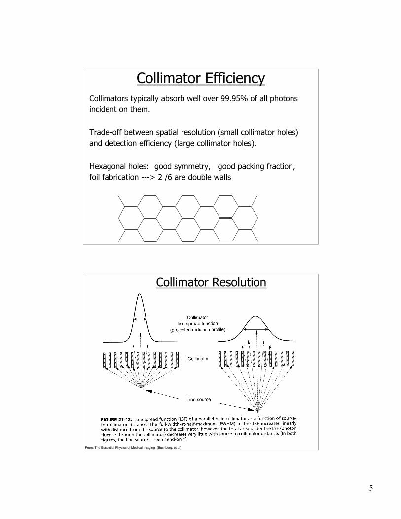
Collimator EfficiencyCollimators typically absorb well over 99.95% of all photonsincident on them.
Trade-off between spatial resolution (small collimator holes)and detection efficiency (large collimator holes).
Hexagonal holes: good symmetry, good packing fraction,foil fabrication ---> 2 /6 are double walls
From: The Essential Physics of Medical Imaging (Bushberg, et al)
Collimator Resolution

6
Gamma Camera - spatial resolution
Rs
= Ri
2+ R
c
2( )
From: Physics in Nuclear Medicine (Cherry, Sorenson and Phelps)
Types of Collimators
magnification

7
From: Physics in Nuclear Medicine (Cherry, Sorenson and Phelps)
Collimator: Resolution and Sensitivity
Collimator: Resolution and Sensitivity
From: The Essential Physics of Medical Imaging (Bushberg, et al)
8
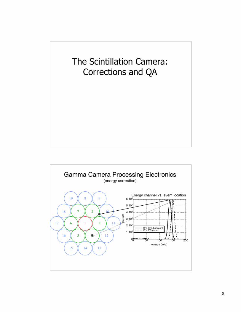
The Scintillation Camera:Corrections and QA
Gamma Camera Processing Electronics(energy correction)
2
3
45
6
7
8
10
11
12
131415
9
16
17
18
19
1
0
1 104
2 104
3 104
4 104
5 104
6 104
0 50 100 150 200
Energy channel vs. event location
10% ER (between)10% ER (over)
Co
un
ts
energy (keV)

9
Gamma Camera Processing Electronics(with and without energy correction)
Gamma Camera Processing Electronics(linearity correction)
2
3
45
6
7
8
10
11
12
131415
9
16
17
18
19
1
From: Physics in Nuclear Medicine (Cherry, Sorenson and Phelps)

10
Gamma Camera Processing Electronics(linearity correction)
Additional Gamma Camera Corrections(sensitivity / uniformity)
Acquired from long uniform flood after energyand linearity corrections have been applied
Multiplicative correction
Adjusts for slight variation in the detectionefficiency of the crystal
Compensates for small defects or damage tothe collimator
Should not be used to correct for largeirregularities

11
Daily Gamma Camera QA Tests
Photopeak window
Flood uniformity
From: The Essential Physics of Medical Imaging (Bushberg, et al)
Multienergy spatial registration (e.g., Ga-67 (93-, 185-, and 300 keV) gamma rays)
From: The Essential Physics of Medical Imaging (Bushberg, et al)
properly adjusted improperly adjusted

12
Pulse Pile-up
From: Physics in Nuclear Medicine (Sorenson and Phelps) and (Cherry, Sorenson and Phelps)
Energy spectra
Pile-up in image
Image Acquisition• Frame mode (data stored as an image)
- static- single image acquisition- can have multiple energy windows
- dynamic- series of images acquired sequentially
- gated- repetitive, dynamic imaging- used for cardiac imaging
• List-mode (data stored event by event)- time stamps are included within data stream- allows for flexible post-acquisition binning- can result in very large data files

13
Region of Interest (ROI) and Time-ActivityCurves (TAC)
From: The Essential Physics of Medical Imaging (Bushberg, et al)
To evaluate the hyperparathyroidism double phase technetium-99m sestamibiparathyroid scintigraphy was performed.Parathyroid Scintigraphy was performed 20 minutes and 2 hours after injection oftechnetium-99m-sestamibi.The 20 minute scan showed uptake in a normal appearing thyroid glandas well as uptake in two ovoid areas in the upper mediastinum.The 2 hour image showed wash out of activity from the thyroid, and persistence ofactivity in the upper mediastinum
Example Clinical Images

14
131I uptake in primary differentiatedthyroid carcinoma (arrow) and in riband pelvic metastases (arrowheads)
99mTc-MDP bone scintigraphy demonstratingmulti-focal increased uptake due to skeletalmetastases from a renal carcinoma – noteright nephrectomy
Example Clinical Images
Collimator artifacts(from high energy gammas
- 364 keV)
99mTc-MIBI scintimammography (supineand prone left lateral views) showing aprimary tumor in the left breast (arrow)and axillary lymph node metastases(arrowhead)
Example Clinical Images

15
201Tl
Example Clinical Images
99mTc
renal excretionwhole body