Overview of Revised CMC EMS System CE; 12 Lead EKG’s February 2009 CE Site Code #107200E1209...
107
Overview of Revised Overview of Revised CMC EMS System CE; CMC EMS System CE; 12 Lead EKG’s 12 Lead EKG’s February 2009 CE February 2009 CE Site Code #107200E1209 Site Code #107200E1209 Prepared by: Prepared by: Bill Glade, DC Wauconda Fire Departmen Bill Glade, DC Wauconda Fire Departmen Sharon Hopkins, RN, BSN Sharon Hopkins, RN, BSN
-
Upload
adele-martin -
Category
Documents
-
view
215 -
download
0
Transcript of Overview of Revised CMC EMS System CE; 12 Lead EKG’s February 2009 CE Site Code #107200E1209...

Overview of Revised Overview of Revised CMC EMS System CE;CMC EMS System CE;
12 Lead EKG’s12 Lead EKG’s
February 2009 CEFebruary 2009 CESite Code #107200E1209Site Code #107200E1209
Prepared by:Prepared by: Bill Glade, DC Wauconda Fire Department Bill Glade, DC Wauconda Fire Department
Sharon Hopkins, RN, BSNSharon Hopkins, RN, BSN

ObjectivesObjectives
Upon successful completion of this module, the Upon successful completion of this module, the EMS provider will be able to accomplish the EMS provider will be able to accomplish the following:following: Identify changes in the Advocate Condell EMS System Identify changes in the Advocate Condell EMS System
CE program as taught in class.CE program as taught in class. Identify the appropriate components of the cardiac Identify the appropriate components of the cardiac
conduction system with the correct wave form on a conduction system with the correct wave form on a rhythm strip.rhythm strip.
Identify when it is appropriate to obtain an EKGIdentify when it is appropriate to obtain an EKG Identify the criteria for significant ST elevation following Identify the criteria for significant ST elevation following
guidelines reviewed in class.guidelines reviewed in class. Identify EKG leads that view the anterior, inferior, Identify EKG leads that view the anterior, inferior,
lateral walls, and septumlateral walls, and septum

ObjectivesObjectives
Recognize the patterns of an MI after viewing the Recognize the patterns of an MI after viewing the components of a 12 lead EKGcomponents of a 12 lead EKG
Identify typical and atypical presentations of AMIIdentify typical and atypical presentations of AMI Identify complications associated with an inferior wall MIIdentify complications associated with an inferior wall MI Identify complications associated with an anterior/septal Identify complications associated with an anterior/septal
wall MIwall MI Identify complications associated with a lateral wall MIIdentify complications associated with a lateral wall MI Identify interventions for complications related to heart Identify interventions for complications related to heart
block, pulmonary edema, and cardiogenic shockblock, pulmonary edema, and cardiogenic shock Identify the SOP guidelines for the patient presenting with Identify the SOP guidelines for the patient presenting with
acute coronary syndrome asacute coronary syndrome as written in the Region X written in the Region X SOP’sSOP’s

ObjectivesObjectives
State dosing and precautions for Aspirin, State dosing and precautions for Aspirin, Nitroglycerin, and MorphineNitroglycerin, and Morphine
Identify ED staff expectations of EMS personnel when Identify ED staff expectations of EMS personnel when calling the hospital to report a patient with ST calling the hospital to report a patient with ST elevation identified on a 12 lead EKGelevation identified on a 12 lead EKG
Identify EMS expectations when delivering a patient Identify EMS expectations when delivering a patient to a hospital after ST elevation has been identified on to a hospital after ST elevation has been identified on a 12 lead EKGa 12 lead EKG
Actively participate in 12 lead EKG scenario practice Actively participate in 12 lead EKG scenario practice and discussionand discussion
Given a picture, correctly trace the order of the Given a picture, correctly trace the order of the cardiac conduction system.cardiac conduction system.
Given a manikin, correctly place electrodes to obtain Given a manikin, correctly place electrodes to obtain a 12 lead EKG.a 12 lead EKG.

CMC EMS CE Process For 2009CMC EMS CE Process For 2009
Educational committee formed to develop a new Educational committee formed to develop a new CE process that will be evolvingCE process that will be evolving
7 CE’s presented by EMS staff7 CE’s presented by EMS staff 2 CE’s presented by department members2 CE’s presented by department members Total of 27 hours of CE per year Total of 27 hours of CE per year Objectives and references for each CE sent to Objectives and references for each CE sent to
departments for previewdepartments for preview Each department will receive a detailed copy of Each department will receive a detailed copy of
the CE material for referencethe CE material for reference CE power points will continue to be posted on CE power points will continue to be posted on
the Condell websitethe Condell website

2009 CE Process2009 CE Process
All CE’s must be completed by year’s endAll CE’s must be completed by year’s end Medical Officer will oversee the completion for Medical Officer will oversee the completion for
those not completing during EMS staff those not completing during EMS staff presentationpresentation
There will no longer be biannual examsThere will no longer be biannual exams Quizzes will be administered at the completion Quizzes will be administered at the completion
of each CEof each CE Successful completion is at 80%Successful completion is at 80% Number of quiz questions may be variable dependent Number of quiz questions may be variable dependent
on topic and will be based on objectiveson topic and will be based on objectives Handouts at class will only be material Handouts at class will only be material
applicable to complete that topic and no longer applicable to complete that topic and no longer the full power points the full power points

Why Are We doing Pre-hospital Why Are We doing Pre-hospital EKG’s?EKG’s?
Early recognition and fast, appropriate Early recognition and fast, appropriate treatment can prevent the extension of an treatment can prevent the extension of an MIMI
Early recognition = early interventionEarly recognition = early intervention
An important diagnostic tool will also be An important diagnostic tool will also be the patient’s general appearancethe patient’s general appearance

Cardiac Conduction SystemCardiac Conduction System
Electrical cells arranged in a systematic Electrical cells arranged in a systematic pathwaypathway
Predominant pacemaker starting the Predominant pacemaker starting the electrical flow comes from the SA nodeelectrical flow comes from the SA node
Electrical cells are part of the conduction Electrical cells are part of the conduction systemsystem
Muscle cells are the mechanical cellsMuscle cells are the mechanical cells
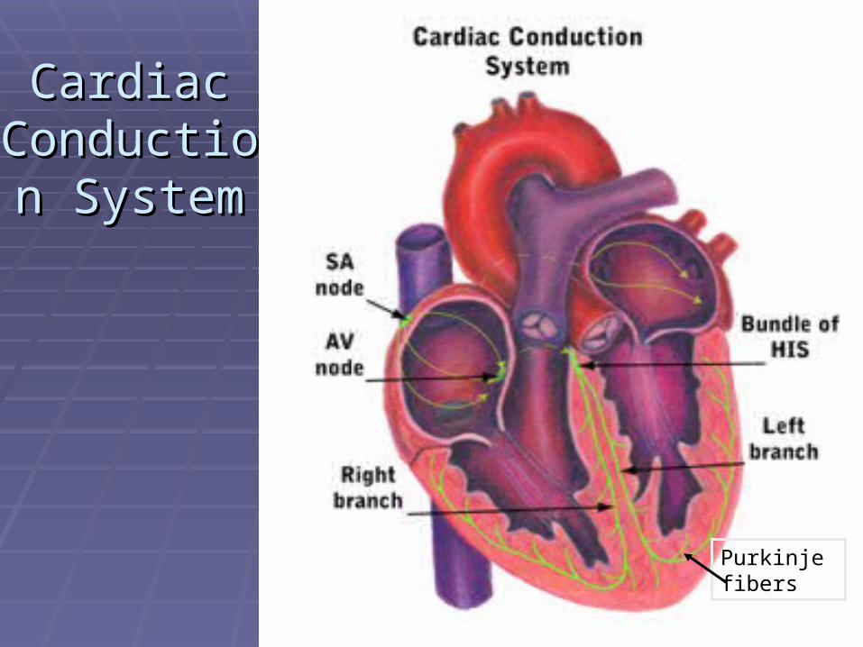
Cardiac Cardiac Conduction Conduction
SystemSystem
Purkinje fibers

EKG WaveformsEKG Waveforms
P wave represents atrial stimulationP wave represents atrial stimulation P wave is rounded and uprightP wave is rounded and upright
PR intervalPR interval Includes the P wave and the isoelectric PR Includes the P wave and the isoelectric PR
segmentsegment PR interval is the time it takes for an impulse PR interval is the time it takes for an impulse
to travel from the SA node through the to travel from the SA node through the internodal pathways toward the ventriclesinternodal pathways toward the ventricles
Includes delay time in the AV nodeIncludes delay time in the AV node Normal PR interval is Normal PR interval is 0.12 – 0.20 seconds0.12 – 0.20 seconds

PR PR IntervalInterval

PR Interval AbnormalitiesPR Interval Abnormalities
PR interval <0.12 secondsPR interval <0.12 seconds Impulse did not begin in the normal Impulse did not begin in the normal
pacemaker site of the SA node but pacemaker site of the SA node but somewhere in the atriasomewhere in the atria
PR interval >0.20 secondsPR interval >0.20 seconds There was a longer than normal delay There was a longer than normal delay
transmitting the impulse through the AV nodetransmitting the impulse through the AV node A change in the PR interval measurement A change in the PR interval measurement
generally will not make the patient symptomaticgenerally will not make the patient symptomatic

EKG Wave Forms cont’dEKG Wave Forms cont’d
QRS complexQRS complex Consists of the Q, R, and S waves collectivelyConsists of the Q, R, and S waves collectively Represents ventricular depolarization or discharge of Represents ventricular depolarization or discharge of
electrical energy throughout ventricular muscleelectrical energy throughout ventricular muscle Larger than the P wave because ventricular Larger than the P wave because ventricular
depolarization involves a larger muscle mass than depolarization involves a larger muscle mass than atrial depolarizationatrial depolarization
Palpation of a pulse is generated by ventricular Palpation of a pulse is generated by ventricular depolarization (seen as the QRS complex)depolarization (seen as the QRS complex)
Normal timing usually considered between 0.06 Normal timing usually considered between 0.06 and 0.11 secondsand 0.11 seconds Normal is less than 0.12 secondsNormal is less than 0.12 seconds

QRS ComplexQRS Complex

QRS Complex MeasurementQRS Complex Measurement
From beginning of Q wave – usually fairly From beginning of Q wave – usually fairly straight forwardstraight forward
Stop measurement at end of S wave; not Stop measurement at end of S wave; not necessarily where QRS intersects baseline necessarily where QRS intersects baseline
On S wave, watch for small notch or other On S wave, watch for small notch or other indicator that electrical flow is changingindicator that electrical flow is changingNot always so easy to determine stop pointNot always so easy to determine stop point
Do not include ST segment or T waveDo not include ST segment or T wave Abnormally wide QRS indicates delay in Abnormally wide QRS indicates delay in
conduction time through the ventriclesconduction time through the ventricles

EKG Wave Forms cont’dEKG Wave Forms cont’d
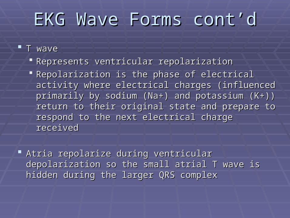
T waveT wave Represents ventricular repolarizationRepresents ventricular repolarization Repolarization is the phase of electrical activity Repolarization is the phase of electrical activity
where electrical charges (influenced primarily by where electrical charges (influenced primarily by sodium (Na+) and potassium (K+)) return to their sodium (Na+) and potassium (K+)) return to their original state and prepare to respond to the next original state and prepare to respond to the next electrical charge receivedelectrical charge received
Atria repolarize during ventricular depolarization so the Atria repolarize during ventricular depolarization so the small atrial T wave is hidden during the larger QRS small atrial T wave is hidden during the larger QRS complexcomplex

When To Obtain a 12-Lead EKGWhen To Obtain a 12-Lead EKG
Any patient presenting with signs and/or symptoms of Any patient presenting with signs and/or symptoms of an acute coronary syndromean acute coronary syndrome
Consider atypical AMI presentationsConsider atypical AMI presentations ElderlyElderly WomenWomen Patient with long standing history of diabetesPatient with long standing history of diabetes
Any patient presenting with a Second degree Type II Any patient presenting with a Second degree Type II (classical) or 3(classical) or 3rdrd degree heart block degree heart block Consider the origin from an AMI until proven Consider the origin from an AMI until proven
otherwiseotherwise

What Are We Looking For?What Are We Looking For? Abnormalities that indicate interruption in the blood Abnormalities that indicate interruption in the blood
flow to the myocardiumflow to the myocardium Plaque formation diminishes blood flow through the Plaque formation diminishes blood flow through the
coronary arteriescoronary arteriesPatients may be asymptomatic while damage Patients may be asymptomatic while damage
silently developssilently develops Plaque rupture begins a cascade of events that Plaque rupture begins a cascade of events that
further compromises blood flow through the injured further compromises blood flow through the injured vessel(s)vessel(s)
This cascade of events could lead to an acute This cascade of events could lead to an acute coronary syndrome (ie: acute MI)coronary syndrome (ie: acute MI)

Coronary CirculationCoronary Circulation
Coronary arteries and veinsCoronary arteries and veins Myocardium extracts the largest amount of Myocardium extracts the largest amount of
oxygen as blood moves into general oxygen as blood moves into general circulationcirculation
Oxygen uptake by the myocardium can Oxygen uptake by the myocardium can only improve by increasing blood flow only improve by increasing blood flow through the coronary arteriesthrough the coronary arteries
If the coronary arteries are blocked, they If the coronary arteries are blocked, they must be reopened if circulation is going to must be reopened if circulation is going to be restored to that area of tissue suppliedbe restored to that area of tissue supplied


12-Lead Electrodes12-Lead Electrodes
A lead is a tracing of the electrical activity A lead is a tracing of the electrical activity between 2 electrodesbetween 2 electrodes
Leads view the heart from the front of the bodyLeads view the heart from the front of the body Top, bottom, right, and left side of heartTop, bottom, right, and left side of heart
Leads view the heart as if it were sliced in half Leads view the heart as if it were sliced in half horizontallyhorizontally Front, back, right, and left sides of heartFront, back, right, and left sides of heart
Each lead has a positive and a negative Each lead has a positive and a negative electrodeelectrode

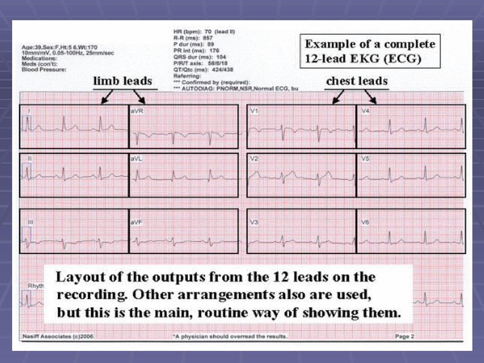
Standard 12-Lead EKGStandard 12-Lead EKG
Six limb leadsSix limb leads Leads I, II, III, aVR, aVL, aVFLeads I, II, III, aVR, aVL, aVF
Six chest leads (precordial leads)Six chest leads (precordial leads) V1, V2, V3, V4, V5, V6V1, V2, V3, V4, V5, V6
Information from 12 leads obtained Information from 12 leads obtained from the attachment of only 10 from the attachment of only 10 electrodeselectrodes
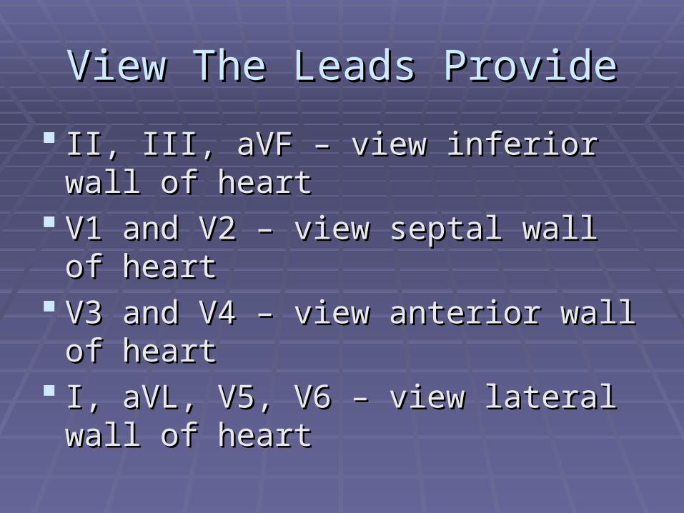
View The Leads ProvideView The Leads Provide
II, III, aVF – view inferior wall of heartII, III, aVF – view inferior wall of heart V1 and V2 – view septal wall of heartV1 and V2 – view septal wall of heart V3 and V4 – view anterior wall of V3 and V4 – view anterior wall of
heartheart I, aVL, V5, V6 – view lateral wall of I, aVL, V5, V6 – view lateral wall of
heartheart

Preparation for 12 Lead EKG Preparation for 12 Lead EKG Skin preparationSkin preparation
Hair removalHair removalclip hair if necessary so electrodes adhereclip hair if necessary so electrodes adhere
Clean and dry skin surfaceClean and dry skin surfacegently rub skin area with gauze padgently rub skin area with gauze pad
need to remove skin oils & dead skinneed to remove skin oils & dead skinif diaphoretic patient wipe with if diaphoretic patient wipe with
towel/gauze or use antiperspirant spraytowel/gauze or use antiperspirant spray

Patient positioningPatient positioning Preferably flatPreferably flat
Heart rotates position as the patient Heart rotates position as the patient position changesposition changes
If patient is elevated, note that If patient is elevated, note that information on the EKGinformation on the EKG

Precordial Chest LeadsPrecordial Chest Leads
For For everyevery person, each precordial lead placed in person, each precordial lead placed in the same relative positionthe same relative position
V1 - 4V1 - 4thth intercostal space, R of sternum intercostal space, R of sternumV2 - 4V2 - 4thth intercostal space, L of sternum intercostal space, L of sternumV4 - 5V4 - 5thth intercostal space, midclavicular intercostal space, midclavicularV3 - between V2 and V4, on 5V3 - between V2 and V4, on 5thth rib rib V5 - 5V5 - 5thth intercostal space, anterior axillary line intercostal space, anterior axillary lineV6 - 5V6 - 5thth intercostal space, mid-axillary line intercostal space, mid-axillary line

Precordial Precordial LeadsLeads
2nd ICS
3rd ICS
1st ICS


12 Lead EKG Printout12 Lead EKG Printout
Standard format 8Standard format 811//22 x 11 x 11 paper paper
12 lead format:12 lead format:
II aVR aVR V1 V1 V4 V4
IIII aVL aVL V2 V2 V5 V5
IIIIII aVF aVF V3 V3 V6 V6
Machines can analyze data obtained Machines can analyze data obtained but humans must interpret databut humans must interpret data

Lateral View – I, aVL, V5, V6Lateral View – I, aVL, V5, V6
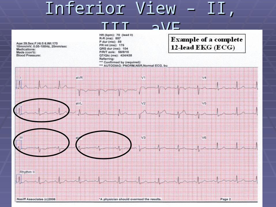
Inferior View – II, III, aVFInferior View – II, III, aVF

Septal View – V1 & V2Septal View – V1 & V2

Anterior View – V3 & V4Anterior View – V3 & V4

Myocardial InsultMyocardial Insult IschemiaIschemia
lack of oxygenationlack of oxygenation ST depression or T wave inversionST depression or T wave inversion permanent damage avoidablepermanent damage avoidable
InjuryInjury prolonged ischemiaprolonged ischemia ST elevationST elevation permanent damage avoidablepermanent damage avoidable
InfarctInfarct death of myocardial tissue; damage permanent; may have death of myocardial tissue; damage permanent; may have
Q waveQ wave

Why A Pre-hospital EKG?Why A Pre-hospital EKG?
EMS looking for ST segment elevationEMS looking for ST segment elevation Indicates injury that can be reversible if found Indicates injury that can be reversible if found
early and acted upon earlyearly and acted upon early TIME IS MUSCLETIME IS MUSCLE The earlier the discovery of an acute cardiac The earlier the discovery of an acute cardiac
event, the quicker the patient can receive the event, the quicker the patient can receive the most appropriate caremost appropriate care
EKG’s sent to the ED before patient arrival EKG’s sent to the ED before patient arrival allows for the right personnel to be available to allows for the right personnel to be available to properly care for the patientproperly care for the patient in the most time in the most time efficient mannerefficient manner
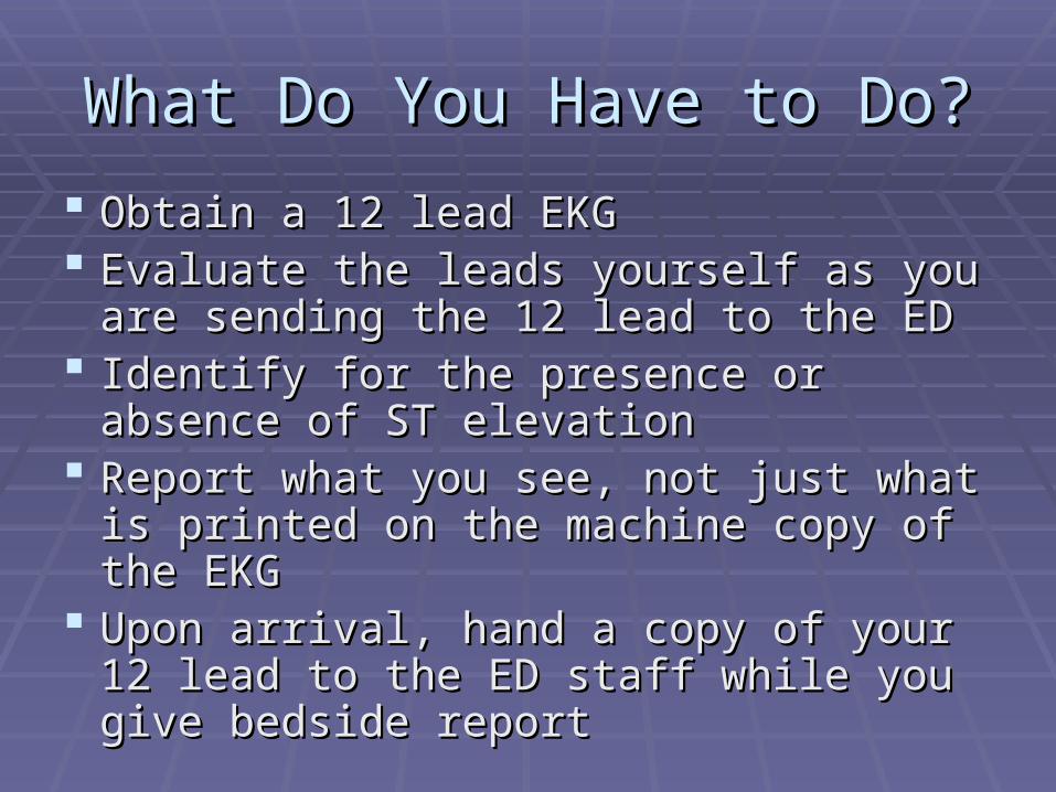
What Do You Have to Do?What Do You Have to Do?
Obtain a 12 lead EKGObtain a 12 lead EKG Evaluate the leads yourself as you are Evaluate the leads yourself as you are
sending the 12 lead to the EDsending the 12 lead to the ED Identify for the presence or absence of ST Identify for the presence or absence of ST
elevationelevation Report what you see, not just what is Report what you see, not just what is
printed on the machine copy of the EKGprinted on the machine copy of the EKG Upon arrival, hand a copy of your 12 lead Upon arrival, hand a copy of your 12 lead
to the ED staff while you give bedside to the ED staff while you give bedside reportreport
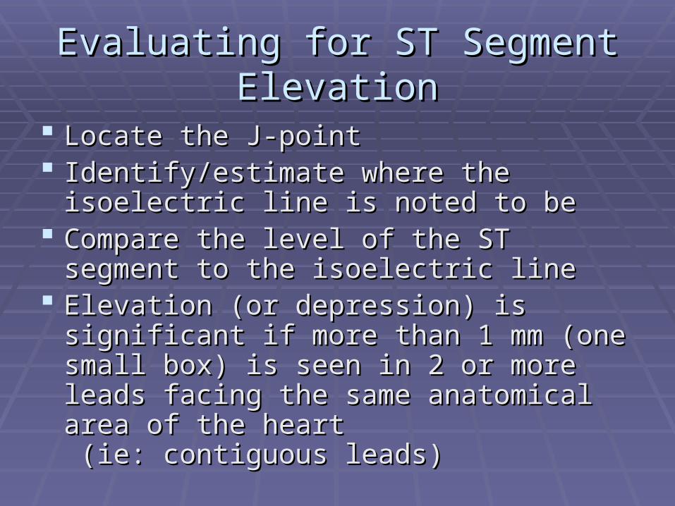
Evaluating for ST Segment Evaluating for ST Segment ElevationElevation
Locate the J-pointLocate the J-point Identify/estimate where the isoelectric line Identify/estimate where the isoelectric line
is noted to beis noted to be Compare the level of the ST segment to Compare the level of the ST segment to
the isoelectric linethe isoelectric line Elevation (or depression) is significant if Elevation (or depression) is significant if
more than 1 mm (one small box) is seen in more than 1 mm (one small box) is seen in 2 or more leads facing the same 2 or more leads facing the same anatomical area of the heart anatomical area of the heart (ie: contiguous leads)(ie: contiguous leads)

J point – where the QRS complex and ST J point – where the QRS complex and ST segment meetsegment meet
ST segment elevation - evaluated 0.04 seconds ST segment elevation - evaluated 0.04 seconds (one small box) after J point (one small box) after J point
The J PointThe J Point

Coved Coved shape shape usually usually indicates indicates acute injuryacute injury
Concave Concave shape is shape is usually usually benign benign especially if especially if patient is patient is asympto-asympto-matic matic
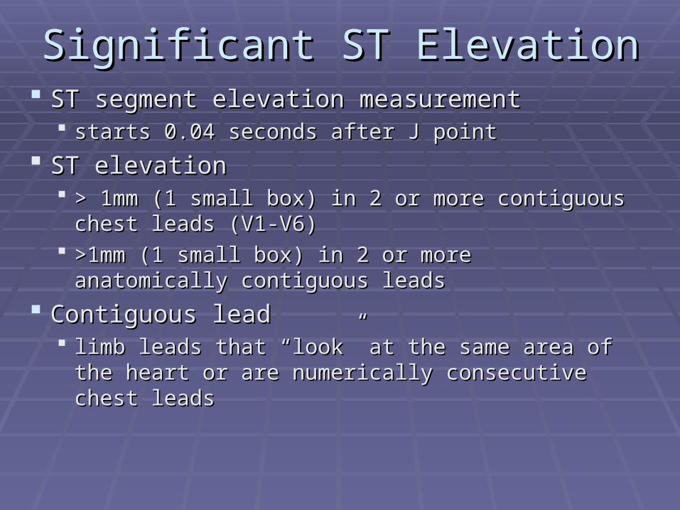
Significant ST ElevationSignificant ST Elevation ST segment elevation measurementST segment elevation measurement
starts 0.04 seconds after J pointstarts 0.04 seconds after J point
ST elevationST elevation > 1mm (1 small box) in 2 or more contiguous chest > 1mm (1 small box) in 2 or more contiguous chest
leads (V1-V6)leads (V1-V6) >1mm (1 small box) in 2 or more anatomically >1mm (1 small box) in 2 or more anatomically
contiguous leadscontiguous leads
Contiguous leadContiguous lead limb leads that “look” at the same area of the heart limb leads that “look” at the same area of the heart
or are numerically consecutive chest leadsor are numerically consecutive chest leads

Contiguous LeadsContiguous Leads
Lateral wall: I, aVL, V5, V6Lateral wall: I, aVL, V5, V6 Inferior wall: II, III, avFInferior wall: II, III, avF Septum: V1 and V2Septum: V1 and V2 Anterior wall: V3 and V4Anterior wall: V3 and V4 Posterior wall: V7-V9 (leads placed Posterior wall: V7-V9 (leads placed
on the patient’s back 5on the patient’s back 5thth intercostal intercostal space creating a 15 lead EKG)space creating a 15 lead EKG)

Evolution of AMIEvolution of AMIA - pre-infarct (normal)A - pre-infarct (normal)
B - Tall T wave (B - Tall T wave (first few first few minutes of infarctminutes of infarct))
C - Tall T wave C - Tall T wave andand ST ST elevation (elevation (injuryinjury))
D - Elevated ST (D - Elevated ST (injuryinjury), ), inverted T wave (inverted T wave (ischemiaischemia), ), Q wave (Q wave (tissue deathtissue death))
E - Inverted T wave E - Inverted T wave ((ischemiaischemia), Q wave (), Q wave (tissue tissue deathdeath))
F - Q wave (F - Q wave (permanent permanent marking) marking)

ST Segment ST Segment ElevationElevation

EKG monitoringEKG monitoring Evaluates electrical activity of the heartEvaluates electrical activity of the heart Can indicate myocardial insult and locationCan indicate myocardial insult and location
ischemiaischemia - initial insult; ST depression seen - initial insult; ST depression seeninjuryinjury - prolonged myocardial hypoxia or - prolonged myocardial hypoxia or
ischemia; ST elevation seenischemia; ST elevation seeninfarctioninfarction - tissue death - tissue death
dead tissue no longer contractsdead tissue no longer contracts amount of dead tissue directly relates to amount of dead tissue directly relates to
degree of muscle impairmentdegree of muscle impairment may show Q waves may show Q waves

Contiguous ECG LeadsContiguous ECG Leads EKG changes are EKG changes are
significant when they significant when they are seen in at least two are seen in at least two contiguouscontiguous leads leads
Two leads are Two leads are contiguous if they look contiguous if they look at the same area of the at the same area of the heart or they are heart or they are numerically consecutive numerically consecutive chest leadschest leads

Groups of EKG LeadsGroups of EKG Leads Inferior wall - II, III, aVFInferior wall - II, III, aVF Septal wall - V1, V2Septal wall - V1, V2 Anterior wall - V3, V4Anterior wall - V3, V4 Lateral wall - I, aVL, V5, V6 Lateral wall - I, aVL, V5, V6
aVR is not evaluated in typical groups aVR is not evaluated in typical groups Standard lead placement does not look at Standard lead placement does not look at
posterior wall or right ventricle of the heart - need posterior wall or right ventricle of the heart - need special lead placement for these viewsspecial lead placement for these views

Basic 12-Lead EKG FormatBasic 12-Lead EKG Format
Lead ILateral wall
aVR not evaluated
V1
Septum
V4
Anterior wall
Lead II Inferior wall
aVLLateral wall
V2
Septum
V5
Lateral wall
Lead III Inferior wall
aVFInferior wall
V3
Anterior
V6
Lateral wall
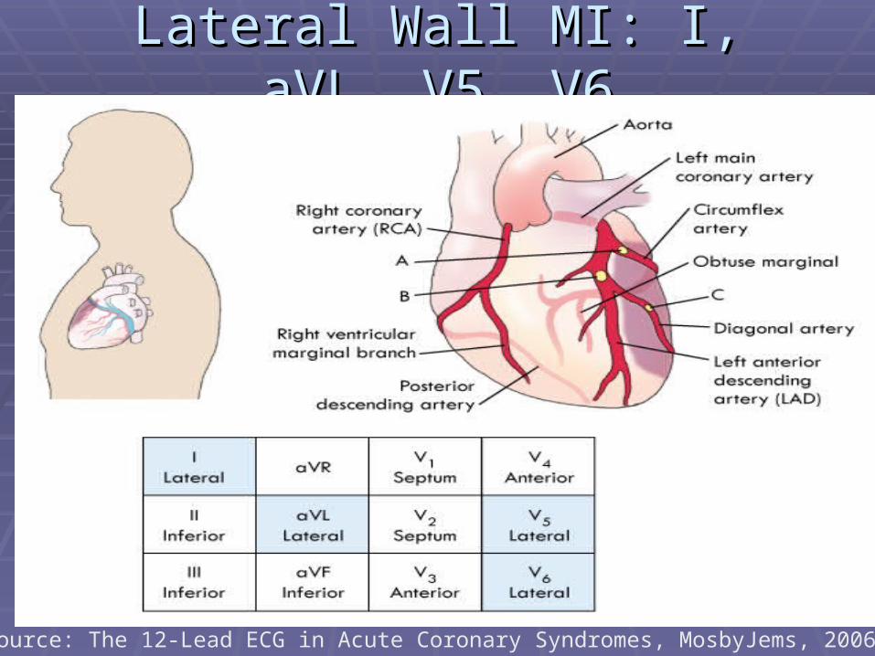
Lateral Wall MI: I, aVL, V5, V6Lateral Wall MI: I, aVL, V5, V6
Source: The 12-Lead ECG in Acute Coronary Syndromes, MosbyJems, 2006.

Inferior Wall MI II, III, aVFInferior Wall MI II, III, aVF
Source: The 12-Lead ECG in Acute Coronary Syndromes, MosbyJems, 2006.

Septal MI: Leads V1 and V2Septal MI: Leads V1 and V2
Source: The 12-Lead ECG in Acute Coronary Syndromes, MosbyJems, 2006.

Anterior Wall MI V3, V4Anterior Wall MI V3, V4
Source: The 12-Lead ECG in Acute Coronary Syndromes, MosbyJems, 2006.

Posterior MI – Reciprocal Changes Posterior MI – Reciprocal Changes ST Depression V1, V2, V3, poss V4ST Depression V1, V2, V3, poss V4
Source: The 12-Lead ECG in Acute Coronary Syndromes, MosbyJems, 2006.

Complications of Lateral Wall MIComplications of Lateral Wall MI
I, aVL, V5,V6I, aVL, V5,V6 Complications arise due to the conduction Complications arise due to the conduction
components that are in the septumcomponents that are in the septum Conduction dysrhythmias most commonConduction dysrhythmias most common
Second degree Type II – classicalSecond degree Type II – classical 33rdrd degree – complete heart block degree – complete heart block Bundle branch blocksBundle branch blocks
Monitor patient closely for these blocksMonitor patient closely for these blocks 22ndnd degree Type II and 3 degree Type II and 3rdrd degree are serious degree are serious
dysrhythmias that need to be treated aggressively dysrhythmias that need to be treated aggressively with TCPwith TCP

Complications of Inferior Wall MIComplications of Inferior Wall MI
II, III, aVFII, III, aVF 40% of patients with inferior MI’s have right ventricular 40% of patients with inferior MI’s have right ventricular
infarcts infarcts In the presence of a right ventricular infarct, there is a In the presence of a right ventricular infarct, there is a
high likeliness of both ventricles being damagedhigh likeliness of both ventricles being damaged Contraction capabilities will be negatively affected Contraction capabilities will be negatively affected
Patients may present hypotensivePatients may present hypotensive Nitrates and Morphine alone will dilate blood vessels Nitrates and Morphine alone will dilate blood vessels
worsening hypotensionworsening hypotension Under Medical Control direction patients are often Under Medical Control direction patients are often
treated with a fluid challenge with the nitrates treated with a fluid challenge with the nitrates 11stst degree heart block and Second degree Type I degree heart block and Second degree Type I
Wenckebach most common heart blocksWenckebach most common heart blocks

Complications of Septal Wall MIComplications of Septal Wall MI
V1 and V2V1 and V2 Significant amount of conduction components Significant amount of conduction components
are in the septal areaare in the septal area Patient predisposed to dysrhythmiaPatient predisposed to dysrhythmia
Second degree Type II – classicalSecond degree Type II – classical 33rdrd degree heart block degree heart block Bundle branch blockBundle branch block
Lethal heart blocks treated aggressively - TCPLethal heart blocks treated aggressively - TCP Rare to have a septal MI aloneRare to have a septal MI alone
Common to have anterior or lateral involvement along Common to have anterior or lateral involvement along with septal areawith septal area
Complications of Anterior Wall MIComplications of Anterior Wall MI
V3, V4V3, V4 Known as the “widowmaker” due to the potential Known as the “widowmaker” due to the potential
for a massive area of infarction from blockage of for a massive area of infarction from blockage of the large amount of myocardium supplied by the the large amount of myocardium supplied by the LAD (left anterior descending artery)LAD (left anterior descending artery)
Often the septal or lateral walls are also involvedOften the septal or lateral walls are also involved Watch for lethal ventricular dysrhythmias and Watch for lethal ventricular dysrhythmias and
cardiogenic shockcardiogenic shock Second degree Type II and 3Second degree Type II and 3rdrd degree heart degree heart
block are more common than other blocksblock are more common than other blocks

Anterior Wall MI - V3, V4Anterior Wall MI - V3, V4
Early death within a few days often from CHFEarly death within a few days often from CHF Massive area of ventricular tissue infarcted if LAD Massive area of ventricular tissue infarcted if LAD
totally occludedtotally occluded
Important to obtain history of recent MI Important to obtain history of recent MI diagnosis and hospital dischargediagnosis and hospital discharge Increased incidence of ventricular tachycardia Increased incidence of ventricular tachycardia
(VT) and ventricular fibrillation (VF) up to 1 -2 (VT) and ventricular fibrillation (VF) up to 1 -2 weeks post acute anterior MIweeks post acute anterior MI

Additional Complications Additional Complications
Acute pulmonary edemaAcute pulmonary edema Nitroglycerin to dilate blood vessels and Nitroglycerin to dilate blood vessels and
reduce preloadreduce preload Lasix to dilate blood vessels and reduce Lasix to dilate blood vessels and reduce
preload; as a diureticpreload; as a diuretic Morphine to dilate blood vessels and reduce Morphine to dilate blood vessels and reduce
preload; reduce anxietypreload; reduce anxiety

Additional ComplicationsAdditional Complications Cardiogenic shockCardiogenic shock
Ineffective pumping from the damaged heartIneffective pumping from the damaged heart IV fluid challenge if lung sounds are clearIV fluid challenge if lung sounds are clear Dopamine drip titrated to maintain a systolic Dopamine drip titrated to maintain a systolic
blood pressure of blood pressure of >>100 mmHg100 mmHgStart at a low dose (5mcg/kg/min)Start at a low dose (5mcg/kg/min)
Estimate the patient’s pounds (ie: 100 #)Estimate the patient’s pounds (ie: 100 #) Take the 1Take the 1stst 2 numbers dropping the last 2 numbers dropping the last
number (“10”)number (“10”) This is the starting point for This is the starting point for
minidrips/minute (8 minidrips/minute)minidrips/minute (8 minidrips/minute)

Common Terms Patients Common Terms Patients Use To Describe Chest Pain Use To Describe Chest PainHeavinessHeaviness
Pressing Pressing
Suffocating Suffocating
Squeezing Squeezing
StranglingStrangling
Burning Burning
Constricting bandConstricting band
A weight in the A weight in the center of my chest center of my chest
A vise tightening A vise tightening around my chestaround my chest

Additional Patient Complaints or Additional Patient Complaints or PresentationsPresentations
Difficulty breathingDifficulty breathingExcessive sweatingExcessive sweatingUnexplained nausea Unexplained nausea
or vomitingor vomitingGeneralized Generalized
weaknessweaknessDizzinessDizziness
Syncope or near-Syncope or near-syncopesyncope
PalpitationsPalpitationsIsolated arm or jaw Isolated arm or jaw
painpainFatigueFatigueDysrhythmiasDysrhythmias
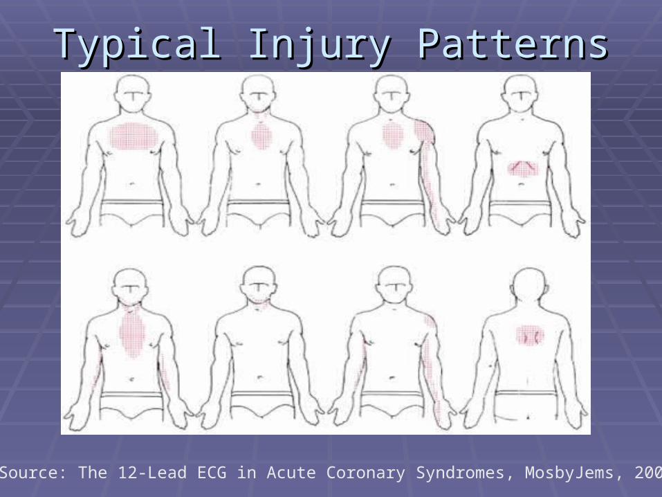
Typical Injury PatternsTypical Injury Patterns
Source: The 12-Lead ECG in Acute Coronary Syndromes, MosbyJems, 2006.

Atypical PresentationAtypical Presentation in the in the ElderlyElderly Most frequent symptoms of acute MI:Most frequent symptoms of acute MI:
Shortness of breathShortness of breath
Fatigue and weakness (“I just don’t feel well”)Fatigue and weakness (“I just don’t feel well”)
Abdominal or epigastric discomfortAbdominal or epigastric discomfort
Often have preexisting conditions making this an Often have preexisting conditions making this an already vulnerable populationalready vulnerable population
HypertensionHypertension
CHFCHF
Previous AMIPrevious AMI
Likely to delay seeking treatmentLikely to delay seeking treatment

Atypical PresentationAtypical Presentation in in WomenWomen Discomfort described as:Discomfort described as:
AchingAchingTightnessTightnessPressurePressureSharpnessSharpnessBurningBurningFullnessFullnessTingling Tingling
Often have no actual chest pain to offer as a Often have no actual chest pain to offer as a complaint. Often the pain is in the back, complaint. Often the pain is in the back, shoulders, or neckshoulders, or neck
Frequent acute Frequent acute symptoms:symptoms:Shortness of breathShortness of breathWeaknessWeaknessUnusual fatigueUnusual fatigueCold sweatsCold sweatsDizzinessDizzinessNausea/vomitingNausea/vomiting

Atypical PresentationAtypical Presentation in the in the Patient With Patient With DiabetesDiabetes
Atypical presentation due to autonomic Atypical presentation due to autonomic dysfunctiondysfunction
Common signs/symptoms:Common signs/symptoms: Generalized weaknessGeneralized weakness Generalized feeling of not being wellGeneralized feeling of not being well SyncopeSyncope LightheadednessLightheadedness Change in mental statusChange in mental status
Region X SOP – Acute Coronary Region X SOP – Acute Coronary SyndromeSyndrome
A 12 lead EKG is obtained on all patients A 12 lead EKG is obtained on all patients presenting with signs and symptoms of presenting with signs and symptoms of acute MIacute MI
OROR For patients where suspicions are raised For patients where suspicions are raised
that the patient may be experiencing an that the patient may be experiencing an acute MI (ie: heart block)acute MI (ie: heart block)

12-Lead Electrode Placement12-Lead Electrode Placement

Region X SOP – Acute Coronary Region X SOP – Acute Coronary SyndromeSyndrome
Determine if the patient is stable or Determine if the patient is stable or unstable to proceed with interventionsunstable to proceed with interventions
Easiest way to determine stability is to Easiest way to determine stability is to evaluate blood flow evaluate blood flow What is the level of consciousness?What is the level of consciousness? What is the blood pressure / is there a radial What is the blood pressure / is there a radial
pulse?pulse? Remember: A B/P reading of 100/systolic Remember: A B/P reading of 100/systolic
does not necessarily indicate the presence does not necessarily indicate the presence or absence of symptomsor absence of symptoms

OxygenOxygen
In the presence of an acute MI, the In the presence of an acute MI, the myocardium is being deprived of blood myocardium is being deprived of blood flow and therefore adequate oxygen levelsflow and therefore adequate oxygen levels
Provide what the patient needsProvide what the patient needs Evaluate each individual clinical Evaluate each individual clinical
presentationpresentation All patients deserve some form of oxygen All patients deserve some form of oxygen
in this early period of myocardial starvation in this early period of myocardial starvation for itfor it
AspirinAspirin
Used to prevent platelet aggregationUsed to prevent platelet aggregation When a plague ruptures, chemicals are released. When a plague ruptures, chemicals are released.
Platelets congregate to the area to seal the rupture. Platelets congregate to the area to seal the rupture. Platelet aggregation further increases the degree of Platelet aggregation further increases the degree of vessel blockage.vessel blockage.
Dosage is 4 – 81 mg (324 mg total) baby aspirin Dosage is 4 – 81 mg (324 mg total) baby aspirin chewedchewed Chewing breaks down the aspirin and allows for faster Chewing breaks down the aspirin and allows for faster
absorptionabsorption Give dose if patient not reliable about taking Give dose if patient not reliable about taking
their own dose or has not taken any aspirintheir own dose or has not taken any aspirin

NitroglycerinNitroglycerin VenodilatorVenodilator
Improves coronary blood flowImproves coronary blood flow By dilating blood vessels, pools blood away By dilating blood vessels, pools blood away
from the heart which decreases preload. This from the heart which decreases preload. This decreases the work load of a stressed heart.decreases the work load of a stressed heart.
Carefully monitor blood pressure before and Carefully monitor blood pressure before and after dosagesafter dosages
Dosage is 0.4 mg tablet slDosage is 0.4 mg tablet sl Dosage can be repeated in 5 minutes if blood Dosage can be repeated in 5 minutes if blood
pressure remains stablepressure remains stable FYI: Pain level will not drop to “0” until the clot FYI: Pain level will not drop to “0” until the clot
is removedis removed
For CMC EMS System ParticipantsFor CMC EMS System Participants
If the patient is <35 years of ageIf the patient is <35 years of age Follow Acute coronary Syndrome SOP by Follow Acute coronary Syndrome SOP by
administering aspirinadministering aspirin Contact Medical control prior to administration of Contact Medical control prior to administration of
nitroglycerin or morphinenitroglycerin or morphine
There should be no delay in obtaining a 12 lead There should be no delay in obtaining a 12 lead EKG in the field and transmitting it to the EDEKG in the field and transmitting it to the ED
Your visual interpretation is to be given during Your visual interpretation is to be given during report to the receiving hospital report to the receiving hospital

MorphineMorphine CNS depressant to reduce anxietyCNS depressant to reduce anxiety Venodilates blood vessels to reduce the Venodilates blood vessels to reduce the
volume of blood returning to the heart to volume of blood returning to the heart to decrease the heart’s workloaddecrease the heart’s workload
Dosage is 2 mg slow IVPDosage is 2 mg slow IVP Dosage started when the 2Dosage started when the 2ndnd dose of dose of
nitroglycerin proves ineffectivenitroglycerin proves ineffective Dosage may be repeated every 2 minutes as Dosage may be repeated every 2 minutes as
neededneeded Maximum dosage is 10 mgMaximum dosage is 10 mg
Watch for hypotension Watch for hypotension

Receiving Hospital ReportReceiving Hospital Report
When sending a 12 lead EKG, inform the When sending a 12 lead EKG, inform the receiving hospital what identifiers have receiving hospital what identifiers have been usedbeen used Department ID numberDepartment ID number Patient sex (M / F)Patient sex (M / F) Patient agePatient age Any other identifierAny other identifier
Always give your visual interpretation of Always give your visual interpretation of what you have observed for ST elevationwhat you have observed for ST elevation

Activating a Cardiac AlertActivating a Cardiac Alert The ED activates a cardiac alert to prepare the The ED activates a cardiac alert to prepare the
cardiac team to provide optimal care for the cardiac team to provide optimal care for the patientpatient
Typical cardiac alert team membersTypical cardiac alert team members ED staff – MD, RN, tech, secretaryED staff – MD, RN, tech, secretary CardiologistCardiologist Cath lab personnelCath lab personnel EKG tech (may be an ED staff member)EKG tech (may be an ED staff member) Lab techLab tech X-ray techX-ray tech
Not all hospitals use all members in a formalized Not all hospitals use all members in a formalized team but all of these members are somehow team but all of these members are somehow integrated into the care of the patientintegrated into the care of the patient

When Does a Cardiac Alert Get When Does a Cardiac Alert Get Called?Called?
When you send a 12 lead EKG with ST When you send a 12 lead EKG with ST elevation, the team gets activatedelevation, the team gets activated
When you confirm what you see on the 12 When you confirm what you see on the 12 lead, whether the EKG is sent or not, may lead, whether the EKG is sent or not, may trigger a cardiac alerttrigger a cardiac alert
There is a direct link in your accuracy, There is a direct link in your accuracy, completeness in patient report, and EKG completeness in patient report, and EKG interpretation with pre-hospital activation interpretation with pre-hospital activation of the cardiac alert teamof the cardiac alert team

Transferring Care of The Patient to Transferring Care of The Patient to The EDThe ED
Bedside report is restated to the ED Bedside report is restated to the ED personnel in the roompersonnel in the room The main report must be to an RN or MDThe main report must be to an RN or MD
Rhythm strips and 12 lead EKG are Rhythm strips and 12 lead EKG are presentedpresented
Important to note positive and negative Important to note positive and negative changes in the patient conditionchanges in the patient condition Pain level has decreasedPain level has decreased Blood pressure has droppedBlood pressure has dropped

DocumentationDocumentation
Follow OPQRST guidelinesFollow OPQRST guidelines Some of this information is added into a check Some of this information is added into a check
box or other prompt; otherwise the information is box or other prompt; otherwise the information is written into the narrativewritten into the narrative
OOnset – what was the patient doing when the nset – what was the patient doing when the problem/pain began? Any contributing factors? problem/pain began? Any contributing factors?
Add this information to the narrative.Add this information to the narrative. PProvocation/palliation – what makes the pain rovocation/palliation – what makes the pain
worse/makes it better; added to narrative worse/makes it better; added to narrative QQuality- in the patient’s own words; added to uality- in the patient’s own words; added to
narrativenarrative

RRegion/egion/RRadiation – where is the problem/pain; adiation – where is the problem/pain; radiation is typically to the jaw, down an arm, felt radiation is typically to the jaw, down an arm, felt in the back; added to narrativein the back; added to narrative
SSeverity – on a scale of 0-10, 0 being no pain everity – on a scale of 0-10, 0 being no pain and 10 being the worse pain the patient has and 10 being the worse pain the patient has experienced; use the “pain scale” boxexperienced; use the “pain scale” box
TTime – when did the problem/pain begin and ime – when did the problem/pain begin and how long has it lasted? Use the “time of onset” how long has it lasted? Use the “time of onset” box.box.
Include associated symptoms like dyspnea or Include associated symptoms like dyspnea or nauseanausea
EKG PracticeEKG Practice
Practice reviewing the following 12 lead Practice reviewing the following 12 lead EKG’s for ST segment elevationEKG’s for ST segment elevation
Evaluate the ST segment at the J pointEvaluate the ST segment at the J point Note: A peaked T wave is Note: A peaked T wave is notnot equivalent equivalent
with ST elevationwith ST elevation Consider potential complications to Consider potential complications to
monitor for based on the location of the monitor for based on the location of the acute MIacute MI

Practice Identifying ST Segment Practice Identifying ST Segment ElevationElevation
> 1mm (1 small box) above the baseline in 2 leads > 1mm (1 small box) above the baseline in 2 leads from any group or 2 or more contiguous leadsfrom any group or 2 or more contiguous leads
(>2 mm (2 small boxes) in limb leads considered (>2 mm (2 small boxes) in limb leads considered alternative elevation by some) measured 0.04 alternative elevation by some) measured 0.04 seconds after J pointseconds after J point


Case #1Case #1

Case #1Case #1 52 year-old patient complains of 52 year-old patient complains of
indigestion after pizza & beer dinner.indigestion after pizza & beer dinner. VS: 124/82; P – 108; R - 18VS: 124/82; P – 108; R - 18 Is there ST elevation:Is there ST elevation:
I, aVL, V5, V6?I, aVL, V5, V6? II, III, aVF?II, III, aVF? V1, V2?V1, V2? V3, V4?V3, V4?
What are you going to do for this patient?What are you going to do for this patient?

Case #2Case #2

Case #2Case #2
62 year-old female developed chest & jaw 62 year-old female developed chest & jaw pain while in the showerpain while in the shower
VS: 110/62; P – 66; R – 20VS: 110/62; P – 66; R – 20 Is there ST elevation:Is there ST elevation:
I, aVL, V5, V6?I, aVL, V5, V6? II, III, aVF?II, III, aVF? V1, V2?V1, V2? V3, V4?V3, V4?
What are you going to do for this patient?What are you going to do for this patient?

Case #3Case #3
Case #3Case #3
45 year-old patient who complains of chest 45 year-old patient who complains of chest heaviness & lightheadednessheaviness & lightheadedness
VS: 90/56; P – 86; R - 22VS: 90/56; P – 86; R - 22 Is there ST elevation:Is there ST elevation:
I, aVL, V5, V6?I, aVL, V5, V6? II, III, aVF?II, III, aVF? V1, V2?V1, V2? V3, V4?V3, V4?
What are you going to do for this patient?What are you going to do for this patient?
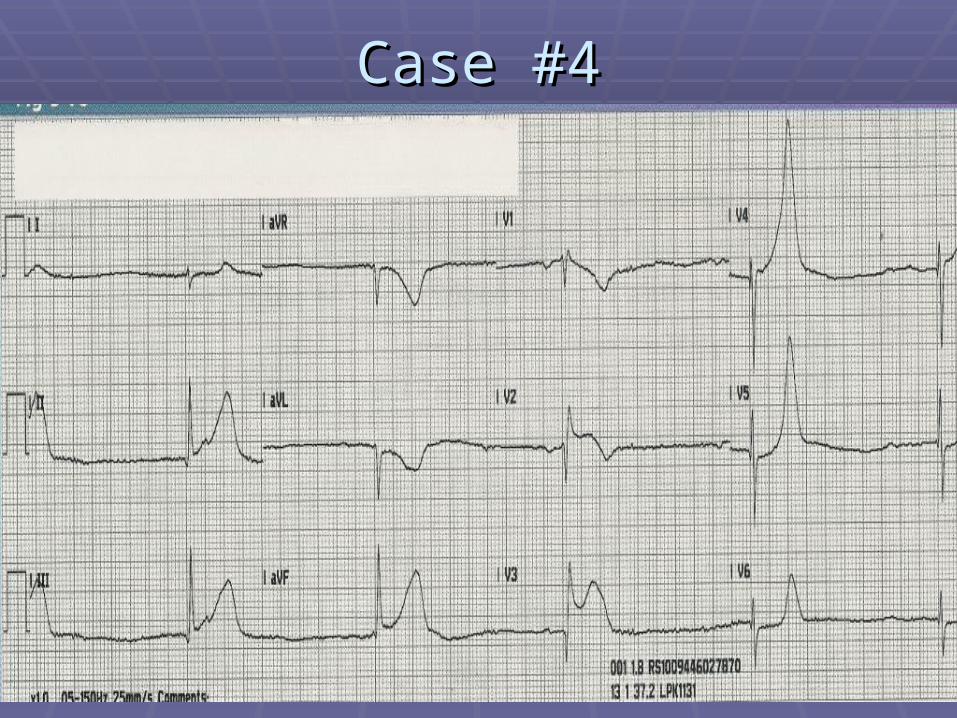
Case #4Case #4

Case #4Case #4
87 year-old female patient complains of 87 year-old female patient complains of dizziness and being extremely tireddizziness and being extremely tired
VS: 88/52; P – 30; R - 16VS: 88/52; P – 30; R - 16 Is there ST elevation:Is there ST elevation:
I, aVL, V5, V6?I, aVL, V5, V6? II, III, aVF?II, III, aVF? V1, V2?V1, V2? V3, V4?V3, V4?
What are you going to do for this patient?What are you going to do for this patient?

Case #5Case #5

Case #5Case #5
58 year-old male patient who complains of chest 58 year-old male patient who complains of chest pain radiating down the left arm after working out pain radiating down the left arm after working out in the gymin the gym
VS: 110/72; P – 100; R - 18VS: 110/72; P – 100; R - 18 Is there ST elevation:Is there ST elevation:
I, aVL, V5, V6?I, aVL, V5, V6? II, III, aVF?II, III, aVF? V1, V2?V1, V2? V3, V4?V3, V4?
What are you going to do for this patient?What are you going to do for this patient?

Case #6Case #6

Case #6Case #6
92 year-old patient complaining of 92 year-old patient complaining of pounding in her chest for one hourpounding in her chest for one hour
VS: 98/66; P – 110; R- 16VS: 98/66; P – 110; R- 16 Is there ST elevation:Is there ST elevation:
I, aVL, V5, V6?I, aVL, V5, V6? II, III, aVF?II, III, aVF? V1, V2?V1, V2? V3, V4?V3, V4?
What are you going to do for this patient?What are you going to do for this patient?

Case #7Case #7

Case #7Case #7
66 year-old patient with history of diabetes 66 year-old patient with history of diabetes for 25 years complains of being for 25 years complains of being lightheaded and is sweatylightheaded and is sweaty
Is there ST elevation:Is there ST elevation: I, aVL, V5, V6?I, aVL, V5, V6? II, III, aVF?II, III, aVF? V1, V2?V1, V2? V3, V4?V3, V4?
What are you going to do for this patient?What are you going to do for this patient?

Case #8Case #8

Case #8Case #8
70 year-old patient had a syncopal episode 70 year-old patient had a syncopal episode when they stood up from the couchwhen they stood up from the couch
VS: 156/98; P – 76; R - 16VS: 156/98; P – 76; R - 16 Is there ST elevation:Is there ST elevation:
I, aVL, V5, V6?I, aVL, V5, V6? II, III, aVF?II, III, aVF? V1, V2?V1, V2? V3, V4?V3, V4?
What are you going to do for this patient?What are you going to do for this patient?

Case #9Case #9

Case #9Case #9
82 year-old patient complains of sudden onset of 82 year-old patient complains of sudden onset of slurred speech, inability to grasp a coffee cup, slurred speech, inability to grasp a coffee cup, and inability to follow simple commandsand inability to follow simple commands
VS: 122/84; P – 110; R - 18VS: 122/84; P – 110; R - 18 Is there ST elevation:Is there ST elevation:
I, aVL, V5, V6?I, aVL, V5, V6? II, III, aVF?II, III, aVF? V1, V2?V1, V2? V3, V4?V3, V4?
What are you going to do for this patient?What are you going to do for this patient?

Case #10Case #10
Case #10Case #10
36 year-old patient who passed out 36 year-old patient who passed out standing in line at a bankstanding in line at a bank
VS: 128/78; P – 80; R - 20VS: 128/78; P – 80; R - 20 Is there ST elevation:Is there ST elevation:
I, aVL, V5, V6?I, aVL, V5, V6? II, III, aVF?II, III, aVF? V1, V2?V1, V2? V3, V4?V3, V4?
What are you going to do for this patient?What are you going to do for this patient?

BibliographyBibliography Aehlert, B. EKG’s Made Easy third Edition. Aehlert, B. EKG’s Made Easy third Edition.
Elsevier Mosby. 2006.Elsevier Mosby. 2006. Beasley, B. Understanding EKG’s A Beasley, B. Understanding EKG’s A
Practical Approach. Brady. 2003.Practical Approach. Brady. 2003. Bledsoe, B., Porter, R., Cherry, R. Bledsoe, B., Porter, R., Cherry, R.
Paramedic Care Principles and Practices. Paramedic Care Principles and Practices. Third Edition. Brady. 2009.Third Edition. Brady. 2009.
Ellis, K. EKG Plain and Simple. Prentice Ellis, K. EKG Plain and Simple. Prentice Hall. 2002.Hall. 2002.
Page, B. 12 Lead EKG for Acute and Page, B. 12 Lead EKG for Acute and Critical Care Providers. Brady. 2005.Critical Care Providers. Brady. 2005.
Phalen, T., Aehlert, B. The 12 Lead EKG in Phalen, T., Aehlert, B. The 12 Lead EKG in Acute Coronary Syndromes. Second Edition, Acute Coronary Syndromes. Second Edition, Elsevier Mosby. 2006.Elsevier Mosby. 2006.
Region X SOP’s. March 2007, Amended Region X SOP’s. March 2007, Amended January 1, 2008.January 1, 2008.
freemd.com (Acute Coronary Syndrome 9/2008)freemd.com (Acute Coronary Syndrome 9/2008) www.anaesthetist.com/icu/organs/heart/ecg/www.anaesthetist.com/icu/organs/heart/ecg/
Findex.htmFindex.htm www.ecglibrary.com/www.ecglibrary.com/ www.gwc.maricopa.edu/class/bio202/cyberheartwww.gwc.maricopa.edu/class/bio202/cyberheart
/ekgqzr.htm/ekgqzr.htm www.madsci.com/manu/ekg_mi.htmwww.madsci.com/manu/ekg_mi.htm