Overview of Anatomy and Physiology Anatomy – the study of the structure of body parts and their...
36
Overview of Anatomy and Physiology Anatomy – the study of the structure of body parts and their relationships to one another Gross or macroscopic Microscopic Developmental Physiology – the study of the function of the body’s structural machinery
-
Upload
gervais-golden -
Category
Documents
-
view
214 -
download
0
Transcript of Overview of Anatomy and Physiology Anatomy – the study of the structure of body parts and their...

Overview of Anatomy and Physiology
Anatomy – the study of the structure of body parts and their relationships to one another
Gross or macroscopic
Microscopic
Developmental
Physiology – the study of the function of the body’s structural machinery
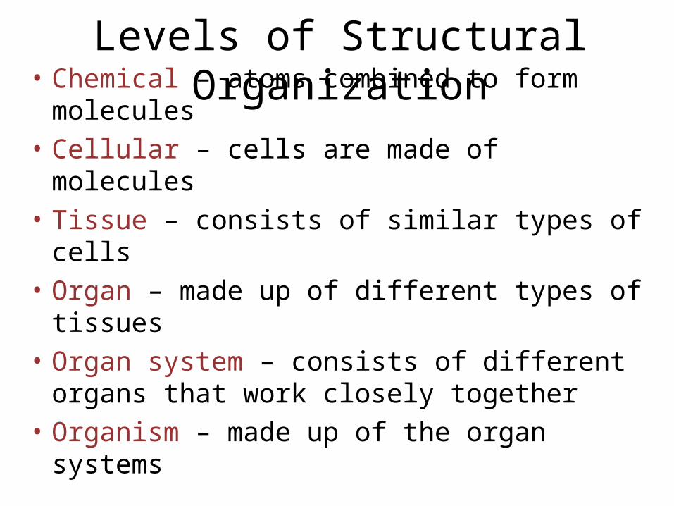
Levels of Structural Organization
• Chemical – atoms combined to form molecules
• Cellular – cells are made of molecules
• Tissue – consists of similar types of cells
• Organ – made up of different types of tissues
• Organ system – consists of different organs that work closely together
• Organism – made up of the organ systems

Organ Systems of the Body
• Lymphatic system– Picks up fluid leaked
from blood vessels and returns it to blood
– Disposes of debris in the lymphatic stream
– Houses white blood cells involved with immunity

Organ Systems of the Body
• Integumentary system– Forms the external body covering– Composed of the skin, sweat glands, oil
glands, hair, and nails– Protects deep tissues from
injury and synthesizes vitamin D

Organ Systems of the Body• Urinary system
– Composed of kidneys, ureters, urinary bladder, and urethra
– Eliminates nitrogenous wastes from the body
– Regulates water, electrolyte, and pH balance of the blood

Anatomical Position
• Body erect, feet slightly apart, palms facing forward, thumbs point away from body

Directional Terms
• Superior and inferior – toward and away from the head, respectively
• Anterior and posterior – toward the front and back of the body
• Medial, lateral, and intermediate – toward the midline, away from the midline, and between a more medial and lateral structure
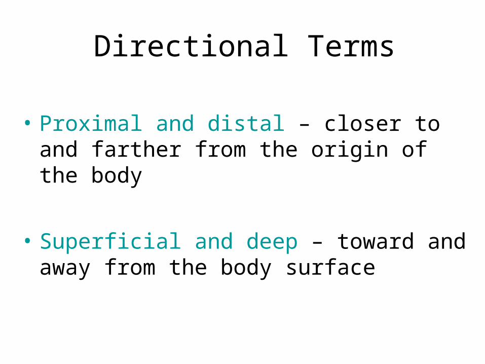
Directional Terms
• Proximal and distal – closer to and farther from the origin of the body
• Superficial and deep – toward and away from the body surface
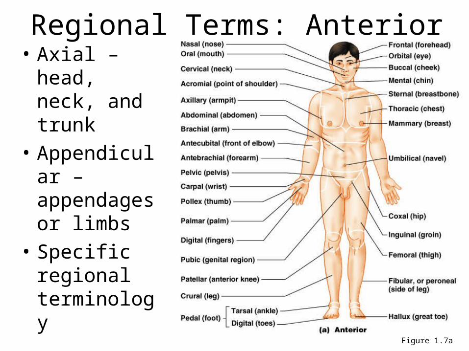
Regional Terms: Anterior View
• Axial – head, neck, and trunk
• Appendicular – appendages or limbs
• Specific regional terminology
Figure 1.7a

Regional Terms: Posterior View
Figure 1.7b

Body Planes• Sagittal – divides the body into right and left
parts
• Midsagittal or medial – sagittal plane that lies on the midline
• Frontal or coronal – divides the body into anterior and posterior parts
• Transverse or horizontal (cross section) – divides the body into superior and inferior parts
• Oblique section – cuts made diagonally
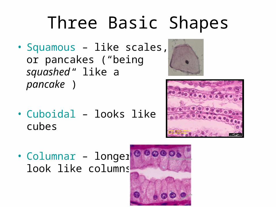
Three Basic Shapes• Squamous – like scales, or
pancakes (“being squashed like a pancake”)
• Cuboidal – looks like cubes
• Columnar – longer and look like columns

Cell Organization
• Simple – single layer of cells; typically found where absorption and filtration occur or a single layer of epithelial is needed
simple squamoussimple cuboidal
simple columnar• Stratified – layers of cells; common in areas
where protection is needed like the skinstratified squamousstratified cuboidalstratified columnar

Review What You’ve Learned…
Muscle Type Striated? # of nucleiVoluntary or Involuntary
Skeletal
Cardiac
Smooth
Yes
Yes
No
Single Nucleus
Single Nucleus
Multi-nucleated Voluntary
Involuntary
Involuntary

Smooth Muscle Cells
– are small and pointed at their ends– can divide and regenerate new cells– non-striated, involuntary, and single nucleus– found in hollow organs like the intestine,
bladder, lungs, and blood vessels– move substances through hollow opening
by contracting slowly; they squeeze things through like a tube of toothpaste

Skeletal Muscle Cells
– Striated, voluntary, and multinucleated– are long, thin and cylindrical; they are
attached to bones and move our skeleton– are usually called “muscle fibers”– do not divide to create new cells– new fibers are produced by stem cells– Striations -- cross stripes (bands) run
perpendicular to the cells
Cardiac Muscle Cells
– are called “cardiocytes” and found in heart walls
– branching cells connect at intercalated disks which allow contractions to occur faster
– are regulated by pacemaker cells which control contraction of the heart muscles
– striated, involuntary, and single nucleus

Synovial Membranes (Connective)
• Some joints are surrounded by a membrane (synovium) that produces a thick, synovial fluid. This fluid nourish the cartilage and keeps it slippery.
Knee Joint

Appendages of the Skin• Sweat glands
– Produce sweat widely distributed in skin (2.5 million per person)
– helps cool the body– Two types
• Eccrine– Opens up to skin through a duct (pore) on skin surface– Most numerous on the body
• Apocrine– Ducts empty into hair follicles– Found mostly in armpits and genital areas– Precise function is unknown but are they are activated
during pain, stress and during sexual foreplay.

Seven Functions of Skin• Mechanical/Chemical damage – keratin
toughens cells; fats cells cushion blows; and pressure receptors to measure possible damage
• Bacterial damage – skin secretions are acidic and inhibit bacteria.
• Ultraviolet radiation – melanin produced to protect from UV damage

Skin Functions
• Thermal control – regulates body temperature– Heat loss: sweat to cool the skin– Heat retention: prevents blood to rush into capillary beds
• Waterproofing – contains lipids to prevent drying out• Excretion of waste – urea and uric acid secreted in
sweat• Makes vitamin D – modifies cholesterol molecules in
skin and converts it to vitamin D

Melanin
• Pigment (melanin) produced by melanocytes
• Melanocytes are mostly in the stratum basale
• Color is yellow to red to brown to black
• Amount of melanin produced depends upon genetics and exposure to sunlight
Layers of the Epidermis
• Stratum lucidum– Formed from dead cells of the deeper layers– Occurs only in thick, hairless skin of the palms
of hands and soles of feet
• Stratum corneum– Outermost layer of epidermis– Scale-like dead cells are filled with keratin
which is a protective protein preventing water loss from skin
Appendages of the Skin
• Sebaceous glands– Produce oil
• Lubricant for skin which keeps skin soft and moist• Prevents brittle hair• Kills bacteria (slightly acidic)
– Most have ducts that empty into hair follicles; others open directly onto skin surface
– Glands are activated at puberty and this is what causes teenage acne

Sweat and its Function
• Composition– Mostly water– Salts and excess vitamin C– Some metabolic waste (urea and uric acid)– Fatty acids and proteins (apocrine only)
• Function– Helps rid body of excess heat– Excretes waste products– Acidic nature inhibits bacteria growth
• Odor is from associated bacteria

Classification of Bones Based of Shape

Anatomy of a Long Bone
• Epiphyseal plate– Flat plate of hyaline cartilage seen in young,
growing bone (a.k.a. = growth plate)
• Epiphyseal line– Remnant of the epiphyseal plate– Seen in adult bones

Bone Surface Markings• Depressions and openings fro blood vessels
and nerves to pass through– Foramen – round or
oval opening in a bone– Meatus – canal-like
– Fossa – shallow depression
mostly to form a joint

Types of Bone Cells
• Osteocytes—mature bone cells
• Osteoblasts—bone-forming cells
• Osteoclasts—bone-destroying cells– Break down bone matrix for remodeling and
release of calcium in response to parathyroid hormone
• Bone remodeling is performed by both osteoblasts and osteoclasts

Bones of the Skeleton
• Know all of the bones of your skeleton!

Simple and Compound Fractures
• Simple – Clean break in the
bone
• Compound– Bone breaks and
pierces skin

Microscopic Anatomy of Skeletal Muscle
• Sarcomere — contractile unit of a muscle fiber (muscle cell)
• Organization of the sarcomere– Myofilaments
• Thick filaments = myosin filaments• Thin filaments = actin filaments

Microscopic Anatomy of Skeletal Muscle
Figure 6.3c
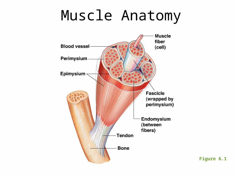
Muscle Anatomy
Figure 6.1

Know your Muscles

Naming Skeletal Muscles
1 – Location of the muscle
2 – Shape of the muscle
3 – Size of the muscle
4 – Direction/Orientation of the muscle
fibers/cells
5 – Number of Origins
6 – Location of the Attachments
7 – Action of the muscle