
Overview - Jones & Bartlett Learningems.jbpub.com/stoy/emt/lecturenotes/Chapter_07.pdf · when...
26
9/11/2012 1 Slide 1 Chapter 7 Assisting the ALS Provider Slide 2 Overview The Team Concept ALS Procedures and Equipment Electrocardiogram (ECG) Monitoring Intravenous Therapy Endotracheal Intubation Slide 3 The Team Concept Prehospital care involves many individuals and entities Providing proper quality care relies heavily on teamwork Copyright © 2013 by Jones & Bartlett Learning, LLC, an Ascend Learning Company
Transcript of Overview - Jones & Bartlett Learningems.jbpub.com/stoy/emt/lecturenotes/Chapter_07.pdf · when...

9/11/2012
1
Slide 1
Chapter 7
Assisting the ALS Provider
Slide 2
Overview
The Team Concept
ALS Procedures and Equipment Electrocardiogram (ECG) Monitoring
Intravenous Therapy
Endotracheal Intubation
Slide 3
The Team Concept
Prehospital care involves many individuals and entities
Providing proper quality care relies heavily on teamwork
Copyright © 2013 by Jones & Bartlett Learning, LLC, an Ascend Learning Company

9/11/2012
2
Slide 4
The Team Concept
Failure to adequately assess and intervene in the management of the airway, breathing, and circulation of a patient in distress will often result in an undesired patient outcome
The ability of the EMT to quickly and efficiently assess these areas remains paramount as the foundation of quality care
Slide 5
The Team Concept
EMTs may work with ALS providers ALS providers may elect to initiate additional
interventions, including: Cardiac monitoring Intravenous therapy Endotracheal intubation
The EMT who is able to anticipate the need or is requested to assist the ALS provider with such skills and procedures will greatly enhance the functioning
of the team.
Slide 6
ALS Procedures and Equipment
ECG monitoring
Intravenous therapy
Endotracheal intubation
Copyright © 2013 by Jones & Bartlett Learning, LLC, an Ascend Learning Company

9/11/2012
3
Slide 7
ECG Monitoring
The ability to assess the electrical activity of the heart may guide what interventions are appropriate for the presenting patient condition
Assisting with the application of the cardiac monitor will expedite the assessment and any necessary interventions
Slide 8
ECG Monitoring
Typical patients requiring monitoring Altered mental status Unresponsive patient Cardiac arrest Chest pain Respiratory distress Abdominal pain or discomfort Traumatic injuries to the chest Hypotension Diabetes
Slide 9
ECG Monitoring
There are several common types of cardiac monitors used by ALS providers in the out-of-hospital setting
Become familiar with the type, application, and start-up features of the monitor used by the ALS services in your area
Copyright © 2013 by Jones & Bartlett Learning, LLC, an Ascend Learning Company

9/11/2012
4
Slide 10
ECG Monitoring
Many cardiac monitors offer multiple electronic features ECG monitoring Defibrillation Synchronized cardioversion Transcutaneous pacing
Other features may include Automatic noninvasive blood pressure Pulse oximetry Capnometry/CO2 monitoring
Slide 11
ECG Monitoring
Electrode placement Cardiac monitoring involves the application of
electrodes to the skin of a patient and connection of the electrical cables to the cardiac monitor
The number of electrodes used and their placement on the patient will be based on the number of monitor cables
Slide 12
ECG Monitoring
The most common electrode placements used to view the electrical activity of the heart are three-, five-, or twelve-lead systems
Copyright © 2013 by Jones & Bartlett Learning, LLC, an Ascend Learning Company

9/11/2012
5
Slide 13
ECG Monitoring
Slide 14
ECG Monitoring
Following placement of the electrode on the patient’s skin, the electrical cables are connected to the appropriate electrodes
Each connector may be identified based on color and letter indications on each snap connector.
On three-cable systems, the identifiers include:Right arm (RA) – WHITE Left arm (LA) – BLACK Left leg (LL) – RED
Slide 15
ECG Monitoring
Five-cable systems Addition of the right leg (RL) – BROWN
Central ground (G) – GREEN
Copyright © 2013 by Jones & Bartlett Learning, LLC, an Ascend Learning Company

9/11/2012
6
Slide 16
ECG Monitoring
Twelve-lead ECG Ten cable-electrode connections
Slide 17
ECG Monitoring
Electrode placement Placed on the chest, abdomen or arms, and legs
Often based on practicality and accessibility
Slide 18
ECG Monitoring
With three- and five-lead systems Right arm (RA) – WHITE
• Just below the lateral clavicular area
Left arm (LA) – BLACK• Just below the lateral clavicular area
Left leg (LL) – RED• Right lateral abdominal area
Copyright © 2013 by Jones & Bartlett Learning, LLC, an Ascend Learning Company

9/11/2012
7
Slide 19
ECG Monitoring
Twelve-lead systems Ten electrodes are placed on the patient
Limb leads are placed distally on the extremities
Remaining six electrodes are placed across the anterior to left lateral chest wall
Slide 20
ECG Monitoring
The electrode must be firmly secured to the skin to obtain an adequate view of the electrical activity of the heart
Preparation of the skin Removal of any moisture that may be present
Removal of excess hair may be necessary to allow the adhesive portion of the electrode to completely adhere
Slide 21
ECG Monitoring
When indicated, the electrical activity of the heart may be monitored through the application of defibrillator/pacer pads as used in hands-free operation of the monitor/defibrillator
These pads are placed in the same manner as AED pads
Copyright © 2013 by Jones & Bartlett Learning, LLC, an Ascend Learning Company

9/11/2012
8
Slide 22
ECG Monitoring
Identify need for cardiac monitoring
Identify electrode placement based on monitor cable system
Expose patient skin in the area of electrode placement to ensure it is dry and free of excess hair
Remove protective cover from electrode and apply to patient’s skin
Connect electrical cables to electrodes
Turn monitor to on position and ensure appropriate monitoring view is selected
Slide 23
Intravenous Therapy
Administration of medications and fluids may improve a patient’s condition and outcome
Intravenous access is obtained by placement of a small catheter in a vein
Slide 24
Intravenous Therapy
The catheter is left in place as the access port to the circulatory system
This port may also be used to obtain blood samples for glucose checks and laboratory studies
Copyright © 2013 by Jones & Bartlett Learning, LLC, an Ascend Learning Company

9/11/2012
9
Slide 25
Intravenous Therapy Standard intravenous
access equipment Sharps container
Tourniquet
2×2 or 4×4 dressing
Alcohol prep
Intravenous catheter
Heparin port or “saline lock”
Administration set
Intravenous fluid
Tape
Biooclusive dressing
Gloves
Slide 26
Intravenous Therapy
The ALS provider will decide what type of fluid will be administered to the patient
Establishing vascular access without fluid administration is a common practice
Slide 27
Intravenous Therapy
The end of the catheter is capped with a heparin port or “saline lock”
Medications may be introduced through this port without connecting the catheter to a fluid administration set
Copyright © 2013 by Jones & Bartlett Learning, LLC, an Ascend Learning Company

9/11/2012
10
Slide 28
Intravenous Therapy
The EMT must always check the appropriate fluid is prepared, as well as the expiration date and clarity of the fluid
If the fluid is discolored, expired, or the bag is leaking, another bag must be used
Slide 29
Intravenous Therapy
To access the fluid bag, tear or carefully cut the protective covering and remove the fluid bag
Fluid bags may have multiple ports located on the inferior end One port will be for connecting the administration
One permits medication to be added to the fluid
Slide 30
Intravenous Therapy
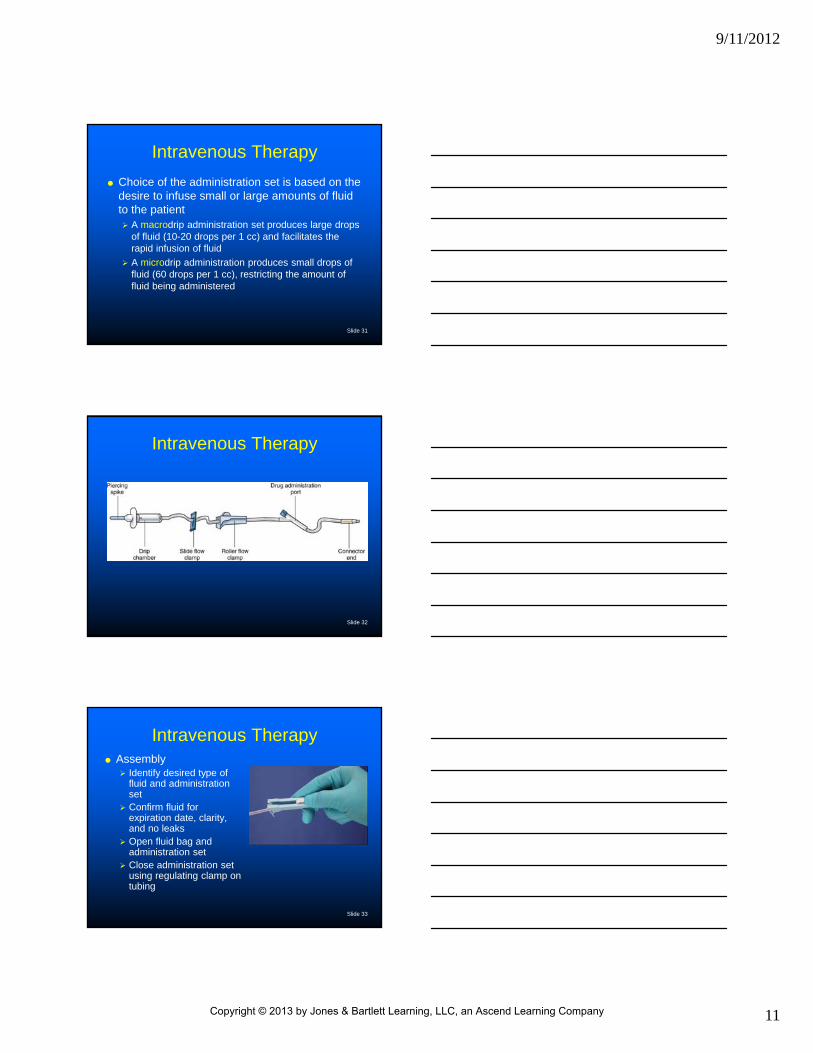
An administration set must be attached to the fluid bag
This tubing transports the fluid to the intravenous catheter placed in the patient’s circulatory system
Two common types of administration sets are available as intravenous tubing
Copyright © 2013 by Jones & Bartlett Learning, LLC, an Ascend Learning Company
9/11/2012
11
Slide 31
Intravenous Therapy
Choice of the administration set is based on the desire to infuse small or large amounts of fluid to the patient A macrodrip administration set produces large drops
of fluid (10-20 drops per 1 cc) and facilitates the rapid infusion of fluid
A microdrip administration produces small drops of fluid (60 drops per 1 cc), restricting the amount of fluid being administered
Slide 32
Intravenous Therapy
Slide 33
Intravenous Therapy Assembly
Identify desired type of fluid and administration set
Confirm fluid for expiration date, clarity, and no leaks
Open fluid bag and administration set
Close administration set using regulating clamp on tubing
Copyright © 2013 by Jones & Bartlett Learning, LLC, an Ascend Learning Company

9/11/2012
12
Slide 34
Intravenous Therapy
Assembly Ensure that sterility is
maintained
Remove protective covers from fluid bag and administration chamber side of tubing
Insert administration set into fluid bag
Slide 35
Intravenous Therapy Assembly
Squeeze administration set chamber to fill approximately one half
Open regulating clamp on tubing to fill tubing with fluid
Ensuring sterility is maintained, remove distal tubing protective cap if needed
Close regulating clamp on tubing when tubing is filled
Ensure all air is removed from tubing
Replace distal tubing protective cap if removed
Slide 36
Intravenous Therapy
Patient preparation The ALS provider will most likely be the individual
preparing the patient and intravenous insertion site for the procedure
Copyright © 2013 by Jones & Bartlett Learning, LLC, an Ascend Learning Company

9/11/2012
13
Slide 37
Intravenous Therapy The area where the intravenous catheter will
be introduced through the skin is prepared by swabbing with an alcohol prep
Swab over the area in a circular motion, beginning at the center of the area and moving outward
The area cleansed should be approximately 3 × 3 inches
Slide 38
Intravenous Therapy
Securing BSI
The ALS provider will remove the needle and insert the distal tubing end into the catheter
• The needle should immediately be placed in a sharps container
The tourniquet should be released
Any excess fluid or blood should be removed from the catheter area
Slide 39
Intravenous Therapy
Securing and monitoring The regulating clamp is moved to the open
position and the site is observed for infiltration of the fluid
Copyright © 2013 by Jones & Bartlett Learning, LLC, an Ascend Learning Company

9/11/2012
14
Slide 40
Intravenous Therapy
Monitoring Common signs and symptoms of intravenous
infiltration• Increased pain in the area of the catheter
• Swelling
• Discoloration
• Intravenous fluid infuses slowly or not at all
Slide 41
Intravenous Therapy
Monitoring If signs of infiltration are noted, immediately close
the regulation clamp The ALS provider will remove the catheter from the
skin and gentle pressure with a dressing is applied to the insertion area
Slide 42
Intravenous Therapy
The catheter is secured to the skin using a bioocclusive dressing and/or tape
Follow the instructions given by the ALS provider when assisting with this procedure
Copyright © 2013 by Jones & Bartlett Learning, LLC, an Ascend Learning Company

9/11/2012
15
Slide 43
Intravenous Therapy A small loop is placed in the
administration set tubing and secured to the patient’s skin
This loop will assist in preventing dislodgment of the catheter from the insertion site
In areas where the insertion site is over a joint, placement of a splint to stabilize the extremity may be necessary to maintain infusion continuity
Slide 44
Intravenous Therapy After securing the catheter and administration
tubing, the site should be rechecked for signs of infiltration
The rate of fluid infusion should be confirmed by the ALS provider
Frequent reassessment of the infusion site should occur during all aspects of patient contact
Slide 45
Video Clip: Assembly of Intravenous Administration Set and Fluid
Copyright © 2013 by Jones & Bartlett Learning, LLC, an Ascend Learning Company

9/11/2012
16
Slide 46
Endotracheal Intubation
A procedure that is performed when a patient is unable to maintain a patent or secure airway without assistance
Slide 47
Endotracheal Intubation
Conditions potentially requiring endotracheal intubation Altered mental status/unresponsive
Respiratory distress/arrest
Cardiopulmonary arrest
Traumatic injuries disrupting the airway
Status epilepticus
Slide 48
Endotracheal Intubation
The three primary methods of intubation in the out-of-hospital setting include Direct laryngoscopy
Nasaotracheal
Digital
Copyright © 2013 by Jones & Bartlett Learning, LLC, an Ascend Learning Company

9/11/2012
17
Slide 49
Endotracheal Intubation
Equipment and patient preparation Two of the three prominent areas where the EMT
can assist are equipment and patient preparation
Some ALS providers may want you to manage the airway while preparations are made for intubation
Others may want you to set up the equipment and prepare the patient for the procedure
Slide 50
Endotracheal Intubation
It is important that the EMT preoxygenate the patient prior to the procedure
Ventilate with a normal rate and tidal volume for a minimum of 2 minutes prior to beginning the endotracheal intubation procedure
Slide 51
Endotracheal Intubation
When the ALS provider is ready to perform the endotracheal intubation procedure: Stop assisting with ventilations
Remove the oropharyngeal airway
Perform any oral suctioning that may be required
Copyright © 2013 by Jones & Bartlett Learning, LLC, an Ascend Learning Company
9/11/2012
18
Slide 52
Endotracheal Intubation
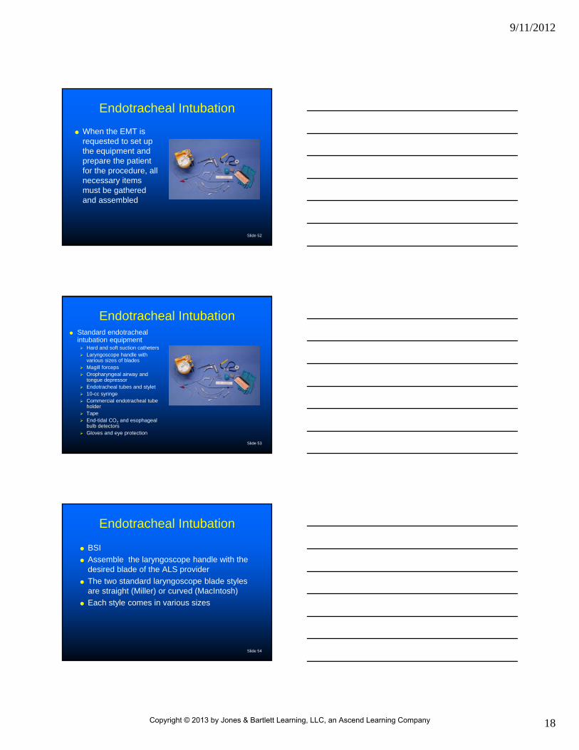
When the EMT is requested to set up the equipment and prepare the patient for the procedure, all necessary items must be gathered and assembled
Slide 53
Endotracheal Intubation Standard endotracheal
intubation equipment Hard and soft suction catheters Laryngoscope handle with
various sizes of blades Magill forceps Oropharyngeal airway and
tongue depressor Endotracheal tubes and stylet 10-cc syringe Commercial endotracheal tube
holder Tape End-tidal CO2 and esophageal
bulb detectors Gloves and eye protection
Slide 54
Endotracheal Intubation
BSI
Assemble the laryngoscope handle with the desired blade of the ALS provider
The two standard laryngoscope blade styles are straight (Miller) or curved (MacIntosh)
Each style comes in various sizes
Copyright © 2013 by Jones & Bartlett Learning, LLC, an Ascend Learning Company
9/11/2012
19
Slide 55
Endotracheal Intubation
The blade is attached to the laryngoscope by placing the blade into the top of the handle in an unopened position and securing it to the pin with a downward motion
A “click” may be heard when using metal equipment
Slide 56
Endotracheal Intubation
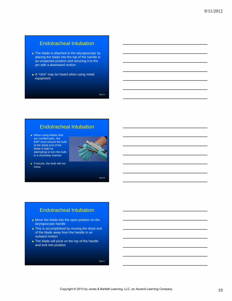
When using blades that are nonfiberoptic, the EMT must ensure the bulb at the distal end of the blade is tight by attempting to turn the bulb in a clockwise manner
If secure, the bulb will not move
Slide 57
Endotracheal Intubation
Move the blade into the open position on the laryngoscope handle
This is accomplished by moving the distal end of the blade away from the handle in an outward motion
The blade will pivot on the top of the handle and lock into position
Copyright © 2013 by Jones & Bartlett Learning, LLC, an Ascend Learning Company
9/11/2012
20
Slide 58
Endotracheal Intubation
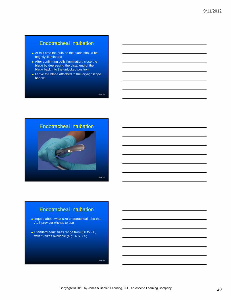
At this time the bulb on the blade should be brightly illuminated
After confirming bulb illumination, close the blade by depressing the distal end of the blade back into the unlocked position
Leave the blade attached to the laryngoscope handle
Slide 59
Endotracheal Intubation
Slide 60
Endotracheal Intubation
Inquire about what size endotracheal tube the ALS provider wishes to use
Standard adult sizes range from 6.0 to 9.0, with ½ sizes available (e.g., 6.5, 7.5)
Copyright © 2013 by Jones & Bartlett Learning, LLC, an Ascend Learning Company

9/11/2012
21
Slide 61
Endotracheal Intubation
Remove the tube from the packaging and attach a 10-cc syringe without a needle to the cuff connection port near the top of the tube
Inflate the distal cuff using no more than 10 cc of air and disconnect the syringe
Check to make sure the distal cuff remains inflated
Slide 62
Endotracheal Intubation
Check the endotracheal cuff for air leaks
Slide 63
Endotracheal Intubation
Reattach the 10-cc syringe to the cuff connection port and withdraw all the air in the cuff
If any air remains in the cuff, this may cause complications during the endotracheal intubation procedure
Copyright © 2013 by Jones & Bartlett Learning, LLC, an Ascend Learning Company

9/11/2012
22
Slide 64
Endotracheal Intubation
A stylet is a fairly rigid devise that assists in maintaining a desired shape of the endotracheal tube
If instructed, place the stylet into the top of the endotracheal tube and advance it until the end is just proximal to the Murphy hole or “eye” on the side of the distal end of the endotracheal tube
Slide 65
Endotracheal Intubation
Bend the stylet over the top of the endotracheal tube and reconfirm the distal end of the stylet is not beyond the landmark identified
The EMT may also be instructed to apply a lubricant to the distal end of the endotracheal tube
Slide 66
Endotracheal Intubation Following stylet and lubrication as directed, place the
endotracheal tube back into the packaging and keep the 10-cc syringe with the tube
The esophageal detection, end-tidal CO2, and commercial tube-securing devices, along with the prepared endotracheal tube, laryngoscope handle and blade, and suction unit, should be placed within reach of the ALS provider performing the endotracheal procedure
Copyright © 2013 by Jones & Bartlett Learning, LLC, an Ascend Learning Company

9/11/2012
23
Slide 67
Endotracheal Intubation
The application of cricoid pressure decreases the risk of aspiration by occluding the esophagus
During spinal restrictive maneuvers for suspected neck injuries, the EMT will often take a position above the head of the patient and hold spinal restrictive measures from this position
Slide 68
Endotracheal Intubation
Confirmation of tube placement Failure to ensure appropriate placement of an
endotracheal tube will often result in the decompensation and ultimately death of a patient
Verification of placement is through the assessment and evaluation of multiple methods
The EMT may be requested to confirm the endotracheal tube placement by auscultation of breath sounds, placement of the esophageal and/or end-tidal CO2 detectors
Slide 69
Endotracheal Intubation
Squeeze the esophageal detector device to remove the air
Attach it to the 15-mm connection at the top of the endotracheal tube
After it is attached to the tube, release the detector; it should reinflate at this time
The absence of inflation may indicate esophageal placement of the endotracheal tube
Copyright © 2013 by Jones & Bartlett Learning, LLC, an Ascend Learning Company

9/11/2012
24
Slide 70
Endotracheal Intubation
Commercially available end-tidal CO2
detectors are used to detect the presence of carbon dioxide during the expiration phase of respiration/ventilation
Attach the device directly to the 15-mm connector; provide four or five ventilations
Slide 71
Endotracheal Intubation
A color change indicates the presence of carbon dioxide
Commonly used devices change from purple to yellow in the presence of carbon dioxide, but not all devices use this color scheme
Slide 72
Endotracheal Intubation Signs of proper placement
Symmetrical chest rise and fall
Absence of epigastric sounds
Presence of bilateral breath sounds
Color of patient skin
Reinflation of the esophageal detector
Color change from purple to yellow on the CO2
detector
Endotracheal tube is free of large amounts of secretions, blood, and vomit
Copyright © 2013 by Jones & Bartlett Learning, LLC, an Ascend Learning Company
9/11/2012
25
Slide 73
Endotracheal Intubation
Following confirmation for correct placement, securing the tube is the next priority
Assist the ALS provider in applying a commercially available endotracheal tube holder or by using a preferred taping method
Note the depth of the endotracheal tube in reference to the lips or teeth of the patient
Slide 74
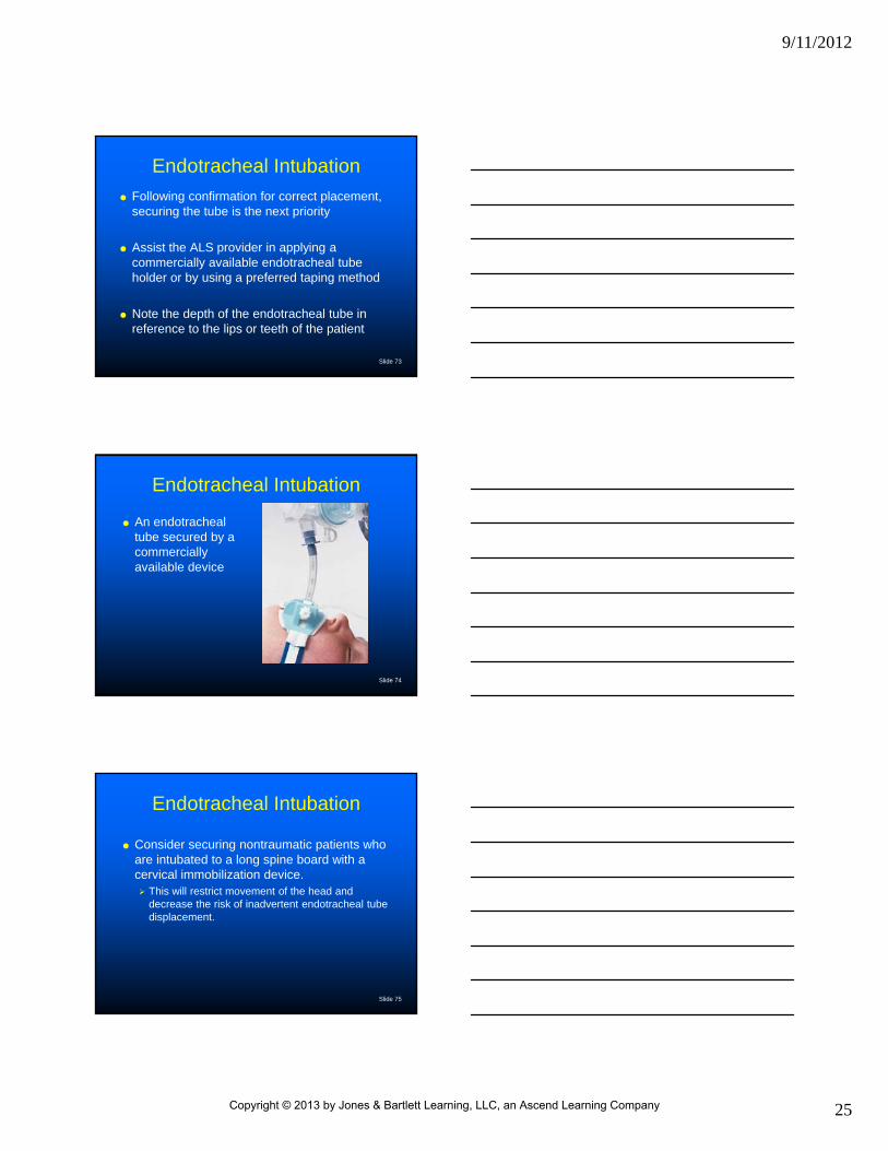
Endotracheal Intubation
An endotracheal tube secured by a commercially available device
Slide 75
Endotracheal Intubation
Consider securing nontraumatic patients who are intubated to a long spine board with a cervical immobilization device. This will restrict movement of the head and
decrease the risk of inadvertent endotracheal tube displacement.
Copyright © 2013 by Jones & Bartlett Learning, LLC, an Ascend Learning Company

9/11/2012
26
Slide 76
Endotracheal Intubation
Ventilate the patient with the bag-mask device
Attach the bag-mask device to the 15-mm endotracheal tube
Perform ventilations
Slide 77
Summary
The Team Concept
ALS Procedures and Equipment Electrocardiogram (ECG) Monitoring
Intravenous Therapy
Endotracheal Intubation
Copyright © 2013 by Jones & Bartlett Learning, LLC, an Ascend Learning Company


![Chapter 12 LO - Jones & Bartlett Learningems.jbpub.com/chapleau/emt/docs/PPT_lectures/Chap… · · 2014-05-05Title: Microsoft PowerPoint - Chapter_12_LO [Compatibility Mode] Author:](https://static.fdocuments.in/doc/165x107/5af12e897f8b9abc788e1b9b/chapter-12-lo-jones-bartlett-2014-05-05title-microsoft-powerpoint-chapter12lo.jpg)


