Overexpression of UTX promotes tumor progression in Oral ...
14
RESEARCH Open Access Overexpression of UTX promotes tumor progression in Oral tongue squamous cell carcinoma patients receiving surgical resection: a case control study Yen-Hao Chen 1,2,3 , Chang-Han Chen 4 , Chih-Yen Chien 5 , Yan-Ye Su 5 , Sheng-Dean Luo 5 and Shau-Hsuan Li 1* Abstract Background: Ubiquitously transcribed tetratricopeptide repeat on chromosome X (UTX) has been identified as a histone 3 lysine 27 (H3K27) demethylase and acted as a tumor suppressor gene or oncogenic function. The current study was to explore the significance of UTX in oral tongue squamous cell carcinoma (OTSCC) patients who received surgical resection. Methods: A total of 148 OTSCC patients who underwent surgical resection were identified, including 64 patients (43%) with overexpression of UTX and 84 patients (57%) harboring low expression of UTX. We also used two OTSCC cell lines, SAS and Cal 27, to determine the modulation of cancer. Chi-square test was used to investigate the difference of categorical variables between the groups; survival outcome was analyzed using the Kaplan–Meier method in univariate analysis, and a Cox regression model was performed for multivariate analyses. Results: Univariate and multivariate analyses showed overexpression of UTX were significantly related to worse disease-free survival (P = 0.028) and overall survival (P = 0.029). The two OTSCC cell lines were treated with GSK-J4, a potent inhibitor of UTX, and transwell migration and invasion assays showed an inhibitory effect with a dose- dependent manner. In addition, western blot analyses also revealed the inhibition of cell cycle and epithelial- mesenchymal transition. Conclusion: Our study suggests that UTX plays an important role in the process of OTSCC and overexpression of UTX may predict poor prognosis in OTSCC patients who received surgical resection. Keywords: UTX, Tongue cancer, Squamous cell carcinoma, Surgery Background Oral cavity cancer is one of the most aggressive cancers worldwide. The tongue is the most commonly occurring site for oral cavity cancers; such cancers are usually squamous cell carcinomas. Oral tongue squamous cell carcinoma (OTSCC) has the highest incidence, and while the incidence generally has increased, the five-year survival rates have not improved over the last 20 years [1, 2]. OTSCC usually migrates rapidly to the adjacent structures and disseminates to other organs through the bloodstream and lymphatic drainage. Surgical resection remains the gold standard for operable disease, and ad- juvant chemotherapy, radiotherapy, or combination ther- apy is indicated for high-risk populations, in clinical practice [3]. The important risk factors of poor © The Author(s). 2021 Open Access This article is licensed under a Creative Commons Attribution 4.0 International License, which permits use, sharing, adaptation, distribution and reproduction in any medium or format, as long as you give appropriate credit to the original author(s) and the source, provide a link to the Creative Commons licence, and indicate if changes were made. The images or other third party material in this article are included in the article's Creative Commons licence, unless indicated otherwise in a credit line to the material. If material is not included in the article's Creative Commons licence and your intended use is not permitted by statutory regulation or exceeds the permitted use, you will need to obtain permission directly from the copyright holder. To view a copy of this licence, visit http://creativecommons.org/licenses/by/4.0/. The Creative Commons Public Domain Dedication waiver (http://creativecommons.org/publicdomain/zero/1.0/) applies to the data made available in this article, unless otherwise stated in a credit line to the data. * Correspondence: [email protected] 1 Department of Hematology-Oncology, Kaohsiung Chang Gung Memorial Hospital and Chang Gung University College of Medicine, No.123, Dapi Rd., Niaosong Dist, Kaohsiung City 833, Taiwan Full list of author information is available at the end of the article Chen et al. BMC Cancer (2021) 21:979 https://doi.org/10.1186/s12885-021-08726-3
Transcript of Overexpression of UTX promotes tumor progression in Oral ...
RESEARCH Open Access
Overexpression of UTX promotes tumorprogression in Oral tongue squamous cellcarcinoma patients receiving surgicalresection: a case control studyYen-Hao Chen1,2,3, Chang-Han Chen4, Chih-Yen Chien5, Yan-Ye Su5, Sheng-Dean Luo5 and Shau-Hsuan Li1*
Abstract
Background: Ubiquitously transcribed tetratricopeptide repeat on chromosome X (UTX) has been identified as ahistone 3 lysine 27 (H3K27) demethylase and acted as a tumor suppressor gene or oncogenic function. The currentstudy was to explore the significance of UTX in oral tongue squamous cell carcinoma (OTSCC) patients whoreceived surgical resection.
Methods: A total of 148 OTSCC patients who underwent surgical resection were identified, including 64 patients(43%) with overexpression of UTX and 84 patients (57%) harboring low expression of UTX. We also used two OTSCCcell lines, SAS and Cal 27, to determine the modulation of cancer. Chi-square test was used to investigate thedifference of categorical variables between the groups; survival outcome was analyzed using the Kaplan–Meiermethod in univariate analysis, and a Cox regression model was performed for multivariate analyses.
Results: Univariate and multivariate analyses showed overexpression of UTX were significantly related to worsedisease-free survival (P = 0.028) and overall survival (P = 0.029). The two OTSCC cell lines were treated with GSK-J4, apotent inhibitor of UTX, and transwell migration and invasion assays showed an inhibitory effect with a dose-dependent manner. In addition, western blot analyses also revealed the inhibition of cell cycle and epithelial-mesenchymal transition.
Conclusion: Our study suggests that UTX plays an important role in the process of OTSCC and overexpression ofUTX may predict poor prognosis in OTSCC patients who received surgical resection.
Keywords: UTX, Tongue cancer, Squamous cell carcinoma, Surgery
BackgroundOral cavity cancer is one of the most aggressive cancersworldwide. The tongue is the most commonly occurringsite for oral cavity cancers; such cancers are usuallysquamous cell carcinomas. Oral tongue squamous cell
carcinoma (OTSCC) has the highest incidence, andwhile the incidence generally has increased, the five-yearsurvival rates have not improved over the last 20 years[1, 2]. OTSCC usually migrates rapidly to the adjacentstructures and disseminates to other organs through thebloodstream and lymphatic drainage. Surgical resectionremains the gold standard for operable disease, and ad-juvant chemotherapy, radiotherapy, or combination ther-apy is indicated for high-risk populations, in clinicalpractice [3]. The important risk factors of poor
© The Author(s). 2021 Open Access This article is licensed under a Creative Commons Attribution 4.0 International License,which permits use, sharing, adaptation, distribution and reproduction in any medium or format, as long as you giveappropriate credit to the original author(s) and the source, provide a link to the Creative Commons licence, and indicate ifchanges were made. The images or other third party material in this article are included in the article's Creative Commonslicence, unless indicated otherwise in a credit line to the material. If material is not included in the article's Creative Commonslicence and your intended use is not permitted by statutory regulation or exceeds the permitted use, you will need to obtainpermission directly from the copyright holder. To view a copy of this licence, visit http://creativecommons.org/licenses/by/4.0/.The Creative Commons Public Domain Dedication waiver (http://creativecommons.org/publicdomain/zero/1.0/) applies to thedata made available in this article, unless otherwise stated in a credit line to the data.
* Correspondence: [email protected] of Hematology-Oncology, Kaohsiung Chang Gung MemorialHospital and Chang Gung University College of Medicine, No.123, Dapi Rd.,Niaosong Dist, Kaohsiung City 833, TaiwanFull list of author information is available at the end of the article
Chen et al. BMC Cancer (2021) 21:979 https://doi.org/10.1186/s12885-021-08726-3
prognosis in OTSCC include depth of invasion, perineu-ral invasion, lymphovascular invasion, increasing patho-logical T and N stage, extracapsular extension, andsurgical margin [4–8]. These risk factors lead to an in-creasing incidence of locoregional failure and distantmetastasis, contributing to poor prognosis and impairedquality of life [9, 10]. Thus, it is of great significance toidentify a potential biomarker associated with tumorprogression to improve prognosis in OTSCC patients.Ubiquitously transcribed tetratricopeptide repeat on
chromosome X (UTX)—also known as KDM6A—is ahistone demethylase that targets di- and tri-methylatedhistone H3 lysine 27 (H3K27); it is involved in embry-onic development, tissue-specific differentiation, andcancer growth [11, 12]. Growing evidence has shownthat UTX mutations or deregulation are associated withseveral cancer types, including breast cancer, bladdercancer, colon cancer, and B-cell lymphoma [13–17].However, the role of UTX in tumor suppression or inthe enhancement of cancer cell proliferation still re-mains controversial. In breast cancer, blocking UTX re-sulted in a significant decrease in tumor cellproliferation and invasion in cell lines and in a mousexenograft model. In addition, breast cancer patientsshowing overexpression of UTX were reported to havepoor prognosis [13]. On the other hand, UTX is oftenassociated with somatic loss-of-function mutations inseveral cancer types such as renal carcinoma, acuteleukemia, medulloblastoma, etc. [18]. In addition, UTXtranscriptionally activates Retinoblastoma (Rb) genes toinhibit tumor cell proliferation in several tumor types,suggestion that UTX is a tumor suppressor [19].However, the role of UTX in OTSCC still remains un-
clear. We suppose that UTX overexpression is a novelmechanism contributing to the promotion of tumor cellproliferation in OTSCC patients. The aim of the currentstudy was to investigate the role of UTX in the progno-sis of OTSCC patients who underwent surgicalresection.
MethodsPatient selectionBetween January 2006 and December 2015, 1059 pa-tients who were diagnosed with OTSCC at KaohsiungChang Gung Memorial Hospital were retrospectivelyreviewed. Patients with a history of a second primarymalignancy or a distant metastasis—whether before orafter the diagnosis of OTSCC—were excluded. Subse-quently, those who received neoadjuvant treatmentsuch as chemotherapy, radiotherapy, or combinationtherapy were also excluded, and patients who under-went curative surgical resection were selected. Amongthese, only patients with available paraffin embedded
tissue blocks were enrolled. Finally, a total of 148OTSCC patients were identified.The Eastern Cooperative Oncology Group (ECOG)
Scale of Performance Status (PS) is one such meas-urement and describes a patient’s level of functioningin terms of their ability to care for themselves, dailyactivity, and physical ability. All patients in our studywere ECOG PS 0 or 1; PS 0 means normal activityand PS 1 means some symptoms, but still near fullyambulatory.
ImmunohistochemistryIn our study, all patients received glossectomy. Thetissue blocks from the formalin-fixed paraffin wax-embedded OTSCC tissue (from glossectomy sample,not from biopsy sample) were cut to prepare sec-tions with 4-μm thickness. For each patient, allslides from the glossectomy sample were carefullyreviewing by two pathologists (WT Huang and SLWang) and one slide which is the most significantfor the OTSCC was selected for further investiga-tion. First, the sections were subjected to deparaffini-zation by incubating them in a dry oven at 60 °C for1 h, antigen retrieval using 10 mM citrate buffer(pH 6.0), followed by incubation in a hot water bath(95 °C) for 20 min, and peroxidase blocking using0.3% hydrogen peroxide for 5 min. Then, a primaryantibody against UTX (ab235989, 1:2000, Abcam,Cambridge, MA, USA) was added to the sectionsand allowed to react; subsequently, a ready-to-usevisualization reagent consisting of a goat secondaryantibody was added to the sections and was allowedto react. The tissue sections were then incubatedwith a polymer for 8 min, followed by staining with3,3′-diaminobenzidine for 10 min, and counterstain-ing with hematoxylin. The negative control groupsamples were stained using an identical procedure,while a slide of hepatocellular carcinoma cells wasused as a positive control. The slides were scored bytwo pathologists (WT Huang and SL Wang) whowere blinded to the clinicopathological features orprognosis. The method used for scoring the expres-sion of UTX was determined according to a previouspublished study [20].The proportions of UTX-expressing tumor cells were
scored using the immunoreactive score (IRS) systemwhich is calculated by the product of the multiplying thestaining intensity (0: none; 1: weak; 2: moderate; and 3:strong) and the percentage of positively stained cells (0:no staining; 1: < 10% of the cells; 2: 11–50%; 3: 51–80%;and 4: > 81%), resulting in IRS scores between 0 (nostaining) and 12 (maximum staining) [21]. A specimenwith a sum score of > 6 was regarded as having positivestaining.
Chen et al. BMC Cancer (2021) 21:979 Page 2 of 14
Cell lines and cultureThe OTSCC cell lines—SAS and Cal 27—were pur-chased from American Type Culture Collection and cul-tured in Dulbecco’s modified Eagle’s medium-nutrientmixture F-12 (Sigma–Aldrich). All culture media con-tained 10% fetal bovine serum. The cells were then cul-tured at 37 °C.
Migration and invasion assaysTranswell inserts (pore size 8 mm; Corning, Glendale,AZ, USA) were used to evaluate cell migration, andMatrigel (BD Biosciences, San Jose, CA, USA) coatedporous filters were used to examine cell invasion. Cells(1 × 10⁴) in 200 ml DMEM medium containing 10% FBSwere seeded into inserts, and 600 ml was added in lowerpart of the well. Cells were incubated for 24 h. Cells onthe upper side of the membrane were wiped, and cellsmoving to the other side of the filters were stained bycrystal violet and counted using a microscope in threerandomly selected fields. Independent experiments wererepeated three times.
Western blot analysisWhole-cell lysates of GSK-J4-treated cells were extractedwith 300 μL of RIPA buffer (50 mM Tris, 150mM NaCl,1% NP40, 0.5% sodium deoxycholate, and 0.1% sodiumdodecyl sulfate [SDS]), and subjected to western blotanalysis. The membranes were incubated with polyclonalantibodies against UTX (ab36938, 1:2000, Abcam, Cam-bridge, MA, USA), Tri-methylation of histone H3 lysine27 (H3K27me3) (A-4039, 1:2000, EpiGentek, Farming-dale, New York, USA), CDK4 (#12790, 1:1000, cell sig-naling, Danvers, Massachusetts, USA), Cyclin D1 (#2978,1:1000, cell signaling, Danvers, Massachusetts, USA), E-cadherin (GTX124178, 1:5000, Genetex, Irvine, CA,USA), N-cadherin (sc-7939, 1:500, Santa Cruz Biotech-nology, Santa Cruz, California, USA), Twist1 (#46702S,1:500, cell signaling, Danvers, Massachusetts, USA), andβ-actin (A5441, 1:10000, Sigma-Aldrich, St. Louis, Mis-souri, USA). Horseradish peroxidase-conjugated anti-rabbit secondary antibody was added to detect primaryantibodies, and blots were developed with a chemilumin-escence system (Pierce). All resolved protein bands weredeveloped using the Western Lightning Chemilumines-cence Reagent Plus system (Amersham Biosciences). Allthe experiments were repeated at least three times withsimilar results.
Ethics statementEthical approval for this study was obtained from theChang Gung Medical Foundation Institutional ReviewBoard (201901388B0). All procedures used in studies in-volving human participants were performed in accord-ance with the ethical standards of the institutional
research committee and the World Medical AssociationDeclaration of Helsinki. Written informed consent waswaived by the Chang Gung Medical Foundation Institu-tional Review Board.
Statistical analysisData of baseline characteristics were expressed asnumber and percentages appropriately. The chi-square test was used for comparing categorical vari-ables. Disease-free survival (DFS) was defined as thetime from surgery to recurrence of tumor or deathfrom any cause without evidence of recurrence. Over-all survival (OS) was calculated from the time of diag-nosis of OTSCC to death or to the time of last livingcontact. Univariate analysis was performed using theKaplan–Meier method, and differences were assessedwith the log-rank test. The Cox proportional hazardsmodel was used to identify independent prognosticfactors in multivariate analysis. The statistical analysiswas performed according to the protocol described inthe previously published study [22].We carried out all statistical analyses using SPSS soft-
ware (International Business Machines Corp., New York,USA). A two-tailed p value of < 0.05 was considered toindicate statistical significance in all analyses.
ResultsPatient characteristicsBetween January 2006 and December 2015, a total of148 OTSCC patients who received surgical resection atKaohsiung Chang Gung Memorial Hospital were en-rolled in the study. All of these patients had an EasternCooperative Oncology Group performance status ≤1. Inour study, we enrolled 135 male patients and 13 femalepatients with a median age of 53 years (range: 26–86years). A history of smoking was found in 122 patients(82%), alcohol consumption in 118 patients (80%), andbetel-nut chewing was mentioned in 112 patients (76%).Forty-two patients (29%) had pathological T1 status, 48patients (32%) had pathological T2 status, 12 patients(8%) had pathological T3 status, and 46 patients (31%)had pathological T4 status; the pathological N statusdata revealed 79 patients (54%) diagnosed as N0, 24 pa-tients (16%) as N1, 42 patients (28%) as N2, and threepatients (2%) as N3. There were 31 patients (21%) withstage I, 29 patients (20%) with stage II, 23 patients (15%)with stage III, 58 patients (39%) with stage IVA, andseven patients (5%) with stage IVB disease. The tumorgrade data showed that 84 patients (57%) had grade 1,59 patients (40%) had grade 2, and five patients (3%) hadgrade 3 tumors.In our study, the median period of follow–up was
82.5 months (range: 60.7–110.6 months) for the 63 livingsurvivors and 52.4 months (range: 1.0–110.6 months) for
Chen et al. BMC Cancer (2021) 21:979 Page 3 of 14

Table 1 Characteristics of 148 patients with oral tongue squamous cell carcinoma receiving surgical resection
Age (years) 53 (range: 26–86)
Sex
male 135 (91%)
female 13 (9%)
Pathological T status
T1 42 (29%)
T2 48 (32%)
T3 12 (8%)
T4a 41 (28%)
T4b 5 (3%)
Pathological N status
N0 79 (54%)
N1 24 (16%)
N2 42 (28%)
N3 3 (2%)
Pathological 8th AJCC Stage
I 31 (21%)
II 29 (20%)
III 23 (15%)
IVA 58 (39%)
IVB 7 (5%)
Histologic grade
1 84 (57%)
2 59 (40%)
3 5 (3%)
UTX expression
Overexpression 64 (43%)
Low expression 84 (57%)
Vascular invasion
Absent 124 (84%)
Present 24 (16%)
Perineural invasion
Absent 81 (55%)
Present 67 (45%)
Extracapsular extension
Absent 111 (75%)
Present 37 (25%)
Surgical margin
Negative 137 (93%)
Positive 11 (7%)
Smoking
Absent 26 (18%)
Present 122 (82%)
Chen et al. BMC Cancer (2021) 21:979 Page 4 of 14
all 148 patients. The five-year DFS and OS rates were44.6 and 47.3%, respectively. The relevant details areshown in Table 1.
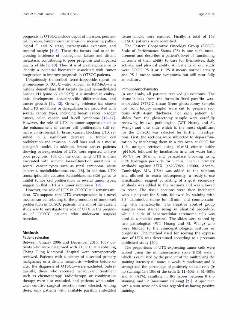
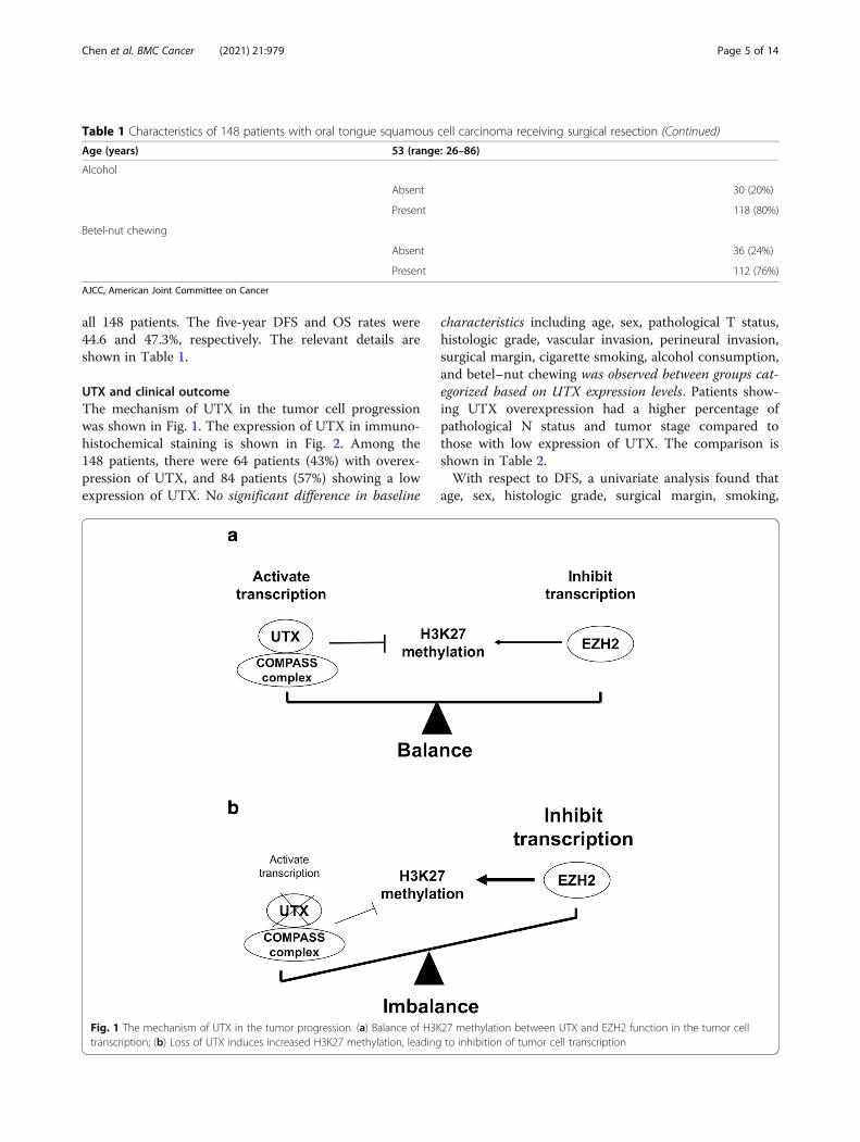
UTX and clinical outcomeThe mechanism of UTX in the tumor cell progressionwas shown in Fig. 1. The expression of UTX in immuno-histochemical staining is shown in Fig. 2. Among the148 patients, there were 64 patients (43%) with overex-pression of UTX, and 84 patients (57%) showing a lowexpression of UTX. No significant difference in baseline
characteristics including age, sex, pathological T status,histologic grade, vascular invasion, perineural invasion,surgical margin, cigarette smoking, alcohol consumption,and betel–nut chewing was observed between groups cat-egorized based on UTX expression levels. Patients show-ing UTX overexpression had a higher percentage ofpathological N status and tumor stage compared tothose with low expression of UTX. The comparison isshown in Table 2.With respect to DFS, a univariate analysis found that
age, sex, histologic grade, surgical margin, smoking,
Table 1 Characteristics of 148 patients with oral tongue squamous cell carcinoma receiving surgical resection (Continued)
Age (years) 53 (range: 26–86)
Alcohol
Absent 30 (20%)
Present 118 (80%)
Betel-nut chewing
Absent 36 (24%)
Present 112 (76%)
AJCC, American Joint Committee on Cancer
Fig. 1 The mechanism of UTX in the tumor progression. (a) Balance of H3K27 methylation between UTX and EZH2 function in the tumor celltranscription; (b) Loss of UTX induces increased H3K27 methylation, leading to inhibition of tumor cell transcription
Chen et al. BMC Cancer (2021) 21:979 Page 5 of 14

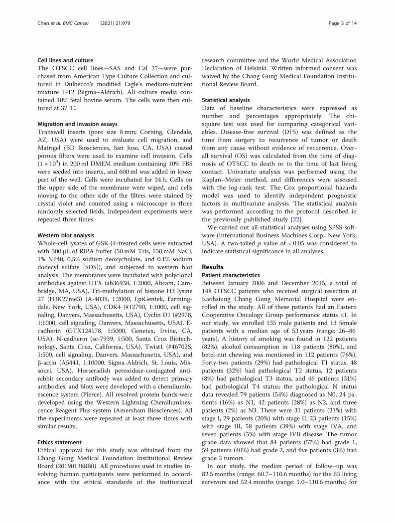
alcohol consumption, and betel–nut chewing were notstatistically significant predictors of DFS. The 58 patientswho had pathological T3–4 were found to have worseDFS than did the other 90 patients who had pathologicalT1–-2 disease (11.4 months versus 69.8 months, P =0.004); 69 patients with nodal metastasis had shorterDFS in comparison with the other 79 patients withoutnodal metastasis (13.2 months versus 87.9 months, P <0.001). Significantly inferior DFS was found in the 88 pa-tients who had pathological stage III–IV than in the 60patients who had pathological stage I–II disease (13.4months versus not reached, P = 0.001). Patients who hadpositive vascular invasion, perineural invasion, and extra-capsular extension were mentioned to have inferior DFScompared to those who did not (P = 0.018, P = 0.008,P < 0.001, respectively). The 64 patients who showed anoverexpression of UTX had worse DFS than did theother 84 patients with a low expression of UTX (20.6months versus 69.8 months, P = 0.006, Fig. 3A). In amultivariate analysis, extracapsular extension (P < 0.001,hazard ratio (HR): 2.22, 95% confidence interval (CI):1.43–3.47) and overexpression of UTX (P = 0.028, HR:1.61, 95% CI: 1.05–2.45) were independent prognosticparameters of worse DFS.In the analysis of OS, no statistically significant differ-
ences in parameters such as age, sex, histologic grade, al-cohol consumption, and betel–nut chewing wereobserved in univariate analysis. The 58 patients withpathological T3–4 were found to have inferior OS com-pared to the other 90 patients with pathological T1–2disease (13.2 months versus 82.2 months, P < 0.001); 69patients who had nodal metastasis had worse OS com-pared to the other 79 patients without nodal metastasis(14.8 months versus not reached, P < 0.001). Significantlyinferior OS was observed in the 88 patients with patho-logical stage III–IV compared to that in the 60 patients
with pathological stage I–II disease (22.5 months versusnot reached, P < 0.001). Patients who had positive vascu-lar invasion, perineural invasion, extracapsular extension,surgical margin, and smoking history were found to haveshorter OS than those who did not (P = 0.004, P = 0.022,P < 0.001, P = 0.031, and P = 0.031, respectively). The 64patients who showed an overexpression of UTX hadworse OS than did the other 84 patients with a low ex-pression of UTX (23.0 months versus 69.8 months, P =0.006, Fig. 3B). Multivariate analysis showed that ad-vanced pathological T status (P = 0.015, HR: 1.73, 95%CI: 1.11–2.69), extracapsular extension (P = 0.003, HR:2.01, 95% CI: 1.26–3.21) and overexpression of UTX(P = 0.029, HR: 1.61, 95% CI: 1.05–2.48) were independ-ent prognostic factors of worse OS. The survival out-comes in the univariate and multivariate analyses areshown in Tables 3 and 4.
Inhibition of UTX by GSK-J4 decrease the abilities ofmigration and invasion of OTSCCAccumulating evidence indicated that GSK-J4, apharmacologic inhibitor, is able to inhibit UTX activitythat acts on specifically H3K27me3 which participates incancer progression. To analyze the effect of UTX inOTSCC, we determined the cellular motility of OTSCCwith GSK-J4 treatment by Transwell assay. Transwellassay results revealed that cells treated with GSK-J4 in adose-dependent manner significantly reduced the num-ber of invaded and migrated cells, compared to cellswithout GSK-J4 treatment (Fig. 4). The observationdemonstrated that, at least in SAS and Cal27 cell lines,inhibition of UTX by GSK-J4 could suppress the motilityof OTSCC. Furthermore, the Western blot analyses wereperformed to determine the expressions of UTX,H3K27me3 status, EMT (epithelial-mesenchymal transi-tion), and GSK-J4-regulated targets in the OTSCC cell
Fig. 2 Results of the immunohistochemical analysis of UTX in oral tongue squamous cell carcinoma patients
Chen et al. BMC Cancer (2021) 21:979 Page 6 of 14

Table 2 Associations between UTX expression and clinicopathological parameters in 148 patients with oral tongue squamous cellcarcinoma receiving surgical resection
Parameters UTX expression
Overexpression (N = 64) Low expression (N = 84) P value
Age
<53y/o 31 (48%) 39 (46%) 0.81
≧53y/o 33 (52%) 45 (54%)
Sex
male 58 (91%) 77 (92%) 0.82
female 6 (9%) 7 (8%)
Pathological T status
T1 + T2 34 (53%) 56 (67%) 0.10
T3 + T4 30 (47%) 28 (33%)
Pathological N status
N0 27 (42%) 52 (62%) 0.017*
N1 + 2 + 3 37 (58%) 32 (38%)
Pathological 8th AJCC Stage
I + II 19 (30%) 41 (49%) 0.019*
III + IV 45 (70%) 43 (51%)
Histologic grade
1 38 (59%) 46 (55%) 0.58
2 + 3 26 (41%) 38 (45%)
Vascular invasion
Absent 53 (83%) 71 (85%) 0.78
Present 11 (17%) 13 (15%)
Perineural invasion
Absent 31 (48%) 50 (60%) 0.18
Present 33 (52%) 34 (40%)
Extracapsular extension
Absent 43 (67%) 68 (81%) 0.06
Present 21 (33%) 16 (19%)
Surgical margin
Negative 58 (91%) 79 (94%) 0.43
Positive 6 (9%) 5 (6%)
Smoking history
Absent 12 (19%) 14 (17%) 0.74
Present 52 (81%) 70 (83%)
Alcohol history
Absent 11 (17%) 19 (23%) 0.42
Present 53 (83%) 65 (77%)
Betel-nut chewing history
Absent 11 (17%) 25 (30%) 0.08
Present 53 (83%) 59 (70%)
AJCC, American Joint Committee on Cancer. *Statistically significant
Chen et al. BMC Cancer (2021) 21:979 Page 7 of 14
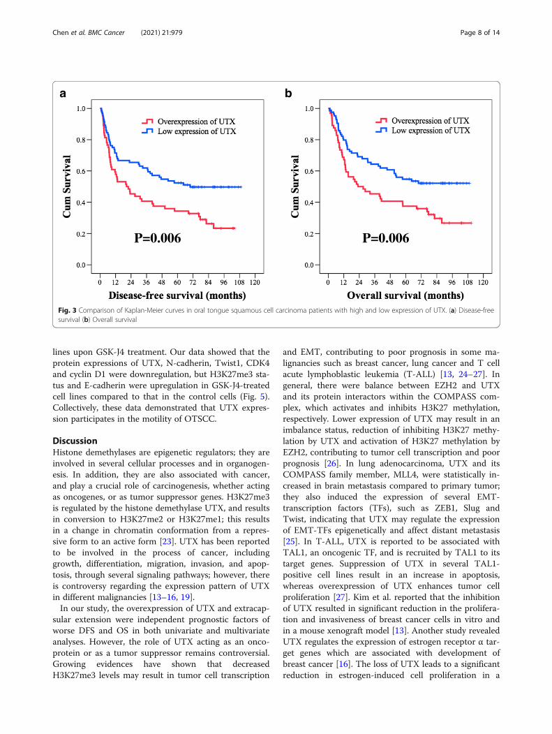
lines upon GSK-J4 treatment. Our data showed that theprotein expressions of UTX, N-cadherin, Twist1, CDK4and cyclin D1 were downregulation, but H3K27me3 sta-tus and E-cadherin were upregulation in GSK-J4-treatedcell lines compared to that in the control cells (Fig. 5).Collectively, these data demonstrated that UTX expres-sion participates in the motility of OTSCC.
DiscussionHistone demethylases are epigenetic regulators; they areinvolved in several cellular processes and in organogen-esis. In addition, they are also associated with cancer,and play a crucial role of carcinogenesis, whether actingas oncogenes, or as tumor suppressor genes. H3K27me3is regulated by the histone demethylase UTX, and resultsin conversion to H3K27me2 or H3K27me1; this resultsin a change in chromatin conformation from a repres-sive form to an active form [23]. UTX has been reportedto be involved in the process of cancer, includinggrowth, differentiation, migration, invasion, and apop-tosis, through several signaling pathways; however, thereis controversy regarding the expression pattern of UTXin different malignancies [13–16, 19].In our study, the overexpression of UTX and extracap-
sular extension were independent prognostic factors ofworse DFS and OS in both univariate and multivariateanalyses. However, the role of UTX acting as an onco-protein or as a tumor suppressor remains controversial.Growing evidences have shown that decreasedH3K27me3 levels may result in tumor cell transcription
and EMT, contributing to poor prognosis in some ma-lignancies such as breast cancer, lung cancer and T cellacute lymphoblastic leukemia (T-ALL) [13, 24–27]. Ingeneral, there were balance between EZH2 and UTXand its protein interactors within the COMPASS com-plex, which activates and inhibits H3K27 methylation,respectively. Lower expression of UTX may result in animbalance status, reduction of inhibiting H3K27 methy-lation by UTX and activation of H3K27 methylation byEZH2, contributing to tumor cell transcription and poorprognosis [26]. In lung adenocarcinoma, UTX and itsCOMPASS family member, MLL4, were statistically in-creased in brain metastasis compared to primary tumor;they also induced the expression of several EMT-transcription factors (TFs), such as ZEB1, Slug andTwist, indicating that UTX may regulate the expressionof EMT-TFs epigenetically and affect distant metastasis[25]. In T-ALL, UTX is reported to be associated withTAL1, an oncogenic TF, and is recruited by TAL1 to itstarget genes. Suppression of UTX in several TAL1-positive cell lines result in an increase in apoptosis,whereas overexpression of UTX enhances tumor cellproliferation [27]. Kim et al. reported that the inhibitionof UTX resulted in significant reduction in the prolifera-tion and invasiveness of breast cancer cells in vitro andin a mouse xenograft model [13]. Another study revealedUTX regulates the expression of estrogen receptor α tar-get genes which are associated with development ofbreast cancer [16]. The loss of UTX leads to a significantreduction in estrogen-induced cell proliferation in a
P=0.006 P=0.006
a b
Fig. 3 Comparison of Kaplan-Meier curves in oral tongue squamous cell carcinoma patients with high and low expression of UTX. (a) Disease-freesurvival (b) Overall survival
Chen et al. BMC Cancer (2021) 21:979 Page 8 of 14
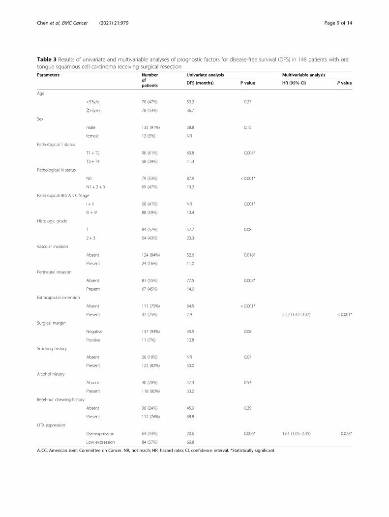
Table 3 Results of univariate and multivariable analyses of prognostic factors for disease-free survival (DFS) in 148 patients with oraltongue squamous cell carcinoma receiving surgical resectionParameters Number
ofpatients
Univariate analysis Multivariable analysis
DFS (months) P value HR (95% CI) P value
Age
<53y/o 70 (47%) 50.2 0.27
≧53y/o 78 (53%) 36.7
Sex
male 135 (91%) 38.8 0.15
female 13 (9%) NR
Pathological T status
T1 + T2 90 (61%) 69.8 0.004*
T3 + T4 58 (39%) 11.4
Pathological N status
N0 79 (53%) 87.9 < 0.001*
N1 + 2 + 3 69 (47%) 13.2
Pathological 8th AJCC Stage
I + II 60 (41%) NR 0.001*
III + IV 88 (59%) 13.4
Histologic grade
1 84 (57%) 57.7 0.08
2 + 3 64 (43%) 23.3
Vascular invasion
Absent 124 (84%) 52.6 0.018*
Present 24 (16%) 11.0
Perineural invasion
Absent 81 (55%) 77.5 0.008*
Present 67 (45%) 14.0
Extracapsular extension
Absent 111 (75%) 64.5 < 0.001*
Present 37 (25%) 7.9 2.22 (1.42–3.47) < 0.001*
Surgical margin
Negative 137 (93%) 45.9 0.08
Positive 11 (7%) 12.8
Smoking history
Absent 26 (18%) NR 0.07
Present 122 (82%) 33.0
Alcohol history
Absent 30 (20%) 47.3 0.54
Present 118 (80%) 33.0
Betel-nut chewing history
Absent 36 (24%) 45.9 0.29
Present 112 (76%) 38.8
UTX expression
Overexpression 64 (43%) 20.6 0.006* 1.61 (1.05–2.45) 0.028*
Low expression 84 (57%) 69.8
AJCC, American Joint Committee on Cancer. NR, not reach; HR, hazard ratio; CI, confidence interval. *Statistically significant
Chen et al. BMC Cancer (2021) 21:979 Page 9 of 14

Table 4 Results of univariate and multivariable analyses of prognostic factors for overall survival (OS) in 148 patients with oraltongue squamous cell carcinoma receiving surgical resectionParameters Number
ofpatients
Univariate analysis Multivariable analysis
OS (months) P value HR (95% CI) P value
Age
<53y/o 70 (47%) 57.7 0.43
≧53y/o 78 (53%) 51.1
Sex
male 135 (91%) 51.1 0.07
female 13 (9%) NR
Pathological T status
T1 + T2 90 (61%) 82.2 < 0.001*
T3 + T4 58 (39%) 13.2 1.73 (1.11–2.69) 0.015*
Pathological N status
N0 79 (53%) NR < 0.001*
N1 + 2 + 3 69 (47%) 14.8
Pathological 8th AJCC Stage
I + II 60 (41%) NR < 0.001*
III + IV 88 (59%) 22.5
Histologic grade
1 84 (57%) 64.5 0.18
2 + 3 64 (43%) 33.0
Vascular invasion
Absent 124 (84%) 68.4 0.004*
Present 24 (16%) 11.4
Perineural invasion
Absent 81 (55%) 82.2 0.022*
Present 67 (45%) 30.7
Extracapsular extension
Absent 111 (75%) 76.5 < 0.001*
Present 37 (25%) 11.7 2.01 (1.26–3.21) 0.003*
Surgical margin
Negative 137 (93%) 57.6 0.031*
Positive 11 (7%) 13.2
Smoking history
Absent 26 (18%) NR 0.031*
Present 122 (82%) 40.3
Alcohol history
Absent 30 (20%) 68.4 0.34
Present 118 (80%) 41.2
Betel-nut chewing history
Absent 36 (24%) NR 0.16
Present 112 (76%) 45.5
UTX expression
Overexpression 64 (43%) 23.0 0.006* 1.61 (1.05–2.48) 0.029*
Low expression 84 (57%) 69.8
AJCC, American Joint Committee on Cancer. NR, not reach; HR, hazard ratio; CI, confidence interval. *Statistically significant
Chen et al. BMC Cancer (2021) 21:979 Page 10 of 14

human breast cancer cell line; in contrast, overexpres-sion of UTX promotes cell migration [16]. Therefore,overexpression of UTX may lead to reduce H3K27methylation, inhibit apoptosis, promote tumor cell tran-scription and distant metastasis and enhance EMT, con-tributing to poor prognosis. Our study concluded theoverexpression of UTX was found to be associated with
worse DFS and OS, indicating the significance of UTXas a tumor oncogene in OTSCC.There were some studies which reported sex-specific
difference in the expression of UTX. Xu et al. showedthat higher expression of UTX was found in female micethan in male mice in most brain regions except in theamygdala [28]. This discordance may result from
***
***
***
***
*
*
*
*
Fig. 4 Transwell migration and invasion assays using SAS and Cal 27 cell lines treated with GSK–J4 at different concentrations. Columns, mean;bars, standard deviation. Significant difference: *P < 0.05 and ***P < 0.001
Chen et al. BMC Cancer (2021) 21:979 Page 11 of 14

differences in chromatin remodeling on the X and Ychromosomes. In addition, a Belgian study demonstratedthat UTX mutations were exclusively present in male T-ALL patients and allelic expression analysis showedUTX escapes X-inactivation in female T-ALL lympho-blasts and normal T cells [29]. In T-ALL, UTX functionsas a tumor suppressor and T-ALL driven by UTX inacti-vation displays collateral sensitivity to pharmacologicH3K27me3 inhibition [29]. In contrast, there were nosignificant difference of gender distribution in our study,the percentage of female patients in the UTX overex-pression and low expression groups were 9 and 8%, re-spectively (Table 2). Although female patients werefound to have longer DFS and OS than male patients inthe univariate and multivariate analyses, there was nostatistical difference; the better DFS and OS in the fe-male patients may be caused by too lower proportion offemale patients (only 9%), resulting in biases existed.GSK–J4 is a potent dual inhibitor of the H3K27me3
demethylases JMJD3 and UTX, and has been reportedto be involved in many physiological and pathologicalprocesses. Growing evidence has addressed the effectof this drug in immune cells [30]. GSK–J4 was ableto modulate inflammation by affecting dendritic cells,causing an increase in the expression of tolerogenicmolecules, and a decrease in the secretion of proin-flammatory cytokines [31]. GSK–J4 was also found to
inhibit the activity of H3K27 demethylase to suppressT helper 17 cell differentiation as seen in in vitrostudies, which suggests that it may be considered as apotential novel therapeutic target for suppressingautoimmune or inflammatory responses [32].H3K27me3 has been related to the differentiation ofnormal stem cells and cancer cells, and H3K27methylation may play a crucial role in inhibiting themaintenance of cancer stem cells [33]. In non-smallcell lung cancer, GSK–J4 was able to induce celldeath and inhibit the proliferation of tumor cells, ir-respective of the genetic mutation status or chemo-therapy resistance [34]. Therefore, GSK–J4 mayrepresent a promising anticancer agent.Our study also had several limitations. First, the small
size of the study population may limit this study’s statis-tical significance. Second, the percentage of female pa-tients were relatively low (only 9%) so it was difficult toavoid selection bias in this retrospectively designedstudy. Third, the association between UTX and down-stream pathways was not fully investigated; additionally,the mechanism of modulation of cancer cell growth andmodulation by UTX was not examined. However, to thebest of our knowledge, this current study enrolled thelargest number of OTSCC patients who underwent sur-gical resection, and may be helpful to understand therole of UTX in the prognosis of OTSCC.
Fig. 5 Western blot analysis of UTX expression and the downstream signaling pathway in the SAS and Cal 27 cell lines. The protein expressionprofiles of UTX, H3K27me3 status, EMT markers, cyclin D1 and CDK4 were examined in the presence or absence of GSK-J4 treatment in theOTSCC cells by Western blotting
Chen et al. BMC Cancer (2021) 21:979 Page 12 of 14

ConclusionOur study suggests that UTX plays an important role inthe process of OTSCC, and that the overexpression ofUTX is an independent prognostic factor of poor prog-nosis in OTSCC patients who received surgical resec-tion. Further research with a large population is neededto confirm our findings, and to clarify the complexmechanism of UTX action in OTSCC.
AbbreviationsOTSCC: oral tongue squamous cell carcinoma; UTX: ubiquitously transcribedtetratricopeptide repeat on chromosome X; H3K27: H3 lysine 27;Rb: Retinoblastoma; ECOG: Eastern Cooperative Oncology Group;PS: Performance Status; IRS: immunoreactive score; H3K27me3: tri-methylation of histone H3 lysine 27; DFS: disease-free survival; OS: overallsurvival; HR: hazard ratio; CI: confidence interval; EMT: epithelial-mesenchymal transition; T-ALL: T cell acute lymphoblastic leukemia;TF: transcription factor
Supplementary InformationThe online version contains supplementary material available at https://doi.org/10.1186/s12885-021-08726-3.
Additional file 1. Supplementary Fig. S1. The original data of westernblot analyses.
AcknowledgementsThe authors thank Drs. WT Huang and SL Wang for assessment of theimmunohistochemical staining. We thank Chang Gung Medical FoundationKaohsiung Chang Gung Memorial Hospital Tissue Bank Core Laboratory forexcellent technical support.
Authors’ contributionsConceptualization, Y-H Chen; methodology, C-H Chen; software, C-Y Chien;validation, S-H Li; formal analysis, Y-Y Su; investigation, S-D Luo; resources, C-Y Chien; data curation, Y-Y Su and S-D Luo; writing—original draft prepar-ation, S-H Li; writing—review and editing, Y-H Chen; visualization, C-H Chen;supervision, Y-H Chen; project administration, Y-H Chen; funding acquisition,Y-H Chen. All authors have read and agreed to the published version of themanuscript.
FundingThis work was supported in part by a grant from Chang Gung MemorialHospital, grant number CMRPG8K0601.
Availability of data and materialsThe datasets used and analyzed during the current study are available fromthe corresponding author on reasonable request.
Declarations
Ethics approval and consent to participateEthical approval for this study was obtained from the Chang Gung MedicalFoundation Institutional Review Board (201901388B0). All procedures used instudies involving human participants were performed in accordance withthe ethical standards of the institutional research committee and the WorldMedical Association Declaration of Helsinki. Written informed consent waswaived by the Chang Gung Medical Foundation Institutional Review Board.
Consent for publicationNot applicable.
Competing interestsThe authors declare that they have no competing interests
Author details1Department of Hematology-Oncology, Kaohsiung Chang Gung MemorialHospital and Chang Gung University College of Medicine, No.123, Dapi Rd.,Niaosong Dist, Kaohsiung City 833, Taiwan. 2School of Medicine, Chung ShanMedical University, Taichung 402, Taiwan. 3Department of Nursing, MeihoUniversity, Pingtung 912, Taiwan. 4Institute of Medicine, Chung Shan MedicalUniversity, Department of Medical Research, Chung Shan Medical UniversityHospital, Taichung 402, Taiwan. 5Department of Otolaryngology, KaohsiungChang Gung Memorial Hospital and Chang Gung University College ofMedicine, Kaohsiung, Taiwan.
Received: 20 January 2021 Accepted: 23 August 2021
References1. The Surveillance E, and End Results (SEER) Cancer Stat Facts: Oral Cavity and
Pharynx Cancer. 2013.2. Ng JH, Iyer NG, Tan MH, Edgren G. Changing epidemiology of oral
squamous cell carcinoma of the tongue: a global study. Head Neck. 2017;39(2):297–304. https://doi.org/10.1002/hed.24589.
3. Price KA, Cohen EE. Current treatment options for metastatic head and neckcancer. Curr Treat Options in Oncol. 2012;13(1):35–46. https://doi.org/10.1007/s11864-011-0176-y.
4. Aivazian K, Ebrahimi A, Low TH, Gao K, Clifford A, Shannon K, et al.Perineural invasion in oral squamous cell carcinoma: quantitativesubcategorisation of perineural invasion and prognostication. J Surg Oncol.2015;111(3):352–8. https://doi.org/10.1002/jso.23821.
5. Daniell J, Udovicich C, Rowe D, McDowell L, Vital D, Bressel M, et al. Impactof histological Oral tongue Cancer margins on locoregional recurrence: amulti-Centre retrospective analysis. Oral Oncol. 2020;111:105004. https://doi.org/10.1016/j.oraloncology.2020.105004.
6. Fagan JJ, Collins B, Barnes L, D'Amico F, Myers EN, Johnson JT. Perineuralinvasion in squamous cell carcinoma of the head and neck. ArchOtolaryngol Head Neck Surg. 1998;124(6):637–40. https://doi.org/10.1001/archotol.124.6.637.
7. Larson AR, Kemmer J, Formeister E, El-Sayed I, Ha P, George J, et al. Beyonddepth of invasion: adverse pathologic tumor features in early Oral tonguesquamous cell carcinoma. Laryngoscope. 2020;130(7):1715–20. https://doi.org/10.1002/lary.28241.
8. Sharma K, Ahlawat P, Gairola M, Tandon S, Sachdeva N, Sharief MI.Prognostic factors, failure patterns and survival analysis in patients withresectable oral squamous cell carcinoma of the tongue. Radiat Oncol J.2019;37(2):73–81. https://doi.org/10.3857/roj.2018.00577.
9. Camisasca DR, Silami MA, Honorato J, Dias FL, de Faria PA, Lourenco Sde Q.Oral squamous cell carcinoma: clinicopathological features in patients withand without recurrence. ORL J Otorhinolaryngol Relat Spec. 2011;73(3):170–6. https://doi.org/10.1159/000328340.
10. Lindenblatt Rde C, Martinez GL, Silva LE, Faria PS, Camisasca DR, LourencoSde Q. Oral squamous cell carcinoma grading systems--analysis of the bestsurvival predictor. J Oral Pathol Med. 2012;41(1):34–9. https://doi.org/10.1111/j.1600-0714.2011.01068.x.
11. Lan F, Bayliss PE, Rinn JL, Whetstine JR, Wang JK, Chen S, et al. A histone H3lysine 27 demethylase regulates animal posterior development. Nature.2007;449(7163):689–94. https://doi.org/10.1038/nature06192.
12. van Haaften G, Dalgliesh GL, Davies H, Chen L, Bignell G, Greenman C, et al.Somatic mutations of the histone H3K27 demethylase gene UTX in humancancer. Nat Genet. 2009;41(5):521–3. https://doi.org/10.1038/ng.349.
13. Kim JH, Sharma A, Dhar SS, Lee SH, Gu B, Chan CH, et al. UTX and MLL4coordinately regulate transcriptional programs for cell proliferation andinvasiveness in breast cancer cells. Cancer Res. 2014;74(6):1705–17. https://doi.org/10.1158/0008-5472.CAN-13-1896.
14. Lang A, Yilmaz M, Hader C, Murday S, Kunz X, Wagner N, et al.Contingencies of UTX/KDM6A Action in Urothelial Carcinoma. Cancers.2019;11(4).
15. Li X, Zhang Y, Zheng L, Liu M, Chen CD, Jiang H. UTX is an escape from X-inactivation tumor-suppressor in B cell lymphoma. Nat Commun. 2018;9(1):2720. https://doi.org/10.1038/s41467-018-05084-w.
16. Xie G, Liu X, Zhang Y, Li W, Liu S, Chen Z, et al. UTX promotes hormonallyresponsive breast carcinogenesis through feed-forward transcriptionregulation with estrogen receptor. Oncogene. 2017;36(39):5497–511. https://doi.org/10.1038/onc.2017.157.
Chen et al. BMC Cancer (2021) 21:979 Page 13 of 14

17. Zhou Z, Zhang HS, Liu Y, Zhang ZG, Du GY, Li H, et al. Loss of TET1facilitates DLD1 colon cancer cell migration via H3K27me3-mediated down-regulation of E-cadherin. J Cell Physiol. 2018;233(2):1359–69. https://doi.org/10.1002/jcp.26012.
18. Suva ML, Riggi N, Bernstein BE. Epigenetic reprogramming in cancer.Science. 2013;339(6127):1567–70. https://doi.org/10.1126/science.1230184.
19. Wang JK, Tsai MC, Poulin G, Adler AS, Chen S, Liu H, et al. The histonedemethylase UTX enables RB-dependent cell fate control. Genes Dev. 2010;24(4):327–32. https://doi.org/10.1101/gad.1882610.
20. Shen Y, Guo X, Wang Y, Qiu W, Chang Y, Zhang A, et al. Expression andsignificance of histone H3K27 demethylases in renal cell carcinoma. BMCCancer. 2012;12(1):470. https://doi.org/10.1186/1471-2407-12-470.
21. Remmele W, Stegner HE. Recommendation for uniform definition of animmunoreactive score (IRS) for immunohistochemical estrogen receptordetection (ER-ICA) in breast cancer tissue. Pathologe. 1987;8(3):138–40.
22. Chen YH, Chien CY, Fang FM, Huang TL, Su YY, Luo SD, et al. Nox4overexpression as a poor prognostic factor in patients with oral tonguesquamous cell carcinoma receiving surgical resection. J Clin Med. 2018;7(12).
23. Zhang F, Xu L, Xu L, Xu Q, Li D, Yang Y, et al. JMJD3 promotes chondrocyteproliferation and hypertrophy during endochondral bone formation inmice. J Mol Cell Biol. 2015;7(1):23–34. https://doi.org/10.1093/jmcb/mjv003.
24. Gameiro SF, Kolendowski B, Zhang A, Barrett JW, Nichols AC, Torchia J, et al.Human papillomavirus dysregulates the cellular apparatus controlling themethylation status of H3K27 in different human cancers to consistently altergene expression regardless of tissue of origin. Oncotarget. 2017;8(42):72564–76. https://doi.org/10.18632/oncotarget.19885.
25. Lee YM, Kim SH, Kim MS, Kim DC, Lee EH, Lee JS, et al. Epigenetic role ofhistone lysine methyltransferase and demethylase on the expression oftranscription factors associated with the epithelial-to-mesenchymaltransition of lung adenocarcinoma metastasis to the brain. Cancers. 2020;12(12).
26. Wang L, Shilatifard A. UTX mutations in human Cancer. Cancer Cell. 2019;35(2):168–76. https://doi.org/10.1016/j.ccell.2019.01.001.
27. Benyoucef A, Palii CG, Wang C, Porter CJ, Chu A, Dai F, et al. UTX inhibitionas selective epigenetic therapy against TAL1-driven T-cell acutelymphoblastic leukemia. Genes Dev. 2016;30(5):508–21. https://doi.org/10.1101/gad.276790.115.
28. Xu J, Deng X, Watkins R, Disteche CM. Sex-specific differences in expressionof histone demethylases Utx and Uty in mouse brain and neurons. JNeurosci. 2008;28(17):4521–7. https://doi.org/10.1523/JNEUROSCI.5382-07.2008.
29. Van der Meulen J, Sanghvi V, Mavrakis K, Durinck K, Fang F, Matthijssens F,et al. The H3K27me3 demethylase UTX is a gender-specific tumorsuppressor in T-cell acute lymphoblastic leukemia. Blood. 2015;125(1):13–21.https://doi.org/10.1182/blood-2014-05-577270.
30. Donas C, Carrasco M, Fritz M, Prado C, Tejon G, Osorio-Barrios F, et al. Thehistone demethylase inhibitor GSK-J4 limits inflammation through theinduction of a tolerogenic phenotype on DCs. J Autoimmun. 2016;75:105–17. https://doi.org/10.1016/j.jaut.2016.07.011.
31. Kruidenier L, Chung CW, Cheng Z, Liddle J, Che K, Joberty G, et al. Aselective jumonji H3K27 demethylase inhibitor modulates theproinflammatory macrophage response. Nature. 2012;488(7411):404–8.https://doi.org/10.1038/nature11262.
32. Liu Z, Cao W, Xu L, Chen X, Zhan Y, Yang Q, et al. The histone H3 lysine-27demethylase Jmjd3 plays a critical role in specific regulation of Th17 celldifferentiation. J Mol Cell Biol. 2015;7(6):505–16. https://doi.org/10.1093/jmcb/mjv022.
33. Sakaki H, Okada M, Kuramoto K, Takeda H, Watarai H, Suzuki S, et al. GSKJ4,a selective Jumonji H3K27 demethylase inhibitor, effectively targets ovarianCancer stem cells. Anticancer Res. 2015;35(12):6607–14.
34. Watarai H, Okada M, Kuramoto K, Takeda H, Sakaki H, Suzuki S, et al. Impactof H3K27 demethylase inhibitor GSKJ4 on NSCLC cells alone and incombination with metformin. Anticancer Res. 2016;36(11):6083–92. https://doi.org/10.21873/anticanres.11198.
Publisher’s NoteSpringer Nature remains neutral with regard to jurisdictional claims inpublished maps and institutional affiliations.
Chen et al. BMC Cancer (2021) 21:979 Page 14 of 14