OVENANT UNIVERSITYcovenantuniversity.edu.ng/content/download/49915/339068/version/2... ·...
26
COVENANT UNIVERSITY ALPHA SEMESTER TUTORIAL KIT (VOL. 2) PROGRAMME: BIOCHEMISTRY 400 LEVEL
Transcript of OVENANT UNIVERSITYcovenantuniversity.edu.ng/content/download/49915/339068/version/2... ·...
COVENANT UNIVERSITY
ALPHA SEMESTER TUTORIAL KIT (VOL. 2)
P R O G R A M M E : B I O C H E M I S T R Y
400 LEVEL

1
DISCLAIMER
The contents of this document are intended for practice and learning purposes at the undergraduate
level. The materials are from different sources including the internet and the contributors do not
in any way claim authorship or ownership of them. The materials are also not to be used for any
commercial purpose.
2
LIST OF COURSES
*BCH411: Advanced Enzymology
BCH412: Nutritional Biochemistry
*BCH413: Industrial Biochemistry
BCH414: Advanced Biochemical Genetics
*BCH415: Biochemical Reasoning and Research Methods
BCH417: Biochemistry of Macromolecules
BCH431: Tissue Biochemistry
BCH432: Techniques in Molecular Biology
BCH433: Trends in Medical Biochemistry
*Not included

3
DEPARTMENT OF BIOLOGICAL SCIENCES
TUTORIAL QUESTIONS FOR BCH 412
1. Define the term ‘Nutrition’ and give in outline form the importance of proteins in human
nutrition. State any 4 major sources of food proteins in human diet.
Nutrition is defined as the Science of foods and the nutrients and other substances they contain, and of their actions within the body, including ingestion, digestion, absorption, transport, metabolism, and excretion.
A broader definition includes the social, economic, cultural, and physiological implications of food and eating. Biological Importance of Proteins in Human Nutrition
1. Proteins are essential construction elements of the organism – bone, connective tissue, muscle, skin and hair;
2. Proteins, together with lipids, constitute the membranes of cells; 3. Specific proteins (immunoglobulins) assume defense functions against infections and diseases; 4. Blood serum proteins assume transport functions (serum albumin transports lipids,
caeruloplasmin transports copper); 5. The respiratory system of vertebrates is based on the oxygen binding properties of specific iron-
rich proteins, hemoglobin, present in erythrocyte and myoglobin, present in muscle; 6. Proteins, as specific enzymes, functions in catalytic activity which enable and control metabolism
on the cellular level; 7. Proteins function as hormones regulating metabolic processes at tissue and cellular level. Insulin
and glucagon, to name only two, regulate the level of blood glucose. Major sources of food proteins Milk, Egg, Meat, Fish and Soybean
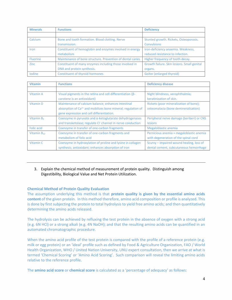
2. Distinguish between major and micro nutrients. State briefly the functions and deficiency
symptoms of Iron, Iodine, Calcium, Zinc, Fluorine, Vitamin A, Vitamin D, Vitamin B1, Vitamin B12 and Folic acid.
Carbohydrate, fat and protein are required in relatively large amounts (many gm daily) hence are called macronutrients. On the other hand, vitamins and minerals are referred to as micronutrients because they are needed in small amounts (mostly in milligrams or indeed micrograms). Incidentally, the three macronutrients are energy-yielding nutrients of the food. In normal and equilibrated diet, most energy is furnished by carbohydrates (55 – 60%). Protein should account for about 12% of total caloric intake, lipids for about 30%.
4
Minerals Functions Deficiency
Calcium Bone and tooth formation. Blood clotting. Nerve
transmission.
Stunted growth. Rickets, Osteoporosis.
Convulsions
Iron Constituent of hemoglobin and enzymes involved in energy
metabolism
Iron-deficiency anaemia. Weakness,
reduced resistance to infection.
Fluorine Maintenance of bone structure. Prevention of dental caries Higher frequency of tooth decay.
Zinc Constituent of many enzymes including those involved in
DNA and protein synthesis.
Growth failure. Skin lesions. Small genital
organs.
Iodine Constituent of thyroid hormones Goiter (enlarged thyroid)
Vitamin Functions Deficiency disease
Vitamin A Visual pigments in the retina and cell differentiation (β-
carotene is an antioxidant)
Night blindness, xerophthalmia;
keratinization of skin.
Vitamin D Maintenance of calcium balance; enhances intestinal
absorption of Ca2+ and mobilizes bone mineral; regulation of
gene expression and cell differentiation.
Rickets (poor mineralization of bone);
osteomalacia (bone demineralisation)
Vitamin B1 Coenzyme in pyruvate and α-ketoglutarate dehydrogenases
and transketolase; regulate Cl- channel in nerve conduction
Peripheral nerve damage (beriberi) or CNS
lesions
Folic acid Coenzyme in transfer of one-carbon fragments Megaloblastic anemia
Vitamin B12 Coenzyme in transfer of one-carbon fragments and
metabolism of folic acid
Pernicious anemia = megaloblastic anemia
with degeneration of the spinal cord
Vitamin C Coenzyme in hydroxylation of proline and lysine in collagen
synthesis; antioxidant; enhances absorption of iron
Scurvy – impaired wound healing, loss of
dental cement, subcutaneous hemorrhage
3. Explain the chemical method of measurement of protein quality. Distinguish among Digestibility, Biological Value and Net Protein Utilization.
Chemical Method of Protein Quality Evaluation The assumption underlying this method is that protein quality is given by the essential amino acids content of the given protein. In this method therefore, amino acid composition or profile is analyzed. This is done by first subjecting the protein to total hydrolysis to yield free amino acids; and then quantitatively determining the amino acids released. The hydrolysis can be achieved by refluxing the test protein in the absence of oxygen with a strong acid (e.g. 6N HCl) or a strong alkali (e.g. 4N NaOH); and that the resulting amino acids can be quantified in an automated chromatographic procedure. When the amino acid profile of the test protein is compared with the profile of a reference protein (e.g. milk or egg protein) or an ‘ideal’ profile such as defined by Food & Agriculture Organization, FAO / World Health Organization, WHO / United Nation University, UNU expert consultation, then we arrive at what is termed ‘Chemical Scoring’ or ‘Amino Acid Scoring’. Such comparison will reveal the limiting amino acids relative to the reference profile. The amino acid score or chemical score is calculated as a ‘percentage of adequacy’ as follows:

5
𝑎𝑚𝑖𝑛𝑜 𝑎𝑐𝑖𝑑 𝑠𝑐𝑜𝑟𝑒 =𝑚𝑔 𝑜𝑓 𝑎𝑚𝑖𝑛𝑜 𝑎𝑐𝑖𝑑 𝑖𝑛 1𝑔 𝑡𝑒𝑠𝑡 𝑝𝑟𝑜𝑡𝑒𝑖𝑛
mg of amino acid in reference protein𝑥 100
Digestibility, Biological Value and Net Protein Utilization The underlying assumption here is that nitrogen retained in the growing animals is representative of net protein synthesis. When the value of nitrogen retained is expressed as a percentage of protein intake or ingested, it is referred to as Net Protein Utilization (NPU). In nitrogen balance studies where ingested nitrogen (I), urinary nitrogen (U) and fecal nitrogen (F) are carefully measured, it is possible to calculate the nitrogen fraction absorbed by the gut (I – F) and the fraction retained in the body (I – F – U). From these data, the apparent digestibility, biological value and net protein utilization can be calculated: Digestibility, D = [(absorbed N)/(intake N)] x 100 = [(I – F)/I] x 100 Digestibility represents the ability of food protein to be hydrolyzed by the intestinal enzymes and, following hydrolysis, to be absorbed by the gut. Biological value, BV = [(retained N)/(absorbed N)] x 100 = [(I – F – U)/(I – F)] x 100 Biological value represents the ability of the absorbed amino acids to be utilized in protein synthesis and depends of the amino acid profile of absorbed protein nitrogen. Net protein utilization, NPU = [(retained N)/(intake N)] x 100 = [(I – F – U)/I] x 100 If correction for endogenous losses of nitrogen is made in the nitrogen balance study, then apparent digestibility and biological value become ‘true digestibility’ and ‘true biological value’.
4. Define the term ‘Basal Metabolic Rate, BMR’. List and explain any 5 factors that may influence BMR.
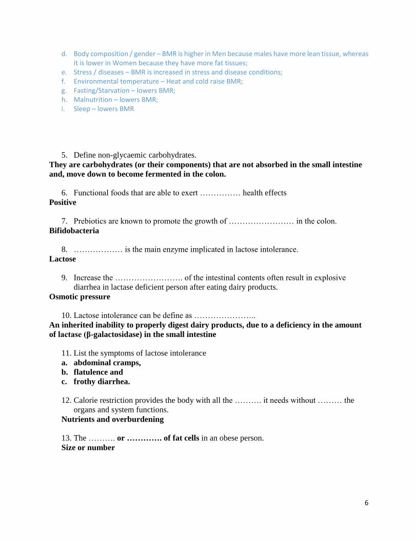
Basal Metabolic Rate (BMR) is defined as the rate of energy use for metabolism under specified conditions: after a 12-hour fast and restful sleep, without any physical activity or emotional excitement, and in a comfortable setting. It is usually expressed as Kcal/kg Body Weight/hr. This energy is expended for metabolic activities that support all the basic processes of life. That is, maintenance of body temperature, breathing (and movement of the lungs), production of Red Blood Cells, heart beating, kidneys functions, etc. Variability of BMR among different people is due largely to the following factors:
a. Age – BMR reduces with age; b. Height – BMR increases with height; c. Growth – BMR is higher in children;
6
d. Body composition / gender – BMR is higher in Men because males have more lean tissue, whereas it is lower in Women because they have more fat tissues;
e. Stress / diseases – BMR is increased in stress and disease conditions; f. Environmental temperature – Heat and cold raise BMR; g. Fasting/Starvation – lowers BMR; h. Malnutrition – lowers BMR; i. Sleep – lowers BMR
5. Define non-glycaemic carbohydrates.
They are carbohydrates (or their components) that are not absorbed in the small intestine
and, move down to become fermented in the colon.
6. Functional foods that are able to exert …………… health effects
Positive
7. Prebiotics are known to promote the growth of …………………… in the colon.
Bifidobacteria
8. ……………… is the main enzyme implicated in lactose intolerance.
Lactose
9. Increase the ……………………. of the intestinal contents often result in explosive
diarrhea in lactase deficient person after eating dairy products.
Osmotic pressure
10. Lactose intolerance can be define as …………………..
An inherited inability to properly digest dairy products, due to a deficiency in the amount
of lactase (β-galactosidase) in the small intestine
11. List the symptoms of lactose intolerance
a. abdominal cramps,
b. flatulence and
c. frothy diarrhea.
12. Calorie restriction provides the body with all the ………. it needs without ……… the
organs and system functions.
Nutrients and overburdening
13. The ………. or …………. of fat cells in an obese person.
Size or number
7
BCH 414 –ADVANCED GENETICS TUTORIAL QUESTIONS
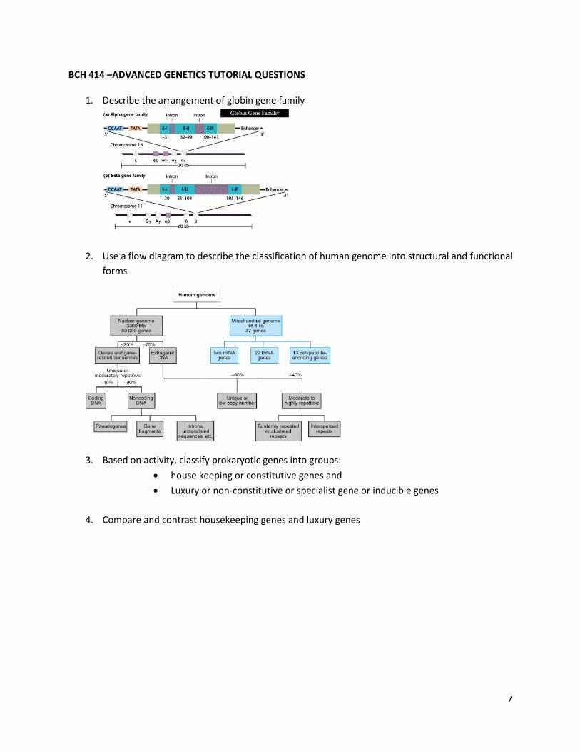
1. Describe the arrangement of globin gene family
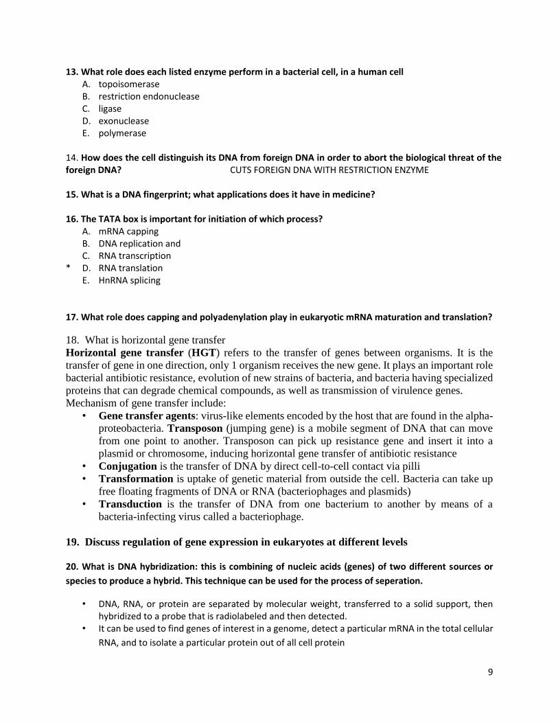
2. Use a flow diagram to describe the classification of human genome into structural and functional
forms
3. Based on activity, classify prokaryotic genes into groups:
house keeping or constitutive genes and
Luxury or non-constitutive or specialist gene or inducible genes
4. Compare and contrast housekeeping genes and luxury genes

8
5. Draw the classical mode of transcription in bacteria
6. Describe the prokaryotic promoter site
7. What are histone proteins?
8. What are non-histone proteins?
9. Describe the organization eukaryotic chromosomes.
10. Describe DNA compaction in eukaryotes.
11. What are conserved genes and how can these be used to identify species ANSWER- A highly conserved gene has very similar or sometimes even identical sequence across different species. This generally suggests that the species are closely related. If 5 DNA sequences from different species are available, compare the conserved region of these DNAs and identify closely related species or the ones that deviate the most from others sequence. Phylogenic trees are built based on these information and organisms have been classified based on relatedness of their conserved region. E.g. using the 16S rRNA to differentiate bacteria from homosapeins 12. In medicine, how can gene modulation be useful? In an organism, a gene whose encoded protein does something very important in the organism, and whose function would be altered by almost any change to its sequence can be identified, altered and re-engineered into the organism. This can be used to make a pathogenic organism become non-pathogenic when they lack the protein that result in infections. In gene cloning, a promoter can be used to increase the expression of a beneficial gene such that the product is produced in larger quantities, and this can extracted and used for medical purposes.
9
13. What role does each listed enzyme perform in a bacterial cell, in a human cell A. topoisomerase B. restriction endonuclease C. ligase D. exonuclease E. polymerase 14. How does the cell distinguish its DNA from foreign DNA in order to abort the biological threat of the foreign DNA? CUTS FOREIGN DNA WITH RESTRICTION ENZYME 15. What is a DNA fingerprint; what applications does it have in medicine? 16. The TATA box is important for initiation of which process? A. mRNA capping B. DNA replication and C. RNA transcription * D. RNA translation E. HnRNA splicing 17. What role does capping and polyadenylation play in eukaryotic mRNA maturation and translation?
18. What is horizontal gene transfer
Horizontal gene transfer (HGT) refers to the transfer of genes between organisms. It is the
transfer of gene in one direction, only 1 organism receives the new gene. It plays an important role
bacterial antibiotic resistance, evolution of new strains of bacteria, and bacteria having specialized
proteins that can degrade chemical compounds, as well as transmission of virulence genes.
Mechanism of gene transfer include:
• Gene transfer agents: virus-like elements encoded by the host that are found in the alpha-
proteobacteria. Transposon (jumping gene) is a mobile segment of DNA that can move
from one point to another. Transposon can pick up resistance gene and insert it into a
plasmid or chromosome, inducing horizontal gene transfer of antibiotic resistance
• Conjugation is the transfer of DNA by direct cell-to-cell contact via pilli
• Transformation is uptake of genetic material from outside the cell. Bacteria can take up
free floating fragments of DNA or RNA (bacteriophages and plasmids)
• Transduction is the transfer of DNA from one bacterium to another by means of a
bacteria-infecting virus called a bacteriophage.
19. Discuss regulation of gene expression in eukaryotes at different levels 20. What is DNA hybridization: this is combining of nucleic acids (genes) of two different sources or
species to produce a hybrid. This technique can be used for the process of seperation.
• DNA, RNA, or protein are separated by molecular weight, transferred to a solid support, then hybridized to a probe that is radiolabeled and then detected.
• It can be used to find genes of interest in a genome, detect a particular mRNA in the total cellular
RNA, and to isolate a particular protein out of all cell protein
10
Southern Blot: DNA-DNA hybrid: A single-stranded DNA molecule (ssDNA probe) + ssDNA target form a double-stranded base-pair hybrid. If the probe sequence is the reverse complement of the target sequence. A radiolabeled DNA probe can be applied to DNA from a gel transferred to a membrane.
Northern blot: DNA-RNA hybrid: ssDNA probe + ssRNA target. It form a double-stranded, base-pair hybrid Western blot: Protein-Protein hybrid: An antibody probe + target protein. If the antibody's antigen-
binding site can bind to an epitope on the target protein. In this case, the hybrid is called an 'antigen-
antibody complex. A radiolabeled antibody can probe membrane-bound proteins

11
BCH 417 Tutorial Questions
1. Explain the hierarchical organization of cells
2. Describe the major classes of macromolecules, their hydrolysis and condensation
3. Discuss the structure and function of complex carbohydrates
4. Describe structure and function and types of complex lipids
5. Describe the structure and synthesis of principal storage polysaccharides
6. Explain the chemical and structural differences between the cell wall and other organelles in
polymers of plants, fungi and bacteria.
7. Write on the protein structures, their conformations, denaturations and diseases
8. Discuss the roles of lipids in the structure and function of the cell membrane.
9. a) Describe the nature of lipoproteins and other blood lipids in relation to the incidence of
arteriosclerosis and heart diseases.
b) Write on the effect of steroids on health
10. Write elaborately on the structure and functions of these plant lipids below
a)Terpenes
b) Polyphenols: bioavailability and metabolism
c) Saponins
d) Tannins
Answers
Que. 2:
All living things are made up of four classes of large biological molecules: carbohydrates, lipids,
proteins, and nucleic acids
Molecular function relates to molecular structure
Diversity of molecular structure is the basis for the diversity of life.
Polymer = Large molecule consisting of many identical or similar subunits (monomers) connected
together.
Three of the four classes of life’s organic molecules are polymers:
Carbohydrates
12
Proteins
Nucleic acids
Condensation (dehydration) reactions = polymerization reactions during which monomers are covalently
linked
Hydrolysis = reaction process that breaks covalent bonds between monomers by the addition of
water molecules.
Que. 7:
4 levels of protein structure Primary --unique A.A. sequence Secondary-- regular repeated coiling or folding
Alpha helix Beta pleated sheet
Tertiary -- irregular contortions due to bonding between side chains (R groups) Weak interactions
Hydrogen bonds Ionic bonds Hydrophobic interactions
Covalent bonds – Disulfide bridges Quaternary -- results when two or more polypeptide chains form one macromolecule
For example, Sickle-cell disease, an inherited blood disorder, results from a single amino acid substitution in the protein hemoglobin
Que. 9 b)
Steroids in medicine
•Corticosteroids: used to treat a huge array of diseases and symptoms
•Anabolic steroids: mimic the effect of testosterone
–Increase the rate of protein synthesis in cells
–Result in increased muscle mass and secondary sex characteristics
–Excess testosterone converted to estradiol, which causes gynomastia in men
–Natural testosterone synthesis is suppressed, resulting in testicular atrophy and reduced
sperm production Que. 10
Terpenes and terpenoids are the most important constituents in essential oils
Terpenes are built from C5 isoprene units
Saponins are high-molecular-weight glycosides, consisting of a sugarunit(s) linked to a triterpene or a
steroid aglycone. Many saponinshave detergent properties. They lower the surface tension of
aqueoussolutions and therefore give stable foams when in contact with water.

13
The non-sugar or the aglycone unit of the saponin molecule is calledthe sapogenin or just the genin. The
saponins can be divided intothree major classes according to the structure of genin: Triterpeneglycosides,
steroid glycosides and steroid alkaloid glycosides.
Triterpenes and steroids are both built up from six isoprene units,both having in common that they are
derived from squalene. Themechanism is probably via a ring-opening of squalene-2,3-epoxide,followed
by a concerted cyclization
11. Describe the structure of cellulose
12. Describe the structure of starch
13. Describe the structure of glycogen
14. Describe the synthesis of Ascorbic acid
15. Describe the structure of inulin

14
16. Explain why rats cannot synthesis ascorbic acid.
17. Describe the chemistry of inulin
18. What are pectins?
19. Describe the chemistry of Hyaluronic acid.
20. What are the biological importance of chondroitin sulfates?

15
BCH 431 TUTORIAL QUESTION (Tissue Biochemistry)
1. “The gastrointestinal tract is well designed for digestion and absorption of food”.
Discuss
GIT is a series of hollow organs joined in a tubular system from the mouth to the anus and
includes accessory organs such as the teeth, tongue, salivary gland, liver, gall bladder and
pancreas and is associated with blood vessels, muscles and nerves. GIT and its glands and
accessory organs release secretions and enzymes to facilitate digestion such as bile, pancreatic
juice, lipases etc. Nerves generate impulses that facilitate release of juices from glands. Blood
vessels facilitate absorption, distribution and assimilation. Muscles (smooth) facilitate
movement of food, homogenization and expulsion. GIT walls (from esophagus to anus) the
same 4 layer of cells - the mucosa, submucosa, muscularis externa, and serosa. Mouth has
salivary glands and secrets saliva which contains water, salts, mucus, amylase etc for
moistening and digesting food. Mouth has teeth for biting off and chewing food to facilitate
chemical digestion. Esophagus is a muscular tube that secrets mucus and facilitate movement
of food via peristalsis. Stomach contains gastric gland, chief and parietal cells that produce
gastric juice, HCl and pepsin for digestion of protein. Duodenum has two juices namely
pancreatic and bile poured into it. Small intestine carries out digestion using pancreatic
enzymes and bile. Ileum contains intestinal juice and villi for final digestion and absorption.
Pancreas produces pancreatic juice while liver produces bile and stores in the gall bladder.
Large intestine ferments, reabsorbs water and converts undigested food to feces.
2. Discuss the role of liver in the digestion and distribution of nutrients in the body
3. Describe the relationship between carbohydrate metabolism and adipose tissue
metabolism
Adipose tissues is majorly used for synthesis and storage of TAGs. Adipose tissue also
functions in protection of internal organs and insulation. Adipose tissue carries out
reesterification of fatty acids. Adipose tissue carries out lipolysis. Adipose tissue carries
out carbohydrate (glucose ) metabolism. During reesterification, FFAs are activated to
fattyacylcoA (acylcoA synthase) and re-esterified on G-3-P to form TAG which is stored.
G-3-P used for reesterification is obtained from glycolysis ie. Carbohydrate metabolism
(glucose)increases G-3-P and indirectly increase esterification and storage of fats in the
AT. Insulin released during carbohydrate metabolism also stimulates re esterification and
TAG storage in adipose tissue by inhibiting HSL. Carbohydrate metabolism reduces
lipolysis by increasing re esterification
4. Discuss the digestion and absorption of protein in the gastrointestinal tract and its
regulation
5. Describe the underlying principles of liver function tests
16
Alanine transaminase ALT (serum glutamic pyruvate transaminase) is involved in
metabolism of amino acids ie transamination. It is produced only by the liver and hence
specific test for liver function. Aspartate transaminase AST (serum glutamic oxaloacetic
acid transaminase) is involved in amino acid metabolism. Its produced by the liver and
other tissues such as muscle, bone, heart, RBC so its not specific. Alkaline phosphatase
ALP is involved in energy metabolism. It is produced by cells in the biliary tract as well as
in bones, so not specific. Gamma glutamyl transpeptidase GGT is involved in oxygen
metabolism. It is produced by liver cells in the biliary tract . 5’ Nucleotidase is involved in
nucleotide metabolism. It is produced by liver cells. Lactate dehydrogenase LDH is
involved in carbohydrate metabolism. It is produced in different tissues as isozymes
including liver, muscle, heart, so not specific. Prothrombin time PT or clotting/coagulation
time is the time it takes for blood to clot. PT is used to assess liver function ie production
of clotting proteins. Glucose-galactose test assesses level of glucose and galactose. Liver
synthesizes glucose (gluconeogenesis) and converts galactose to glucose
6. Discuss the effects of hormones and caffeine on lipolysis and esterification of
triacylglycerol in the adipose tissue
7. Mention the functions of the kidney.
Excretion of metabolic waste products and foreign chemicals, Regulation of water and electrolyte
balances, Regulation of body fluid osmolality and electrolyte concentrations, Regulation of
arterial pressure, Regulation of acid-base balance, Secretion, metabolism, and excretion of
hormones and Gluconeogenesis.
8. Draw the structure of the nephron and describe the processes involved in urine formation.
9. Mention the major functions of blood and its components.
Transport and distribution of essential nutrients to tissues, removal of waste products, defense
against internal insult and repair of damaged tissues. The major components of the blood consist
of the liquid phase known as the plasma and the formed elements which include red cells
10. Give biochemical reasons why an elderly woman with chronic kidney disease would have
anemia. What is this type of anemia called?
11. Mention the major proteins found in the plasma and their functions.
Albumin, Transferrin and Ceruloplasmin are transport proteins, Enzymes – coagulation
enzymes and complement factors, C-reactive protein – acute phase reactant Immunoglobulins –
humoral immunity
12. Differentiate between the plasma and serum and describe how they can be gotten from
blood. Describe the blood clotting cascade.

17
13. Mention two major electrolytes in biological systems and their functions.
Sodium and potassium. Functions of sodium include Blood pressure and volume maintenance,
Major cation in the extracellular fluid, Transmission of nerve impulses. Functions of Potassium
include Major cation of intracellular fluid, With sodium, involved in maintaining normal water
balance, osmotic equilibrium and acid-base regulation, Neuromuscular activity, Promotes cellular
growth, Na/K ATPase pump.
14. Which chemical substance that is an additional reserve of energy for muscle contraction?
15. What compound is the secondary backup energy reserve the muscle cell used to regenerate
ATP, which is rapidly depleted during short bursts of contraction thereby giving time for
activation of glycogenolysis to supply glucose thereafter?
CREATINE PHOSPHATE
16. Differentiate between skeletal and smooth muscles and state examples of where they can be
found?
17. What is the primary source of energy for muscle contraction?
ATP HYDROLYSIS
18. Differentiate between neuromuscular and musculoskeletal disorder.
19. Describe the process of muscle contraction
Muscle contraction is coupled to nerve impulses: A nervous impulse at the neuromuscular junction
causes a release of Acetylcholine (neurotransmitter). Acetylcholine causes depolarisation of the
motor end plate causing release of Ca²⁺ ion from the sarcoplasmic reticulum. Contraction depends on
ATP and Ca+ level in muscles: Ca²⁺ binds to Troponin, changing its shape, moving Tropomyosin
from the active site of the Actin. The Myosin filaments can now attach to the Actin, forming a cross-
bridge. ATP is used to power movement of the myosin heads: ATP breakdown releases energy which
enables the Myosin to pull the Actin filaments inwards, shortening the muscle. Myosin detaches from
Actin and the cross-bridge is broken when an ATP molecule binds to the Myosin head. Repeated
sliding of Actin and myosin filaments over each other to cause muscle contraction is called sliding
filament theory
20. What is Phenylketonuria
18
DEPARTMENT OF BIOLOGICAL SCIENCES TUTORIAL QUESTIONS
Techniques in Molecular Biology BCH 432
1. Outline the protocols for extracting genomic and plasmid DNA from E. coli.
2. List the materials required to isolate DNA from rat liver, human blood and yeast.
3. How do you ensure that you have successfully isolated pure DNA after following the
appropriate protocol for extraction?
4. Discuss the action mechanism of the four types or restriction endonucleases.
5. What is the function of nanodrop machine; how does it operate?
6. Present the protocol for carrying out restriction enzyme digestion, listing the materials
required.
7. How would you analyze the sample you have just digested with HindIII and EcoRV to
estimate the size of the resulting fragments?
8. List the major analytical equipment used in the Molecular Biology Laboratory and what
they are used for.
9. Describe in detail the technique used to separate large DNA fragments of over 6Mb in
size in the Laboratory.
10. Compare and contrast agarose gel electrophoresis and SDS-PAGE.
11. Illustrate with appropriate diagram and discuss the amplification process in a thermal
cycler.
12. Fabric and hair strand from a crime scene were taken to a forensic laboratory for
analysis: outline and discuss the molecular biology techniques that will be carried out
on the samples to identify the culprit from three prime suspects.
13. Outline the protocol for transforming E. coli by both the chemical and electroporation
method.
14. Discuss the protocol for cloning human insulin (humulin) gene for increased production
in bacteria.
15. Discuss the protocols and materials needed for both Western and Southern blotting.

19
ANSWERS (ODD NUMBERS)
Question 1
Outline the protocols for extracting genomic and plasmid DNA from E. coli.
i. Label the tube with the sample number on the top and the name of enzyme by
the side
ii. Add 30 µl of sterile water into the eppendorf tube
iii. Add 5 µl of fast digest buffer
iv. Add 10 µl of the DNA
v. Add 5 µl of the RE
vi. Mix gently and spin down
vii. Incubate at 37oC in a heat block for 1 hour
viii. Inactivate enzyme at 80oC for 5 minutes
ix. Carry out agarose gel electrophoresis using 0.6 or 0.7% agarose.
Question 3
How do you ensure that you have successfully isolated pure DNA after following the appropriate
protocol for extraction?
i. Checking for percentage purification on a nanodrop machine.
ii. Running a gel electrophoresis to check.
Question 5
i. To estimate the purification of DNA and percentage contamination with RNA.
ii. NanoDrop technology is based on an innovative sample retention system that uses the
surface tension to hold and measure microvolume samples between two optical
pedestals without the use of cuvettes or capillaries. The NanoDrop 2000c
spectrophotometer uses this technology to quickly and easily measure 0.5-2 μL droplets
of proteins, DNA, RNA, and other biomolecules. The microsample is placed directly
on top of the detection surface and a liquid column is created between the ends of the
optical fibers by surface tension. This liquid column forms a vertical optical path. A
xenon flash lamp provides the light source and a spectrometer utilizing a linear CCD
20
array is used to analyze the light that passes through the sample (Desjardins et al.,
2009).
Question 7
How would you analyze the sample you have just digested with HindIII and EcoRV to estimate
the size of the resulting fragments?
i. Running an agarose gel electrophoresis to check and then estimating the size by
comparing bands in the sample with the DNA ladder (molecular weight marker) at
either or both sides (1st and/or last well) of the gel.
ii. Give further detail on running agarose gel electrophoresis.
Question 9
Describe in detail the technique used to separate large DNA fragments of over 15-20kb in size in
the Laboratory.
i. The technique is Pulsed-field gel electrophoresis (PFGE)
ii. It is relatively similar to carrying out a standard agarose GE except that the voltage is
periodically switched among three directions: one runs through the central axis of the
gel and two run at an angle of 60 degrees either side. The pulse times are equal for each
direction resulting in a net forward migration of the DNA.
iii. Present further details on PFGE from texts and review journal articles.
Question 11
Illustrate with appropriate diagram and discuss the amplification process in a thermal cycler.
i. In order to perform PCR, you must know at least a portion of the sequence of the DNA
molecule that you wish to replicate. You must then synthesize primers which are short
oligonucleotides (containing about 18-24 nucleotides) that are precisely complementary
to the sequence at the 3' end of each strand of the DNA you wish to amplify.
ii. The DNA sample is heated to separate (DENATUR ATION) its strands and mixed with
the primers.
iii. If the primers find their complementary sequences in the DNA, they bind
(ANNEALING) to them.
iv. Synthesis begins (as always 5' -> 3') using the original strand as the template
(EXTENSION).
v. Materials required
a. DNA to be amplified
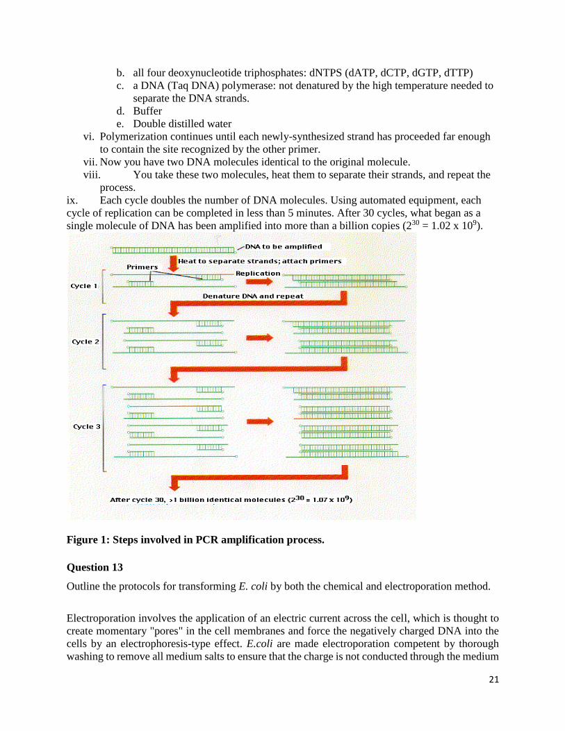
21
b. all four deoxynucleotide triphosphates: dNTPS (dATP, dCTP, dGTP, dTTP)
c. a DNA (Taq DNA) polymerase: not denatured by the high temperature needed to
separate the DNA strands.
d. Buffer
e. Double distilled water
vi. Polymerization continues until each newly-synthesized strand has proceeded far enough
to contain the site recognized by the other primer.
vii. Now you have two DNA molecules identical to the original molecule.
viii. You take these two molecules, heat them to separate their strands, and repeat the
process.
ix. Each cycle doubles the number of DNA molecules. Using automated equipment, each
cycle of replication can be completed in less than 5 minutes. After 30 cycles, what began as a
single molecule of DNA has been amplified into more than a billion copies (230 = 1.02 x 10⁹).
Figure 1: Steps involved in PCR amplification process.
Question 13
Outline the protocols for transforming E. coli by both the chemical and electroporation method.
Electroporation involves the application of an electric current across the cell, which is thought to
create momentary "pores" in the cell membranes and force the negatively charged DNA into the
cells by an electrophoresis-type effect. E.coli are made electroporation competent by thorough
washing to remove all medium salts to ensure that the charge is not conducted through the medium
22
and the electroporation is carried out at 0°C to miminize heat damage to the cells. Chemical
competence is conferred to E.coli by re-suspension in CaCl2 solution at 0°C. Under these
conditions, the Ca2+ ion is thought to create pores in the membrane, assist binding of the DNA to
the cell membrane and mask the negative charge on the DNA, easing its passage through the
hydrophobic cell membrane. The DNA is forced into the cells by applying a short 42°C heat shock,
which results in a thermal current that sweeps the DNA into the cells (Oswald, 2007).
Electroporation: Formation of transient holes in the cell membranes using electric shock; this
allows DNA to enter the bacteria (Schiestl et al., 1993).
Bacterial Transformation Heat Shock Protocol (Cawood and Su, 2014: Oxford Genetics)
1. Thaw one tube of your pre-made competent cells per DNA/ligation reaction or control
reaction on ice and push the tube deep into the ice. Thawing takes about 5-10 minutes.
2. Keep the cells as cold as possible and avoid touching the part of the tube containing the
cells; a small amount of heat can significantly decrease the transformation process.
3. Pre-chill 15ml Falcon Tubes (Sigma-Aldrich: SIAL0791) on ice and transfer 3-4 μl of the
ligation reaction (or control reaction) into each tube.
4. Add 95 μl of competent cells into each ligation reaction and incubate on ice for 20 minutes
(minimum); may be longer: max 45 minutes.
5. Heat shock at 42°C for 90 seconds in either a heat block or water bath.
6. Then add 1 ml of LB or SOC medium without antibiotic and incubate the cells in an
incubation shaker at 37°C, 227RPM for 1 hour.
7. Pour all the LB containing the transformed competent cells onto an agar plate containing
the correct antibiotic.
8. Leave the plate upright to dry with the lid slightly off in a class 1 hood or in a 37°C
incubator for about 5-10 minutes. Do not do this in a hood that is used for mammalian
tissue culture. Your colleagues will not be happy. Do not leave the plate to dry for too long
as some of the bacteria may die.
9. Incubate the plate overnight at 37°C. The colonies that will appear originate from single
transformed cells and are resistant to the antibiotic due to the presence of the plasmid. Each
colony will contain millions of identical copies of the same cell, hence the term clone.
10. Now move on to picking your colony.
Preparation of electrocompetent E. coli cells
1. Prepare all the media (SOB, 10 % glycerol); reserve the centrifuge; prechill a GS3 rotor
4°C; sterilize centrifuge tubes by autoclaving and prechill them!
2. Inoculate 30 mL SOB with single colony from a fresh plate and grow them o/n at 37°C.
3. Inoculate 1 L SOB (1:100 ie. 10 mL of the starting culture per 1 L SOB) and grow it at
37°C on a shaker with 200 rpm until it reaches an OD600 of 0.4-0.5 (approx. after 4 h).
continue with all working steps on ice or 4°C!
4. Centrifuge the cells at 4.000 rpm / 20 min / 4°C.
5. Wash with 1 vol. 10 % glycerol.
6. Centrifuge at 4.000 rpm / 20 min / 4°C.
7. Wash with 0.5 vol. 10 % glycerol.
8. Centrifuge at 4.000 rpm / 20 min / 4°C.

23
9. Wash with 0.1 vol. 10 % glycerol.
10. Centrifuge at 4.000 rpm / 20 min / 4°C.
11. Go to get liquid nitrogen.
12. Add 0.5 - 1 mL 10 % glycerol (per liter liquid culture started with) and resuspend pellet
and prepare aliquots of 100 µL in 1.5 mL reaction tubes and freeze them immediately in
liquid nitrogen.
13. Store the electrocompetent cells at -80°C.
Question 15
Discuss the protocols and materials needed for both Western and Southern blotting.
Consult with lecture materials for detailed protocols.

24
BCH 433 TUTORIAL QUESTIONS
1. What is the basis of diagnosis of clinical conditions and what determines the choice of test to be
used?
Clinical biochemistry detects anomalies based on anatomical and physiological differences of normal and
disease states of the body. Clinical signs and symptoms presented in many conditions may be similar with
different diseases, however this can be used to first suspect a condition and then confirmed by clinical
tests. The choice of test is based on the underlying cause of disease. There are Clinical tests that are
qualitative, quantitative, semi-quantitative, Observational/Physical test. The test equipment can vary
from simple to complex equipment E.g. to detect hyperglycaemia in a diabetic patient, blood glucose level
can be measured using different equipment such as use of point of care system (fast and can be operated
by unskilled persons) rather than laboratory spectrophotometry (operated by trained personnel)
2. Discuss how the underlying cause of Ebola viral disease, symptoms and how it can be diagnosed
confirmatively
3. What is the basis for diagnosing blood disorder
Blood disorders are caused by anolmalies that affect red blood morphology, production or functioning.
There are many blood disorders and detection is based on the underlying cause of the disorder and basis
for diagnosis of blood disorder is based on the following:
Abnormal erythrocyte based on morphology: abnormal erythrocytes can determine the underlying
cause of such disease. E.g. Sickle-cell anemia causes a sickle-shaped RBCs, thus Morphological test
compared with normal red blood cell morphology; reddish, biconcave disc-shaped when stained with
appropriate dye can be used to detect the condition.
Anemias based on RBC levels - a symptom that results when blood has lower than normal ability to carry
oxygen. Anemia can be detected by checking the RBC levels. This can be caused by infection,
dyserythropoeisis or haemorrhage. Normal packed RBC range level in an individual ranges from 35-50%.
Abnormally elevated levels or very low levels can indicate a blood disorder. Likewise, differential count
of WBC; neutrophil, Leucocyte, eosinophils or Platelet count of a patient can be compared with the
physiological level and used to infer presence of an infection such as bacterial, viral, parasitic infection
can be detected from these counts.
Haemoglobin concentration: normal haemoglobin concentration of an adult male ranges from 13-18g/dl
Thrombosis based on Blood clots: platelets clump at site of tissue injury thus indicating thrombosis.
25
Cancers e.g. Leukemia: rapid production of abnormal white blood cells and Lymphoma which prevents
lymphatic system function; which removes excess fluids from body and produces immune cells can be
detected by bone marrow evaluation and by antigenic tests.
4. Discuss the detection of two blood disorders using 2 different point of care systems and discuss the
underlying basis for detection of such condition
5. Discuss 4 different tests that can be used for the detection of a pathogenic disease such as tuberculosis
6. What are immunologic tests and their uses: These are tests used to detect Antigen through Antibody
binding. Immunologic reactions are usually coupled with a colorimetric test. It is based on specific antigen-
antibody interactions and uses known quantity of antibody that binds to antigens, this detects presence
and level of antigen. It uses probes to label antibodies, proteins, amino acids and peptides or nucleic acids
in tissue or body fluid. Fluorescent dyes such as: cyanine, fluorescein, rhodamine, Alexa Fluors, Dylight
fluors for proteins are used.
7. What are recombinant DNA techniques and how can they be useful in clinical biochemistry, mention
two different techniques and their application in disease diagnosis
8. What are genetic diseases?
These are diseases that are caused by genes transmitted from parent to offspring, these diseases manifest
in the offspring based on different environmental and other factors. Some diseases caused by pathogens
can be identified based on the foreign gene/DNA material of the infecting organism detectable in the host.
E.g. some pathogenic diseases
9. What is forensic science and how can it be applied in clinical biochemistry?
10. Disucss 1 presumptive and 1 confirmatory test that can be used in forensic biochemistry using bodily
fluids obtained from a patient.
Acid Phosphatase Test (Walker or Brentamine spot test): male prostate gland secrets into semen high
amount of acid phosphatase enzyme(AP), this can be detected in semen. AP reacts with α-Naphthyl acid
phosphate and Brentamine Fast Blue dyes, AP produces dark purple color in less than a minute. This can
be used to detect prostate cancer. It is a presumptive test, other test can be conducted.
ABO typing: It is an immunologic test showing the reaction of Anti-A and Anti-B antibodies. Serum can be
used, mixed with blood that is known to be Type A or Type B and the reaction is observed for agglutination.
This is a confirmatory test.
11. What are controlled substances, describe 1 each in the schedule 1-3, discuss the regulation of soft and
hard drugs in Nigeria