Ovary-Sparing Radiation Planning Techniques Can Achieve...
7
Research Article Ovary-Sparing Radiation Planning Techniques Can Achieve Ovarian Dose Reduction for Soft Tissue Sarcoma of the Buttock and Thigh Konstantin A. Kovtun, 1 Wee-Pin Yeo, 2 Catherine H. Phillips, 3 Akila Viswanathan, 2 and Elizabeth H. Baldini 2 1 Harvard Radiation Oncology Program, Brigham and Women’s Hospital/Dana Farber Cancer Institute, Boston, MA, USA 2 Department of Radiation Oncology, Brigham and Women’s Hospital/Dana Farber Cancer Institute, Boston, MA, USA 3 Department of Radiology, Brigham and Women’s Hospital/Dana Farber Cancer Institute, Boston, MA, USA Correspondence should be addressed to Konstantin A. Kovtun; [email protected] Received 21 May 2017; Accepted 8 August 2017; Published 18 September 2017 Academic Editor: Valerae O. Lewis Copyright © 2017 Konstantin A. Kovtun et al. is is an open access article distributed under the Creative Commons Attribution License, which permits unrestricted use, distribution, and reproduction in any medium, provided the original work is properly cited. Background and Objectives. Attention to ovary dose is important for premenopausal women undergoing radiation therapy (RT) and must not be overlooked when treating extremity sarcoma. We assessed whether ovary-sparing RT plans could decrease ovary dose without compromising target coverage. Methods. Standard sarcoma target volumes and organs at risk (OAR) were contoured by a sarcoma dedicated radiation oncologist on CT planning scans for 23 women with thigh or buttock sarcoma. IMRT plans (50Gy) with and without attempted ovary-sparing were created by an expert sarcoma dosimetrist. Results. All plans met target coverage goals. Compared to standard plans, ovary-sparing plans had lower mean bilateral ovary doses (MBOD) (652 versus 483 cGy, = 0.007) but higher bone doses (mean V50: 8.5% versus 6.9%, = 0.049) and lower conformity indexes (1.12 versus 1.19, = 0.009). Tumors < 8 cm from the pubic symphysis had significant MBOD reduction with ovary-sparing plans (376 cGy versus 619 cGy, = 0.0184). On multivariate analysis, distance to pubic symphysis and proximal medial thigh site were associated with MBOD reduction with ovary-sparing plan. Conclusions. For preoperative IMRT, ovary-sparing planning significantly reduces ovarian dose in women with sarcoma of the proximal thigh and near the pubic symphysis. 1. Introduction e median age of diagnosis for soſt tissue sarcoma (STS) in women ranges from 15 to 65 years based on histologic subtype [1]. Accordingly, many of these women are of child-bearing age and/or premenopausal. Radiation therapy (RT) is a key component of local management for extremity STS [2–4] and wide clinical target volume (CTV) margins are required for optimal local control [5]. For premenopausal women receiv- ing RT who desire fertility or preserved estrogen production, ovarian dose is an important consideration for functional preservation. Ovaries are not generally discussed as organs at risk (OAR) when planning radiation therapy for thigh and but- tock STS. However, studies have shown that increasing ovar- ian dose is associated with acute ovarian failure, infertility, and premature menopause [6]. It is estimated that 50% oocyte destruction occurs at doses less than 2 Gy [7]. Additionally, effective sterilization doses are reported to decrease with age with 14.3Gy leading to ovarian failure in 97.5% of patients [8] and 6 Gy leading to an intermediate risk of dysfunction in the average 30-year-old woman. [9]. Given that the ovary is such a radiosensitive organ, careful attention to ovarian dose is imperative when delivering RT to premenopausal women. We performed an analysis of intensity-modulated RT (IMRT) plans designed with and without intent to spare ovaries for STS of the thigh and buttock to assess whether the ovary-sparing plans could achieve ovary dose reduction without compromising target coverage and to determine if there are clinical predictors of ovarian dose reduction with the new plans. Hindawi Sarcoma Volume 2017, Article ID 2796925, 6 pages https://doi.org/10.1155/2017/2796925
Transcript of Ovary-Sparing Radiation Planning Techniques Can Achieve...

Research ArticleOvary-Sparing Radiation Planning Techniques CanAchieve Ovarian Dose Reduction for Soft Tissue Sarcoma ofthe Buttock and Thigh
Konstantin A. Kovtun,1 Wee-Pin Yeo,2 Catherine H. Phillips,3
Akila Viswanathan,2 and Elizabeth H. Baldini2
1Harvard Radiation Oncology Program, Brigham and Women’s Hospital/Dana Farber Cancer Institute, Boston, MA, USA2Department of Radiation Oncology, Brigham and Women’s Hospital/Dana Farber Cancer Institute, Boston, MA, USA3Department of Radiology, Brigham and Women’s Hospital/Dana Farber Cancer Institute, Boston, MA, USA
Correspondence should be addressed to Konstantin A. Kovtun; [email protected]
Received 21 May 2017; Accepted 8 August 2017; Published 18 September 2017
Academic Editor: Valerae O. Lewis
Copyright © 2017 Konstantin A. Kovtun et al. This is an open access article distributed under the Creative Commons AttributionLicense, which permits unrestricted use, distribution, and reproduction in any medium, provided the original work is properlycited.
Background andObjectives. Attention to ovary dose is important for premenopausal women undergoing radiation therapy (RT) andmust not be overlooked when treating extremity sarcoma. We assessed whether ovary-sparing RT plans could decrease ovary dosewithout compromising target coverage.Methods. Standard sarcoma target volumes and organs at risk (OAR) were contoured by asarcoma dedicated radiation oncologist on CT planning scans for 23 women with thigh or buttock sarcoma. IMRT plans (50Gy)with and without attempted ovary-sparing were created by an expert sarcoma dosimetrist. Results. All plans met target coveragegoals. Compared to standard plans, ovary-sparing plans had lower mean bilateral ovary doses (MBOD) (652 versus 483 cGy, 𝑝 =0.007) but higher bone doses (mean V50: 8.5% versus 6.9%, 𝑝 = 0.049) and lower conformity indexes (1.12 versus 1.19, 𝑝 = 0.009).Tumors < 8 cm from the pubic symphysis had significant MBOD reduction with ovary-sparing plans (376 cGy versus 619 cGy,𝑝 = 0.0184). On multivariate analysis, distance to pubic symphysis and proximal medial thigh site were associated with MBODreduction with ovary-sparing plan. Conclusions. For preoperative IMRT, ovary-sparing planning significantly reduces ovarian dosein women with sarcoma of the proximal thigh and near the pubic symphysis.
1. Introduction
The median age of diagnosis for soft tissue sarcoma (STS) inwomen ranges from 15 to 65 years based on histologic subtype[1]. Accordingly, many of these women are of child-bearingage and/or premenopausal. Radiation therapy (RT) is a keycomponent of local management for extremity STS [2–4] andwide clinical target volume (CTV) margins are required foroptimal local control [5]. For premenopausal women receiv-ing RT who desire fertility or preserved estrogen production,ovarian dose is an important consideration for functionalpreservation.
Ovaries are not generally discussed as organs at risk(OAR) when planning radiation therapy for thigh and but-tock STS. However, studies have shown that increasing ovar-ian dose is associated with acute ovarian failure, infertility,
and prematuremenopause [6]. It is estimated that 50%oocytedestruction occurs at doses less than 2Gy [7]. Additionally,effective sterilization doses are reported to decrease with agewith 14.3 Gy leading to ovarian failure in 97.5% of patients[8] and 6Gy leading to an intermediate risk of dysfunctionin the average 30-year-old woman. [9]. Given that the ovaryis such a radiosensitive organ, careful attention to ovariandose is imperative when delivering RT to premenopausalwomen.
We performed an analysis of intensity-modulated RT(IMRT) plans designed with and without intent to spareovaries for STS of the thigh and buttock to assess whetherthe ovary-sparing plans could achieve ovary dose reductionwithout compromising target coverage and to determine ifthere are clinical predictors of ovarian dose reduction withthe new plans.
HindawiSarcomaVolume 2017, Article ID 2796925, 6 pageshttps://doi.org/10.1155/2017/2796925
2 Sarcoma
2. Materials and Methods
2.1. Patient Data. The study cohort comprised 23 womenwith STS of the thigh or buttock treated with preoperativeRT between September 2010 and February 2016.With Institu-tional ReviewBoard approval,medical recordswere reviewedto ascertain patient and tumor characteristics. Women withprior unilateral or bilateral oophorectomy were excluded.All women had CT simulation scans from the pelvis to theknee. An extremity board was used for immobilization, andpatients were positioned in a supine position with either astraight leg or slightly frog-legged position. Bolus was notused.
2.2. Definition of Target Volumes and Critical Structures.Standard sarcoma gross target volumes (GTV), clinical targetvolumes (CTV), and planning target volumes (PTV) werecontoured by a sarcoma dedicated radiation oncologist onCT planning scans for all patients. T1 postgadolinium MRseries were used to contour GTV. GTV to CTV expansionswere typically 3.5 cm in the longitudinal directions and 1.5 cmradially on each axial slice with editing for normal tissueinterfaces and as per established consensus guidelines [5, 10].CTV to PTV expansions were 5mm in all directions. Targetvolumes were edited from skin surface 3–5mm. Standardorgans at risk (OARs) including bone, bowel, bladder, andrectum were contoured, and the dosimetrist identified anappropriate strip of limb circumference as anOARduring theplanning process.
Bilateral ovaries were contoured as separate structures bytwo radiation oncologists (Konstantin A. Kovtun and AkilaViswanathan) and one radiologist (Catherine H. Phillips)using CT simulation scans as well as pelvic MR scans whenavailable. Final ovary structures were defined by consensus.
2.3. Comparative Intensity-Modulated RT (IMRT) Plans. Allpatients were initially planned without the presence of ovarycontours and without regard to ovary dose (standard plan) byan experienced sarcoma planner (Wee-Pin Yeo). All patientswere planned with an IMRT technique to a preoperative doseof 50Gy in 2Gy fractions. General sarcoma guidelines forbeam arrangementwere followed including beam selection inattempt to maximize target coverage and spare a longitudinalstrip of limb circumference (<20Gy) and, to the extentpossible, avoid beams traveling through the contralaterallimb and abdominal and pelvic structures. Coverage criteriawere PTV V95 (volume receiving at least 95% of prescriptiondose) greater than or equal to 95%. OAR constraints includedthe following: bone (mean dose < 37Gy, maximum dose <59Gy, V40 < 64%), bowel bag (V45 < 195 cc), rectum (V50 <50%), and anus/vulva/perineum (V30 < 50%)
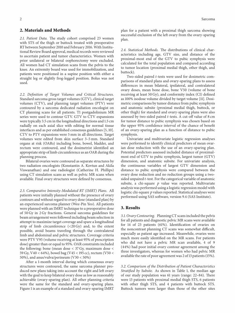
After a 1-month interval during which consensus ovarystructures were contoured, the same sarcoma planner pro-duced new plans taking into account the right and left ovarywith the goal to keep bilateral ovary dose as low as reasonablyachievable (ovary-sparing plan). All other planning criteriawere the same for the standard and ovary-sparing plans.Figure 1 is an example of a standard and ovary-sparing IMRT
plan for a patient with a proximal thigh sarcoma showingsuccessful exclusion of the left ovary from the ovary-sparingplan.
2.4. Statistical Methods. The distributions of clinical char-acteristics including age, GTV size, and distance of theproximal-most end of the GTV to pubic symphysis werecalculated for the total population and compared accordingto tumor location (proximal medial thigh, other thigh, andbuttock).
Two-sided paired 𝑡-tests were used for dosimetric com-parisons of standard plans and ovary-sparing plans to assessdifferences in mean bilateral, ipsilateral, and contralateralovary doses, mean bone dose, bone V50 (volume of bonereceiving at least 50Gy), and conformity index (CI) definedas 100% isodose volume divided by target volume [11]. Dosi-metric comparisons by tumor distance from pubic symphysisand anatomic subsite (proximal medial thigh, buttock, orother thigh) for standard and ovary-sparing plans were alsoassessed by two-sided paired 𝑡-tests. A cut-off value of 8 cmfor tumor distance to pubic symphysis was chosen based onthe upper 95% confidence interval of the chance of benefitof an ovary-sparing plan as a function of distance to pubicsymphysis.
Univariate and multivariate logistic regression analyseswere performed to identify clinical predictors of mean ovar-ian dose reduction with the use of an ovary-sparing plan.Potential predictors assessed included distance of proximal-most end of GTV to pubic symphysis, largest tumor (GTV)dimension, and anatomic subsite. For univariate analysis,the continuous variables of largest GTV dimension anddistance to pubic symphysis were compared between theovary dose reduction and no reduction groups using a two-sided unpaired 𝑡-test. For the categorical variable of anatomicsubsite, a chi-square 𝑝 value was reported. Multivariateanalysis was performed using a logistic regressionmodel withlogistic chi-square 𝑝 values reported. Statistical analyses wereperformed using SAS software, version 9.4 (SAS Institute).
3. Results
3.1. OvaryContouring. PlanningCT scans included the pelvisfor all patients and diagnostic pelvic MR scans were availablefor 14 of 23 patients (61%). Identification of ovaries onthe noncontrast planning CT scans was somewhat difficult,especially as patient age increased. Meanwhile, ovaries weremuch more easily identified on the MR scans. For patientswho did not have a pelvic MR scan available, 4 of 9(44%) had poor initial ovary contour agreement among thethree investigators, whereas for women who had pelvic MRavailable the rate of poor agreementwas 2 of 13 patients (15%).
3.2. Comparison of the Distribution of Patient CharacteristicsStratified by Subsite. As shown in Table 1, the median ageof our study population was 61 years (range: 22–84). Therewere 13 patients with proximal medial thigh STS, 6 patientswith other thigh STS, and 4 patients with buttock STS.Buttock tumors were larger than those of the other sites

Sarcoma 3
(A)
(C)(D)
(B)
Left ovary
Left ovary
(a) Comparison of axial (A) and coronal (B) slices of a standard plan and ovary-sparing plan (C, D) for apatient with a right proximal medial thigh soft tissue sarcoma
Dose (cGy)
Relative dose (%)
Stru
ctur
e vol
ume (
cG3)
4
2
0
121086420
6005004003002001000
(b) Dose volume histogram comparison of the right (blue) and left (red)ovary for a standard plan (square) and an ovary avoidance plan (triangle)for a patient with a right proximal medial thigh soft tissue sarcoma
Figure 1
Table 1: Patient characteristics (𝑛 = 23).
Clinicalcharacteristics
Proximal medial thigh(𝑛 = 13)
Other thigh(𝑛 = 6)
Buttock(𝑛 = 4)
Allpatients(𝑛 = 23)
Median age (years)(range)
55(38–76)
58(22–84)
65(49–70)
61(22–84)
Median largest GTVdimension (cm)(range)
7.5(2.4–15.8)
7.7(2.5–15)
15.0(7.9–20.9)
8.3(2.4–20.9)
Median distance oftumor to pubicsymphysis (cm)(range)
5.2(1.5–10.9)
16.5(6.9–27)
11.0(9.3–12.7)
8.0(1.5–27)
Pelvic MRI available,number (%) 8 (61%) 2 (33%) 4 (100%) 14 (61%)
MRI: Magnetic Resonance Imaging; cm: centimeter.
(median largest dimension of 15.0 cm for buttock versus7.5 cm for proximal medial thigh versus 7.7 cm for otherthigh). Proximal medial thigh tumors were closest to thepubic symphysis (median distance: 5.2 cm versus 16.5 cm forother thigh versus 11.0 cm for buttock).
3.3. Dosimetric Comparisons of Standard Plans and Ovary-Sparing Plans. Target volume coverage goals were met for allstandard and ovary-sparing plans and OAR constraints forlimb circumference, bone, bowel, bladder, and rectum wereall met as well. As shown in Table 2, compared with standard

4 Sarcoma
Table 2: Dosimetric comparisons of standard plans and ovary-sparing plans (𝑛 = 23).
Standard plan Ovary-sparing plan 𝑝 valueMean bilateral ovary dose (MBOD, cGy) (SD) 652 (100) 483 (84) 0.007Ipsilateral ovary dose, mean (cGy) (SD) 1072 (1606) 888 (1596) 0.03Contralateral ovary dose, mean (cGy) (SD) 232 (460) 78(86) 0.075Mean bone dose (cGy) (SD) 161 (77) 165 (79) 0.19Mean bone V50 (SD) 6.86% (7.70) 8.45% (10.1) 0.049Conformity index, mean (SD) 1.12 (0.07) 1.19 (0.13) 0.009MBOD: mean bilateral ovary dose; SD: standard deviation; cGy: centigray.
Table 3: Dosimetric comparison of mean bilateral ovary dose (MBOD) and mean contralateral ovary dose (MCOD) for standard plans andovary-sparing plans by anatomic location (𝑛 = 23).
Tumor distance from pubic symphysis 𝑁 Standard plan Ovary-sparing plan 𝑝 valueTumor < 8 cm from pubic symphysis, MBOD (cGy) (SD) 10 619 (765) 376 (540) 0.018Tumor ≥ 8 cm from pubic symphysis, MBOD (cGy) (SD) 13 678 (1184) 566 (1022) 0.17Tumor < 8 cm from pubic symphysis, MCOD (cGy) (SD) 10 249 (575) 72 (109) 0.128Tumor ≥ 8 cm from pubic symphysis, MCOD (cGy) (SD) 13 211 (277) 86 (442) 0.222Tumor subsite, MBOD (cGy)
Proximal medial thigh (SD) 13 445 (721) 293 (494) 0.008Other thigh, MBOD (SD) 6 95 (217) 19 (309) 0.33Buttock, MBOD (SD) 4 2161 (1170) 1800 (1116) 0.20
Tumor subsite, MCOD (cGy)Proximal medial thigh (SD) 13 167 (255) 70 (50) 0.12Other thigh, MBOD (SD) 6 8.8 (15.6) 7.2 (11.6) 0.36Buttock, MBOD (SD) 4 781 (881) 210 (104) 0.26
MBOD: mean bilateral ovary dose; SD: standard deviation; cGy: centigray.
plans, the ovary-sparing plans had lowermean bilateral ovarydoses (MBOD) (mean dose: 652 cGy versus 483 cGy, 𝑝 =0.007), lower ipsilateral ovary doses (mean dose: 1072 cGyversus 888 cGy,𝑝 = 0.03), and lower contralateral ovary doses(mean dose: 232 cGy versus 78 cGy, 𝑝 = 0.075). These lowerovarian doses came at the expense of decreased conformalityin the ovary-sparing plans compared with the standard plans(mean conformity index: 1.12 versus 1.19, 𝑝 = 0.009) as well asa small increased bone dose in the ovary-sparing plans (meanV50 bone: 8.5% versus 6.9%, 𝑝 = 0.049).
As shown in Table 3, comparison of ovary-sparing andstandard plans by anatomic subsite showed that patients withproximal medial thigh lesions had significant reductions inMBOD with an ovary-sparing plan (MBOD: 293 cGy versus445 cGy, 𝑝 = 0.008). There were no statistically significantreductions in ovarian dose for other thigh or buttock subsites.Tumors less than 8 cm from the pubic symphysis were alsoassociated with a significant MBOD reduction on ovary-sparing plans (619 cGy versus 376 cGy, 𝑝 = 0.0184). Com-parisons of mean contralateral ovary dose (MCOD) basedon anatomic subsite and the 8 cm pubic symphysis cut-offshowed reductions in MCOD with ovary-sparing, but thesecomparisons did not reach statistical significance (Table 3).
3.4. Univariate and Multivariate Analysis for Predictorsof Ovary-Sparing Dosimetric Reduction. Closer distance topubic symphysis was associated with benefit in MBOD with
closer tumors more likely to achieve ovary dose reductionfrom an ovary-sparing plan. Mean distance to pubic sym-physis for plans benefiting from ovary-sparing was 6.3 cm(95% CI: 4.5–8.1 cm) versus 14.1 cm (95% CI: 8.2–19.9 cm)for plans that demonstrated no MBOD reduction (MVA 𝑝value: 0.0038). Subsite was also associated with ovary dosebenefit with 77% of proximal medial thigh and 75% ofbuttock plans achieving ovary dose reductions (UVA 𝑝 value:0.054; MVA 𝑝 value: 0.048). Only one of the other thighsubsite plans achieved dose reduction with the ovary-sparingtechnique. Largest GTV dimension was not associated with astatistically significant ovary dose reduction with the ovary-sparing technique (Table 4).
4. Discussion
In this study, we compared standard and ovary-sparingIMRT treatment plans for 23 women with STS of the thighand buttock. We demonstrated that ovary-sparing treatmentplans significantly reduced ovary dose albeit at the expenseof decreased plan conformality resulting in a slightly lowerconformity index and slightly higher bone V50. Ovariandoses for standard and ovary-sparing plans were highest fortumors located in the buttock, followed by proximal medialthigh and other thigh.The ability to achieve significant ovarydose reduction with ovary-sparing plans compared withstandard plans was most evident for proximal medial thigh

Sarcoma 5
Table 4: Univariate and multivariate analysis of likelihood of reduction in mean bilateral ovary dose (MBOD) with an ovary-sparing plan.
Reduction inMBOD
No reductionin MBOD
UVA𝑝 value
MVA𝑝 value
Mean distance to pubicsymphysis (cm) (95% CI)
6.3(4.5, 8.1)
14.1(8.2, 19.9) 0.0079∗ 0.0038∧
Site, number (%)Proximal medial thigh 10 (77%) 3 (23%)Buttock 3 (75%) 1 (25%)Other thigh 1 (17%) 5 (83%) 0.054# 0.048∧
Largest GTV dimension (cc)(95% CI)
8.7(5.6, 11.8)
9.5(6.0, 13.0) 0.72∗ 0.054∧
MBOD: mean bilateral ovary dose; SD: standard deviation; UVA: univariate analysis; MVA: multivariate analysis; cm: centimeter; cc: cubic centimeter. ∗𝑡-test𝑝 value. #Fisher’s exact 𝑝 value. ∧Logistic regression chi-square 𝑝 value.
tumors and tumors located less than 8 cm from the pubicsymphysis.
Several points require further discussion. For bothwomen andmen interested in fertility and/or who are activelyproducing sex hormones, the radiation oncologist mustbe cognizant of minimizing dose to ovaries and testicles,respectively, during the treatment planning process. Sincemale genitalia are externalized, the importance of attentionto testicular placement at the time of simulation, contouringtesticles, and setting testicle OAR avoidance constraints tendsto be readily apparent. However, for women, since ovaries arein the pelvis, one could imagine that attention to ovaries asan important OAR avoidance structure could be overlooked,particularly when treating tumors of the thigh.This planningstudy shows that ovarian doses for tumors of the buttockand thigh are frequently high enough to ablate fertility aswell as estrogen production, and in several cases, ovarian-sparing plans achieved significant ovary dose reduction.Furthermore, contouring ovaries on noncontrast planningCT scans is often difficult. The availability of pelvic MR scansoptimizes the ability to localize ovaries on the simulationscan.
Further study is needed to determine how specific ovarydosesmight translate into clinical outcomes such as infertilityor early menopause. For example, for proximal medial thightumors, ovary-sparing plans reducedMBOD from445 cGy to293 cGy and reduced MCOD from 167 cGy to 70 cGy. Whileit is likely that these dose differences would affect ovaryfunction based on data for oocyte radiation sensitivity at 2Gy[7], further clinical correlation is needed to assess whethersuch dose differences would be associated with meaningfulclinical endpoints such as fertility and/or estrogen produc-tion. This is a complicated topic to study. Many factors inaddition to radiation dose affect oocyte number and functionparticularly if systemic therapy is used [12–14]. For example,oocyte number and function decrease with advancing age[15]. Furthermore, since ovaries are paired organs, as is thecase for kidneys, potentially acceptable dose constraints willlikely vary from maximal sparing of one ovary to a certaindose delivered to both ovaries as series have shown that, inpatients receiving ovarian doses of at least 15 Gy excluding atleast one ovary, approximately half of the patients developed
ovarian dysfunction as opposed to all patients where thecontralateral ovary is not spared [16].
There are several limitations of this study. There washeterogeneity of sites and relatively small case numbers.Median age was 61, and thus many of the patients in thesample were postmenopausal. This may partly explain thedifficulty encounteredwith contouring ovaries on some of theplanning CT scans. Despite the advanced age of many of thepatients in our series, we feel that the ovary-sparing planningexercisewas still valid. In addition, the ovarian doses reportedare specific to the standard and ovary-sparing plans generatedby one experienced dosimetrist. Other planners would likelygenerate plans with some variations to those reported herein.Lastly, calculation of peripheral dosewith treatment planningsystems is not always correct and ovary doses so calculatedshould always be interpreted with caution. However, thepoint of this report was to highlight the importance of payingattention to ovary dose in premenopausal women receivingradiation in proximity to the ovaries and to show that, withattention to ovary-sparing, for many cases, ovarian dosecan be reduced. Despite the above limitations, our studydemonstrates the feasibility and potential benefits of ovaryavoidance plans in appropriately selected patients.
The findings of our report serve to reinforce the followingpractice guidelines for any premenopausal woman about toundergo radiation to a site in proximity to the ovaries (trunk,abdomen, pelvis, buttock, and thigh):
(1) For women interested in future fertility, a reproduc-tive endocrinology consult is recommended.
(2) A diagnostic pelvic MR should be performed to helpdelineate ovary location on the planning CT scan.
(3) The simulation CT scan should include the wholepelvis so ovaries can be contoured and ovary dosecalculations performed.
(4) Treatment planning beam arrangements should bechosen with ovary-sparing in mind. Target volumecoverage should not be compromised. However, it isreasonable to accept minor trade-offs of other OARconstraints in attempt to minimize ovarian dose.

6 Sarcoma
(5) For custom prescription templates with OAR con-straints, inclusion of ovary and testicle constraintson all templates of trunk, abdomen, pelvis, andextremity is recommended in order to minimize thechance that ovary or testicle dose assessment could beinadvertently overlooked.
5. Conclusion
Ovary-sparing planning techniques significantly reduceovarian dose in women with STS of the proximal thigh ortumors less than 8 cm from the pubic symphysis. Furtherstudy is needed to determine accurate ovary constraints,likely stratified by patient age, associated with infertility andpremature ovarian failure. In the meantime, when deliveringradiation to the trunk, abdomen, pelvis, or thigh for pre-menopausal women, ovary dose should be calculated andtreatment plans that minimize ovary dose selected.
Conflicts of Interest
The authors declare that there are no conflicts of interestregarding the publication of this article.
References
[1] A. Ferrari, I. Sultan, T. T. Huang et al., “Soft tissue sarcomaacross the age spectrum: a population-based study from thesurveillance epidemiology and end results database,” PediatricBlood and Cancer, vol. 57, no. 6, pp. 943–949, 2011.
[2] S. A. Rosenberg, J. Tepper, E. Glatstein et al., “The treatment ofsoft-tissue sarcomas of the extremities: prospective randomizedevaluations of (1) limb-sparing surgery plus radiation ther-apy compared with amputation and (2) the role of adjuvantchemotherapy,” Annals of Surgery, vol. 196, no. 3, pp. 305–315,1982.
[3] P. W. Pisters, L. B. Harrison, D. H. Leung, J. M. Woodruff,E. S. Casper, and M. F. Brennan, “Long-term results of aprospective randomized trial of adjuvant brachytherapy in softtissue sarcoma,” Journal of Clinical Oncology, vol. 14, no. 3, pp.859–868, 1996.
[4] J. C. Yang, A. E. Chang, A. R. Baker et al., “Randomizedprospective study of the benefit of adjuvant radiation therapy inthe treatment of soft tissue sarcomas of the extremity,” Journalof Clinical Oncology, vol. 16, no. 1, pp. 197–203, 1998.
[5] R. L. M. Haas, T. F. Delaney, B. O’Sullivan et al., “Radiotherapyfor management of extremity soft tissue sarcomas: Why, when,and where?” International Journal of Radiation Oncology Biol-ogy Physics, vol. 84, no. 3, pp. 572–580, 2012.
[6] D. M. Green, C. A. Sklar, J. D. Boice Jr. et al., “Ovarian failureand reproductive outcomes after childhood cancer treatment:results from the childhood cancer survivor study,” Journal ofClinical Oncology, vol. 27, no. 14, pp. 2374–2381, 2009.
[7] W. H. B. Wallace, A. B. Thomson, and T. W. Kelsey, “Theradiosensitivity of the human oocyte,” Human Reproduction,vol. 18, no. 1, pp. 117–121, 2003.
[8] W. H. B. Wallace, A. B. Thomson, F. Saran, and T. W. Kelsey,“Predicting age of ovarian failure after radiation to a fieldthat includes the ovaries,” International Journal of RadiationOncology Biology Physics, vol. 62, no. 3, pp. 738–744, 2005.
[9] J. Y. Wo and A. N. Viswanathan, “Impact of Radiotherapy onFertility, Pregnancy, and Neonatal Outcomes in Female CancerPatients,” International Journal of Radiation Oncology BiologyPhysics, vol. 73, no. 5, pp. 1304–1312, 2009.
[10] D. Wang, W. Bosch, D. Roberge et al., “RTOG Sarcoma Radi-ation Oncologists Reach Consensus on Gross Tumor Volume(GTV) andClinical TargetVolume (CTV) onComputedTomo-graphic Images for Preoperative Radiotherapy of Primary SoftTissue Sarcoma of Extremity in RTOG Studies,” InternationalJournal of RadiationOncology, Biology, Physics, vol. 81, pp. e525–e528, 2011.
[11] L. Feuvret, G. Noel, J.-J. Mazeron, and P. Bey, “Conformityindex: a review,” International Journal of Radiation OncologyBiology Physics, vol. 64, no. 2, pp. 333–342, 2006.
[12] A. W. Loren, P. B. Mangu, L. N. Beck et al., “Fertility preser-vation for patients with cancer: American Society of ClinicalOncology clinical practice guideline update,” Journal of ClinicalOncology, vol. 31, no. 19, pp. 2500–2510, 2013.
[13] C. R. Gracia,M. D. Sammel, E. Freeman et al., “Impact of cancertherapies on ovarian reserve,” Fertility and Sterility, vol. 97, no.1, pp. 134–e1, 2012.
[14] T. T. Sy Ortin, C. A. Shostak, and S. S. Donaldson, “Gonadalstatus and reproductive function following treatment forHodgkin’s disease in childhood: The Stanford experience,”International Journal of Radiation Oncology, Biology, Physics,vol. 19, no. 4, pp. 873–880, 1990.
[15] M. J. Faddy, R. G. Gosden, A. Gougeon, S. J. Richardson,and J. F. Nelson, “Accelerated disappearance of ovarian folliclesin mid-life: implications for forecasting menopause,” HumanReproduction, vol. 7, no. 10, pp. 1342–1346, 1992.
[16] A. Schuck, V. Hamelman, J. H. Bramswig et al., “Ovarian func-tion following pelvic irradiation in prepubertal and pubertalgirls and young adult women,” Strahlentherapie und Onkologie,vol. 181, no. 8, pp. 534–539, 2005.

Submit your manuscripts athttps://www.hindawi.com
Stem CellsInternational
Hindawi Publishing Corporationhttp://www.hindawi.com Volume 2014
Hindawi Publishing Corporationhttp://www.hindawi.com Volume 2014
MEDIATORSINFLAMMATION
of
Hindawi Publishing Corporationhttp://www.hindawi.com Volume 2014
Behavioural Neurology
EndocrinologyInternational Journal of
Hindawi Publishing Corporationhttp://www.hindawi.com Volume 2014
Hindawi Publishing Corporationhttp://www.hindawi.com Volume 2014
Disease Markers
Hindawi Publishing Corporationhttp://www.hindawi.com Volume 2014
BioMed Research International
OncologyJournal of
Hindawi Publishing Corporationhttp://www.hindawi.com Volume 2014
Hindawi Publishing Corporationhttp://www.hindawi.com Volume 2014
Oxidative Medicine and Cellular Longevity
Hindawi Publishing Corporationhttp://www.hindawi.com Volume 2014
PPAR Research
The Scientific World JournalHindawi Publishing Corporation http://www.hindawi.com Volume 2014
Immunology ResearchHindawi Publishing Corporationhttp://www.hindawi.com Volume 2014
Journal of
ObesityJournal of
Hindawi Publishing Corporationhttp://www.hindawi.com Volume 2014
Hindawi Publishing Corporationhttp://www.hindawi.com Volume 2014
Computational and Mathematical Methods in Medicine
OphthalmologyJournal of
Hindawi Publishing Corporationhttp://www.hindawi.com Volume 2014
Diabetes ResearchJournal of
Hindawi Publishing Corporationhttp://www.hindawi.com Volume 2014
Hindawi Publishing Corporationhttp://www.hindawi.com Volume 2014
Research and TreatmentAIDS
Hindawi Publishing Corporationhttp://www.hindawi.com Volume 2014
Gastroenterology Research and Practice
Hindawi Publishing Corporationhttp://www.hindawi.com Volume 2014
Parkinson’s Disease
Evidence-Based Complementary and Alternative Medicine
Volume 2014Hindawi Publishing Corporationhttp://www.hindawi.com