
OVARIAN RESERVE
40
Ovarian Reserve SOGC, 2011 NICE, 2013 ESHRE, ACOG, 2015 Aboubakr Elnashar Benha university, Egypt ABOUBAKR ELNASHAR
-
Upload
aboubakr-elnashar -
Category
Health & Medicine
-
view
1.183 -
download
0
Transcript of OVARIAN RESERVE
Ovarian
Reserve
SOGC, 2011
NICE, 2013
ESHRE, ACOG, 2015
Aboubakr ElnasharBenha university, Egypt
ABOUBAKR ELNASHAR
CONTENTS
1.OVARIAN AGING
2.OVARIAN RESERVE
3.OVARIAN RESERVE TESTS
ABOUBAKR ELNASHAR
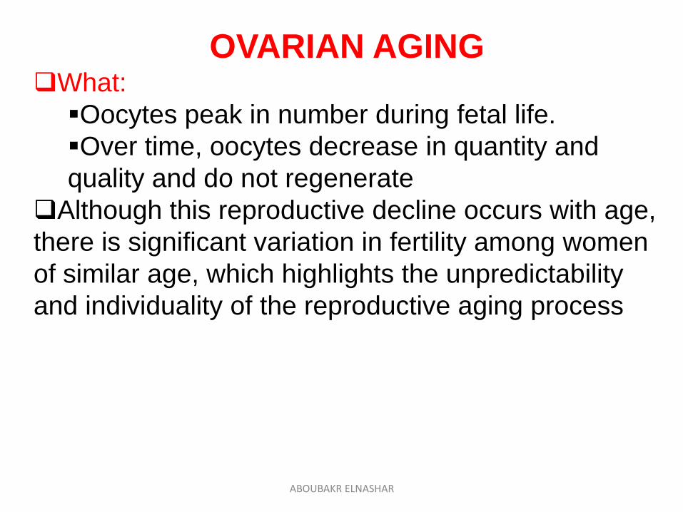
OVARIAN AGINGWhat:
Oocytes peak in number during fetal life.
Over time, oocytes decrease in quantity and
quality and do not regenerate
Although this reproductive decline occurs with age,
there is significant variation in fertility among women
of similar age, which highlights the unpredictability
and individuality of the reproductive aging process
ABOUBAKR ELNASHAR
ABOUBAKR ELNASHAR
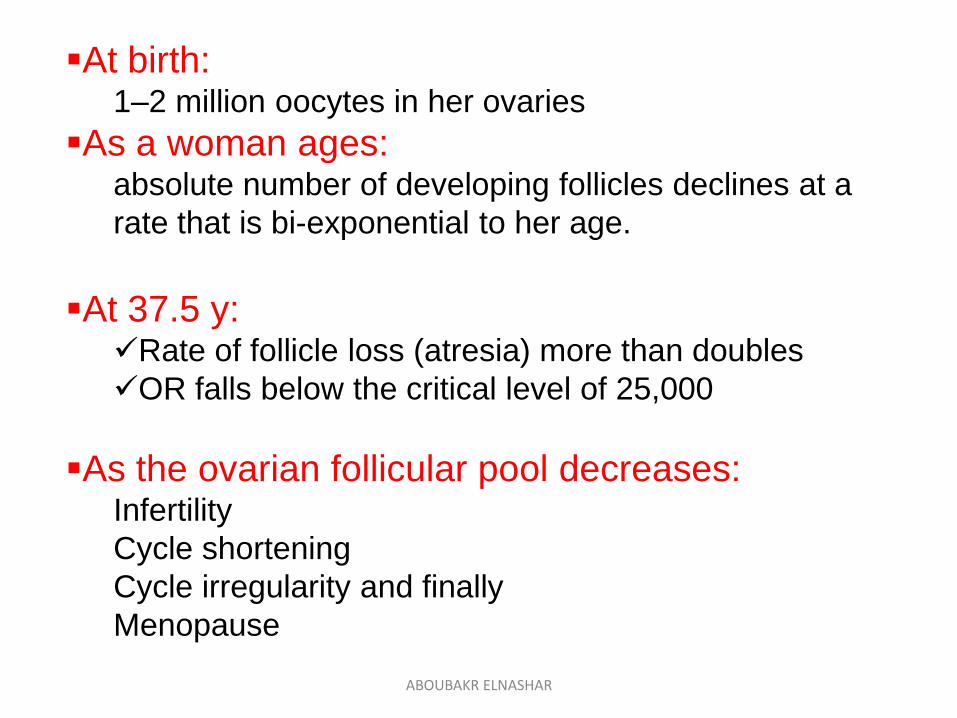
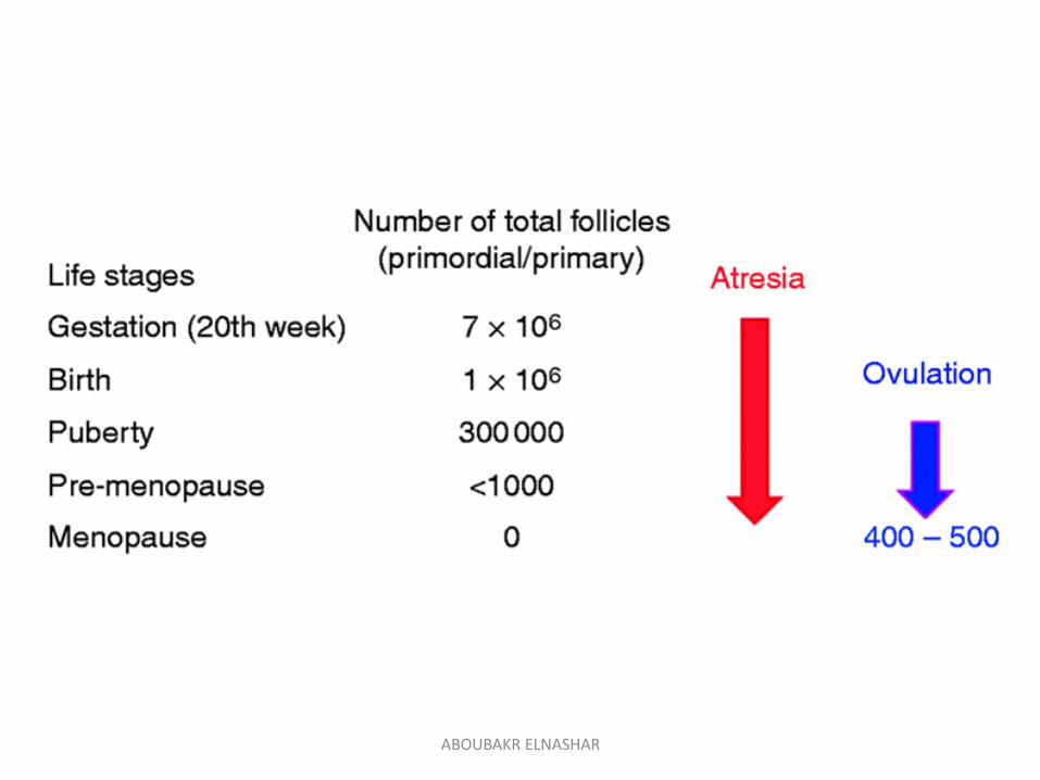
At birth: 1–2 million oocytes in her ovaries
As a woman ages:absolute number of developing follicles declines at a
rate that is bi-exponential to her age.
At 37.5 y:Rate of follicle loss (atresia) more than doubles
OR falls below the critical level of 25,000
As the ovarian follicular pool decreases: Infertility
Cycle shortening
Cycle irregularity and finally
Menopause
ABOUBAKR ELNASHAR
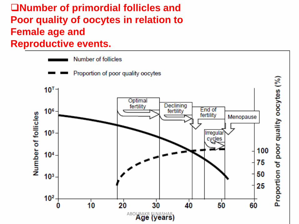
Number of primordial follicles and
Poor quality of oocytes in relation to
Female age and
Reproductive events.
ABOUBAKR ELNASHAR
ABOUBAKR ELNASHAR
OVARIAN RESERVE
What:
Reproductive potential
function of the number and quality of remaining
oocytes.
ABOUBAKR ELNASHAR
Predictors of ovarian reserve
1. Age
2. History of poor response or cancelled cycles
3. Menstrual cycle length
4. ORT:
Most important: AFC &AMH
best test for quantitative OR
(Broer et al., 2009; IMPORT study 2013)
ABOUBAKR ELNASHAR
Decreased or diminished ovarian reserve (DOR)
What:
Commonly have regular menses but
reduced quantity of ovarian follicles:
limited response to ovarian stimulation
reduced fecundity
(probability of achieving a live birth in a single
reproductive cycle).
Distinct from:
menopause or
premature ovarian failure (also referred to as
primary ovarian insufficiency)
ABOUBAKR ELNASHAR
Causes:
In most cases, are unknown.
It is unclear whether DOR represents a pathologic condition resulting from
1. abnormally rapid atresia in a normal pool of oocytes,
2. normal atresia of an abnormally small initial pool of oocytes, or
3. simply the extreme end of a normal bell-shaped population distribution of the number of oocytes at a given age.
ABOUBAKR ELNASHAR
Risk Factors
• Advanced reproductive age (older than 35 years)
• Family history of early menopause
• Genetic conditions:
45,X mosaicism
• FMR1 (Fragile X) premutation carrier
• Conditions that can cause ovarian injury endometriosis
pelvic infection
• Previous ovarian surgery (eg, for endometriomas)
• Oophorectomy
• History of
gonadotoxic therapy or
pelvic irradiation
• SmokingABOUBAKR ELNASHAR
OVARIAN RESERVE TESTS:
The purpose
Predict ovarian reserve and/or reproductive potentialIdentify infertility patients at risk for DOR, who
are more likely to
exhibit a ‘‘poor’’ response to gonadotropin
stimulation
have a lesser chance of achieving pregnancy
with ART.
Prognosis
Dose of the drugs
Safety considerationABOUBAKR ELNASHAR
Indications:
≥ 35 ys not conceived after 6 months or
< 35 ysEndometriosis
Unexplained infertility
Single ovary
Previous ovarian surgery,
Poor response to FSH,
Previous exposure to chemotherapy or
radiation (Iii-b) SOGC, 2011
ABOUBAKR ELNASHAR
Types:
Biochemical tests
reflect the biology of the aging ovary, the one component of the reproductive system most closely related to decreased fecundity.Basal measurements
FSH, AMH, E2, inhibin B
Dynamic= Provocative tests
assess the response of the
hypothalamic–pituitary– ovarian axis to
a stimulus.
CCCT.
ABOUBAKR ELNASHAR
Ultrasonographic
AFC
ovarian volume.
ABOUBAKR ELNASHAR
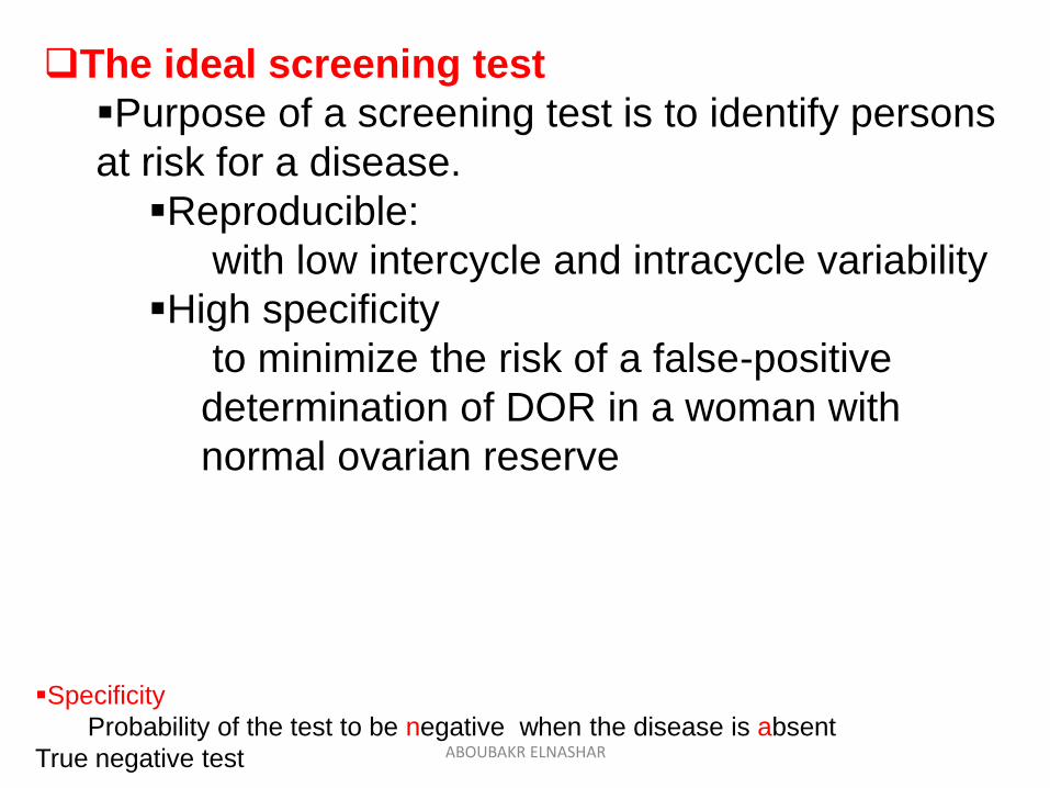
The ideal screening test
Purpose of a screening test is to identify persons
at risk for a disease.
Reproducible:
with low intercycle and intracycle variability
High specificity
to minimize the risk of a false-positive
determination of DOR in a woman with
normal ovarian reserve
Specificity
Probability of the test to be negative when the disease is absent
True negative test ABOUBAKR ELNASHAR
ABOUBAKR ELNASHAR
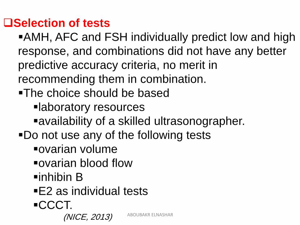
Selection of tests
AMH, AFC and FSH individually predict low and high
response, and combinations did not have any better
predictive accuracy criteria, no merit in
recommending them in combination.
The choice should be based
laboratory resources
availability of a skilled ultrasonographer.
Do not use any of the following tests
ovarian volume
ovarian blood flow
inhibin B
E2 as individual tests
CCCT.(NICE, 2013) ABOUBAKR ELNASHAR
The most appropriate ORT to use in practice are
basal FSH plus E2 levels or
AMH levels.
AFC, also may be useful if there is an indication
to perform TVS(ESHRE, ACOG, 2015)
ABOUBAKR ELNASHAR
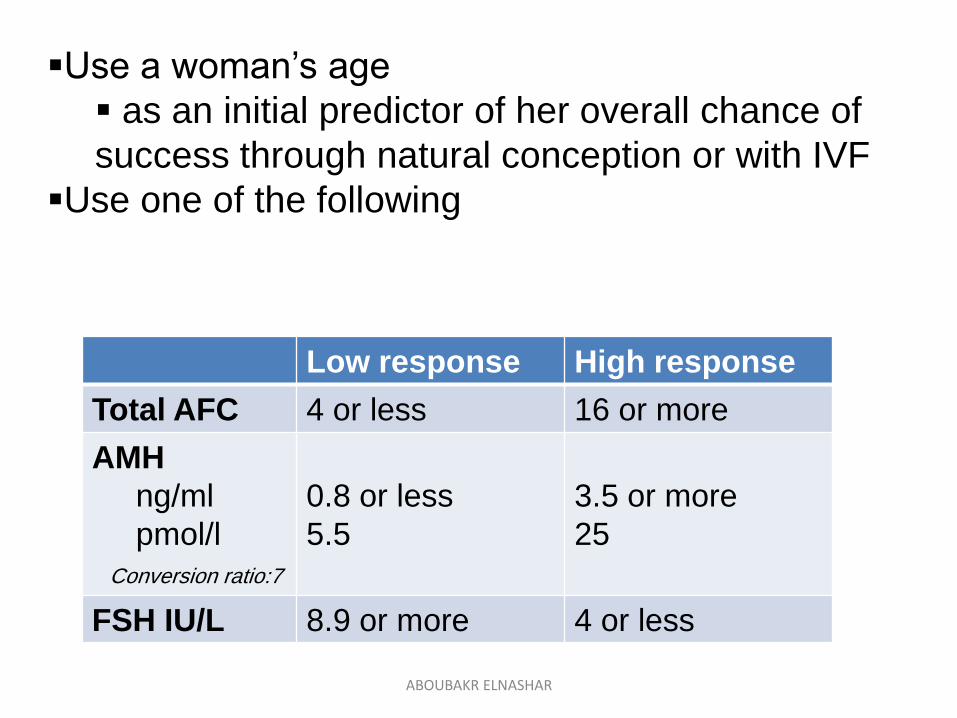
Use a woman’s age
as an initial predictor of her overall chance of
success through natural conception or with IVF
Use one of the following
High responseLow response
16 or more4 or lessTotal AFC
3.5 or more
25
0.8 or less
5.5
AMH
ng/ml
pmol/l
Conversion ratio:7
4 or less8.9 or moreFSH IU/L
ABOUBAKR ELNASHAR
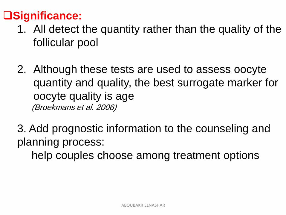
Significance:
1. All detect the quantity rather than the quality of the
follicular pool
2. Although these tests are used to assess oocyte
quantity and quality, the best surrogate marker for
oocyte quality is age(Broekmans et al. 2006)
3. Add prognostic information to the counseling and
planning process:
help couples choose among treatment options
ABOUBAKR ELNASHAR
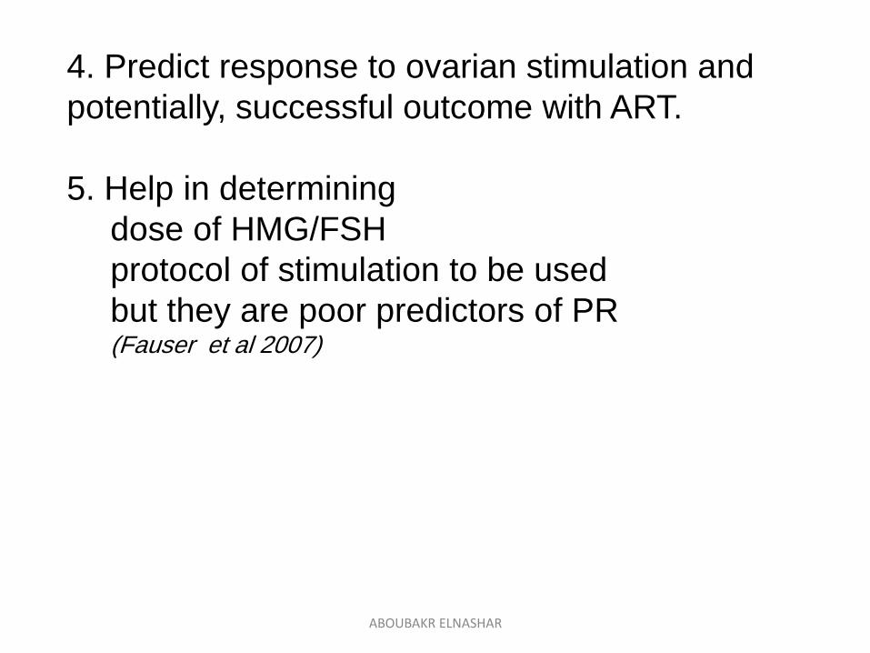
4. Predict response to ovarian stimulation and
potentially, successful outcome with ART.
5. Help in determining
dose of HMG/FSH
protocol of stimulation to be used
but they are poor predictors of PR (Fauser et al 2007)
ABOUBAKR ELNASHAR
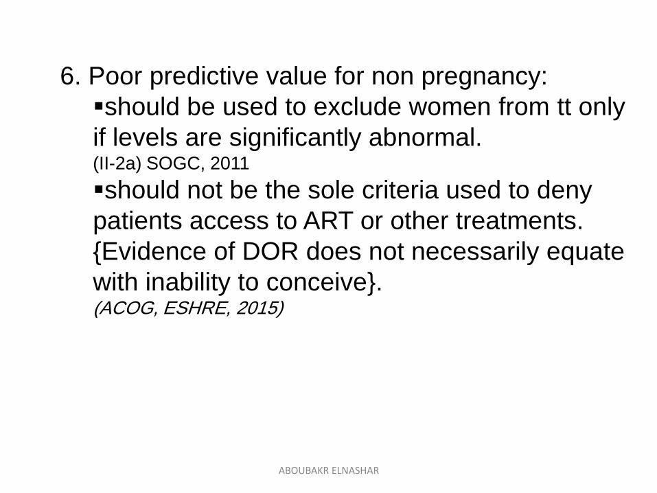
6. Poor predictive value for non pregnancy:
should be used to exclude women from tt only
if levels are significantly abnormal. (II-2a) SOGC, 2011
should not be the sole criteria used to deny
patients access to ART or other treatments.
{Evidence of DOR does not necessarily equate
with inability to conceive}.(ACOG, ESHRE, 2015)
ABOUBAKR ELNASHAR
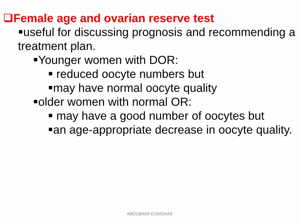
Female age and ovarian reserve test
useful for discussing prognosis and recommending a
treatment plan.
Younger women with DOR:
reduced oocyte numbers but
may have normal oocyte quality
older women with normal OR:
may have a good number of oocytes but
an age-appropriate decrease in oocyte quality.
ABOUBAKR ELNASHAR
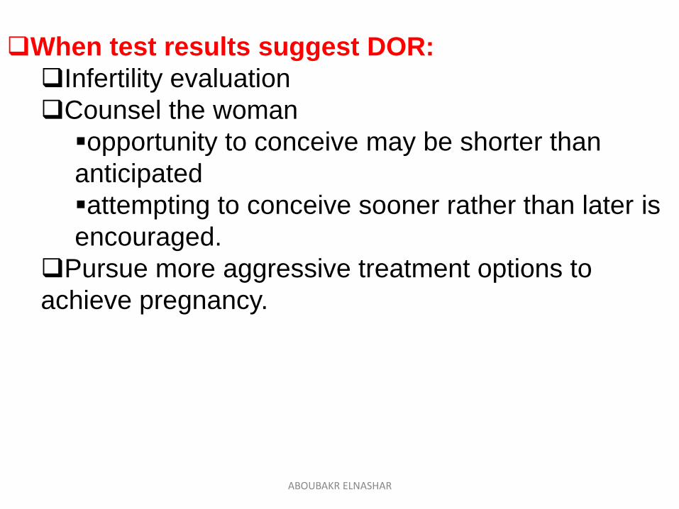
When test results suggest DOR:
Infertility evaluation
Counsel the woman
opportunity to conceive may be shorter than
anticipated
attempting to conceive sooner rather than later is
encouraged.
Pursue more aggressive treatment options to
achieve pregnancy.
ABOUBAKR ELNASHAR
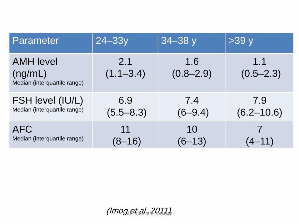
>39 y34–38 y24–33yParameter
1.1
(0.5–2.3)
1.6
(0.8–2.9)
2.1
(1.1–3.4)
AMH level
(ng/mL)Median (interquartile range)
7.9
(6.2–10.6)
7.4
(6–9.4)
6.9
(5.5–8.3)
FSH level (IU/L)Median (interquartile range)
7
(4–11)
10
(6–13)
11
(8–16)
AFCMedian (interquartile range)
(Imog et al ,2011) ABOUBAKR ELNASHAR
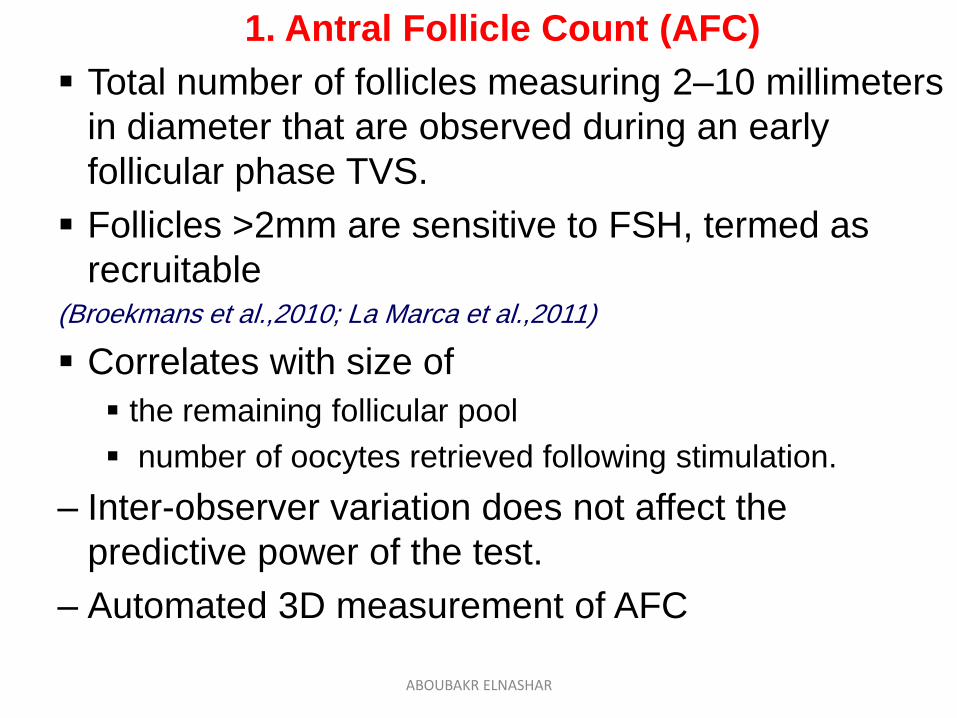
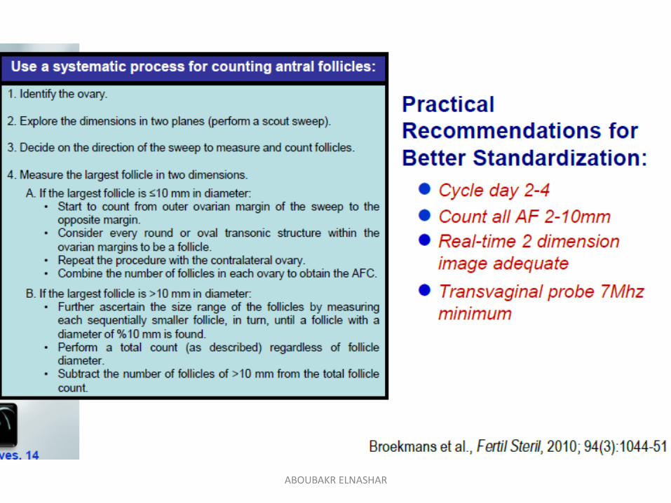
1. Antral Follicle Count (AFC)
Total number of follicles measuring 2–10 millimeters
in diameter that are observed during an early
follicular phase TVS.
Follicles >2mm are sensitive to FSH, termed as
recruitable(Broekmans et al.,2010; La Marca et al.,2011)
Correlates with size of
the remaining follicular pool
number of oocytes retrieved following stimulation.
– Inter-observer variation does not affect the
predictive power of the test.
– Automated 3D measurement of AFC
ABOUBAKR ELNASHAR
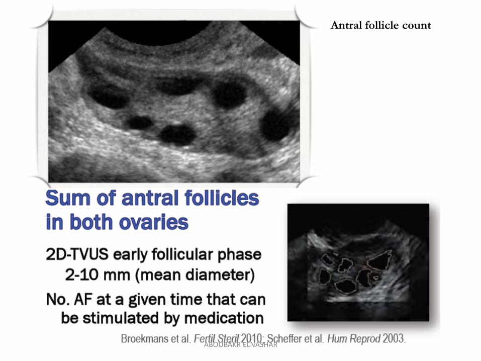
Antral follicle count
ABOUBAKR ELNASHAR
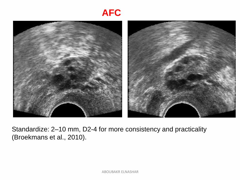
AFC
Standardize: 2–10 mm, D2-4 for more consistency and practicality
(Broekmans et al., 2010).
ABOUBAKR ELNASHAR
ABOUBAKR ELNASHAR
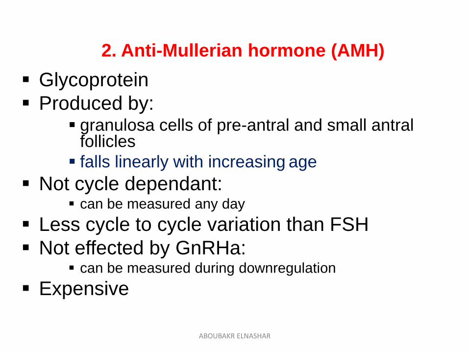
2. Anti-Mullerian hormone (AMH)
Glycoprotein
Produced by: granulosa cells of pre-antral and small antral
follicles
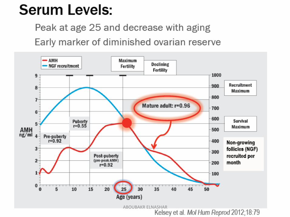
falls linearly with increasing age
Not cycle dependant: can be measured any day
Less cycle to cycle variation than FSH
Not effected by GnRHa: can be measured during downregulation
Expensive
ABOUBAKR ELNASHAR
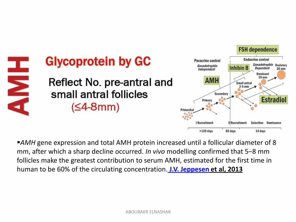
AMH gene expression and total AMH protein increased until a follicular diameter of 8 mm, after which a sharp decline occurred. In vivo modelling confirmed that 5–8 mm follicles make the greatest contribution to serum AMH, estimated for the first time in human to be 60% of the circulating concentration. J.V. Jeppesen et al, 2013
ABOUBAKR ELNASHAR
ABOUBAKR ELNASHAR
ABOUBAKR ELNASHAR
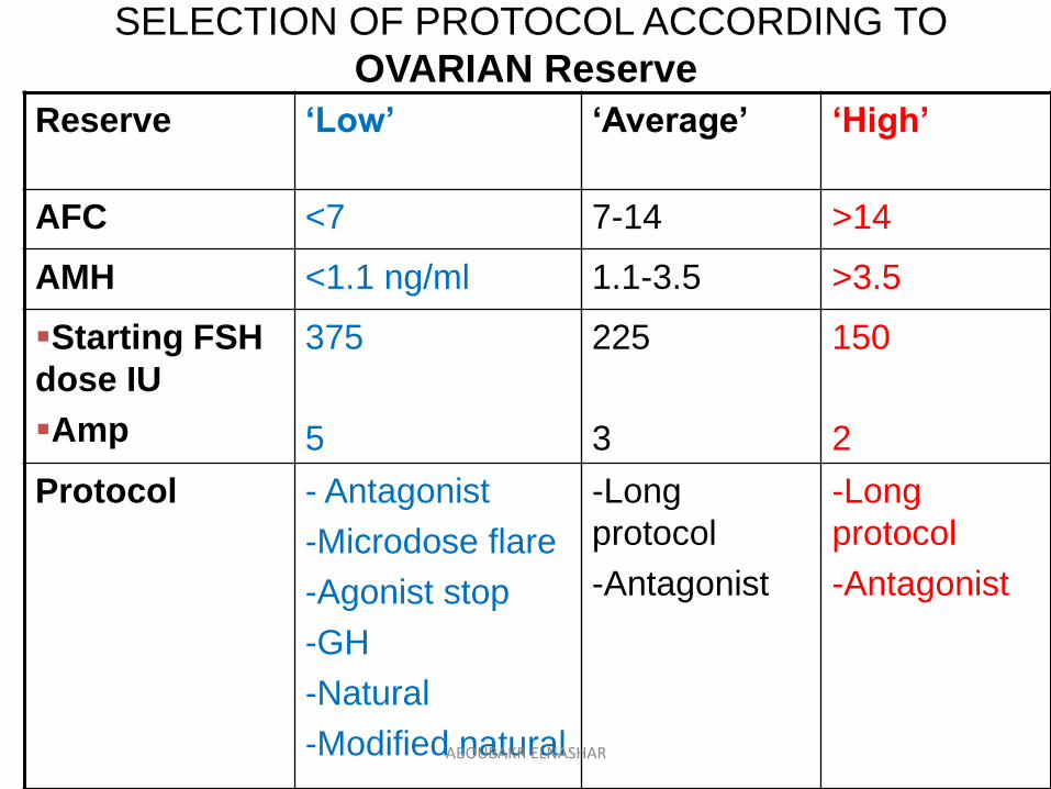
SELECTION OF PROTOCOL ACCORDING TO
OVARIAN Reserve
Reserve ‘Low’ ‘Average’ ‘High’
AFC <7 7-14 >14
AMH <1.1 ng/ml 1.1-3.5 >3.5
Starting FSH
dose IU
Amp
375
5
225
3
150
2
Protocol - Antagonist
-Microdose flare
-Agonist stop
-GH
-Natural
-Modified natural
-Long
protocol
-Antagonist
-Long
protocol
-Antagonist
ABOUBAKR ELNASHAR
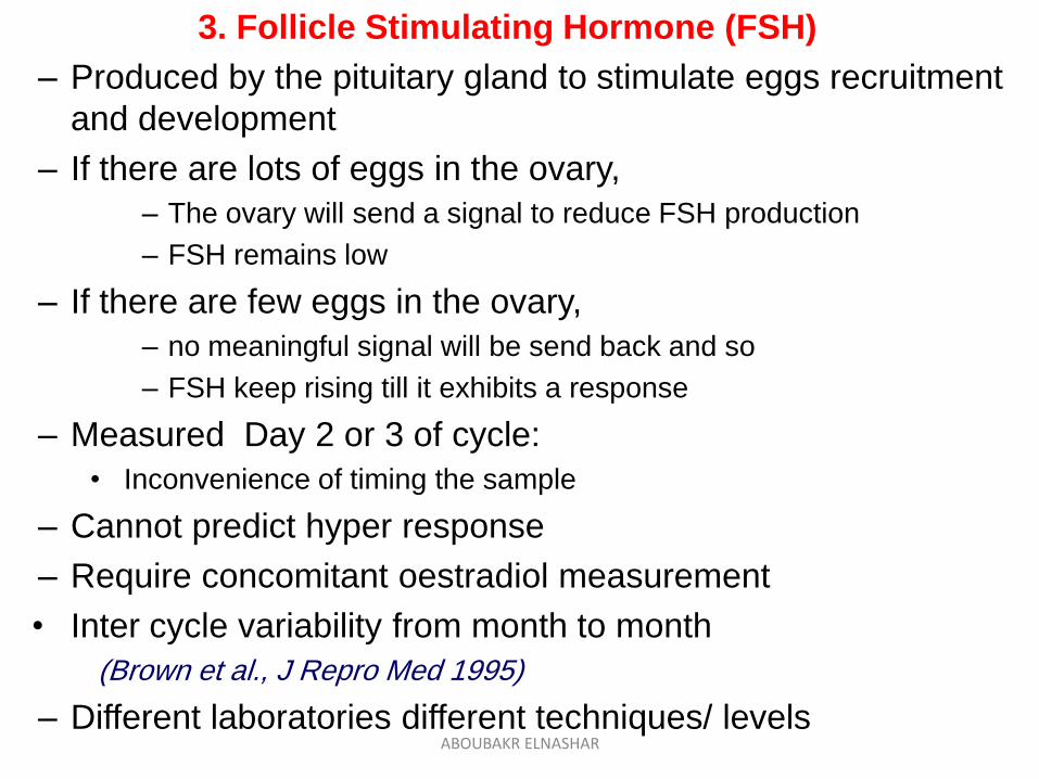
3. Follicle Stimulating Hormone (FSH)
– Produced by the pituitary gland to stimulate eggs recruitment
and development
– If there are lots of eggs in the ovary,
– The ovary will send a signal to reduce FSH production
– FSH remains low
– If there are few eggs in the ovary,
– no meaningful signal will be send back and so
– FSH keep rising till it exhibits a response
– Measured Day 2 or 3 of cycle:
• Inconvenience of timing the sample
– Cannot predict hyper response
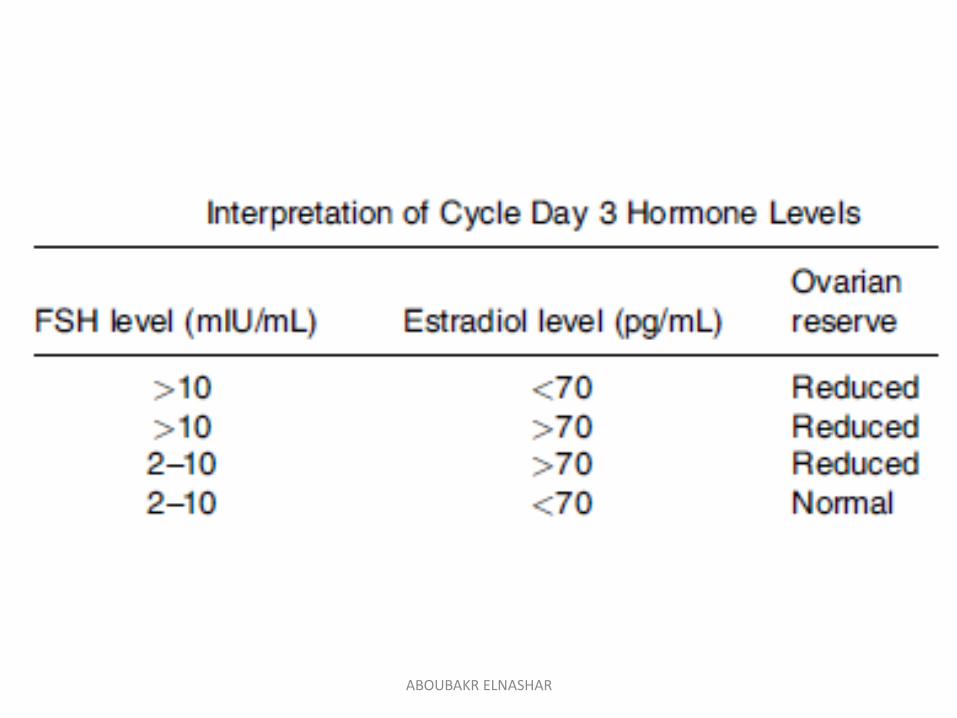
– Require concomitant oestradiol measurement
• Inter cycle variability from month to month
(Brown et al., J Repro Med 1995)
– Different laboratories different techniques/ levelsABOUBAKR ELNASHAR
ABOUBAKR ELNASHAR
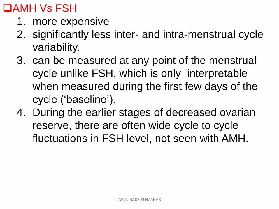
AMH Vs FSH
1. more expensive
2. significantly less inter- and intra-menstrual cycle
variability.
3. can be measured at any point of the menstrual
cycle unlike FSH, which is only interpretable
when measured during the first few days of the
cycle (‘baseline’).
4. During the earlier stages of decreased ovarian
reserve, there are often wide cycle to cycle
fluctuations in FSH level, not seen with AMH.
ABOUBAKR ELNASHAR
Thank youABOUBAKR ELNASHAR


![Research Paper Sigma-1 receptor is involved in diminished ... · ovarian reserve (DOR) is found in approximately 10% of infertile women [2, 3]. Ovarian reserve means the number and](https://static.fdocuments.in/doc/165x107/5f0c6ce07e708231d4355653/research-paper-sigma-1-receptor-is-involved-in-diminished-ovarian-reserve-dor.jpg)