Outlines of Urodynamic Studies and its Clinical … · Outlines of urodynamic studies and its...
13
Review Article The ORION Vol. 10, September 2001 www.orion-group.net/journals Outlines of urodynamic studies and its clinical application Salam MA General consideration Urodynamics is synonymous with hydrodynamk s, which measures the flow and pressure of water. In urological practice urodynamics is a neuro physiological study of urinary tract which measures the pressure, flow and electrical changes in various component 01 urinary tract during the different phases of activity of upper and lower urinary tract. In practice the activity of bladder, urethral sphincter and pelvic muscle is recorded through especial equipment called urodynamic machine. Urinary continence during bladder filling, urine storage in the bladder and the efficiency of subsequent voiding all depend on the accurate co- ordination activity of vesicoureteral segment. The interpretation of urodynamic study is simple in most cases and provides an essential complement to the modern practice of urology, gynaecology, neurology, spinal injury management, and associated specialities. The Dream Machine of Urodynamics Indication of urodynamics study in respective fields You may chose your desired test of bladder function for your patients. Urodynamic test proved to be invaluable to the specialist of the following subjects in varieties of clinical conditions; some of them are outlined below. Urology Urodynamics is essential in the evaluation of patient withNeuropathic bladder due to spinal cord trauma, Spinal cord disease, Central nervous system disease like Parkinsonism, Stress incon-tinene, over flow incontinence. Urge incontinence, LUTS (Lower urinary tract symptoms), Benign Prostatic Hyperplasia, Unstable bladder, Diabetic cvstopathy, Doubtful Pelvic Ureteric Junction obstruction whitaker test). Neurology Patients with Neurological disorder often require urodynamics for evaluation of CNS or PNS disease associated with bladder dysfunction CVA, CVD, Parkinsonism, Spinal disease of almost all type Peripheral Nerve diseases, Autonomic neuropathy. Paediatric surgery Urodynamics is essential for VUR with suspected hyper reflexic bladder PUV with suspected hyper reflexic bladder, Decompensated bladder, Chronic retention of urine, lncontinenue. Endocrinology Urodynamcis is essential for evaluation of bladder function in disease like-Diabetes mellitus (diabetic cvstopathv) with Lower unnarytract symptoms. Neurosurgery Neurosurgeons require urodynamics for evaluation of patients with- Spina hifida., Disc prolapse, Spinal tumors, Spinal diseases e.g. Traumatic diseases, lnfections,Neoplasm, Degenerative diseases, Urinary symptoms following brain surgery etc. Gynaecology Gynaecologists often require urodynamics for evaluation of patients with-Strees urinary incontinence, Lower urinary tract symptoms in women, Recurrent urinary infects. Dr. M A Salam, MBBS, FCPS, FICS (USA), Consultant Urologist and Andrologist. Associate Professor, Department of Urology, Bangabandhu Sheikh Mujib Medical University, Dhaka
-
Upload
nguyendieu -
Category
Documents
-
view
222 -
download
0
Transcript of Outlines of Urodynamic Studies and its Clinical … · Outlines of urodynamic studies and its...

Review Article
The ORION Vol. 10, September 2001 www.orion-group.net/journals
Outlines of urodynamic studies and its clinical application Salam MA
General consideration Urodynamics is synonymous with hydrodynamk s, which measures the flow and pressure of water. In urological practice urodynamics is a neuro physiological study of urinary tract which measures the pressure, flow and electrical changes in various component 01 urinary tract during the different phases of activity of upper and lower urinary tract. In practice the activity of bladder, urethral sphincter and pelvic muscle is recorded through especial equipment called urodynamic machine. Urinary continence during bladder filling, urine storage in the bladder and the efficiency of subsequent voiding all depend on the accurate co-ordination activity of vesicoureteral segment. The interpretation of urodynamic study is simple in most cases and provides an essential complement to the modern practice of urology, gynaecology, neurology, spinal injury management, and associated specialities. The Dream Machine of Urodynamics Indication of urodynamics study in respective fields You may chose your desired test of bladder function for your patients. Urodynamic test proved to be invaluable to the specialist of the following subjects in varieties of clinical conditions; some of them are outlined below.
Urology Urodynamics is essential in the evaluation of patient withNeuropathic bladder due to spinal cord trauma, Spinal cord disease, Central nervous system disease like Parkinsonism, Stress incon-tinene, over flow incontinence. Urge incontinence, LUTS (Lower urinary tract symptoms), Benign Prostatic Hyperplasia, Unstable bladder, Diabetic cvstopathy, Doubtful Pelvic Ureteric Junction obstruction whitaker test). Neurology Patients with Neurological disorder often require urodynamics for evaluation of CNS or PNS disease associated with bladder dysfunction CVA, CVD, Parkinsonism, Spinal disease of almost all type Peripheral Nerve diseases, Autonomic neuropathy. Paediatric surgery Urodynamics is essential for VUR with suspected hyper reflexic bladder PUV with suspected hyper reflexic bladder, Decompensated bladder, Chronic retention of urine, lncontinenue. Endocrinology Urodynamcis is essential for evaluation of bladder function in disease like-Diabetes mellitus (diabetic cvstopathv) with Lower unnarytract symptoms. Neurosurgery Neurosurgeons require urodynamics for evaluation of patients with- Spina hifida., Disc prolapse, Spinal tumors, Spinal diseases e.g. Traumatic diseases, lnfections,Neoplasm, Degenerative diseases, Urinary symptoms following brain surgery etc. Gynaecology Gynaecologists often require urodynamics for evaluation of patients with-Strees urinary incontinence, Lower urinary tract symptoms in women, Recurrent urinary infects.
Dr. M A Salam, MBBS, FCPS, FICS (USA), Consultant Urologist and Andrologist. Associate Professor, Department of Urology, Bangabandhu Sheikh Mujib Medical University, Dhaka

Review Article
The ORION Vol. 10, September 2001 www.orion-group.net/journals
Orthopaedics Urodynamics is essential in the evaluation of patient with-Spinal injury, Hyper reflexic bladder, Hypo rellexic bladder, Areflexic bladder, Spinal diseases like PLID, spinal tumor etc. Paediatrics Evaluation of bladder function with urodynamics is important ui-patients like, Enuresis, Incontinence, Spinal bifida, Cerebral palsy, Myelomeningocele. General practice Urodynamics is essential for. evaluation of peoples complaining of lower urinary tract symptoms, such as Frequency, Urgency, Hesitency, Poor flow, incomplete voiding etc. Incontinence due to any cause, Diabetes mellitus with voiding symptoms. Components of urodynamics Urodynamics consists of a number of complimentary component of varying degrees of complexity, the needs to be tailored to meet the clinical requirements of each case. Some of the tests are clinical and very simple to perform, they are called as simple urodynamics. Other components of urodynamic tests is highly complex and require electronic equipments to record pressure, flow and electrical activity of the urinary tract and pelvic floor muscles they are called as complex urodynarnics. Components of Urodynamics
For simple problem like assessment of voiding symptoms from benign prostatic hyperplasia, simple urodynamics consisting of observation of act of micturation in the toilet, frequency, volume chart analysis, estimation of urine flow by uroflowmetry or analysis of Ultrasound cystodynamogram may be cost effective and highly satisfactory investigation.
For assessing the complex neurourological problem like unexplained lower urinary tract symptoms (LUTS) or assessing the bladder function in spinal injury victims, more complex urodynamics study like Cystometry, Urethral pressure measurement, Sphincter EMG, Upper tract urodynamics (the Whitaker test) etc. would be appropriate. These tests are expansive and slightly invasive but can provide invaluable information for the correct management of the problem. Simple urodynamics Observation of Ad of Miduration Observing the act of micturation in a BPH patient will disclose the degree of discomfort felt by the patient and will allow to have a rough estimation of flow rate. The residual volume can be. measured by clinical test at the end of voiding. This test is possibly the simplest visual or clinical uro dynamics. Frequency Volume Chart Estimation of bladder activity and voided volume can be assessed simply by keeping a frequency volume chart which may give a clear indication about the volume of fluid intake and volume voided in a given time. Many people complaining of frequency, urgency and nocturia and their frequency volume chart may show that they are taking lot of water due to their obsessive drinking habit. Uroflowmetry The first simplest and most useful investigation in the assessment of voiding dysfunction (LUTS) is the measurement of urinary flow rate. On the basis of this test it is often possible objectively to confirm the

Review Article
The ORION Vol. 10, September 2001 www.orion-group.net/journals
presence of bladder outlet obstruction. Studies have demonstrated that simple uroflowmetry by itself is adequate investigation tigatiorc for uncomplicated bladder outflow obstruction in nearly 50% of patients. The urine flow may be reduced also in detrusor under activity which may mislead the clinician. Clear example is a case of chronic retention of urine due to BPH. Here the uroflow tracing will show evidence of obstruction but reliving the obstruction by TURP may not improvi the flow of urine. In such instance a Cystrometrogram (CMG) will confirm a under active detrusor. In addition, it must be remembered that a normal flow rate can be present in the early stages of obstruction as a consequence of a compensatory increase in voiding pressure by hypertrophied detrusor which will maintain of an apparently normal flow rate. Ultrasound scan of the bladder will show a thickened bladder wall and cystoscopy will confirm the bladder out flow obstruction due to BPH or stricture urethra despite of a normal flow rate. In these cases the International Prostatic Symptom Score will guide the clinician.
Indication of Uroflowmetry
Technique The flow rate is measured with a flow meter, which is a device that measures the flow and indicates a quantity of fluid volume or mass) passed per unit time; such machines normally indicate the volumetric flow rate. A flow rate estimation can be carried out either by itself or in combination with other techniques eg.
Ultrasound Cystodynamogram (USCD), Cystometrogram (CMG), Video Cystometrogram (VCMG). The measurement is expressed in mI/s. Type of Uroflowmeter 1. Spinning Disc type 2. Dip stick type 3. Weight transducer type. Spinning disc method The voided fluid is directed on a rotating disc lodged in a large funnel fitted with a comode. The amount flowing urine on the disc produces a retardation in its speed, and the power required to keep the disc rotating at a constant rate is measured, thereby allowing calculation of the flow rate of urine passed the disc. The calculations are plotted in the form of tracing on a thermal paper. This type of uroflowmeter is most dependable and durable and as such most of the urological centre prefers the equipment. Electronic dipstick method A dipstick is mounted in a collecting chamber and as urine accumulates the electrical capacitance of the dipstick changes. This allows calculation of the rate of accumulation of fluid and hence the flow rate. Gravimetric (weight transducer) method Such instruments measure the weight of collected fluid or the hydrostatic pressure at the base of a collecting cylinder. This similarly allows calculation of a flow rate. Practical guidelines
Review Article
The ORION Vol. 10, September 2001 www.orion-group.net/journals
Definitions Comment It is evident that the urinary flow rate provides important and useful information suggesting whether there is obstruction to the outflow tract. The flow rate pattern may indicate a possible aetiology. It should be always done after treatment of BOO to evaluate result and documentation. Drawbacks The flow rate can he misleading in the following conditions- • If the detrusor function is normal or the detrusor is over active, the flow rate is maintained at fairly normal values despite the high urethral resistance due to BPH or other causes of bladder outlet obstruction. • The flow rate is not diminished until the urethral calibre is reduced below 11 Ch. • The flow rate may be misleading in patients where there is significant detrusor decompensation leaving large residual volume. • The ilow rate can be influenced by straining and this should be taken into account when interpreting results. • Irregularities in the measured flow rate can be due to collecting funnel artefacts and to
variations in the direction of the urinary stream. Uroflowmetry by itself is an adequate investigation for uncomplicated prostate outflow obstruction in nearly 50% of patients. It is also invaluable in the assessment of voiding function across a ‘spectrum of urological conditions. It must, however, be emphasised that most reliance should be placed on the observed flow pattern rather than the absolute data obtained. In view of the limitations outlined above, in certain circumstances where doubt remains following a flow rate, or in particular where previous surgery has been carried out, more complex investigations are required. To provide more detailed information about bladder function a simple flow rate can be combined with ultrasound imaging (USCD) or with cystometrogram (Voiding CMG. Ultrasound Cystodynamogram (USCD) Ultrasound scan of urinary tract is combined with a flow rate to provide more practical information of bladder function. This investigation is performed routinely to all patients with lower urinary tract symptoms in men, women & children, BPH, stricture urethra, neuropathic bladder or assessing the result of treatment of bladder outflow obstruction etc. Indication of USCD • Diagnostic indication e.g. Bladder outflow obstruction • Follow up e.g. Post TURP or Urethral surgery Technique The upper tract is scanned, then the bladder is scanned at maximum urgency for the thickness of the bladder wall, any filling defect and the maximum cystometric capacity is measured. The patient voids into a flowmeter in a uroflowmeter to obtain the uroflow tracing. Now a postvoid bladder scan is carried immediately out to measure the residual volume.
Review Article
The ORION Vol. 10, September 2001 www.orion-group.net/journals
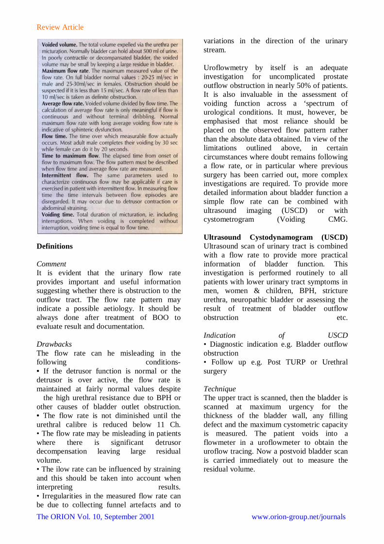
USCD Provides
Complex urodynamics Cystometrogram Cystometry is the method by which the pressurevolume relationship of the bladder is measured. Cystometrogram (.an be added to uroflowmeter to obtain simultaneous flow tracing or can be radiological evaluation of bladder neck during voiding in order to see the bladder neck activity to study incontinene. Based on above facts cystometrogram can be of three types: 1. Simple Cystometry 2. Voiding Cystometry 3. Video Cystometry Simple cystometry The term simple cystometry is usually taken to mean the measurement of detrusor pressure during controlled bladder filling md subsequent voiding to assess detrusor activity, sensation, capacity and compliance
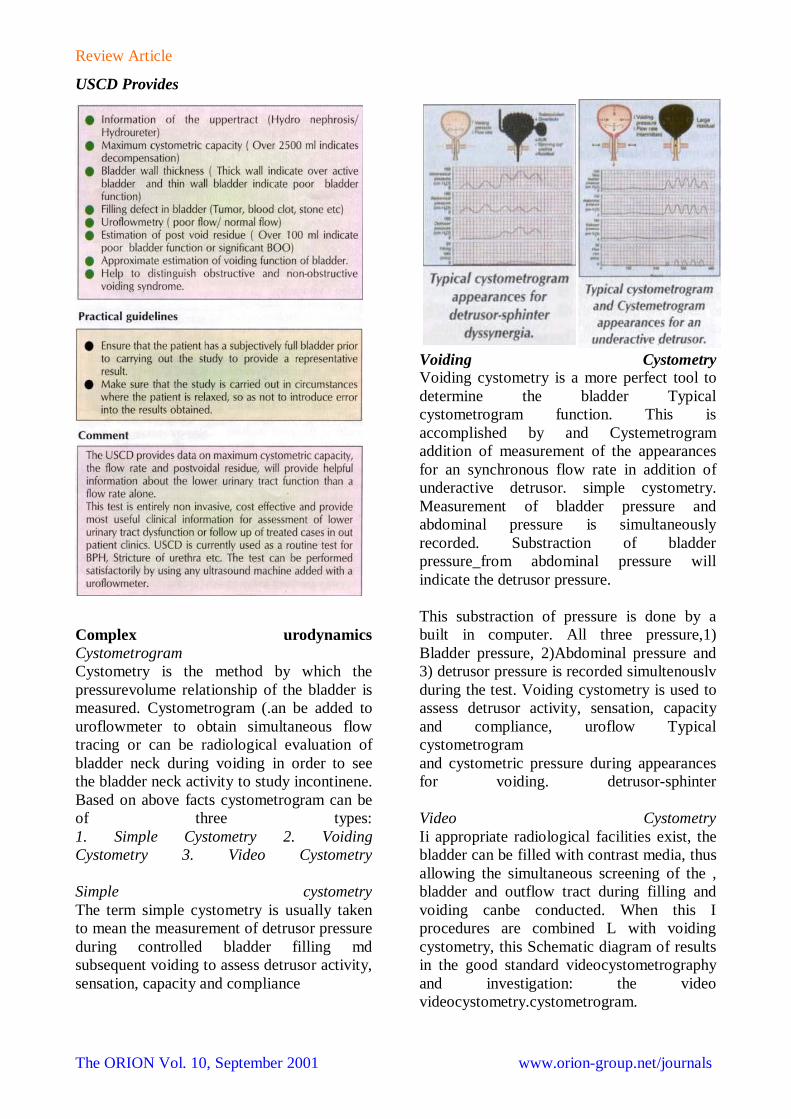
Voiding Cystometry Voiding cystometry is a more perfect tool to determine the bladder Typical cystometrogram function. This is accomplished by and Cystemetrogram addition of measurement of the appearances for an synchronous flow rate in addition of underactive detrusor. simple cystometry. Measurement of bladder pressure and abdominal pressure is simultaneously recorded. Substraction of bladder pressure_from abdominal pressure will indicate the detrusor pressure. This substraction of pressure is done by a built in computer. All three pressure,1) Bladder pressure, 2)Abdominal pressure and 3) detrusor pressure is recorded simultenouslv during the test. Voiding cystometry is used to assess detrusor activity, sensation, capacity and compliance, uroflow Typical cystometrogram and cystometric pressure during appearances for voiding. detrusor-sphinter Video Cystometry Ii appropriate radiological facilities exist, the bladder can be filled with contrast media, thus allowing the simultaneous screening of the , bladder and outflow tract during filling and voiding canbe conducted. When this I procedures are combined L with voiding cystometry, this Schematic diagram of results in the good standard videocystometrography and investigation: the video videocystometry.cystometrogram.

Review Article
The ORION Vol. 10, September 2001 www.orion-group.net/journals
Radiological screening provides valuable additional anatomical information on the appearances of the bladder, the presence of ureteric reflux, the level of any outflossn obstruction in the lower urinary tract, the degree of support to the bladder base during coughing and, by itseli, is more than adequate for the diagnosis of stress urinary incontinence or sphincter weakness incontinence. This information, along with the accompanying pressure flow traces, can be recorded on a video tape or in CD allowing subsequent review and discussion. Videocystometry is, however, essential for the adequate assessment of the complex cases where equivocal results have been obtained from simpler investigations for the (lefinition of neuropathic disorders, and in situations where there has been an apparent failure of a previous surgical procedure. Indication of Video CMG 1. Study of stress incontinence 2. Post TURP incontinence Practical points of urodynamic study A number of variations in technique are currently available and the following points deserve specific consideration. Access Whilst the majority of cystometry is carried out via the transurethral route, percutaneous placement of catheters both for bladder filling and pressure measurement is essential in the investigation of paediatric patients and when the urethral access is not possible.
Type of catheter Fluid filled cather is one of various type and size with single or multiple lumen. Cather tip transducer specifications vary between manufacturers; these catheters tend to be expensive and too long fragile for routine use.
Urodynamic equipment A number of commercial urodynamic systems are currently available. These vary greatly in terms of sampling rate, associated computer software backup and price. Test medium (liquid or gas) Normal saline is the most ideal medium for cystometry. Gas cystometrv is rarely performed now a days. The major drawback with gas cvstometry is aerodynamics not urodynamics. Position of patient eg. Supine, sitting or standing. Type of filling This may be by diuresis or catheter. Filling by catheter may be continuous or incremental; the precise filling rate should be stated. When the incremental method is used the volume increment should be stated. Continuous pressure measurement is of greatest usefulness in clinical practice. Techniques of videocystometrography/cystometry A full urodynamic history is taken before the test is performed. One of the goal during the test is to reproduce the similar symptoms the patient was complaining. The detrusor pressure is estimated by the automatic subtraction of rectal pressure (as an index of intra-abdominal pressure) from the total bladder pressure, thus removing the influence of artefacts produced by abdominal straining. During this study, notice is taken of the initial bladder residual, the bladder volume at the
1. luman Pves Catheter, Infusion Catheter, Rectal Catheter used with Disposable Sphincter Electrode.
Review Article
The ORION Vol. 10, September 2001 www.orion-group.net/journals
time of the patient’s first sensation of filling, the final tolerated bladder volume and the final residual volume after voiding. All systems are zeroed at atmospheric pressure. For external transducers, the reference point is the level of the superior edge of the svmphysis pubis. For catheter mounted transducer the reference point is the transducer itself. 2mm diameter saline-filled catheter is introduced into the rectum, the end of the tube being connected with a rectal baloon or with a finger of a surgical gloves. A slit is cut in this to prevent tamponade producing artefactual results during the study. With the patient in the supine position, the external urethral meatus is cleaned with antiseptic solution. The urethra is anaesthetised with 1% lignocaine gel containing chlorhexidine and then a 5 Fr. Manufacturers double lumen or a 10 Ch Nelaton filling catheter with a 1 mm diameter saline-filled plastic pressure catheter inserted into the bladder and the two catheters end is then disengaged. The bladder is drained of urine and this initial residual volume recorded. The two pressure I measurement lines are then connected to the transducers incorporated in the urodynamic apparatus. The lines are flushed through with saline, great care being taken to exclude .all air bubbles from both the tubing and transducer chambers. Normal saline or normal saline mixed with contrast medium (for video urodynamics) at room temperature is then instilled into the bladder at a predetermined rate under the control a peristaltic pump. Fast-fill (>100 mI/mm) is used routinely in most unit, although slower filling rates approaching the physiological range are mandatory in the assessment of the more complex cases. The bladder is filled initially in the supine position and the volume at first sensation of filling is noted. When the subject first experiences discomfort, the radiographic table is tipped towards the standing position and subsequent bladder filling discontinued when at the maximum tolerated capacity. During bladder filling the patient is asked to consciously suppress bladder contraction. The Nelaton filling catheter is then removed from the bladder and the patient turned to the oblique
position relative to the X-ray machine and asked to void into the flowmeter provided. Throughout the study continuous rectal pressure, total bladder pressure and electronically subtracted detrusor pressure (total bladder pressure minus rectal pressure) measurements are sampled at a predetermined rate (1 Hz on most commercially available contemporary machines) and the results displayed on the video display unit/stored to disc/polygraph chart recorded-depending on the equipment in use. The adjacent X-ray screening apparatus allows the synchronous display of pressure and flow, and also radiographic data relating to bladder morphology, ureteric reflux and the appearances of the bladder outlet and urethra, to be displayed alongside the numerical data on a video display unit. The monitor images are recorded on video tape allowing review and detailed study. Comment Before starting to fill the bladder the residual urine may be measured. However, the removal of a large volume of residual urine may alter detrusor function, especially in the neuropathic disorders. Certain cystometric parameters may be significantly altered by the speed of bladder filling. During cyslometry it is taken for granted that the patient is awake, unanaesthetised and neither sedated, nor taking drugs that eflect bladder function. Any variations from this ideal must he taken into account when interpreting results. The failure of the bladder neck mechanism to relax occurs in a small percentage of young men resulting in a clinical entity of bladder neck obstruction (dys-synergia). This abnormality is evident during videocystometry if the patient is asked to inhibit micturation voluntarily. In a normal urodynamic study, contrast media will be milked back from the distal sphincter mechanism proximally through the bladder neck into the bladder, indicates a normal stop test. It there is any obstruction lies at the level of the bladder neck, contrast will be trapped between the bladder neck and external

Review Article
The ORION Vol. 10, September 2001 www.orion-group.net/journals
sphincter resulting a tear drop appearance of trapped contrast material in prostatic urethra. This is a reliable test for diagnosing the bladder neck hypertrophy. During cystometry, under normal circumstances the bladder should fill to a capacity of approximately 500 ml before a strong desire to void is experienced. During subsequent bladder fifing, whilst the patient is making a conscious effort to inhibit voiding, in practice the subtracted intravesical detrusor pressure should not rise significantly off its baseline value-although the International Continence Society on longer recognises a specific value as being essential to the diagnosis of detrusor instability. In practice, we have found that, if there is pressure rise greater than 15 cm of water, then detrusor instability can be said to be present. The only exception is represented by a gradual linear rise in pressure during detrusor filling-so called low compliance-which, although cIearly defined, remains as an yet poorly classified entity. During subsequent voiding the patient’s bladder empties completely with a maximum detrusor pressure of 30-50 cm of water and a maximum urinary flow rate of 25 -30 mI/s in men and 30-40 nil/s in women. Compliance indicates the change of the volume for a change in pressure. Compliance is calculated by dividing the volume change (dV) by the change in (letrusor pressure (dP det) during the change in bladder volume (C=dV/dP det). Compliance is expressed as ml per cm H20. Filling rate <10 ml per minute is slow till cystometry-physiological filling. 10-100 ml per minute k medium fill cystometry >100 nil per minute is fast till cystometry. During Filling Phase the following parameters are observed during the urodynamic study
Bladder pressure measurements during micturition Opening time is the elapsed time from initial rise in detrusor pressure to onset of flow. This is the initial isovolumetric contraction period of micturition. Time lags should be taken into account. In most urodynamic systems a time lag occurs equal to the time taken for the urine to pass from the point of pressure measurement to the uroflow transducer. Premicturition pres$ureis the pressurdlecprded immediately before the initial isovolumetric contraction. Opening pressure is the pressure recorded at the onset of measured flow. Maximum pressure is the maximum value of the measured pressure. Pressure at maximum flow is the pressure recorded at maximum measured flow rate. Contraction pressure at maximum flow is the difference between pressure at maximum flow and premicturition pressure. Postmicturition events (e;g. after contraction) are not well understood and so cannot be defined as The Stop Test If this demonstrates the presence of contrast trapped in the posterior urethra, it may indicate bladder neck obstruction. Women, particularly those with stress incontinence are often unable to carry out a stop test. Isometric Pressure (PISO) Detrusor pressure measured at the time significance of the PISO is controversial. Residual urine Residual urine is defined as the volume of fluid remaining in the bladder immediately following the completion of micturition.

Review Article
The ORION Vol. 10, September 2001 www.orion-group.net/journals
Normally residual volume should be 0 ml but for practically post void residue more than l00 ml is significant. When residual volume exceeds 300ml the condition may be described as chronic retention of urine. Post Void Residue (PVR) •Higher the PVR, weaker the Bladder function or significant BOO . •Normal volume of PVR is O ml. •PVR of 100ml or above is significant. •PVR over 300 ml indicates chronic retention of urine. The measurement of residual urine forms an integral part of the study of micturition. However, voiding in unfamiliar surroundings may lead to unrepresentative results. Important points in the interpretation of residual volume •When estimating residua[ urine, the measurement of voided volume and the time interval between voiding and residual urine estimation should be recorded: this is particularly important if the patient is in a diuretic phase. • In the Condition of vesicoureteric reflux, urine may re-enter the bladder after micturition and may falsely be interpreted as residual time. •The presence of urine in bladder diverticula following micturition presents special problems of interpretation, since a diverticulum may be regarded either as part of the bladder cavity, or outside the functioning bladder. •The absence of residual urine is usually an observation of clinical value, but does pot exclude intravesical obstruction or bladder disfunction. •An isolated finding of residual urine requjres confirmation. , •PVR less than 100 ml with high IPSS score demands further investigations. Urethral pressure
measurement The urethral pressure and the urethral closure pressure are the opposing force 10 detrusor pressure which represent the ability of the urethra to maintain continence and prevent leakage. In current urodynamic practice, the urethral pressure is measured by a number of different techniques. Technique Measurements may be made qt one point in the urethra over a period of time, or at several points along the urethra consecutively forming a urethral pressure profile (UPP). Image At rest the urethral pressure profile denotes the intraluminal pressure along the length of the urethra. All systems zeroed at atmospheric pressure. For external transducers the reference point is the superior edge of the symphysis pubis. For catheter-mounted transducers the reference point is the transducer itself. Intravesical pressure should be measured to exclude ca simultaneous detrusor contraction. The subtraction of intravesical pressure from urethral pressure produces the urethral closure pressure profile. Intraluminal urethral pressure maybe measured : •At rest (the storage phase), with the bladder at any given volume- resting urethral pressure profile (UPP). • .During coughing or straining-stress uretl\tral pressure profile. The principle of this study is to measure the transmission of pressure from the abdominal cavity to the urethra. lr stress incontitrlence this pressure transmission, which is thought tockeep the normal urethra closed during stress, is in adequate. The utethral closure pressure becomes negative on coughIng. • .During voiding-voiding urethral pressure profilometry; (VUPP). The VUPP is used to rleterminecthe pressure and site of urethral obstruction: Pressure is recorded in the urethra during voiding. The technique is similar to that used in the UPP measured during storage.
Review Article
The ORION Vol. 10, September 2001 www.orion-group.net/journals
Techniques of urethral pressure profilometry Three techniques are described to perform urethral pressure profilometry. The perfusion method, the oldest technique, the balloon catheter profilometry and the most modern catheter mounted transducer technique. Perfusion method. A dual lumen catheter with two hole at the tip 5 mm apart is withdrawn down the urethra while the catheter is constantly perfused at a set rate by using a syringe pump. Continuous pressure is monitored through the other lumen of the catheter. Balloon catheter pressure profilometry This technique uses a soft balloon mounted on a catheter. The pressure is transmitted by a fluid column to the external pressure transducer. Catheter mounted T ransducer A miniature pressure transducer is mounted over the catheter when gently pulling out through the urethra it will record the pressure in the urethra instantly. The catheters are highly expansive and too fragile but can provide most accurate data for studying sphincter weakness incontinence(SWI) or stress urinary Incontinence (SUI) The following parameters recorded in UPP •Maximum urethral pressure is the maximum pressure of the measured profile. •Maximum urethral closure pressure is the maximum difference between the urethral pressure and the intravesical pressure. . •Functional profile length is the length of the urethra along which the urethral pressure exceeds intravesical pressure. •Functional profile length (on stress) is the length over which the urethral pressure exceeds the intravesical pressure on stress. Comment Urethral pressure profilometry has, in recent years, enjoyed a disproportionate amount of attention. The results obtained are extremely susceptible to experimental artifacts and the degree of relaxation of the patient. In
particular, it must be remembered that this study can be distressingly uncomfortable for the patients- especially males. The information gained from urethral pressure measurements in the storage phase is of limited value in the assessment of voiding disorders. Total profile length is not generally regarded as a useful parameter . VUPP is not yet fully developed as a technique and a number of technical, as well as clinical, problems need to be solved before the VUPP is widely used. Electromyography Electromyography (EMG) is the study of electrical potentials generated by the depolarization of muscle and in 1his context, refers to urethral sphincter striated muscle EMG. The functional unit in EMG is the motor unit. This is comprised of a single motor neuron and the muscle fibers which results from activation of a single anterior horn cell. Muscle action potentials may be detected either by needle electrodes, or by surface electrodes. Technique Needle electrodes (concentric, bipolar, monopolar, single fibre) are placed directly into the muscle mass and permit visualization of the individl!al motor unit action potentials. Image Surface electrodes (skin, anal plug, catheter) are applied to an epithelial surface as close to the muscle under study as possible. Surface electrodes detect the action potentials from groups of adjacent motor units underlying the recording surface; they can be difficult to secure adequately and provide less reproducible results. EMG should be interpreted in the light of the patient's symptoms, physical findings and Urological and Urodynamic investigations. The main clinical indication for EMG studies is as an adjunct to video cystometrography to distinguish between striated and smooth muscle in distal urethral obstruction of a neuropathic type. Other EMG studies provide interesting scientific information, which
Review Article
The ORION Vol. 10, September 2001 www.orion-group.net/journals
however, rarely alters the clinical management of patients. Nerve conduction studies Nerve conduction studies involve stimulation of a peripheral nerve and recording the time for a response to occur in muscle, innervated by the nerve under study. Reflex latencies Reflect latencies require stimulation of sensory fields and recordings from the muscle which contracts reflexly in response to the stimulation. Such responses are a test of reflex arcs, which comprise such afferent and efferent limbs and a synaptic region within the central nervous system. The reflex latency expresses the nerve production velocity in both limbs of the arc and the integrity of the central nervous system at the level of the synapse(s). Increased reflex tendency may occur as a result of slowed afferent or efferent nerve production, or due to central nervous system conduction delays. Sensory testing Limited information, of a subjective nature, may be obtained during stometry by recording such parameters as the first desire to micturate, urgency or pain. However, sensory function in the lower urinary tract can be assessed by semi-objective tests, which rely upon the measurement of urethral and/or vesical sensory thresholds to a standard applied stimulus such as a known electrical current. The vesical/urethral sensory threshold is defined as the least current which consistently produces a sensation perceived by the subject during stimulation at the site under investigation; the absolute values will vary in relation to the site of the stimulus, the characteristics of the equipment and the stimulation parameters. Ambulatory urodynamics Urodynamics is the gold standard investigation for the lower urinary tract dysfunction but still a significant number of cases the test is negative. Ambulatory urodynamic monitoring overcomes with some of the limitations with natural bladder filling and the patient is fully ambulant and able to carry out every day activities which may provoke the symptoms.
Techniques of ambulatory urodynamics The equipment needed for the test is vesical and rectal catheter, a portable data storage device and a personal computer for processing of the data. Image after catheter insertion in bladder and in rectum the battery operated data storage system is connected with the catheters and the patient is encouraged to perform normal activity with the storage device. Next morning the patient return back with the recording device. The data are processed by a computer and the graphs are plotted which may show the type of bladder dysfunction during a particular time. Comments Ambulatory urodynamics is highly expansive test with a low sensitivity and specipecity. Despite of high expectation the test has never gained a popularity. For the above reasons this test should be reserved for the most difficult problem. Upper urinary tract urqdynamics (the whitaker test) Under normal circumstances, urine accumulates in the renal pelvis at a resting pressure of less than S cm H20. The pelvic pressure rises to 10 cm H20 once distended, and urine enters the ureter to be transported as a bolus to the bladder by ureteric peristalsis at pressures between 20- 60 cm H20. Efficient peristalsis is Diagram of the Whitker test dependent upon the ureteric walls being able to oppose. Ureteric dilatation, whether obstructive or not, or disorders of wall mobility prevent the ureteric walls opposing, compromising the Image efficient transport of urine and tubular flow. The normal response of the upper tract to obstruction at, or above, the vesicoureteric junction is an increase in the rate of ureteric and pelvic peristalsis and eventual dilatation. Dilatation causes discoordinated peristalsis and inefficient transport of urine. As flow is reduced down the ureter, pressure rises are first transmitted to the collecting ducts, then along the tubules to the glomeruli. If there is not parallel increase in the glomerular hydrostatic pressure, filtration will eventually stop.
Review Article
The ORION Vol. 10, September 2001 www.orion-group.net/journals
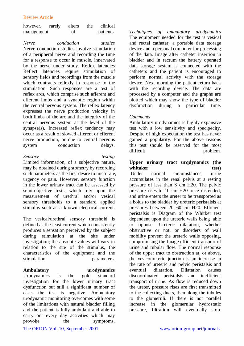
Pressure flow studies involve the perfusion of the kidney with contrast at a known rate, whilst simultaneously measuring the pressure within the renal pelvis and bladder. Significant rises in pressure are indicative of obstruction, whilst free drainage at low pressure excludes obstruction. Technique The Whitaker test is performed in the conscious patient at least 24 hours after insertion of a nephrostomy tube. Bladder pressure is measured via a urethral catheter connected to a transducer. Renal pelvic pressure can be measured through a nephrostomy tube, or through a needle placed in the collecting system at antegrade pyelography. Puncture technique needs to be as good as any jeak for the collecting system degrades the information that pressure studies will provide. Through one arm of a 'V' connector, dilute contrast used at an initial rate of 10 ml/min, whilst the other arm of the 'V' connector is connected to a pressure transducer recording renal pelvic pressure in response to perfusion. Perfusion at 10 ml/min is considered as physiological. The bladder pressure is continuously measured and the subtracted pressure automatically calculated. Such manometric equipment is table in any department performing lower urinary tract dynamics. Simultaneous fluoroscopy defines the anatomy of the bladder tract and spot films can be taken. Practical points Using this technique, a pressure difference between the upper and lower urinary tract of less than 15 cm H20 excludes obstruction. If the renal pelvic pressure exceeds the oladder pressure by more than 12 cm H20, obstruction is confirmed. Pressure differentials between 15 and 22 cm of H20 lie in the equivocal range. If both the bladder and pelvic pressure rise equally
together, vesicoureteric reflux has occurred. Pelvic pressure over 22 cm obstruction is confirmed. Higher rates of perfusion have been advocated, but are of the debatable clinical usefulness. In the patient with a urinary diversion, e.g. ileal loop, loin pain is quite frequent because of the high pressures generated by bowel peristalsis refluxing up the reimplanted ureters. The extent of this can easily be assessed by upper tract urodynamics; Comment The principal value of upper tract urodynamics lies in allowing an accurate objective assessment as to whether there is obstruction to renal drainage. It is an invasive procedure, since a percutaneous nephrostomy tract is required and therefore its use should be reservEId for the case where other investigations, such as excretory urography or isotope renography, have produced equivocal results. Some urodynamic definitions Stable detrusor function During filling the bladder contents increase in volume without a significant corresponding rise in pressure. Normal detrusor contractility Normal voiding occurs by a sustained detrusor contraction, which can be initiated and suppressed voluntarily and results in complete bladder emptying over a normal timespan; the magnitude of the recorded detrusor pressure rise is dependent on the outlet resistance. Overactive detrusor function Involuntary detrusor contractions during bladder filling, either spontaneous or provoked by rapid filling (provocation cystometry), alterations in posture, exercise or coughing. The unstable detrusor is one that is shown objectively to contract either spontaneously or on provocation during the filling phase while the patient is attempting to inhibit micturition. The unstable detrusor may be asymptomatic

Review Article
The ORION Vol. 10, September 2001 www.orion-group.net/journals
and its presence does not necessarily imply a neurological disorder. Detrusor hyper-reflexia Detrusor hyperactivity in the presence of a documented neurological disorder. Low-compliance During normal bladder filling little or no significant rise in pressure occurs so-called normal compliance. Although the term low- compliance is applied to a gradual rise in detrusor pressure during bladder filling and is usually taken to imply a poorly distensible bladder, e.g. a shrunken fibrotic bladder complicating interstitial cystitis or after radiotherapy, it must be remembered that both detrusor instability and hyper-reflexia are forms of low compliance and that, at present, there is insufficient data to define normal, high and low compliance. Underactive (hypocontractile) detrusor function This is taken to be referring to detrusor activity during micturition. The term underactive applies to all situations where a detrusor contraction is inadequate to produce emptying of the bladder. Acontractile detrusor No contractile activity is evident on urodynamic investigation. Areflexic detrusor Acontractility resulting from an abnormality of the central nervous system. A specific type occurs with lesions of the conus medullaris or sacral nerve outflow and is known as a decentralized detrusor where the peripheral ganglia in the wall of the bladder are preserved and peripheral nerves are therefore intact. This subgroup is characterised by involuntary intravesical pressure fluctuations of low amplitude, sometimes called autonomous waves.
Destrusor bladder neck dyssynergia. This refers to a detrusor contraction concurrent with a failure for there to be complete bladder neck opening on micturition. Although rare in the population, it is a common cause of voiding dysfunction in the younger male. In this context it is important to realize that, in recent years, it has been increasingly recognized that an important component of prostatic obstruction results from the contraction of smooth muscle within the pathologically enlarged prostate. Detrusor sphincter dyssynergia (DSD). This describes a detrusor contraction concurrent with an involuntary contraction of the urethral and/or periurethral striated smooth muscle. No similar term has been elaborated for corresponding detrusor/distal urethral smooth muscle dyssynergia. Obstructive overactivity of the striated urethral sphincter muscle may occur in the absence of detrussor contraction, but is not DSD. DSD is usually associated with neurological disorders and, in the absence of a documented neurological deficit, this diagnosis would need to be treated with caution. References I. Emil A. T anagho, Jack W. McAninch; Smith's General Urology; 15th edn.; McGraw Hill 2000. , 2, Walsh, Retik, Vaughan & Wein; Campbell's urology; tth edn; W.B. Saunders Company, 1998, 3. Siroky, Edelstein & Krane; Manual of ur<?logy; 2nd edn. Lippincott Williams & Wilkins, 1999. 4, Gillen Water }Y, Gray hack JT; Adult and Pediatric urology; Mosby 1996.