Outline Clinical Impressions: Diphtheria Diphtheria ... · Diphtheria, Pertussis and Streptococcal...
12
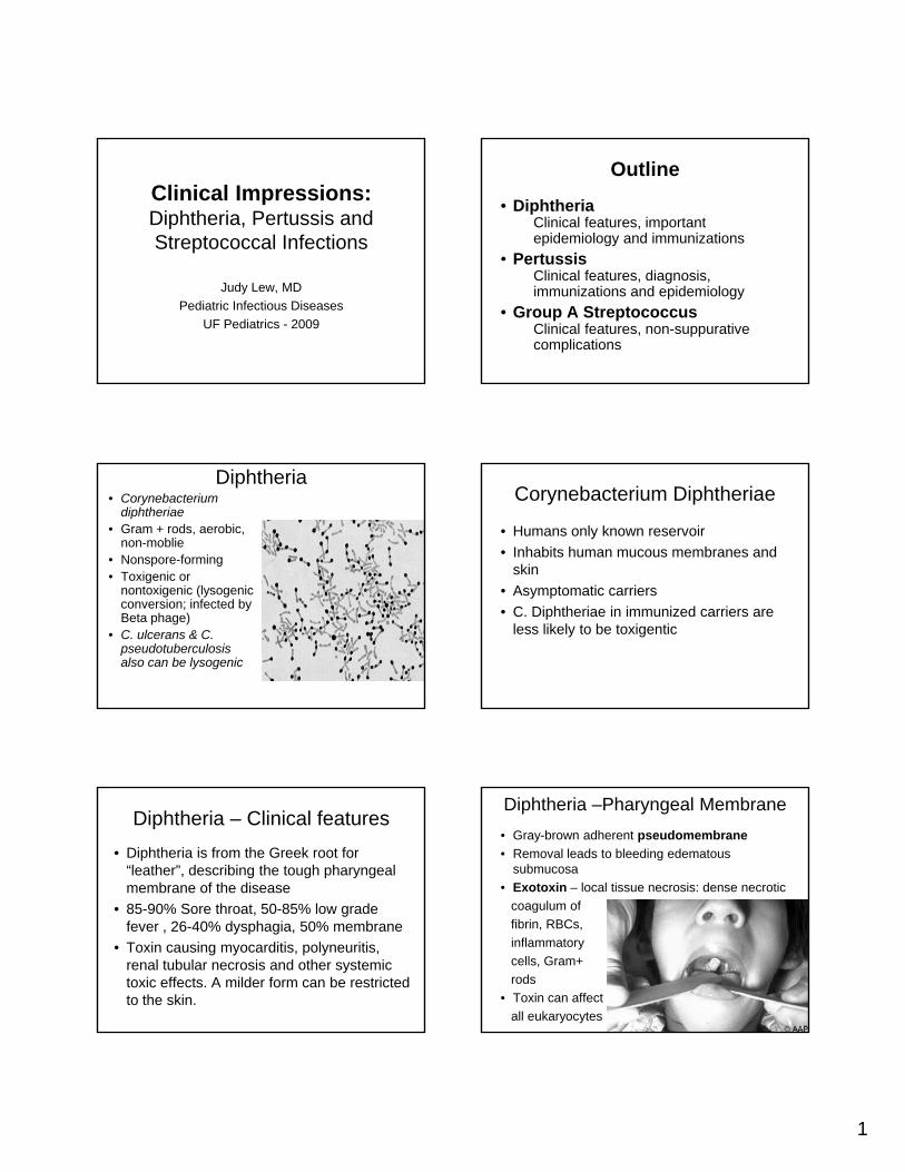
1 Clinical Impressions: Diphtheria, Pertussis and Streptococcal Infections Judy Lew, MD Pediatric Infectious Diseases UF Pediatrics - 2009 Outline • Diphtheria Clinical features, important epidemiology and immunizations • Pertussis Pertussis Clinical features, diagnosis, immunizations and epidemiology • Group A Streptococcus Clinical features, non-suppurative complications Diphtheria • Corynebacterium diphtheriae • Gram + rods, aerobic, non-moblie • Nonspore-forming • Toxigenic or t i i (l i nontoxigenic (lysogenic conversion; infected by Beta phage) • C. ulcerans & C. pseudotuberculosis also can be lysogenic Corynebacterium Diphtheriae • Humans only known reservoir • Inhabits human mucous membranes and skin A t ti i • Asymptomatic carriers • C. Diphtheriae in immunized carriers are less likely to be toxigentic Diphtheria – Clinical features • Diphtheria is from the Greek root for “leather”, describing the tough pharyngeal membrane of the disease • 85-90% Sore throat 50-85% low grade • 85-90% Sore throat, 50-85% low grade fever , 26-40% dysphagia, 50% membrane • Toxin causing myocarditis, polyneuritis, renal tubular necrosis and other systemic toxic effects. A milder form can be restricted to the skin. Diphtheria –Pharyngeal Membrane • Gray-brown adherent pseudomembrane • Removal leads to bleeding edematous submucosa • Exotoxin – local tissue necrosis: dense necrotic coagulum of fibrin, RBCs, inflammatory cells, Gram+ rods • Toxin can affect all eukaryocytes
Transcript of Outline Clinical Impressions: Diphtheria Diphtheria ... · Diphtheria, Pertussis and Streptococcal...
1
Clinical Impressions:Diphtheria, Pertussis and Streptococcal Infections
Judy Lew, MDPediatric Infectious Diseases
UF Pediatrics - 2009
Outline
• DiphtheriaClinical features, important epidemiology and immunizations
• PertussisPertussisClinical features, diagnosis, immunizations and epidemiology
• Group A StreptococcusClinical features, non-suppurativecomplications
Diphtheria• Corynebacterium
diphtheriae• Gram + rods, aerobic,
non-moblie• Nonspore-forming• Toxigenic or
t i i (l inontoxigenic (lysogenic conversion; infected by Beta phage)
• C. ulcerans & C. pseudotuberculosis also can be lysogenic
Corynebacterium Diphtheriae
• Humans only known reservoir• Inhabits human mucous membranes and
skin A t ti i• Asymptomatic carriers
• C. Diphtheriae in immunized carriers are less likely to be toxigentic
Diphtheria – Clinical features• Diphtheria is from the Greek root for
“leather”, describing the tough pharyngeal membrane of the disease
• 85-90% Sore throat 50-85% low grade• 85-90% Sore throat, 50-85% low grade fever , 26-40% dysphagia, 50% membrane
• Toxin causing myocarditis, polyneuritis, renal tubular necrosis and other systemic toxic effects. A milder form can be restricted to the skin.
Diphtheria –Pharyngeal Membrane• Gray-brown adherent pseudomembrane• Removal leads to bleeding edematous
submucosa• Exotoxin – local tissue necrosis: dense necrotic
coagulum ofgfibrin, RBCs, inflammatorycells, Gram+rods
• Toxin can affect all eukaryocytes
Dad
Typewritten Text
October 27, 2009, 11:00
2
Diphtheria – Membrane Formation• “Strangling Angel of Children”• Membrane + edema can cause airway obstruction• Most common cause of death is suffocation due
aspiration of themembranemembrane.
• 2/3 with carditis,10-25% clinicaldysfunction
• Neurotoxicity ishigh in severedisease
Diphtheria – “Bull Neck”• Fatality rate 5 – 10%, but in <5 or >40 year
olds, could be 20%.• 50-60% morality due to suffocation or
cardiac failure L h d iti & d• Lymphadenitis & and edema
• Paralysis of thepalatal muscles
• Larynx
“Bull Neck”• Nasopharyngeal and pharyngeal swab for
culture• Selective media
Loeffler, Tinsdale,with tellurite
T t t ith• Treatment withantibiotics (PCN,EES) andantitoxin
• Early recognition anddiagnosis
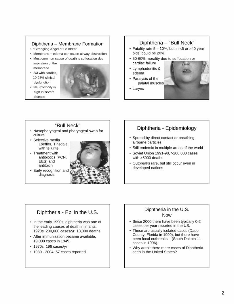
Diphtheria - Epidemiology
• Spread by direct contact or breathing airborne particles
• Still endemic in multiple areas of the worldS i t U i 1991 98 200 000• Soviet Union 1991-98, >200,000 cases with >5000 deaths
• Outbreaks rare, but still occur even in developed nations
Diphtheria - Epi in the U.S.
• In the early 1990s, diphtheria was one of the leading causes of death in infants; 1920s: 200,000 cases/yr, 13,000 deaths.
• After immunization became available• After immunization became available, 19,000 cases in 1945.
• 1970s, 196 cases/yr • 1980 - 2004: 57 cases reported
Diphtheria in the U.S.Now
• Since 2000 there have been typically 0-2 cases per year reported in the US.
• These are usually isolated cases (Dade County, Florida in 1990), but there have y, ),been focal outbreaks – (South Dakota 11 cases in 1996).
• Why aren’t there more cases of Diphtheria seen in the United States?
3
ImmunizationDiphtheria Toxoid-containing Vaccine
• Primary series – 2, 4, 6 months of ageDTaP – Diphtheria, tetanus, andacellular Pertussis
• Boosters at 15 18 months and 4 6 years• Boosters - at 15-18 months and 4-6 yearsDTaP
• Boosters at 11 years of age and every 10 years – Td or Tdap – reduced doses of diphtheria toxoid and acellular pertussis
The spike between 1993 and 1997, is attributable to a drop in vaccine coverage in new Independent States of the former Soviet Union. WHO.
Reasons for Dramatic Diphtheria Decline Unclear
Immunization expected to prevent symptoms of toxoid production, not colonization
• Historical evidence suggests epidemics in cycles with gaps of >100 years
• Immunization could counter hypothesized colonizing advantage of lysogentic strains
• Other unknown virulence factors
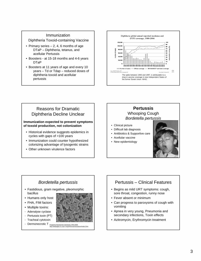
PertussisWhooping Cough
Bordetella pertussis• Clinical picture• Difficult lab diagnosisg• Antibiotics & Supportive care• Acellular vaccine• New epidemiology
Bordetella pertussis• Fastidious, gram negative, pleomorphic
bacillus• Humans only host• FHA FIM factors• FHA, FIM factors• Multiple toxins:- Adenalyse cyclase- Pertussis toxin (PT)- Tracheal cytotoxin- Dermonecrotic T Programa Nacional de Vacinacao, PNV-2006
http://webpages.fc.ul.pt/~mcgomes/vacinacao/pnv/index3.htm
Pertussis – Clinical Features• Begins as mild URT symptoms: cough,
sore throat, congestion, runny nose• Fever absent or minimum• Can progress to paroxysms of cough with• Can progress to paroxysms of cough with
vomiting• Apnea in very young, Pneumonia and
secondary infections, Toxin effects• Azitromycin, Erythromycin treatment
4
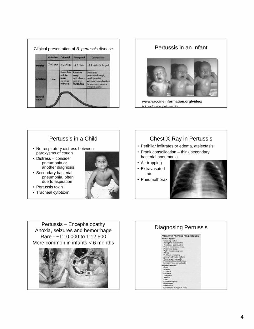
Clinical presentation of B. pertussis disease Pertussis in an Infant
www.vaccineinformation.org/video/look here for some good video clips
Pertussis in a Child
• No respiratory distress between paroxysms of cough
• Distress – considerpneumonia or panother diagnosis
• Secondary bacterialpneumonia, oftendue to aspiration
• Pertussis toxin• Tracheal cytotoxin
Chest X-Ray in Pertussis• Perihilar infiltrates or edema, atelectasis• Frank consolidation – think secondary
bacterial pneumonia• Air trappingpp g• Extravasated
air• Pneumothorax
Pertussis – EncephalopathyAnoxia, seizures and hemorrhage
Rare - ~1:10,000 to 1:12,500More common in infants < 6 months
Diagnosing Pertussis
5
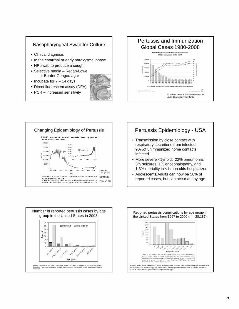
Nasopharyngeal Swab for Culture
• Clinical diagnosis• In the catarrhal or early paroxysmal phase• NP swab to produce a cough• Selective media – Regan-Lowe
or Bordet-Gengou agar• Incubate for 7 – 14 days• Direct fluorescent assay (DFA)• PCR – increased sensitivity
Pertussis and ImmunizationGlobal Cases 1980-2008
50 million cases & 300,000 deaths / YR Up to 3% mortality in infants
Changing Epidemiology of Pertussis
MMWR 12/15/2006
55(RR17)
Pages 1-33
Pertussis Epidemiology - USA
• Transmission by close contact with respiratory secretions from infected; 90%of unimmunized home contacts infectedinfected
• More severe <1yr old: 22% pneumonia, 3% seizures, 1% encephalopathy, and 1.3% mortality in <1 mon olds hospitalized
• Adolescents/Adults can now be 50% of reported cases, but can occur at any age
Number of reported pertussis cases by age group in the United States in 2003.
Adapted from Hopkins RS, Jajosky RA, Hall PA, Adams DA, Connor FJ, Sharp P, et al. Centers for Disease Control and Prevention. Summary of notifiable diseases-United States, 2003. MMWR Morb Mortal Wkly Rep 2005;52:55.
Reported pertussis complications by age group in the United States from 1997 to 2000 (n = 28,187).
Adapted from Centers for Disease Control and Prevention. National Immunization Program. Pertussis and pertussis vaccine. Epidemiology and prevention of vaccine-preventable diseases. Accessed August 30, 2005, at: http://www.cdc.gov/nip/ed/slides/pertussis8p.ppt.
6
Reasons for Pertussis Increase
• Decreasing immunization of the young• Waning immunity in adolescents/adults• Atypical presentation in older patientsAtypical presentation in older patients• Possible carrier state even with
immunization
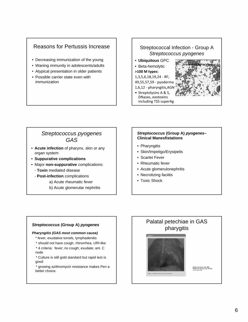
• Ubiquitous GPC • Beta-hemolytic>100 M types:
Streptococcal Infection - Group AStreptococcus pyogenes
>100 M types:1,3,5,6,18,19,24 ‐ RF;49,55,57,59 ‐ pyoderma1,6,12 ‐ pharyngitis,AGN• Streptolysins A & S, DNases, exotoxins including TSS superAg
Streptococcus pyogenesGAS
• Acute infection of pharynx, skin or any organ system
• Suppurative complications• Major non-suppurative complications:
- Toxin mediated disease- Post-infection complications
a) Acute rheumatic feverb) Acute glomerular nephritis
Streptococcus (Group A) pyogenes–Clinical Manesfistations
• Pharyngitis• Skin/Impetigo/Erysipelis• Scarlet Fever• Rheumatic fever• Acute glomerulonephritis• Necrotizing faciitis• Toxic Shock
Streptococcus (Group A) pyogenes
Pharyngitis (GAS most common cause)* fever, exudative tonsils, lymphadenitis* should not have cough, rhinorrhea, URI-like* 4 criteria: fever; no cough; exudate; ant. C node* Culture is still gold standard but rapid test is good* growing azithromycin resistance makes Pen a better choice.
Palatal petechiae in GAS pharygitis
William Gluckman, DO, MBA, FACEP and Jessica Kay, PharmDJUCM Sept 2008
7
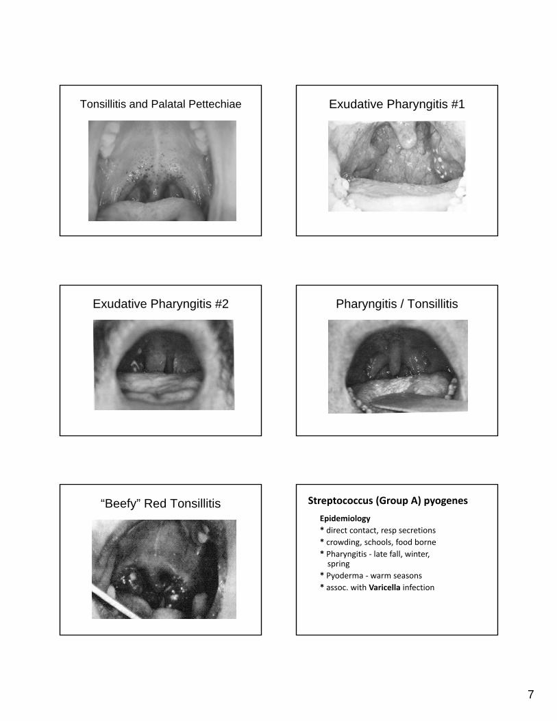
Tonsillitis and Palatal Pettechiae Exudative Pharyngitis #1
Exudative Pharyngitis #2 Pharyngitis / Tonsillitis
“Beefy” Red Tonsillitis Streptococcus (Group A) pyogenes
Epidemiology* direct contact, resp secretions* crowding, schools, food borne* Pharyngitis ‐ late fall, winter, spring
* Pyoderma ‐ warm seasons* assoc. with Varicella infection
8
Streptococcus (Group A) pyogenesDiagnosis* GPC in pairs, chains; catalase negative* beta‐hemolytic in blood agar* Rapid* CultureTreatment* Pen V 2‐3x/d for 10d, Erythomycin x10d, narrow‐spectrum cephalos x10d
Streptococcus (Group A) pyogenes
Suppurative COMPLICATIONS:
•Peritonsilar abscess•Cervical lympanenpathy•Empyema•Osteomyelitis, septic arthritis, endocarditis, or any body site.
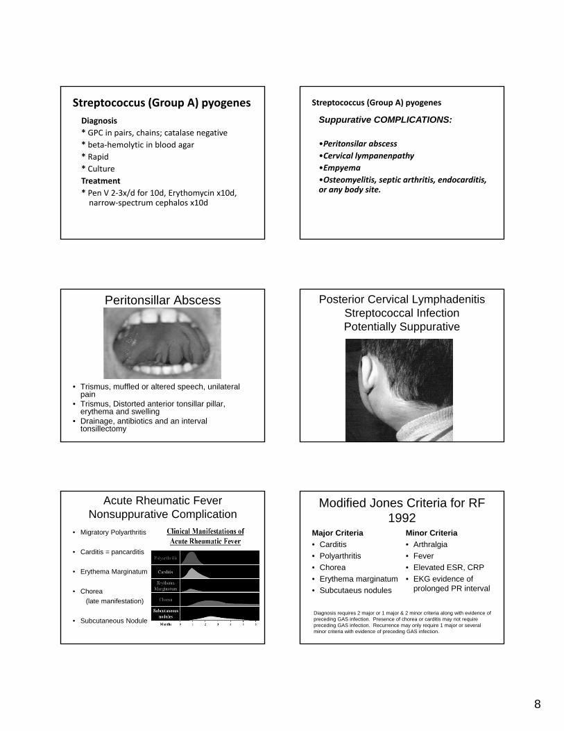
Peritonsillar Abscess
• Trismus, muffled or altered speech, unilateral pain
• Trismus, Distorted anterior tonsillar pillar, erythema and swelling
• Drainage, antibiotics and an interval tonsillectomy
Posterior Cervical LymphadenitisStreptococcal InfectionPotentially Suppurative
Acute Rheumatic FeverNonsuppurative Complication
• Migratory Polyarthritis
• Carditis = pancarditis
• Erythema Marginatum
• Chorea (late manifestation)
• Subcutaneous Nodule
Modified Jones Criteria for RF 1992
Major Criteria• Carditis• Polyarthritis• Chorea
Minor Criteria• Arthralgia• Fever• Elevated ESR CRP• Chorea
• Erythema marginatum• Subcutaeus nodules
• Elevated ESR, CRP• EKG evidence of
prolonged PR interval
Diagnosis requires 2 major or 1 major & 2 minor criteria along with evidence of preceding GAS infection. Presence of chorea or carditis may not require preceding GAS infection. Recurrence may only require 1 major or several minor criteria with evidence of preceding GAS infection.
9
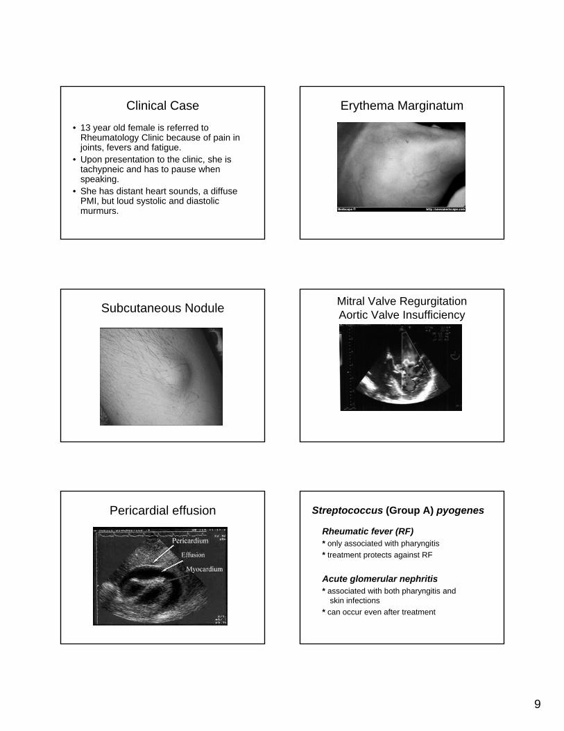
Clinical Case
• 13 year old female is referred to Rheumatology Clinic because of pain in joints, fevers and fatigue.
• Upon presentation to the clinic, she is p p ,tachypneic and has to pause when speaking.
• She has distant heart sounds, a diffuse PMI, but loud systolic and diastolic murmurs.
Erythema Marginatum
Subcutaneous Nodule Mitral Valve RegurgitationAortic Valve Insufficiency
Pericardial effusion Streptococcus (Group A) pyogenes
Rheumatic fever (RF)* only associated with pharyngitis* treatment protects against RF
Acute glomerular nephritis* associated with both pharyngitis and
skin infections* can occur even after treatment
10
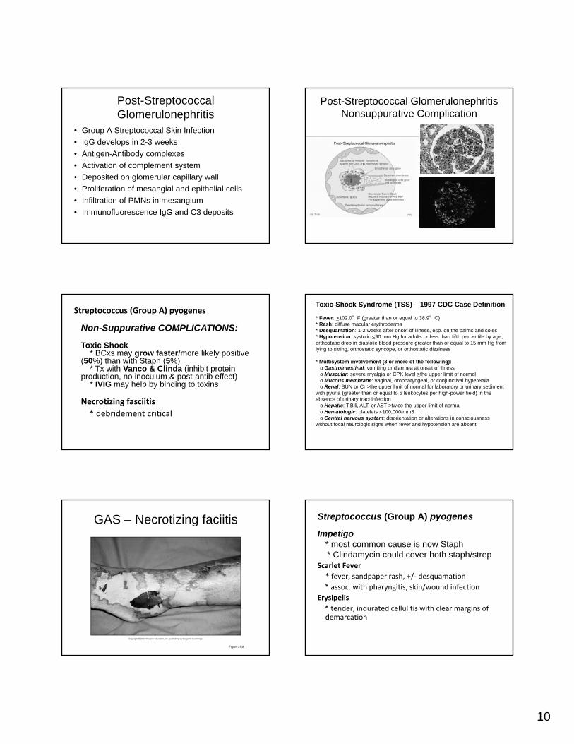
Post-Streptococcal Glomerulonephritis
• Group A Streptococcal Skin Infection• IgG develops in 2-3 weeks• Antigen-Antibody complexes• Activation of complement system• Activation of complement system• Deposited on glomerular capillary wall• Proliferation of mesangial and epithelial cells• Infiltration of PMNs in mesangium• Immunofluorescence IgG and C3 deposits
Post-Streptococcal GlomerulonephritisNonsuppurative Complication
Streptococcus (Group A) pyogenes
Non-Suppurative COMPLICATIONS:
Toxic Shock* BCxs may grow faster/more likely positive
(50%) than with Staph (5%)* Tx with Vanco & Clinda (inhibit protein ( p
production, no inoculum & post-antib effect)* IVIG may help by binding to toxins
Necrotizing fasciitis* debridement critical
Toxic-Shock Syndrome (TSS) – 1997 CDC Case Definition
* Fever: >102.0°F (greater than or equal to 38.9°C)* Rash: diffuse macular erythroderma* Desquamation: 1-2 weeks after onset of illness, esp. on the palms and soles* Hypotension: systolic <90 mm Hg for adults or less than fifth percentile by age; orthostatic drop in diastolic blood pressure greater than or equal to 15 mm Hg from lying to sitting, orthostatic syncope, or orthostatic dizziness
* Multisystem involvement (3 or more of the following):o Gastrointestinal: vomiting or diarrhea at onset of illness o Muscular: severe myalgia or CPK level >the upper limit of normalo Mucous membrane: vaginal, oropharyngeal, or conjunctival hyperemiao Renal: BUN or Cr >the upper limit of normal for laboratory or urinary sediment
with pyuria (greater than or equal to 5 leukocytes per high-power field) in the absence of urinary tract infection
o Hepatic: T.Bili, ALT, or AST >twice the upper limit of normal o Hematologic: platelets <100,000/mm3o Central nervous system: disorientation or alterations in consciousness
without focal neurologic signs when fever and hypotension are absent
GAS – Necrotizing faciitis Streptococcus (Group A) pyogenes
Impetigo* most common cause is now Staph* Clindamycin could cover both staph/strep
Scarlet Fever* fever sandpaper rash +/‐ desquamation fever, sandpaper rash, +/ desquamation* assoc. with pharyngitis, skin/wound infection
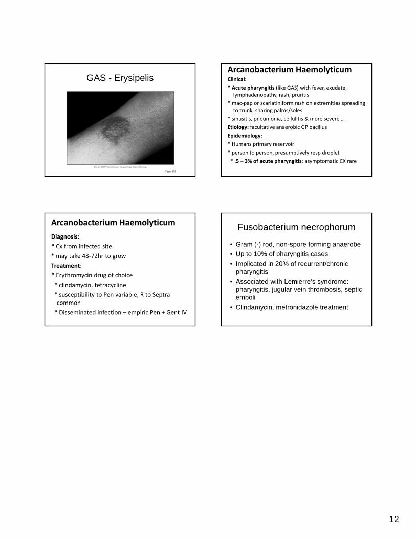
Erysipelis* tender, indurated cellulitis with clear margins of demarcation
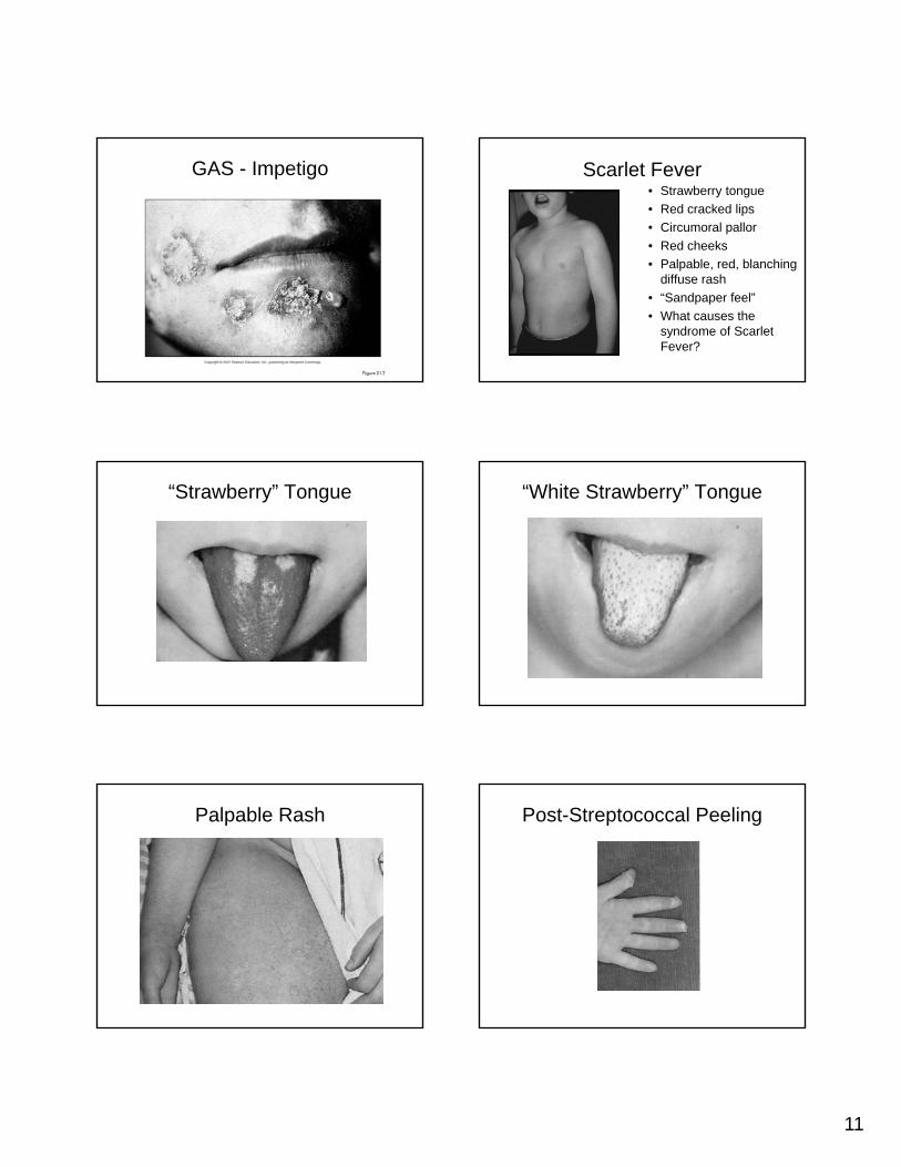
11
GAS - Impetigo Scarlet Fever• Strawberry tongue• Red cracked lips• Circumoral pallor• Red cheeks
P l bl d bl hi• Palpable, red, blanching diffuse rash
• “Sandpaper feel”• What causes the
syndrome of Scarlet Fever?
“Strawberry” Tongue “White Strawberry” Tongue
Palpable Rash Post-Streptococcal Peeling
12
GAS - ErysipelisArcanobacterium HaemolyticumClinical:
* Acute pharyngitis (like GAS) with fever, exudate, lymphadenopathy, rash, pruritis
*mac‐pap or scarlatiniform rash on extremities spreading to trunk, sharing palms/soles
* sinusitis, pneumonia, cellulitis & more severe … sinusitis, pneumonia, cellulitis & more severe …
Etiology: facultative anaerobic GP bacillus
Epidemiology:
* Humans primary reservoir
* person to person, presumptively resp droplet
* .5 – 3% of acute pharyngitis; asymptomatic CX rare
Arcanobacterium Haemolyticum
Diagnosis:
* Cx from infected site
* may take 48‐72hr to grow
Treatment:
* Erythromycin drug of choice
* clindamycin, tetracycline
* susceptibility to Pen variable, R to Septra common
* Disseminated infection – empiric Pen + Gent IV
Fusobacterium necrophorum
• Gram (-) rod, non-spore forming anaerobe• Up to 10% of pharyngitis cases• Implicated in 20% of recurrent/chronic
h itipharyngitis• Associated with Lemierre’s syndrome:
pharyngitis, jugular vein thrombosis, septic emboli
• Clindamycin, metronidazole treatment