Our Facilities We have modern laboratories which are equipped to meet the requirements of a wide...
30
Our Facilities We have modern laboratories which are equipped to meet the requirements of a wide range of experimental research. (But, we still lack a lot !!) - Polymer synthesis laboratory - Polymer characterization laboratory - Cell-culture laboratory - Imaging laboratory - Nanofiltration
-
date post
19-Dec-2015 -
Category
Documents
-
view
212 -
download
0
Transcript of Our Facilities We have modern laboratories which are equipped to meet the requirements of a wide...

Our Facilities
We have modern laboratories which are equipped to meet the requirements of a wide range of experimental research. (But, we still lack a lot !!)
- Polymer synthesis laboratory
- Polymer characterization laboratory
- Cell-culture laboratory
- Imaging laboratory
- Nanofiltration

•Biomaterials/tissue engBiomaterials/tissue eng
•Drug/DNA/RNA deliveryDrug/DNA/RNA delivery
•NanomedicineNanomedicine
•BiosensorsBiosensors
•BiochipsBiochips
•ImagingImaging
•Biocatalyst DevelopmentBiocatalyst Development
•Bioseparations Bioseparations (with emphasis on (with emphasis on
Nanotechniques)Nanotechniques)
Our Research TopicsInvolving NanotechnologyInvolving Nanotechnology

Biomaterials & Tissue
engineering
Microporous Biodegradable PLLA-CL Scaffolds
prepared in supercritical CO2
Microporous Biodegradable
Electruspun PCL Scaffolds

NANOFIBROUS MATERIALSBy Electrospinning
Electrospinning is a unique method
that produces polymeric nanofibers
with diameter in the range of nano to
a few microns using electrically
driven jet of polymer
solutions/melts

Composites for Hard Tissue RepairPoly(DL-Lactide/ε-caprolactone) with nano-size TCP
precipitate
HEAT + NH4OHcentrifugation
TCP gel
Dring and Sintering
H3PO4
Ca(OH)2
Aim: To prepare a hard
tissue filling material that is
biodegradable, easily
applied
Average particle size;
Gel form: 300 nm
Dry form: 10 nm

DRUG/DNA/RNA DRUG/DNA/RNA DELIVERY DELIVERY
At 28ºC28ºCGene Expression
Efficiency: 50%50%

Imaging at Nanoscale
SCANNING PROBE MICROSCOPIESSCANNING PROBE MICROSCOPIES
• Scanning Tunnel Microscopy• Atomic Force Microscopy• SPR Microscopy• Imaging Ellipsometry
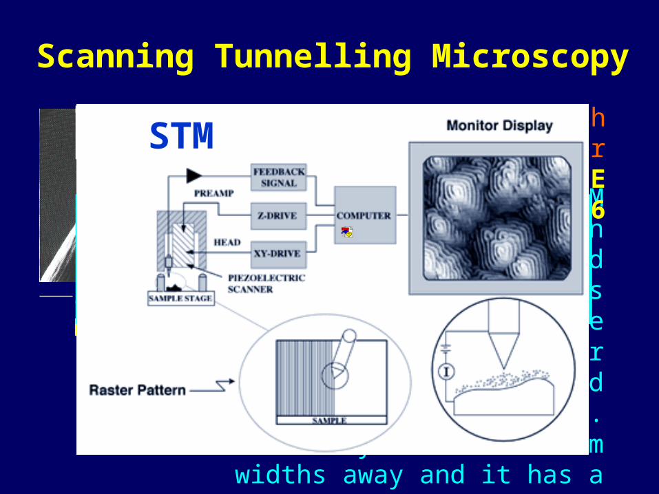
Binning and Rohrer, IBM researcher’s in Switzerland described the STM in 1981. It is essentially a superfine stylus that sweeps over a
surface like a blind person’s walking stick. The stylus is
few atom widths away and it has a molecularly or
perhaps atomically fine tip.
5 µ
Gerd Binnig & Heinrich Rohrer NOBEL PRIZE in
1986ONLY Conducting surfaces and
molecular monolayers
High resolution straightforward
Scanning Tunnelling Microscopy
STM
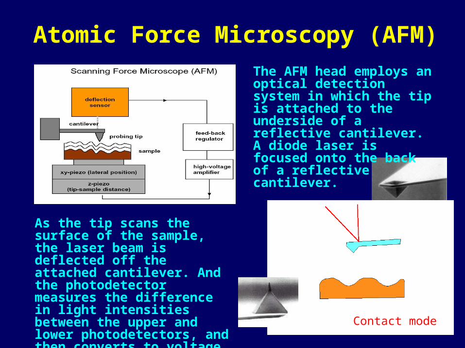
Atomic Force Microscopy (AFM)
Contact mode
The AFM head employs an optical detection system in which the tip is attached to the underside of a reflective cantilever. A diode laser is focused onto the back of a reflective cantilever.
As the tip scans the surface of the sample, the laser beam is deflected off the attached cantilever. And the photodetector measures the difference in light intensities between the upper and lower photodetectors, and then converts to voltage.

ATOMIC FORCE MICROSCOPY (AFM)

DNADNA CromosomeCromosome
AntibodyAntibodyErythrocytesErythrocytes
ATOMIC FORCE MICROSCOPY (AFM)

SPSPRR
SPR Microscope
Θ
Θ
L1
L2
L3
L4
HeNeLaser,
1.5 mW
Polarizer
4 Waveplate
Filter
CCD Camera
Stages
PC/Framegrabber
Stage ControllerSubstrate
Prism
FlowCell
Following: W. Knoll, Colloid Polym. Sci. 1988, 109, 244
Ref
lect
ion
Inte
nsit
y(%
)Angle of Monitoring
I II0
100

Imaging Ellipsometry
Substrate
Analyzer
CCD Camera
Objective
Laser Polarizer
CompensatorSample (e.g. film)
Dextran on silicone
Dye monolayerPatterned surface

FORCE MEASUREMENTS by using Modified AFM Tips

SIGNAL
AN
AL
YT
E
TRANSDUCERTRANSDUCERBIORECEPTORBIORECEPTOR
The bioreceptor is a biomolecule that recognizes the target analyte. The transducer converts the recognition
event into a measurable signal.
EnzymeAntibodyReceptorDNA
ElectrochemicalOpticalPiezoelectric
BIOSENSORBIOSENSORSS

Quartz crystal plate
Au electrodes
Contact
UNIVERSAL SENSORS Inc.
QUARTZ CRYSTAL MICROBALANCE
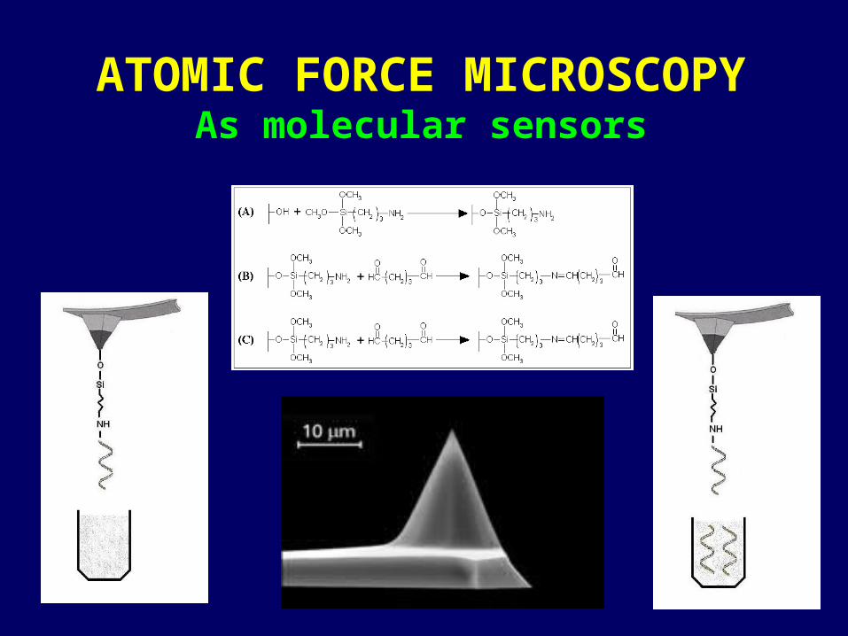
ATOMIC FORCE MICROSCOPYAs molecular sensors

ATOMIC FORCE MICROSCOPYAs molecular sensors
Representative fluorescence
microscopy images: (A) and (B) before
ssDNA immobilization; (C)
and (D) carrying the immobilized
ligand ssDNA

MAGNETICALLY LOADED NANOPARTICLES
Nanoparticles take on special properties because of their small size.
Nanoparticles can be use as carriers or labels with a variety of markers, transported through various media, and interfaced both in vivo and in vitro.
Nanoparticle technology over the last decade plays an important role in the diagnosis and treatment of cancer.
As reporter platformsAs contrast enhancing agents
Early disease detection Imaging Drug delivery As a vehicle
Biomedical Applications

Magnetic and Superparamagnetic Particles
– Colloid size: 1-5 µm
– Magnetite (Fe3O4), Maghemite (γ- Fe2O3)
Colloidal Magnetic Particles– Colloid size: 50-500 nm– Ferrofluids
MAGNETIC NANOPARTICLES
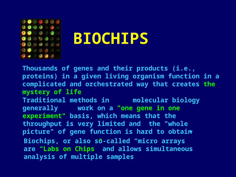
BIOCHIPS
Thousands of genes and their products (i.e., proteins) in a given living organism function in a complicated and orchestrated way that creates the mystery of life
Traditional methods in molecular biology generally work on a "one gene in one experiment" basis, which means that the throughput is very limited and the "whole picture" of gene function is hard to obtain
Biochips, or also so-called “micro arrays” are “Labs on Chips” and allows simultaneous analysis of multiple samples

DNA-CHIPS

PROTEIN-CHIPS

SURFACE PLASMON SURFACE PLASMON RESONANCE RESONANCE
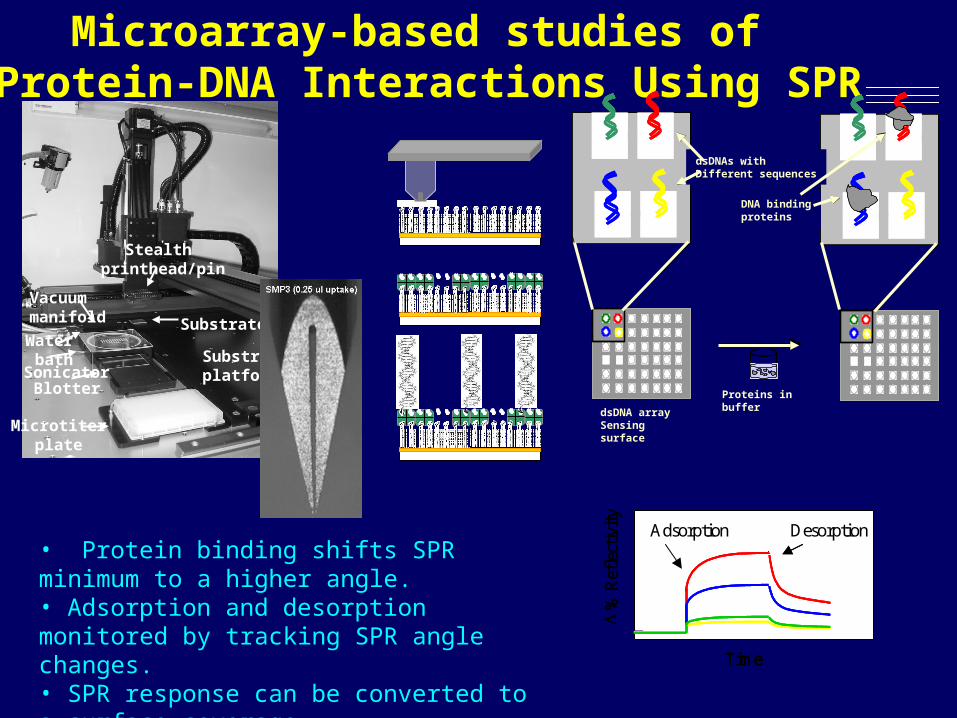
• Protein binding shifts SPR minimum to a higher angle. • Adsorption and desorption monitored by tracking SPR angle changes.• SPR response can be converted to a surface coverage.
%
Ref
lect
ivit
y
Adsorption Desorption
Time
%
Adsorption Desorption
Time
%
Ref
lect
ivit
y
Adsorption Desorption
Time
%
Adsorption Desorption
Time
Microarray-based studies of Protein-DNA Interactions Using SPR
Microtiter plate
BlotterSonicator
Water bath Substrate
platform
Stealth printhead/pin
Substrate
Vacuummanifold
Proteins in bufferdsDNA array sensing surface
dsDNAs withdifferent sequences
DNA-binding proteins
Proteins in bufferdsDNA array sensing surface
Proteins in bufferdsDNA array sensing surface
dsDNAs withdifferent sequences
DNA-binding proteins
dsDNAs with Different sequences
DNA bindingproteins
dsDNA arraySensing surface
Proteins in buffer
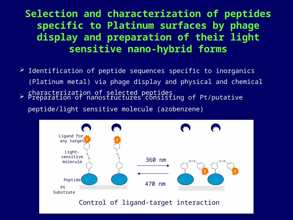
Selection and characterization of peptides specific to Platinum surfaces by phage display and preparation of
their light sensitive nano-hybrid forms
Identification of peptide sequences specific to inorganics (Platinum metal)
via phage display and physical and chemical characterization of selected
peptides Preparation of nanostructures consisting of Pt/putative peptide/light sensitive
molecule (azobenzene)
N
N
N
N
N N N N
Pt Substrate
Peptide
Light-sensitive molecule
Ligand for any target
360 nm
470 nm
Control of ligand-target interaction

Statistical analysis
Alignment (Clustal W)
Charge, (http://us.expasy.org/tools/pi),
Hydrophilicity
Fluorescence microscopy
Cross-specificity with powders and arrays
Binding characteristics with QCM, SPR
Characterization of selected peptides
I. Peptide selection via phage display
SD152PTSTGQA

Characterization:
Functional Grup analysis (FTIR, 1H-NMR, 13C-NMR)
Ninhydrin assay,
Absorption behaviour,
Binding study with QCM ( ka, kd)
Contact angle, elipsometer, AFM on
Platinum surfaces
b) Coupling of peptide to azobenzene molecule
ğ
-Δf (Hz)
time (s)

GENE THERAPY
AIM: To avoid restenosis by Gene Therapy
Proliferation of smooth Proliferation of smooth muscle cells are stopped by muscle cells are stopped by blocking the MMP-2 blocking the MMP-2 enzyme.enzyme.
For this purpose, inhibitor For this purpose, inhibitor gene is transfected to the gene is transfected to the smooth muscle cells.smooth muscle cells.
60x 103 smc/ well
10 g plasmid in NaCI 100 µl (1mg/ml) polymer pH 5.95

GENE EXPRESSION OF SMOOTH MUSCLE CELLS
SMC’s transfected with Poly(NIPA-co-MAH)/plasmid DNA complex (e) light microscope image (f) Fluorescence microscope image.
e fa b