OTOLARYNGOLOGY - Openventio. Imaging of Paranasal Sinus Mucoceles – C. S. Vanaja* and Khandelwal...
26
PUBLISHERS OTOLARYNGOLOGY ISSN 2470-4059 www.openventio.org Editor-in-Chief Arianna Di Stadio, PhD, MD Associate Editors Mohsen Naraghi, MD Mustafa Celik, PhD Sydney Correia Leao, MD Open Journal | June 2016 | Volume 2 | Issue 3 |
Transcript of OTOLARYNGOLOGY - Openventio. Imaging of Paranasal Sinus Mucoceles – C. S. Vanaja* and Khandelwal...
PUBLISHERS
OTOLARYNGOLOGY
ISSN 2470-4059
www.openventio.org
Editor-in-ChiefArianna Di Stadio, PhD, MD
Associate EditorsMohsen Naraghi, MDMustafa Celik, PhD
Sydney Correia Leao, MD
Open Journal
| June 2016 | Volume 2 | Issue 3 |
OTOLARYNGOLOGY
Open Journal
Otolaryngol Open J
ISSN 2470-4059
– Yakup Yegin, Mustafa Çelik*, Baver Maşallah Şimşek, Burak Olgun, Aylin Karahasanoğlu, Ceylan Çolak and Fatma Tülin Kayhan
2. Cortical Auditory Evoked Potentials in Persons Using Hearing Aids
3. The Relationship Between the Degree of the Mastoid Pneumatization and Mean Platelet Volume
4. Rare Localization of Lymphoma
5. Imaging of Paranasal Sinus Mucoceles
– C. S. Vanaja* and Khandelwal Nimisha
– Marrakchi J*, Zhani A, Chahed H, Nefzaoui S, Ben Amor M, Beltaif N, Kchir N and Besbes G
– Jihene Marrakchi*, Alya Zhani, Houda Chahed, Safa Nefzaoui, Mohamed Ben Amor, Najeh Beltaief, Nidhameddine Kchir and Ghazi Besbes
Case Report
Research
80-86
87-90
91-93
94-100
Table of Contents
1. Clear Cell Myoepithelioma - A Rare Presentation in Nasal Cavity – Arpit Saxena*, A. V. Ramesh, Poonam Raj Mehra, Nikhilesh Kumar and Richa Ranjan
Case Report
Research
Research
77-79

OTOLARYNGOLOGY
Open Journalhttp://dx.doi.org/10.17140/OTLOJ-2-117
Otolaryngol Open J
ISSN 2470-4059
Arpit Saxena, MS1*; A. V. Ramesh, MS1; Poonam Raj Mehra, MS1; Nikhilesh Kumar, MD2; Richa Ranjan, MD2
1Department of ENT, Command Hospital (CC), Lucknow, UP, India2Department of Pathology, Command Hospital (CC), Lucknow, UP, India
*Corresponding author Arpit Saxena, MS Senior Resident Department of ENT Command Hospital (CC) Room No. 204, SR Hostel DTH, Saifai Etawa, Lucknow 206130 UP, India Tel. +919532306179 E-mail: [email protected]
Article HistoryReceived: May 9th, 2016 Accepted: May 23rd, 2016 Published: May 23rd, 2016
CitationSaxena A, Ramesh AV, Raj Mehra P, Kumar N, Ranjan R. Clear cell myoepi-thelioma - a rare presentation in nasal cavity. Otolaryngol Open J. 2016; 2(3): 77-79. doi: 10.17140/OTLOJ-2-117
Copyright©2016 Saxena A. This is an open access article distributed under the Creative Commons Attribution 4.0 International License (CC BY 4.0), which permits unrestricted use, distribution, and reproduction in any medium, provided the original work is properly cited.
Volume 2 : Issue 3Article Ref. #: 1000OTLOJ2117
Clear Cell Myoepithelioma - A Rare Presentation in Nasal Cavity
Page 77
Case Report
ABSTRACT
Myoepitheliomas are predominantly tumors of salivary glands constituting less than 1% of all salivary gland tumors. A 65 year old women presented with bleeding unilateral mass for 03 months. Contrast Enhanced Computed Tomography (CECT) revealed a heterogeneously enhancing mass lesion arising from right anterior ethmoid air cells and filling the right nasal cavity entirely. Endoscope guided endoscopic biopsy was performed. Histopathological exami-nation showed a well circumscribed tumour arranged in small glands and sheets. Tumor cell were immunopositive for S100 (Ib), Cytokeratin (CK) and Vimetin (focally ) while negative for chromogranin, synaptophysin, CD 10 and Smooth Muscle Actin (SMA). MIB-Labelling Index was <5%. With these features a diagnosis of clear cell myoepithelioma was made. Nasal myo-epithelioma is an extremely rare low-grade neoplasm. Nasal myoepitheliomas are composed of myoepithelial cells with solid, myxoid or reticular patterns of growth. The cells themselves may be clear-cell type, spindle-shaped, plasmacytoid and epithetlioid. In all reported cases of myoepithelioma, surgery was the mainstay treatment. Partial maxillectomy via a lateral rhinot-omy approach, Caldwell-Luc procedure has been recommended for patients who were suspect-ed to have a low-grade sarcomatous neoplasm. Only five cases have been reported in sinonasal region. We report the second case to be managed endoscopically. We report an extremely rare benign tumor of nasal cavity (clear cell myoepithelioma) which was managed endoscopically with recurrence within 6 months. The recurrence was also managed endoscopically. This case highlights the varied malignancies which may be encountered in sinonasal region.
KEYWORDS: Myoepithelioma; Clear cell tumor; Endoscopic sinus surgery.
INTRODUCTION
Myoepitheliomas are predominantly tumors of salivary glands constituting less than 1% of all salivary gland tumors.1 Only five cases of sinonasal myoepithelioma have been reported in literature till today’s date. We present the first case of clear cell sub type of myoepithelioma in nose and the second case to be managed endoscopically.
CASE REPORT
A 65 years old lady presented with complains of intermittent bleeding for 3 months and nasal obstruction on the right side for 2 months. There were no known comorbidities. On examina-tion patient had a bulge over right lateral wall of nose seen externally. Anterior rhinoscopy revealed a fleshy smooth mass filling right nasal cavity. No blood clots or secretions were seen. Probing revealed the mass to be painless, soft in consistency, arising from right lateral wall of nose and bleeding on touch. Posterior rhinoscopy revealed normal nasopharynx. Contrast Enhanced Computed Tomography (CECT) revealed a heterogeneously enhancing mass lesion arising from right anterior ethmoid air cells and filling the right nasal cavity entirely (Figure 1). No features of bone erosion were seen. Hematolgical and biochemical parameters of pa-tient were normal. It was planned an endoscope guided excision biopsy of mass. Intraopera-

OTOLARYNGOLOGY
Open Journalhttp://dx.doi.org/10.17140/OTLOJ-2-117
Otolaryngol Open J
ISSN 2470-4059
Page 78
tive, mass was seen arising from sphenoethmoidal recess. Brisk bleeding was encountered coming from sphenopalatine artery which was controlled with bipolar cautery. Post op uneventful.
Histopathological examination showed a well circum-scribed tumour arranged in small glands and sheets. The indi-vidual cells were bland with clear cytoplasm. No layering was seen. Mitosis was scant and no necrosis was seen. Few dead bony spicules were noted (Figure 2). Special stains revealed gly-cogen in clear cells. Tumor cell were immunopositive for S100 (Ib), Cytokeratin (CK) and Vimetin (focally ) while negative for chromogranin, synaptophysin, CD 10 and Smooth Muscle Actin (SMA). MIB-Labelling Index was <5%. With these features a diagnosis of clear cell myoepithelioma was made.
Patient was kept on follow up with no further episodes of epistaxis. Endoscopy after 6 months showed fleshy mass aris-ing from middle meatus. Repeat tomography was done which showed recurrent mass lesion arising from right anterior ethmoid air cells. Endoscopic excision of middle turbinate and anterior ethmoid air cells was done and specimen sent for histopathology which also showed clear cell myoepithelioma with no evidence of malignant transformation. There has been no further recur-rence during a follow up period of 1year after second surgery.
DISCUSSION
Primary pleomorphic adenomas of the nasal cavity constitute around 18% of sinonasal nonepithelial neoplasms.2,3 Nasal myo-epithelioma is an extremely rare low-grade neoplasm. The main symptoms of nasal myoepithelioma are rapid enlargement of the tumor mass with nasal obstruction and epistaxis for periods varying from 3 month to 3 years.4,5 The imaging appearance of a myoepithelioma is usually nonspecific.5
Nasal myoepitheliomas are composed of myoepithe-lial cells with solid, myxoid or reticular patterns of growth. The cells themselves may be clear-cell type, spindle-shaped, plas-macytoid and epithelioid.6 Myoepitheliomas are usually devoid of ductal elements. However in three out of five cases of nasal myopepitheliomas small amounts of ductal elements have been reported. Variable degree of nuclear atypia, often mixed with a population of cells with eosinophilic cytoplasm has been noted. Frankly malignant change has not been seen in nasal myoepi-theliomas. Immunohistochemistry (IHC) is an important adjunct in differential diagnosis of myoepitheliomas (Figures 3 and 4). The possible differential diagnosis and their features are as in Table 1.7
In all reported cases of myoepithelioma, surgery was the mainstay treatment. Partial maxillectomy via a lateral rhi-notomy approach, Caldwell-Luc procedure has been recom-mended for patients who were suspected to have a low-grade sarcomatous neoplasm.4,8 With the advent of nasal endoscopes Fijukura and Okubu removed a 10 mm tumor endoscopically.9 In our patient, CECT showed tumor arising from anterior ethmoid air cells, restricted to nasal cavity with no bony erosion. Hence, an endonasal endoscopic approach was planned.
CONCLUSION
We report an extremely rare benign tumor of nasal cavity (clear cell myoepithelioma) which was managed endoscopically with recurrence within 6 months. The recurrence was also managed endoscopically. This case highlights the varied malignancies
Figure 1: CECT Paranasal Sinuses: Heterogenously enhancing soft tissue mass in right nasal cavity and anterior eth-moid air cells.
Figure 2: H&E microphotograph showing clear cell in tubules. No mitosis or atypia noted.

OTOLARYNGOLOGY
Open Journalhttp://dx.doi.org/10.17140/OTLOJ-2-117
Otolaryngol Open J
ISSN 2470-4059
Page 79
which may be encountered in sinonasal region. An in-depth knowledge, high index of suspicion and use of IHC is required for an accurate diagnosis. Endoscopic excision is likely to be the mainstay of management in the future.
CONFLICTS OF INTEREST: None.
CONSENT
Authors obtain written informed consent from the patient for submission of this manuscript for publication.
REFERENCES
1. Sciuba JJ, Brannon RB. Myoepithelioma of salivary glands: Report of 23 cases. Cancer. 1982; 49(3): 562-572. doi: 10.1002/1097-0142(19820201)49:3<562::AID-CNCR2820 490328>3.0.CO;2-6
2. Hyams VJ, Batsakis JG, Michaels L: Tumors of the upper re-spiratory tract and ear. In Atlas of tumor pathology. 2nd series. Washington DC, USA: Armed Forces Institute of Pathology; 1988.
3. Mackle T, Zahirovic A, Walsh M. Pleomorphic adenoma of the nasal septum. Ann Otol Rhinol Laryngol. 2004; 113(3): 210-211. doi: 10.1177/000348940411300307
4. Begin LR, Rochon L, Frenkiel S. Spindle cell Myoepithelio-ma of the nasal cavity. Am J Surg Pathol. 1991; 15(2): 184-190. Web site. http://journals.lww.com/ajsp/abstract/1991/02000/spindle_cell_myoepithelioma_of_the_nasal_cavity.12.aspx. Ac-cessed May 8, 2016
5. Lateef SS, Castillo M, Mukherji SK, Cooper LL. Myoepitheli-oma of the nasal piriform aperture: CT findings. Am J Roentgen-ol. 1999; 173(5): 1413-1414. doi: 10.2214/ajr.173.5.10541134
6. Pilch BZ: Head and neck surgical pathology: Salivary glands, Lippincott Williams and Wilkins, Philadelphia. 2001
7. Fletcher DM: Diagnostic histopathology. Tumors of nasal cavity, paranasal sinuses & Nasopharynx, Churchill Living-stone, Elsevier. 2007.
8. Begin LR, Black MJ. Salivary-type myxoid m yoepithelioma of the sinonasal tract: a potential diagnostic pitfall. Histopa-thology. 1993; 23(3): 283-285. doi: 10.1111/j.1365-2559.1993.tb01204.x
9. Fujikura T, Okubo K. Nasal Myoepithelioma Removed through Endonasal Endoscopic Surgery. J Nippon Med Sch. 2010; 77(5): 273-276. doi: 10.1272/jnms.77.273
S.No. Tumor Morphology IHC & Special stain
1 Clear cell oncocytoma Trabecular pattern in cell with central round nuclei and granular eosinophilic cytoplasm
PTAH +Vimentin +/-
2 Epithelial myoepithelial Carcinoma
Bilayer ductal structure, inner cuboidal & outer clear cell
Inner layer – CK +Outer cells – S 100 +, Actin +
3 Mucopidermoid Carcinoma Squamoid and intermediate cell along with clear cells
Clear cell resistant to diastase, Mucicarmine +
4 Acini cell carcinoma Microcystic pattern cells with peripheral nuclei and basophilic cytoplasm
PAS + (not sensitive to diastase), Amylase +, CEA +
5 Clear cell carcinomaNOS Usually diagnosis of exclusion Negative myoepithelial marker
(SMA, S100, Calponin, P63 – ve)
6 Metastatic RCCSinusoidal pattern clear cells, Hemosiderin deposits,Haemorrhage present.
CK +, Vimentin +, CD 10 +
Figure 3: IHC showing membranous cytokeratin positivity. Figure 4: IHC showing diffuse, strong nuclear positivity for S100.
Table 1: Histopathological and IHC characteristics of differential diagnosis of myoepitheliomas.

OTOLARYNGOLOGY
Open Journalhttp://dx.doi.org/10.17140/OTLOJ-2-118
Otolaryngol Open J
ISSN 2470-4059
C. S. Vanaja, PhD*; Khandelwal Nimisha, MASLP
Department of Audiology and Speech Language Pathology, Bharati Vidyapeeth Deemed University, Pune, Maharashtra, India
*Corresponding authorC. S. Vanaja, PhD Professor Department of Audiology & Speech Language Pathology Bharati Vidyapeeth Deemed University Pune Satara Road Pune-411043 Maharashtra, India E-mail: [email protected]
Article HistoryReceived: March 8th, 2016 Accepted: May 23rd, 2016 Published: May 24th, 2016
CitationVanaja CS, Nimisha K. Cortical audi-tory evoked potentials in persons us-ing hearing aids. Otolaryngol Open J. 2016; 2(3): 80-86. doi: 10.17140/OT-LOJ-2-118
Copyright©2016 Vanaja CS. This is an open access article distributed under the Creative Commons Attribution 4.0 International License (CC BY 4.0), which permits unrestricted use, distribution, and reproduction in any medium, provided the original work is properly cited.
Volume 2 : Issue 3Article Ref. #: 1000OTLOJ2118
Cortical Auditory Evoked Potentials in Persons Using Hearing Aids
Page 80
Research
ABSTRACT
Introduction: A review of literature on usefulness of Cortical Auditory Evoked Potentials (CAEPS) in verifying the usefulness of hearing aid shows equivocal results and a majority of the studies are carried out in a research laboratory. Objective: The aim of the present investigation was to investigate the usefulness of recording CAEPs for verification of hearing aids in a clinical set up. Material And Methods: CAEPs to stimulus /ma/, /ga/ and /ta/ were recorded from 14 persons with normal hearing and nine persons with mild to moderately severe sensorineural hearing loss. For persons with hearing impairment, the testing was carried out without a hearing aid (unaided) and with a hearing aid (aided) programmed based on NAL NL 1 prescriptive formula. Results: The results revealed that in aided condition, the detectability of CAEP responses was more when compared to unaided condition in persons with hearing impairment. There was a significant difference between the unaided CAEP responses of persons with hearing impair-ment and CAEP responses of persons with normal hearing. However, no such difference was observed between aided CAEPS responses of persons with hearing impairment and those of normal hearing. Conclusions: CAEPs can be reliably recorded in a clinical set up from individuals using hear-ing aids. The detectability of responses increases when a person is wearing hearing aid. CAEPs will be helpful in verification of hearing aids especially in persons with moderately severe to severe hearing loss.
KEYWORDS: Long latency response; Aural rehabilitation; Hearing aid fitting.
INTRODUCTION
The advancement in the field of pediatric audiology has resulted in early, efficient and objective measures of hearing threshold estimation for infants. This has provided with the ability to fit appropriate hearing aid at a very young age. Verification of the selected hearing aid in infants and small children is a challenging task as it is difficult to obtain reliable behavioral measures from them. There is a need to use electrophysiological measures for such population.
A review of literature shows that investigators have studied the usefulness of various auditory evoked potentials such as auditory brainstem response (ABR), auditory steady state responses (ASSR) and cortical auditory evoked potentials (CAEPs ) as a tool for verification of selected hearing aid. ABR and ASSR are best elicited by click and tonal stimuli and these stimuli gives very limited information regarding speech perception thus their use is limited. CAEPs can be elicited using speech stimuli and hence can be more useful in verification of hearing aids. CAEPs recorded in persons using hearing aids will also verify if the sounds are sufficiently amplified and processed in the auditory pathway till cortex.
Rapin, Graziani1 were the first to study the effect of sensorineural hearing loss and personal hearing aids on CAEPs. They found that a majority of their participants (5 out of 8) had aided cortical responses better than the unaided cortical responses to clicks and pure tones,

OTOLARYNGOLOGY
Open Journalhttp://dx.doi.org/10.17140/OTLOJ-2-118
Otolaryngol Open J
ISSN 2470-4059
Page 81
however, two of the infants did not show any changes in cortical responses for aided versus unaided condition. Though attempts to record CAEPs in persons wearing hearing aid started 50 years back, it is still not a proven measure of validating hearing aid use in the clinical set up. Some of the investigators have reported that CAEPs demonstrate benefit of hearing aids. It has been re-ported that use of personal hearing aid substantially improve the detectability of CAEPs and a majority of individuals with hear-ing impairment showed reduced latency, increased amplitude and improved morphology when tested in with their hearing aids, The improvement in detectability was especially observed in individuals with higher degree of hearing impairment.2
Recent research has also focused on investigating the usefulness of CAEPs in assessing the benefit from hearing aid in different frequency regions. It has been suggested the recording CAEPs for /m/,/g/ and /t/ stimuli will check the hearing across the speech spectrum, as each of the stimuli represent low, mid and high frequency region respectively.3
Contrary to the studies which support use of CAEPs in hearing aid validation, some researchers reported that CAEPs do not reflect the change in hearing aid gain. Tremblay, Kalstein, Billings, Souza4 observed very subtle enhancement in amplitude of CAEPs when the hearing aid provides mild high frequency gain. Similarly, Billings, Tremblay, Souza, Binns5 reported no significant difference in latency and amplitude of CAEPs when the hearing aid gain was changed by 20 dB.
Thus, though there is evidence in literature suggest-ing that CAEPs can be recorded reliably from persons using hearing aid, there is variability in the results observed in dif-ferent studies. This variability may be due to the variations in the test protocol and the amplification devices used. It has been well established that both stimulus related and acquisition re-lated factors have an effect on CAEPs. In addition the effect of hearing aid related variables on aided cortical potentials is yet to be completely explored. It has been reported that hearing aid processing alters the acoustic properties of the signal used for eliciting CAEPs and the aided CAEPs may not reflect accurately reflect the signal amplified from a hearing.6,7 Also, CAEPs may not reliably reflect hearing aid gain as amplification alters the signal to noise ratio which in turn can affect the CAEPs.8 The effect of amplification on hearing aid output is complicated as it depends on the amplification device or the hearing aids used. Easwar, Purcell, Scollie9 compared the hearing aid processing of phonemes in running speech and phonemes used for recording CAEPs. There was a difference in processing of the two signals by hearing aids. In addition, they observed that the output from the hearing aid varied depending on the hearing aid used.
Thus, it can be inferred from these studies, that the la-tency and amplitude of aided CAEPs may not be good param-eters to measure the benefit from hearing aid/s. However, the presence or absence of waveforms may be better indicator of hearing aid benefit in a clinical situation. Glista, Easwar, Purcell,
Scollie10 investigated the reliability of recording and interpret-ing CAEPs using a commercially available clinical instrument to assess the benefit from hearing aid technology. They observed that for some children frequency compression hearing aids in-creased audibility in certain frequency regions which in turn in-creased the detectability of tone burst CAEPs. An investigation by Billings, Papesh, Penman, Baltzell, Gallun11 corroborate this. They reported that CAEPs are helpful clinically in determining whether audible signals are detected physiologically.
The aim of the present investigation was to probe the feasibility and usefulness of recording CAEPs from persons us-ing hearing aids, in a clinical set up using a commercially avail-able auditory evoked potential system. The present research also investigated if there is a difference in the CAEP responses re-corded from persons with hearing loss and those of normal hear-ing. All the three measures, the latency and amplitude of peaks as well as the detectablity of waveforms were considered for analysis.
MATERIAL AND METHODS
Participants
Nine individuals with hearing impairment and 14 individuals with normal hearing in the age range of 60-70 years participated in the study. Pure tone average for 500 Hz, 1000 Hz and 2000 Hz was less than 25 dB HL and immittance evaluation indicated no middle ear pathology for participants with normal hearing. For participants with hearing loss, pure tone average ranged be-tween 41 to 70 dB HL in the better ear with an air-bone gap of less than 10 dB and immittance evaluation revealed no middle ear pathology. Retro-cochlear pathology was ruled out based on the clinical history and the results of the audiological test battery including pure tone audiometry, speech audiometry, immittance evaluation and auditory brainstem responses. All the partici-pants with hearing loss benefitted from the hearing aid used in the study. Sound field behavioral thresholds with the hearing aid programmed based on NAL NL formula was less than 55 dB HL. Participants were in good general health, with no report of any otologic or neurologic disorders. The study was approved by the Research and Ethics Committee of Bharati Vidyapeeth University, Pune and informed consent was taken from all the participants before collecting data.
Stimuli For Recording Caeps
Stimulus for CAEPs was natural speech sound /ma/, /ga/ and /ta/ recorded in a computer using adobe audition software, version 2.0. The sampling frequency was 48,000 Hz with 16 bit resolu-tion. The sound was spoken by a native, male Marathi speaker into a unidirectional microphone connected to the computer. The duration of each stimulus was approximately 350 msec. The stimuli were loaded into Biologic auditory evoked potential sys-tem for CAEP recording.

OTOLARYNGOLOGY
Open Journalhttp://dx.doi.org/10.17140/OTLOJ-2-118
Otolaryngol Open J
ISSN 2470-4059
Page 82
Hearing Aid
A digitally programmable behind-the-ear hearing aid coupled to an open tube was used throughout the study for all the partici-pants. According to the manufacture’s published specifications the frequency range of the hearing aid extended from 100 to 6000 Hz. The hearing aid had a maximum output of 133 dB SPL with a gain of 0-100 dB. The hearing aid had 4 channels and 8 bands. Hearing aid used for the research was checked for the electroacoustic characteristics using Fonix 7000 hearing aid ana-lyzer. The hearing aid was programmed using NOAH software and hearing aid programmer, HI-PRO.
Procedure
Biologic auditory evoked potential system (Navigator pro) with auditory evoked potential software version 7.0.0 was used to record CAEP. Participants were instructed to sit on a chair in relaxed and comfortable position. Silver coated disc electrodes were placed on testing sites after cleaning the site with skin pre-paring gel. Conduction paste was used to increase the conduc-tivity of the signal. The electrodes were securely placed using a medical tape. The inverting electrode was placed on the mas-toid of the test ear; non-inverting electrode was placed on vertex (Cz), with the common electrode on low forehead (Fpz). It was ensured that electrode impedance and inter-electrode impedance was less than 5 kΩ and 2 kΩ, respectively. CAEPs were record-ed using the protocol given in Table 1. CAEPs were recorded twice to ensure replicability and the waveforms obtained in two recordings were then added to improve the morphology. P1, N1, P2 and N2 peaks were marked independently by two audiolo-gists who were unaware of the test conditions.
For persons with hearing impairment, testing was car-ried out without a hearing aid (unaided) and with a hearing aid (aided). The hearing aid programmed based on NAL NL 1 pre-scriptive formula was fitted to the better ear and the poorer ear was blocked during testing. The obtained data from behavioral and electrophysiological measures were tabulated and statistical analyses were carried out using Statistical Package for Social Sciences (SPSS) version 16.
RESULTS
CAEPs could be reliably recorded for all the three stimuli, how-
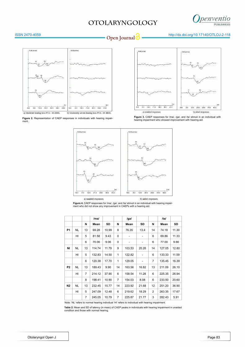
ever all the peaks were not present in all the individuals. The most consistent peaks were P2 and N2. All the four peaks (P1, N1, P2, N2) could be recorded for /ma/ in 13 individuals. For /ga/ sound P2 and N2 were present in all 14 individuals. P1 could be identified in eight individuals and N1 could be identified in only 9 individuals. Responses for /ta/ sound showed P1 and N1 in all 14 individuals, P2 in 13 and N2 in 12 individuals. Figure 1 shows representative waveforms recorded from participants with normal hearing for the three stimuli.
CAEPs were obtained from individuals with hearing impairment without a hearing aid (unaided) and with a hearing aid (aided condition). The responses obtained were compared with those recorded from persons with normal hearing. P2 was the most consistent response and was present in a majority of individuals with hearing impairment. Two individuals showed no response to all the sounds. For /ma/ sound, CAEPs could be recorded from 7 individuals whereas for /ga/ and /ta/ sound, responses could be obtained only from 6 individuals. Figure 2 shows samples of waveforms obtained from persons with hear-ing impairment. Detectability of responses increased in aided condition. However, CAEPs could not be recorded from all the individuals with hearing impairment even in aided condition. A lot of individual variability was observed. Some persons showed improvement in morphology with a hearing aid while a few did not show any improvement. Figure 3 shows CAEP responses for the three stimuli in an individual with hearing impairment who showed improvement with hearing aid while Figure 4 shows responses for a person who did not show any improvement in CAEPs with a hearing aid.
Table 2 shows the mean and standard deviation of the latencies (P1, N1, P2, and N2) for individuals with normal hear-ing and for those with hearing impairment. The table shows la-tencies in both unaided condition and aided conditions for those with hearing impairment. Overall, the mean latencies were lon-ger for individuals with hearing impairment when compared to
Table 1: Protocol for CAEP recording.
Stimuli /ma/, /ga/, /ta/
Stimulus intensity 60 dB SPL
Repetition rate 1.1/sec
Polarity Rarefaction
Filter 0.1-30 Hz
No. of channels Single channel
Amplification 30,000
No. of sweeps 300
Figure 1: CAEPs for /ma/, /ga/ and /ta/ stimuli in normal hearing individu-als.
OTOLARYNGOLOGY
Open Journalhttp://dx.doi.org/10.17140/OTLOJ-2-118
Otolaryngol Open J
ISSN 2470-4059
Figure 4. CAEP responses for /ma/, /ga/, and /ta/ stimuli in an individual with hearing impair-ment who did not show any improvement in CAEPs with a hearing aid.
Figure 2. Representation of CAEP responses in individuals with hearing impair-ment..
Figure 3. CAEP responses for /ma/, /ga/, and /ta/ stimuli in an individual with hearing impairment who showed improvement with hearing aid.
Table 2: Mean and SD of latency (in msec) of CAEP peaks in individuals with hearing impairment in unaided condition and those with normal hearing.
/ma/ /ga/ /ta/
N Mean SD N Mean SD N Mean SD
P1 NL 13 69.28 10.99 8 76.35 13.4 14 74.18 11.30
HI 5 81.58 9.43 0 - - 6 69.86 11.33
6 70.06 9.06 0 - - 6 77.00 9.66
NI NL 13 114.74 11.79 9 103.53 20.26 14 127.05 12.80
HI 5 132.83 14.50 1 122.82 - 6 133.33 11.59
6 120.38 17.70 1 129.05 - 7 135.45 16.39
P2 NL 13 189.43 9.90 14 163.56 16.82 13 211.09 26.10
HI 7 214.12 37.90 6 158.54 11.28 6 225.35 26.94
8 198.41 10.90 7 154.03 8.08 6 233.50 20.60
N2 NL 13 232.45 15.77 14 223.92 21.68 12 251.20 36.90
HI 5 247.09 12.48 6 219.62 18.29 2 263.35 17.67
7 245.05 10.79 7 225.87 21.77 3 282.43 5.91
Note: ‘NL’ refers to normal hearing individual ‘HI’ refers to individual with hearing impairment.
Page 83

OTOLARYNGOLOGY
Open Journalhttp://dx.doi.org/10.17140/OTLOJ-2-118
Otolaryngol Open J
ISSN 2470-4059
those with normal hearing. The latency of peaks in persons with hearing impairment was lesser in aided condition when com-pared with the unaided condition. Table 3 shows the amplitude (P1N1 & P2N2) for the two groups. The amplitude was larger for /ma/ and /ta/ sound and smaller for /ga/ in individuals with hearing impairment when compared to those with normal hear-ing except for P2N2 amplitude for /ta/. With a hearing aid, there was an increase in amplitude of P2N2 of /ta/ and /ga/ sound.
Mann Whitney U test was carried out to check if the latency and amplitude observed in persons with hearing impair-ment was significantly different from those observed in persons with normal hearing. Comparison between unaided responses of individuals with hearing impairment and those of normal hear-ing showed that for /ma/ sound, the latency of all the peaks was significantly different from those of participants with normal hearing but there was no significant difference in the amplitude of the response. For /ga/ sound the latencies of P1 and N1 as well as amplitudes of P1-N1 and P2-N2 differ significantly from those of persons with normal hearing. There was no significant difference between the two groups for latencies and amplitude of all the peaks of /ta/. It can be observed from the table that the latencies and amplitudes of responses obtained in aided were not significantly different from those obtained for individuals with normal hearing except for latency of N2 and amplitude of P2-N2
for /ma/ sound (Table 4).
To summaraise, the result revealed that in aided condi-tion, the detectability of CAEP responses was more when com-pared to unaided condition in persons with hearing impairment. There was a significant difference between the unaided CAEP responses of persons with hearing impairment and CAEP re-sponses of persons with normal hearing. However, no such dif-ference was observed between aided CAEPS responses of per-sons with hearing impairment and those of normal hearing.
DISCUSSION The aim of the present study was to investigate the usefulness of CAEPs in verification of hearing aid. The speech stimuli used in the present study were consonant vowel (CV) syllables with consonants representing low, mid and high frequency region. The duration of all the three stimuli was 350 m sec with a SD of 12 msec. Two of the consonants were voiced (/m/ and /g/) and one was voiceless (/t/), the vowel /a/ was kept constant.
Statistically significant difference observed between CAEP responses of individuals with hearing impairment in un-aided condition and those of normal hearing can be attributed to the loss of audibility in persons with hearing impairment.
Table 3: Mean and SD of amplitude (in µV) of CAEP peaks in individuals with hearing impairment in unaided condition and those with normal hearing.
/ma/ /ga/ /ta/
N Mean SD N Mean SD N Mean SD
P1N1 NL 13 3.77 1.16 8 1.71 1.17 14 6.33 1.80
HI 5 4.40 1.58 0 - - 6 6.76 4.66
6 4.40 3.02 0 - - 6 5.88 3.90
P2N2 NL 13 2.32 0.69 14 5.33 1.76 12 1.75 1.20
HI 5 3.97 1.92 6 4.69 2.46 2 1.11 0.45
7 3.25 1.00 7 4.92 2.13 3 2.27 1.92
Table 4: Results of Mann-Whitney U test (z values) comparing CAEPs of persons with hearing impairment with those of individuals with normal hearing.
/ma/ /ga/ /ta/
Unaided Aided Uniaded Aided Unaided Aided
P1 2.80** -0.48 -2.66 ** - -0.56 -0.54
N1 2.79** -0.48 -2.43* -1.22 -0.76 -1.68
P2 2.36* -1.82 -0.76 -1.27 -1.23 -1.71
N2 2.77** -2.58* -0.82 -0.78 -0.37 -1.16
P1N1 1.54 -0.09 -2.00* - -0.30 -1.32
P2N2 0.51 2.02* -1.96* -1.04 -0.73 -0.72
Note: ‘NL’ refers to normal hearing individual ‘HI’ refers to individual with hearing impairment.
Page 84
Note: *=significant at 0.05 level; **=significant at 0.01 level.

OTOLARYNGOLOGY
Open Journalhttp://dx.doi.org/10.17140/OTLOJ-2-118
Otolaryngol Open J
ISSN 2470-4059
Similar results have been reported by earlier investigators.12,13 No significant difference was obtained between aided responses of individuals with hearing impairment and those of individuals with normal hearing indicates that the audibility has improved with hearing aid. However, the latencies in the aided condition were longer than those obtained for persons with normal hear-ing. Korczak, Kurtzberg, Stapells13 also reported, prolonged la-tencies in aided conditions in comparison to the mean latencies obtained in the normal hearing individuals in persons who were benefitting from hearing aids. They concluded that despite of the benefits provided by the hearing aid, individuals with hearing impairment process speech in less effective manner than their normal hearing counterparts.
Inspection of individual data showed that for /ma/ sound, 8 participants showed improvement in aided condition. For /ga/ 6 participants showed improvement and for /ta/ only 4 individuals showed improvement with hearing aid. Individuals with severe hearing loss (pure tone average greater than 71.6 dB HL) showed marked improvement in CAEP responses when unaided and aided responses were compared. These findings suggesting detectability of CAEPs improve when the degree of hearing loss is high as compared to lesser degree of hearing loss, is similar to the finding’s reported by earlier investigators.13 These results suggest that CAEPs can be used to assess the use-fulness of a hearing aid in those who cannot give a voluntary response. Recording aided CAEPs in infants and children can assure the clinician and the parents/caregivers that the child is hearing with the hearing aid. Longer latency observed in aided condition when com-pared to unaided conditions for some responses could be attrib-uted to fact that CAEP’s are sensitive to the changes in temporal features within milliseconds14 and hearing aids alter the acous-tics of speech stimuli and thus CAEPs.4,5 Billings, Tremblay, Miller8 studied the effect of hearing aid gain settings on latency and amplitude of P1, N1 and P2 waves. They reported that hear-ing aid modifies stimulus characteristics such as SNR, which in turn affects CAEP in a way that does not reliably reflect hearing aid gain.
CONCLUSION
To conclude, the results of the present study reveal that the CAEPs can be reliably recorded in a clinical set up from individ-uals using hearing aids. The detectability of responses increases when a person is wearing hearing aid. CAEPs can be used for verification of hearing aids in difficult-to-test population who are not able to give reliable behavioral responses. CAEPs may be helpful in verifying the usefulness of hearing aids in persons with severe hearing loss.
ACKNOWLEDGEMENT
Authors wish to acknowledge the authorities of Bharati Vidyap-eeth Deemed University School of Audiology and Speech Lan-
guage Pathology for granting permission to carry out the study.
CONFLICTS OF INTEREST: None.
REFERENCES
1. Rapin I, Graziani LJ. Auditory-evoked responses in normal, brain-damaged, and deaf infants. Neurology. 1967; 17(9): 881-894. doi: 10.1212/WNL.17.9.881
2. Korczak PA, Stapells DR. Effects of various articulatory fea-tures of speech on cortical event-related potentials and behavior-al measures of speech-sound processing. Ear hear. 2010; 31(4): 491-504. doi: 10.1097/AUD.0b013e3181d8683d
3. Golding M, Pearce W, Seymour J, Cooper A, Ching T, Dillon H. The relationship between obligatory cortical auditory evoked potentials (CAEPs) and functional measures in young infants. J Am Acad Audiol. 2007; 18(2): 117-125. doi: 10.3766/jaaa.18.2.4
4. Tremblay KL, Kalstein L, Billings CJ, Souza PE. The neu-ral representation of consonant-vowel transitions in adults who wear hearing AIDS. Trends Amplif. 2006; 10(3): 155-162. doi: 10.1177/1084713806292655
5. Billings CJ, Tremblay KL, Souza PE, Binns MA. Effects of hearing aid amplification and stimulus intensity on cortical audi-tory evoked potentials. Audiol Neurootol. 2007; 12(4): 234-246. doi: 10.1159/000101331
6. Jenstad LM, Marynewich S, Stapells DR. Slow Cortical Po-tentials and Amplification-Part II: Acoustic Measures. Int J Oto-laryngol. 2012; 2012: 386542. doi: 10.1155/2012/386542
7. Marynewich S, Jenstad LM, Stapells DR. Slow cortical poten-tials and amplification-part I: n1-p2 measures. Int J Otolaryngol. 2012; 2012: 921513. doi: 10.1155/2012/921513
8. Billings CJ, Tremblay KL, Miller CW. Aided corti-cal auditory evoked potentials in response to changes in hearing aid gain. Int J Audiol. 2011; 50(7): 459-467. doi: 10.3109/14992027.2011.568011
9. Easwar V, Purcell DW, Scollie SD. Electroacoustic Compari-son of Hearing Aid Output of Phonemes in Running Speech ver-sus Isolation: Implications for Aided Cortical Auditory Evoked Potentials Testing. Int J Otolaryngol. 2012; 2012: 518202. doi: 10.1155/2012/518202
10. Glista D, Easwar V, Purcell DW, Scollie S. A Pilot Study on Cortical Auditory Evoked Potentials in Children: Aided CAEPs Reflect Improved High-Frequency Audibility with Frequency Compression Hearing Aid Technology. Int J Otolaryngol. 2012; 2012: 982894. doi: 10.1155/2012/982894
11. Billings CJ, Papesh MA, Penman TM, Baltzell LS, Gal-
Page 85
OTOLARYNGOLOGY
Open Journalhttp://dx.doi.org/10.17140/OTLOJ-2-118
Otolaryngol Open J
ISSN 2470-4059
lun FJ. Clinical use of aided cortical auditory evoked poten-tials as a measure of physiological detection or physiological discrimination. Int J Otolaryngol. 2012; 2012: 365752. doi: 10.1155/2012/365752
12. Wall LG, Dalebout SD, Davidson SA, Fox RA. Effect of hearing impairment on event-related potentials for tone and speech distinctions. Folia Phoniatr (Basel). 1991; 43(6): 265-274. doi: 10.1159/000266137
13. Korczak PA, Kurtzberg D, Stapells DR. Effects of sensori-neural hearing loss and personal hearing AIDS on cortical event-related potential and behavioral measures of speech-sound processing. Ear hear. 2005; 26(2): 165-185. Web site. http://journals.lww.com/ear-hearing/pages/articleviewer.aspx?year=2005&issue=04000&article=00005&type=abstract. Accessed March 7, 2016
14. Aiken SJ, Picton TW. Human cortical responses to the speech envelope. Ear hear. 2008; 29(2): 139-157. doi: 10.1097/AUD.0b013e31816453dc
Page 86

OTOLARYNGOLOGY
Open Journalhttp://dx.doi.org/10.17140/OTLOJ-2-119
Otolaryngol Open J
ISSN 2470-4059
Yakup Yegin, MD1; Mustafa Çelik, MD1*; Baver Maşallah Şimşek, MD1; Burak Olgun, MD1; Aylin Karahasanoğlu, MD2; Ceylan Çolak, MD2; Fatma Tülin Kayhan, MD1
1Department of Otolaryngolgy, Head and Neck Surger, Bakırköy Dr. Sadi Konuk Training and Research Hospital, Istanbul, Turkey 2Department of Radiology, Bakirkoy Dr. Sadi Konuk Research and Training Hospital, Istanbul, Turkey
*Corresponding authorMustafa Çelik, MD Physician Department of Otolaryngology Head and Neck Surgery Bakırköy Dr. Sadi Konuk Training and Research Hospital Zuhuratbaba Mah. Tevfik Sağlam Cad.No: 11, Bakırköy Istanbul, Turkey Tel. 0905335976636 E-mail: [email protected]
Article HistoryReceived: May 21st, 2016 Accepted: June 1st, 2016 Published: June 2nd, 2016
CitationYegin Y, Çelik M, Şimşek BM, et al. The relationship between the degree of the mastoid pneumatization and mean platelet volume. Otolaryngol Open J. 2016; 2(3): 87-90. doi: 10.17140/OT-LOJ-2-119
Copyright©2016 Çelik M. This is an open ac-cess article distributed under the Creative Commons Attribution 4.0 International License (CC BY 4.0), which permits unrestricted use, distribution, and reproduction in any medium, provided the original work is properly cited.
Volume 2 : Issue 3Article Ref. #: 1000OTLOJ2119
The Relationship Between the Degree of the Mastoid Pneumatization and Mean Platelet Volume
Page 87
Research
ABSTRACT
Objectives: To explore the relationships between mean platelet volume (MPV) values and the degree of the mastoid pneumatization.Study Design: A retrospective clinical chart review.Methods: In total, 189 patients (130 females and 59 males; average age, 36.50±15.62 years; age range: 18-65 years) were included in the study. The patients were divided into three groups in terms of the degree of the mastoid pneumatization. The mastoid pneumatization was measured between 0 and 5 cm3 for group A, between 5 and 10 cm3 for group B and 10 cm3 and above for group C, respectively. The MPV values of each groups were compared.Results: The mean mastoid pneumatization in group A, B and C was 3.96±2.72 cm3, 8.93±2.14 cm3 and 11.40±1.36 cm3, respectively. The mean MPV values of group A, B and C were 7.80±1.22 fl, 8.12±1.46 fl and 7.78±1.26 fl, respectively. The mean MPV values did not differ between males and females (p>0.05). The mean mastoid pneumatization was higher in males than in females (p=0.024, p<0.05). The mean MPV values did not differ significantly between the groups (p>0.05).Conclusions: The degree of the mastoid pneumatization did not affect the MPV values. Further studies with larger numbers of patients are needed to evaluate the relationship between the degree of the mastoid pneumatization and MPV values.
KEYWORDS: Mastoid pneumatization; Mean platelet volume; Chronic hypoxia.
INTRODUCTION
The precise functions of the mastoid air cell system are a current and controversial theme. The mastoid air cell system is adopted as an air reservoir for the middle ear. However, knowledge of the physiologic functions of the mastoid air cell system remains unsatisfactory. The potential functions of the mastoid air cell system are:
• protection of the sensitive inner ear structures from external temperature changes,• pressure regulator by impact of the large surface area in accordance with gas exchange.1
The mastoid air cell system enlarges variably to all regions of temporal bone which has a pyramidal shape. The pneumatization of mastoid bone varies individually and its de-velopment alters with age. The mastoid pneumatization has been measured in cadavers via cross-sectional histological analysis. In 1940, Diamant2 is the first to report the mastoid pneu-matization in literature. The mean size of normal adolescent’s mastoid was reported as 12.07 cm3 by him. The development of mastoid air cell system is completely mature at approximately 15 years of age in males and 10 years of age in females.2,3 However, the determination of the exact mastoid pneumatization is difficult although all air cells are interrelated. The mastoid
OTOLARYNGOLOGY
Open Journalhttp://dx.doi.org/10.17140/OTLOJ-2-119
Otolaryngol Open J
ISSN 2470-4059
Page 88
pneumatization has been calculated quantitatively by several methods including such as water-weight method,4 an acoustic method5 and a pressurized transducer.6 Recent and significant advances in computed tomography (CT) provide better images of the anatomical features of the temporal bone.1 Multiplanar reconstruction (MPR) is used to this end. Recent advances in CT allow simple and accurate measurement of the degree of mastoid pneumatization.7 Cadaver studies is associated with more errors than are computer-assisted anatomical approaches. Especially, measurements derived from CT images with the aid of MPR af-ford objective and reliable values. Only a few studies have mea-sured the mastoid pneumatization using this technique.
Mean platelet volume (MPV) is used as a parameter of platelet functions. In literature, increasing MPV levels have been associated with the prognosis of some diseases including such as hypertension, unstable angina pectoralis, neurological diseases, autoimmune diseases and obstructive sleep apnea.8-15 MPV may be used as a marker that indicates chronic intermit-tent hypoxia. In the study of Somuk et al.16 reported that MPV parameter was found high in the children with chronic effusion otitis media. According to Wittmaack’s endodermal theory,17 middle ear diseases in infancy and early childhood are reduced the pneumatization of the mastoid bone. Therefore, hereditary and environmental theories proposed that a small mastoid air cellular system predisposes to chronic or acute otitis media. To our knowledge, there is no reported study that exploring the re-lationships between MPV values and the degree of the mastoid pneumatization. We address this topic in the present study. We explored the relationships between MPV values and the degree of the mastoid pneumatization.
MATERIALS AND METHODS
We retrospectively reviewed data collected from January 2013 to January 2016 on patients which were referred to the Depart-ment of Otolaryngology, Head-and-Neck Surgery, of our hospi-tal for trauma. In total, 189 patients were included in the study. Patients with ossicular chain defects, a cholesteatoma, tympa-nosclerosis, atelectasia, a history of previous ear surgery, his-tory of chronic otitis media and temporal bone fractures were excluded from the study. All patients underwent CT imaging to exclude temporal bone fractures. No any temporal bone fracture
was determined on CT imaging in all of the patients. The side-effects of radiation were explained to all patients prior to CT, as was the reason why CT was planned. All patients were told of the purpose of the study and written informed consent was ob-tained from the patients. The study was conducted in accordance with the principles of Helsinki Declaration. The study protocol was approved by our local Ethics Committee. A multidetector CT system (Siemens Sensation 40, Erlangen, Germany) was used for CT imaging. Imaging parameters included a slice thick-ness and reconstruction interval of 0.5 mm and a field of view of 21.8×28.8 cm; we took at least 150-400 images, which were reconstructed using a classical filtered-back projection. Tempo-ral CT imaging was performed using a Med plus Dicom Wiever system (Med plus Ltd., High Wycombe, UK). No contrast ma-terial was injected. The images were evaluated on a worksta-tion (Leonardo; Siemens) by two experienced radiologists. In this volumetric procedure, mastoid air cells with a gray-scale level similar to air in the temporal bone were determined on the CT imaging. After image processing, only the volumes of the extracted pneumatized parts were measured. The right and left sides were calculated separately in each patient (Figure 1A, 1B and 1C). Routine blood samples were taken from the antecubital vein into tubes with ethylene-diamine-tetracetic acid (EDTA) by a nurse. MPV was measured by hematology analyzer machine. Normal values for MPV were accepted as 6, 0-11, 0 fl. The pa-tients were divided into three groups in terms of the degree of the mastoid pneumatization. The mastoid pneumatization was measured between 0 and 5 cm3 for group A, between 5 and 10 cm3 for group B and 10 cm3 and above for group C, respectively. The MPV values of each group were compared.
Statistical Analysis
Number Cruncher Statistical System (NCSS) 2007 software (Kaysville, UT, USA) was used for all statistical analy-ses. Descriptive statistics (means and standard deviation, medi-ans with interquartile range) were derived. The significance of each intergroup difference was analyzed using Student’s t-test, and the significance of any difference in median values was ex-plored with the aid of the Mann-Whitney U-test and Chi-square test. Qualitative data comparisons were performed using the Pearson χ2 test. A p<0.05 was considered to reflect statistical sig-nificance.
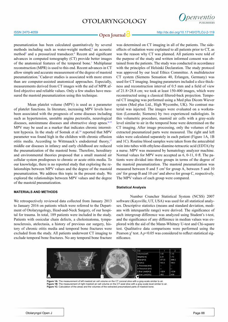
Figure 1A: The measurement of left mastoid air cell volume on the CT coronal slice with a gray-scale similar to air.Figure 1B: The measurement of right mastoid air cell volume on the CT axial slice with a gray-scale level similar to air.Figure 1C: Calculation of the areas and the volumes of the extracted pneumatized parts of mastoid bone.

OTOLARYNGOLOGY
Open Journalhttp://dx.doi.org/10.17140/OTLOJ-2-119
Otolaryngol Open J
ISSN 2470-4059
Page 89
RESULTS
We included 189 patients: 130(68.8 %) females and 59(31.2 %) males. Their average age was 36.50±15.62 years (range: 18-65 years). The mean mastoid pneumatization in group A, B and C was 3.96±2.72 cm3, 8.93±2.14 cm3 and 11.40±1.36 cm3, respec-tively (Table 1). The mean MPV values of group A, B and C were 7.80±1.22 fl, 8.12±1.46 fl and 7.78±1.26 fl, respectively (Table 2). The mean MPV values did not differ between males and females (p>0.05). The mean mastoid pneumatization was higher in males than in females (p=0.024, p<0.05) (Table 3). The mean MPV values did not differ significantly between the groups (p>0.05) (Table 4).
DISCUSSION
The degree of mastoid pneumatization plays a crurical role in middle ear physiologic functions. The development of mastoid pneumatization varies between individuals.1 Two hypothesis have been propounded among inter-individual variations of the degree of the mastoid pneumatization. The first hypothesis is that the degree of mastoid pneumatization is determined geneti-cally. In study of Sade et al18 reported that patients with otoscle-rosis have larger temporal bone pneumatization than do healthy subjects. In another study, Pata et al19 investigated the relation-
ship between presbycusis and mastoid pneumatization consider-ing the etiologies of both are reflected to have genetic factors. The cited authors found no differences between the presbycusis subjects and normal subjects in terms of the volume of mastoid pneumatization.19 Todd et al20 explored the reason why cystic fibrosis patients had significantly less otitis than the normal population. Cystic fibrosis patients frequently have nasal polyps and sinusitis, but interestingly are spared from an increased oc-currence of otitis media. This condition legitimized the authors. The authors reported that mastoid pneumatization of cystic fi-brosis patients was larger than the normal population.20 The sec-ond hypothesis is that the status of the middle ear cavity affects the degree of mastoid pneumatization. The degree of pathologic involvement of the middle ear cavity among childhood states the size of the mastoid pneumatization. Increasing the num-ber of pathologic involvement of the middle ear cavity among childhood decreases the degree of the mastoid pneumatization. Therefore, impact of the degree of mastoid pneumatization on hematological parameters remains unclear. Is there any relation-ship between the poorly mastoid pneumatization and systemic chronic intermittent hypoxia? Or is there any predictive value in hematological parameters for defining poorly mastoid pneumati-zation? These questions remain unclear. Also, it was the consid-eration that legitimizes the present study. MPV may be used as a marker that indicates chronic intermittent hypoxia. In the present study, the patients were divided into three groups in terms of the degree of the mastoid pneumatization. The mean mastoid pneu-matization in group A, B and C was 3.96±2.72 cm3, 8.93±2.14 cm3 and 11.40±1.36 cm3, respectively. The mean MPV values of group A, B and C were 7.80±1.22 fl, 8.12±1.46 fl and 7.78±1.26 fl, respectively. The mean MPV values did not differ signifi-cantly between the groups. To our knowledge, the present study provides the first report of explored the relationships between the mastoid pneumatization and MPV values. However, the re-lationship between systemic chronic hypoxia and mastoid pneu-matization remains unclear. The discrepancies among previous studies with our study may be attributable to the imaging param-eters used, subject data and sample size. Although previous ra-diological studies have been measured the two dimensional size of mastoid pneumatization, in the present study we measured the degree of mastoid pneumatization using a three-dimensional computer-based image reconstruction technique. The value of the technique used in the present study is its high accuracy and easy-to-use. The limitations of our study include the small sam-ple size and the lack of randomization, the lack of assessment of other systemic chronic hypoxia parameters. If assessment of other systemic chronic hypoxia parameters were performed, the study may be more valuable and effective.
CONCLUSIONS
In conclusion, the MPV values did not affect the degree of the mastoid pneumatization. Further studies with larger numbers of patients are needed to evaluate the relationship between the de-gree of the mastoid pneumatization and MPV values.
Mastoid volume (cm3) Patient number % Mean volume
(cm3)
Group A Between 0 and 5 cm3 59 31.2 3.96±2.72
Group B Between 5 and 10 cm3 84 44.4 8.93±2.14
Group C 10 cm3 and higher 46 24.4 11.40±1.36
Groups Mean Platelet volume (fl)
Group A 7.80±1.22 ( 5.50-10.92)
Group B 8.12±1.46 ( 6.22-11.60)
Group C 7.78±1.26 ( 6.24-12.50)
Mastoid pneumatization (cm3)
Males 9.50±2.53 cm3 (2,96-14,89 cm3)
Females 8.69±1.82 cm3( 3,02-13,66 cm3)
*p 0.024
*Mann-Whitney U-test.
*p
Group A Group B Group C
Group A x 0.258 0.622
Group B 0.258 x 0.496
Group C 0.622 0.496 x
*Mann-Whitney U-test.
Table 3: Comparison of the levels of mastoid pneumatiza-tion between males and females.
Table 1: Summary of group characteristics.
Table 2: The mean platelet volume of groups.
Table 4: Comparison of the levels of mean platelet volu-me of each groups.

OTOLARYNGOLOGY
Open Journalhttp://dx.doi.org/10.17140/OTLOJ-2-119
Otolaryngol Open J
ISSN 2470-4059
Page 90
CONFLICTS OF INTEREST
No author has any potential conflicts of interest.
REFERENCES
1. Lee DH, Jun BC, Kim DG, Jung MK, Yeo SW. Volume va-riation of mastoid pneumatization in different age groups: a study by three-dimensional reconstruction based on computed tomography images. Surg Radiol Anat. 2005; 27(1): 37-42. doi: 10.1007/s00276-004-0274-7
2. Diamant M. Otitis and pneumatization of mastoid bone. Acta Otolaryngol. 1940; 41: 10.
3. Koc A, Ekinci G, Bilgili AM, Akpinar IN, Yakut H, Han T. Evaluation of the mastoid air cell system by high resolution computed tomography:three-dimensional multiplanar volume rendering technique. J Laryngol Otol. 2003; 117(8): 595-598. doi: 10.1258/002221503768199906
4. Silbiger H. Uber das Ausmass der Mastoid Pneumatisa-tion beim Menschen. Acta Anat. 1950; 11(1): 215-223. doi: 10.1159/000140504
5. Molvaer OI, Vallersnes FM, Kringlebotn M. The size of the middle ear and the mastoid air cell system measured by an acoustic method. Acta Otolaryngol. 1978; 85(1-2): 24-32. doi: 10.3109/00016487809121419
6. Andreasson L, Mortensson W. Comparison between the area and the volume of the air filled ear space. Acta Radio Diagn. 1975; 16(4): 347-352. Web site. http://europepmc.org/abstract/med/1189960. Aceesed May 20, 2016
7. Ars B, Dirckx J, Ars-Piret N, Buytaert J. Insights in the phy-siology of the human mastoid: message to the surgeon. Int Adv Otol. 2012; 8(2): 296-310. Web site. http://victorslavutsky.com/wp-content/uploads/2014/10/macs-bernard-ars1.pdf. Accessed May 20, 2016
8. Park Y, Schoene N, Harris W. Mean platelet volume as an indicator of platelet activation: methodological issues. Platelets. 2002; 13(5-6): 301-306. doi: 10.1080/095371002220148332
9. Greisenegger S, Endler G, Hsieh K, Tentschert S, Mann-halter C, Lalouschek W. Is elevated mean platelet volume as-sociated with a worse outcome in patients with acute ischemic cerebrovascular events? Stroke. 2004; 35(7): 1688-1691. doi: 10.1161/01.STR.0000130512.81212.a2
10. Ulaşlı SS, Ozyurek BA, Yilmaz EB, Ulubay G. Mean pla-telet volume:an inflammatory marker in acute exacerbation of chronic obstructive pulmonary disease. Pol Arch Med Wewn. 2012; 122(6): 284-290. Web site. http://pamw.pl/en/issue/artic-le/22576316. Accessed May 20, 2016
11. Sagit M, Korkmaz F, Kavugudurmaz M, Somdas MA. Impa-ct of septoplasty on mean platelet volume levels in patients with marked nasal septal deviation. J Craniofac Surg. 2012; 23(4): 974-976. doi: 10.1097/SCS.0b013e31824e2c08 12. Köseoğlu Soyalıç H, Somuk BT, Doğru S, Gürbüzler L, Göktaş G, Eyibilen A. Evaluation of mean platelet volume and its ratio over platelet count in children with obstructive sleep apnea syndrome. Kulak Burun Bogaz Ihtis Derg. 2015; 25(1): 16-21. doi: 10.5606/kbbihtisas.2015.28863
13. Cengiz C, Erhan Y, Murat T, et al. Values of mean platelet volume in patients with chronic tonsillitis and adenoid hypert-rophy. Pak J Med Sci. 2013; 29(2): 569-572. Web site. http://www.ncbi.nlm.nih.gov/pmc/articles/PMC3809227/. Accessed May 20, 2016
14. Varol E, Ozturk O, Gonca T, et al. Mean platelet volu-me is increased in patients with severe obstructive sleep ap-nea. Scand J Clin Lab Invest. 2010; 70(7): 497-502. doi: 10.3109/00365513.2010.520733
15. Vizioli L, Muscari S, Muscari A. The relationship between mean platelet volume with the risk and prognosis of cardiovas-cular disease. Int J Clin Pract. 2009; 63(10): 1509-1515. doi: 10.1111/j.1742-1241.2009.02070.x
16. Somuk BT, Soyalıç H, Koc S, Gürbüzler L, Doğru S, Eyibi-len A. Mean platelet volume as an inflammatory marker of ch-ronic otitis media with effusion. Int J Pediatr Otorhinolaryngol. 2014; 78(11): 1958-1960. doi: 10.1016/j.ijporl.2014.08.037
17. Wittmaack K. Uber die normale pneumatisation des schla-fenbeines einschlieblich ihrer Beziehungen zu den Mittelohrerk-rankungen [In German]. Jena: Ficher; 1918.
18. Sade J, Shatz A, Kremer S, Levit I. Mastoid pneumatization in otosclerosis. Ann Otol Rhinol Laryngol. 1989; 98(6): 451-454. doi: 10.1177/000348948909800611
19. Pata YS, Akbas Y, Unal M, Duce MN, Akbas T, Micozka-dıoglu D. The relationship between presbycusis and mastoid pneumatization. Yonsei Med J. 2004; 45(1): 68-72. doi: 10.3349/ymj.2004.45.1.68 20. Todd NW, Martin WS. Temporal bone pneumatization in cystic-fibrosis patients. Laryngoscope. 1988; 98(10): 1046-1049. doi: 10.1288/00005537-198810000-00004
OTOLARYNGOLOGY
Open Journalhttp://dx.doi.org/10.17140/OTLOJ-2-120
Otolaryngol Open J
ISSN 2470-4059
Marrakchi J, MD1*; Zhani A, MD2; Chahed H, MD1; Nefzaoui S, MD1; Ben Amor M, MD1; Beltaif N, MD1; Kchir N, MD2; Besbes G, MD1
1Department of ENT, La Rabta University Hospital, Tunis, Tunisia2Department of Pathological Anatomy, La Rabta University Hospital, Tunis, Tunisia
*Corresponding authorJihene Marrakchi, MD Clinical Chief ENT Department La Rabta University Hospital Tunis, Tunisia Tel. 0021698260676 E-mail: [email protected]
Article HistoryReceived: May 23rd, 2016 Accepted: June 6th, 2016 Published: June 6th, 2016
CitationMarrakchi J, Zhani A, Chahed H, et al. Rare localization of lymphoma. Otolar-yngol Open J. 2016; 2(3): 91-93. doi: 10.17140/OTLOJ-2-120
Copyright©2016 Marrakchi J. This is an open access article distributed under the Creative Commons Attribution 4.0 International License (CC BY 4.0), which permits unrestricted use, distribution, and reproduction in any medium, provided the original work is properly cited.
Volume 2 : Issue 3Article Ref. #: 1000OTLOJ2120
Rare Localization of Lymphoma
Page 91
Case Report
ABSTRACT
Objective: Primitive Thyroid Lymphomas (PTL) are rare tumors. Women in the sixth or seven-th decade of life are more commonly affected. In the present study, we report a case of primitive thyroid lymphoma and we review the epidemiology, the clinical presentation, the diagnosis, and the treatment of this rare disorder.Case Report: A 47-year-old woman presented to our department reporting a recent-onset neck mass since 3 months. Clinical examination revealed an enlargement of the thyroid gland with a 2.5 cm-firm left nodule. Cervical ultrasound was done. The patient had a thyroidectomy as-sociated with bilateral Central Lymph Node Dissection (CLND). The diagnosis was a transfor-mation of a Mucosa-associated lymphoid tissue (MALT) lymphoma into an aggressive Diffuse Large B-Cell Lymphoma (DLBCL).Conclusion: The most common type of primary thyroid lymphoma (PTL) is diffuse large B-cell lymphoma, which behaves in a more aggressive manner than mucosa-associated lymphoid tissue lymphoma. Treatment and prognosis of PTL depend upon the histology and stage of the tumor at diagnosis.
KEYWORDS: Thyroid; Lymphoma; Mucosa-associated lymphoid tissue (MALT); Diffuse large B-cell lymphoma.
ABBREVIATIONS: PTL: Primitive Thyroid Lymphomas; CLND: Central Lymph Node Dissec-tion; MALT: Mucosa-associated lymphoid tissue; DLBCL: Diffuse Large B-Cell Lymphoma; FNA: Fine Needle Aspiration; NHL: Non-Hodgkin lymphoma.
INTRODUCTION
Primary Lymphoma (PL) of the thyroid is a very rare disease. It is less than 2 to 5% of mali-gnant neoplasms of the thyroid.1 Involvement of the thyroid gland may occur in the context of a systemic disease or rarely be primitive. The most common histological sub-type is a diffuse large B-cell lymphoma (DLBCL) followed by Mucosa-associated lymphoid tissue (MALT) lymphoma.
CASE REPORT
A 47-year-old woman presented to our department reporting a recent-onset neck mass since 3 months. There were no local obstructive symptoms associated such as dyspnea or dysphonia. Her medical past history was unremarkable. She had no history of personal or family thyroid disease or radiation exposure. Clinical examination revealed an enlargement of the thyroid gland with a 2.5 cm-firm left nodule. No cervical nodes were palpable.
Cervical ultrasound showed multinodular goiter with hypoechogenes heterogeneous nodules containing microcalcifictions. Infracentimetric lymph nodes in levels ll, III, IV of the both lateral neck were detected. The patient was in biological eutyhroidie. She underwent thy-roidectomy associated with bilateral central lymph node dissection (CLND).
On macroscopic appearence, the thyroid gland was solid, nodular white-gray colored
OTOLARYNGOLOGY
Open Journalhttp://dx.doi.org/10.17140/OTLOJ-2-120
Otolaryngol Open J
ISSN 2470-4059
Page 92
with a fish-flesh. On microscopic examination, the findings showed a marked diffuse lymphocytic infiltration destroying thyroid follicles. Large lymphatic cells with a typical and large nuclei containing central nucleoli were observed.
Elsewhere, we found a typical lymphocytes with small and centrocyte-like cell. The neighboring thyroid parenchyma contained lesions of thyroiditis. The metastasis of central neck node was evident. Immunohistochemistry showed CD20 posi-tivity and CD15, CD3, CD20 negativity. The diagnosis was a transformation of a MALT lymphoma into an aggressive diffuse large B-cell lymphoma (Figure 1).
Total body Computed Tomography (CT) scan was rea-lized to complete staging according to Ann Arbor classification. It did not document any pathological finding. The diagnosis of primitive thyroid lymphoma was then made. The patient was at stage IIE. A combination of chemotherapy and radiotherapy treatment was adjuncted post-operatively. No tumor recurrence was observed after a mean follow-up of five years.
DISCUSSION
Primitive thyroid lymphoma (PTL) represents 2 to 7% of all extranodal primitive lymphomas. It mainly occur in middle- to older-aged patients with a predilection for females in the 6th de-cade of life.2
Most of the patients with PTL have a previous history of auto immune thyroid it is with or without hypothyroidism. In fact, Hashimoto’s thyroiditis co-exists in 83% of patients with PTL. Furthermore, in patients affected by chronic autoimmune thyroid it is, the probability of developing a PTL is 20 times greater than in the general population.2
Non-Hodgkin lymphoma (NHL) is the most common PLT (93%). Two sub-types are frequent: Diffuse large B-cell lymphoma is the most encountered accounting for more than 50% of cases, followed by mucosa-associated lymphoid tissue (MALT) lymphoma.3 Upto 40% of all diffuse large cell lym-phomas appear to have undergone transformation from a MALT lymphoma.4
Clinically, general symptoms associated with lympho-mas, such as fever, excessive perspiration and weight loss, are present in only 10-20% of patients.2 A rapidly growing (usually within 1-3 months), painless thyroid enlargement, either in the form of goiter or discrete nodule, is the most common clinical presentation in PTL.
Diffuse large B-cell lymphoma (DLBCL) thyroid lym-phoma is considered as a high grade lymphoma with a more ag-gressive clinical course. They present as a painless fast-growing mass causing compressive symptoms like dysphagia, hoarseness or dyspnea. These symptoms overlap with that of the anaplastic thyroïd carcinoma.5
MALT lymphoma is considered low grade tumor with an indolent natural history and presents as a slow-growing tu-mor with early stage disease confined to the thyroid. DLBCL can develop from MALT lymphoma, and these two subtypes can be detected in the same gland. The mixed MALT and DLBCL sub-type shows the same clinical behavior as that of DLBCL.6
The diagnosis of PTL is not always evident. In fact, due to their rarity and clinical polymorphism the diagnosis is often made on definite histology after thyroid surgery as the case of our patient. Effectively, Fine Needle Aspiration (FNA) results are in-consistent due to the histopathological similarities between primary thyroid lymphoma and Hashimoto’s thyroidi-tis.7
Once a diagnosis is made, total body CT scan should be performed to complete staging, according to the Ann Arbor classification. Large series of PTL revealed that about 50% of cases is confined to the gland (stage IE), 45% involved the gland and regional lymph nodes (stage IIE). lymph node involvement above and below the diaphragm (stage IIIE) or extranodal di-sease (stage IV) are found in only 5% of cases.2,4
Treatment depends on the histological subtype and the stage of the disease. Despite controversy regarding the optimal modality for the management of PTL, the combination of che-motherapy and locoregional radiotherapy is the standard treat-ment of localized aggressive lymphoma diffuse large B-cell lym-
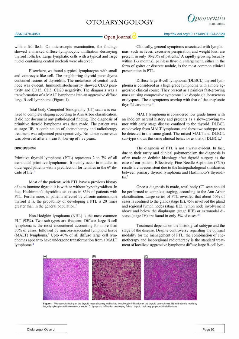
Figure 1: Microscopic finding of the thyroid mass showing. A) Marked lymphocytic infiltration of the thyroid parenchyma. B) Infiltration is made by large lymphocytes with voluminous nuclei. C) Lymphoid infiltration destroying folliclar thyroid realizing lymphoepithelial lesions.
(B) (C)(A)
OTOLARYNGOLOGY
Open Journalhttp://dx.doi.org/10.17140/OTLOJ-2-120
Otolaryngol Open J
ISSN 2470-4059
Page 93
phoma.8 The conventional chemotherapeutic regimen consists of cyclophosphamide, doxorubicin, vincristine, and prednisone (CHOP), and radiotherapy is used for local disease control.6
Surgery is the primary treatment of localized MALT lymphomas1,9 in disseminated or aggressive disease, surgery may be indicated for alleviation of compressive symptoms or protection of the airway.9 Surgical dissection may be more com-plicated than in standard cases of thyroïdectomy due to the pos-sible tight adhesions existing between the gland’s capsule and the surrounding structures.2
The prognosis of PTL is affected by disease stage. In localized tumors, it is usually favorable with a survival rate at 5 years from 70% to 80%.10 However, the prognosis is very poor for lesions with extracapsular invasion (IIE) (20 to 50%). For stages IIIE and IV, the rates are 15 to 35%.2
CONCLUSION
In summary, PTL has excellent prognosis when it is confined to the regional neck area and treated properly according to histo-logic sub-type and stage. The diagnosis should be early evoked in abrupt thyroid enlargement or compression symptoms. Treat-ment of PTL requires a multidisciplinary approach in order to choose the most appropriate therapy.4
CONFLICTS OF INTEREST: None.
CONSENT
The authors article did not publish any personal photo or infor-mation regarding any of the patients in his manuscript. Thus, the consent is not required for the article publication.
REFERENCES
1. Papadakis G, Tertipi A, Papazian M, Moustakas K, Pappas A. Case report: Primary thyroid lymphoma presenting as a rapidly enlarging thyroid mass. Endocrinol Metab Int J. 2015;1(1): 1-3. Web site. http://medcraveonline.com/EMIJ/EMIJ-01-00002.pdf Accessed May 22, 2016
2. Avenia N, Ragusa M, Cirocchi R, et al. Surgical treatment of primitive thyroid lymphoma. Tumori. 2009; 95(6): 712-719. Web site. http://www.tumorijournal.com/article/surgical-treat-ment-of-primitive-thyroid-lymphoma. Accessed May 22, 2016
3. Stein SA, Wartof sky L. Primary thyroid lymphoma: A clinical review. J Clin Endocrinol Metab. 2013, 98(8): 3131-3138. doi: 10.1210/jc.2013-1428 4. Walsh S, Lowery AJ, Evoy D, Mcdermott EW, Prichard RS. Thyroid lymphoma: Recent advances in diagnosis and optimal management strategies. Oncologist. 2013; 18(9): 994-1003. doi: 10.1634/theoncologist.2013-0036
5. Onal C, Li YX, Miller RC. Treatment results and prognos-tic factors in primary thyroid lymphoma patients: A rare cancer network study. Ann Oncol. 2011; 22(1): 156-164. doi: 10.1093/annonc/mdq310
6. Young JC, Jun HH, Do Hoon K. Clinicopathological cha-racteristics and treatment outcomes of 38 cases of primary thy-roid lymphoma: a multicenter study. Ann Surg Treat Res. 2015; 89(6): 295-299. doi: 10.4174/astr.2015.89.6.295
7. Beasley MJ. Lymphoma of the thyroid and head and neck. Clin Oncol. 2012; 24(5): 345-351. doi: 10.1016/j.clon.2012.02.010
8. Kim EH, Young Kim J, Kim T-J. Aggressive primary thyroid lymphoma: Imaging features of two elderly patients. Ultrasono-graphy. 2014; 33(4): 298-302. doi: 10.14366/usg.14025
9. Alzouebi M, Goepel JR, Horsman JM, Hancock BW. Primary thyroid lymphoma: The 40 year experience of a UK lympho-ma treatment centre. Int J Oncol. 2012; 40(6): 2075-2080. doi: 10.3892/ijo.2012.1387
10. Kumar R, Khosla D, Kumar N, et al. Survival and failure outcomes in primary thyroid lymphomas: A single centre ex-perience of combined modality approach. J Thyroid Res. 2013; 2013: 269034. doi: 10.1155/2013/269034

OTOLARYNGOLOGY
Open Journalhttp://dx.doi.org/10.17140/OTLOJ-2-121
Otolaryngol Open J
ISSN 2470-4059
Jihene Marrakchi, MD1*; Safa Nefzaoui, MD1, Dorra Chiboub, MD1, Mariem Jrad, MD2; Mohamed Ben Amor, MD1; Najeh Beltaief, MD1; Habiba Mizouni, MD2; Besbes Ghazi, MD1
1Department of ENT, La Rabta University Hospital, Tunis, Tunisia2Department of Imaging, La Rabta University Hospital, Tunis, Tunisia
*Corresponding authorJihene Marrakchi, MD Clinical Chief ENT Department La Rabta University Hospital Tunis, Tunisia Tel. 0021698260676 E-mail: [email protected]
Article HistoryReceived: May 5th, 2016 Accepted: June 22nd, 2016 Published: June 23rd, 2016
CitationMarrakchi J, Nefzaoui S, Chiboub D, et al. Imaging of paranasal sinus muco-celes. Otolaryngol Open J. 2016; 2(3): 94-100. doi: 10.17140/OTLOJ-2-121
Copyright©2016 Marrakchi J. This is an open access article distributed under the Creative Commons Attribution 4.0 International License (CC BY 4.0), which permits unrestricted use, distribution, and reproduction in any medium, provided the original work is properly cited.
Volume 2 : Issue 3Article Ref. #: 1000OTLOJ2121
Imaging of Paranasal Sinus Mucoceles
Page 94
Research
ABSTRACT
Introduction: Mucoceles are cystic masses developing after obstruction of the sinus ostium. The symptoms are not specific. Computed Tomography scan (CT scan) and Magnetic Reso-nance Imaging (MRI) confirm the diagnosis. Objectives: We herein review the radiologic characteristics of mucoceles in CT scan and MRI.Materials and Methods: We report a retrospective study of 43 patients diagnosed with paranasal sinuses mucoceles. CT scans were performed for all patients, but MRI was carried out only in selected cases.Results: Our study was constituted of 27 males and 16 females with a mean age of 47 years. The CT scan appearence of mucoceles were in all cases as a well circumscribed expansile si-nus mass with an effect on the neighbor bone structure. This mass was hypodense in 26 cases, isodense in 14 cases and hyperdense in 3 patients. The paranasal sinuses most frequently af-fected in our series were the fronto-ethmoidal sinuses. The most affected bone eroded was the lamina papiracea. Intracranial extension was seen in four cases. CT scan allowed to predict the cause of mucoceles in some cases and to provide information about anatomic variants. MRI was realized for 15 patients in addition to the CT scan. It allowed to study the extension of mucoceles to the neighboring organs especially orbital and endocranial ones.Conclusion: The presentations of mucoceles on imaging are quite variable. CT scan provides precious information about the location, bone erosion and extension of the mucoceles. MRI is indicated in some cases especially in cases of orbital or cranial extension.
KEYWORDS: Mucoceles; Paranasal sinuses; Computed Tomography scan (CT scan); Magnetic Resonance Imaging (MRI).
ABBREVIATIONS: CT scan: Computed Tomography scan; MRI: Magnetic Resonance Imaging.
INTRODUCTION
Mucoceles are benign, slow-growing paranasal sinus lesions that develop after obstructions of the sinus ostium.1 Symptoms are variable. The diagnosis is based on imaging. CT scan of the sinuses is the method of the choice. MRI is indicated in some cases and provides much infor-mation of mucocele extensions to adjacent compartments.2
The purpose of this study was to review the role of pre-operative imaging and to il-lustrate the main characteristics imaging findings of paranasal sinuses mucoceles.
MATERIALS AND METHODS
We conducted a retrospective review of the charts of 43 patients diagnosed with paranasal sinus mucoceles who were admitted to our Department of Otolaryngology, between January 1990 and December 2012.
Review of the patients’ medical records including out-patient clinical records and re-ports of imaging were performed.

OTOLARYNGOLOGY
Open Journalhttp://dx.doi.org/10.17140/OTLOJ-2-121
Otolaryngol Open J
ISSN 2470-4059
Page 95
CT scans of the head were performed for all patients. Axial, sagittal, coronal and contrast CT scan with 3 mm slice thickness were reviewed in all cases.
MRI was carried out only in selected cases for the evaluation of the extension of sinonasal mucoceles. MRI find-ings on coronal and axial views in T1, T2 weighted and contrast enhanced images were studied.
RESULTS
Clinical Features
Our study was constituted of 27 males and 16 females (sex ra-tio=1.68) with a mean age of 47 years (from 14 to 77 years).
Rhinosinusitis past history was present in six patients, a facial traumatism in seven patient and eleven patients have undergone prior sinus surgery.
The most commonly reported symptoms were ophthal-mologic one (n=24, 56%), including proptosis (n=16), chronic lacrimation (n=7), diplopia (n=3), visual acuity reduced (n=2) and ptosis (n=1).
Headache was present in twenty three patients. Rhino-logic symptoms were reported in 20 patients and were domi-nated by chronic discharge (17 cases).
On examination, we noted a face swelling in 17 cases, a proptosis in 16 patients and ophtalmoplegia in two cases.
Endoscopic nasal examination revealed an obstructive deviation of nasal septum in 10 cases, a filling of middle meatus in six patients and adhesions between the middle turbinate and the nasal septum in three cases.
Radiologic Findings
CT scan: The CT scan appearence of mucoceles were in all cases as a well circumscribed expansile sinus mass with an effect on the neighbor bone structure. This mass was hypodense in 26 cases, isodense in 14 cases and hyperdense in 3 patients. After injection of contrast agents, we saw a poor enhancement in three cases and a peripherally enhanced image in all others cases.
The paranasal sinuses most frequently affected in our series were the fronto-ethmoidal sinuses (Figure 1). Mucoceles involved both the frontal and ethmoidal sinus in fourteen cases, ten mucoceles were located in the ethmoid sinus, and five were located in the frontal sinus (Figures 2, 3 and 4).
Bone erosion was noted (Figures 5 and 6). The most affected was the lamina papiracea which was eroded in 27 cases (Table 1).
Intracranial extension was seen in four cases and was
Figure 1: The location of the mucoceles in our patients.
Figure 2: CT scan imaging (coronal and sagital sections) showing an expansile mass in the fronto-ethmoidal sinuses with orbital involvement.
Figure 3: Axial CT scan of a mucocele involv-ing the right ethmoid.
Figure 4: Axial and coronal CT scan imaging demonstrating a mucocele of the sphenoid sinus.

OTOLARYNGOLOGY
Open Journalhttp://dx.doi.org/10.17140/OTLOJ-2-121
Otolaryngol Open J
ISSN 2470-4059
Page 96
Figure 7: Axial and coronal CT scan imaging of the sinuses demonstrat-ing a mucocele of the frontal sinus and the ethmoid with important endo-cranial extension.
Figure 5: CT scan with coronal and sagital reconstruction: Mucocele with-in the left frontal sinus with erosion of the roof of the orbite and extension into the orbital cavity.
Figure 6: Axial computed tomographic im-age of the paranasal sinuses showing a completely opacified right maxillary sinus with a medial bulge of the wall of sinus and the septum.
Bone erosion Number of cases
Mucocele location
F E M S FE FEM SE EM
Lamina papiracea 27 2 7 _ _ 12 4 2 _
Orbital roof 9 2 _ _ _ 5 2 _ _
Ethmoidal roof 6 _ _ _ _ 4 2 _ _
Anterior wall of frontal sinus 5 2 _ _ _ 2 1 _ _
Posterior wall of frontal sinus 5 2 _ _ _ 2 1 _ _
Postero-superior wall of sphenoidal sinus 3 _ _ 1 _ _ 2 _
Medial wall of maxillary sinus 2 _ _ 2 _ _ _ _ _
External wall of maxillary sinus 1 _ _ 1 _ _ _ _ _
Table 1: Bone erosion in our study.
F: Frontal sinus; E: Ethmoidal sinus; M: Maxilary sinus; S: Sphenoidal sinus.
consequently to a frontal mucocele in one case, fronto-ethmoidal mococele in two cases, spheno-ethmoidal mucocele in one other case (Figure 7).
Extension into the orbite was observed in 27 cases and concerned predominately the medial wall of the orbite. Optic nerve was repulsed in two cases of fronto-ethmoidal mucoceles and one case of fronto-ethmoido-maxillary mucocele. It was compressed in others three cases of: Ethmoidal, Sphenoidal and Spheno-ethmoidal mucoceles. We noted one case of atrophic op-tic nerve in a sphenoidal mucocele (Figures 8, 9 and 10).
CT scan allowed to note a proptosis in 17 cases (grade 2 in
16 patients and grade 3 in one patient with a fronto-ehtmoidal mucocele associated with an important orbital extension) Others lesions were observed some predicted of the cause of mucoceles:
*Adhesions between the middle turbinate and lateral nasal wall were observed in three patients who had prior sinus surgery and were seen respectively in an ethmoidal, fronto-ethmoidal and fronto-ethmoidal maxillary mucoceles.
*Calcifications of the frontal recess in one patient having a fron-tal mucocele and history of craniofacial traumatism.
*Sinus retention were noted in three cases of maxillary mucoce-
Figure 8: Axial and sagittal images of CT scan demonstrating a mass consistent with a left spheno-ethmoid mucocele. The medial orbital wall is expanded and we note a mass effect on the medial rectus and optic nerve with endocranial extension.
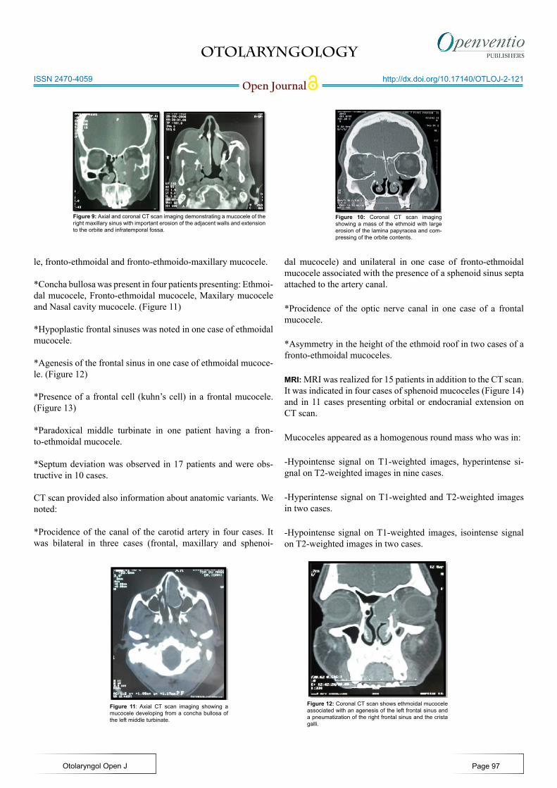
OTOLARYNGOLOGY
Open Journalhttp://dx.doi.org/10.17140/OTLOJ-2-121
Otolaryngol Open J
ISSN 2470-4059
Figure 9: Axial and coronal CT scan imaging demonstrating a mucocele of the right maxillary sinus with important erosion of the adjacent walls and extension to the orbite and infratemporal fossa.
Figure 10: Coronal CT scan imaging showing a mass of the ethmoid with large erosion of the lamina papyracea and com-pressing of the orbite contents.
le, fronto-ethmoidal and fronto-ethmoido-maxillary mucocele.
*Concha bullosa was present in four patients presenting: Ethmoi-dal mucocele, Fronto-ethmoidal mucocele, Maxilary mucocele and Nasal cavity mucocele. (Figure 11)
*Hypoplastic frontal sinuses was noted in one case of ethmoidal mucocele.
*Agenesis of the frontal sinus in one case of ethmoidal mucoce-le. (Figure 12)
*Presence of a frontal cell (kuhn’s cell) in a frontal mucocele. (Figure 13)
*Paradoxical middle turbinate in one patient having a fron-to-ethmoidal mucocele.
*Septum deviation was observed in 17 patients and were obs-tructive in 10 cases.
CT scan provided also information about anatomic variants. We noted:
*Procidence of the canal of the carotid artery in four cases. It was bilateral in three cases (frontal, maxillary and sphenoi-
dal mucocele) and unilateral in one case of fronto-ethmoidal mucocele associated with the presence of a sphenoid sinus septa attached to the artery canal.
*Procidence of the optic nerve canal in one case of a frontal mucocele.
*Asymmetry in the height of the ethmoid roof in two cases of a fronto-ethmoidal mucoceles.
MRI: MRI was realized for 15 patients in addition to the CT scan. It was indicated in four cases of sphenoid mucoceles (Figure 14) and in 11 cases presenting orbital or endocranial extension on CT scan.
Mucoceles appeared as a homogenous round mass who was in:
-Hypointense signal on T1-weighted images, hyperintense si-gnal on T2-weighted images in nine cases.
-Hyperintense signal on T1-weighted and T2-weighted images in two cases.
-Hypointense signal on T1-weighted images, isointense signal on T2-weighted images in two cases.
Figure 11: Axial CT scan imaging showing a mucocele developing from a concha bullosa of the left middle turbinate.
Figure 12: Coronal CT scan shows ethmoidal mucocele associated with an agenesis of the left frontal sinus and a pneumatization of the right frontal sinus and the crista galli.
Page 97

OTOLARYNGOLOGY
Open Journalhttp://dx.doi.org/10.17140/OTLOJ-2-121
Otolaryngol Open J
ISSN 2470-4059
-Isointense signal on T1-weighted images, hyperintense signal on T2-weighted images in one case.
-Isointense signal on T1-weighted and T2-weighted images in one case.
In all cases, we have noted no enhancement after injection of gadolinium.
MRI allowed to study the extension of mucoceles to the neighbo-ring organs especially orbital and endocranial ones. We observed that:
-The orbite contents was repulsed in two cases of fronto-eth-moidi-maxillary mucoceles and one case of fronto-ethmoidal mucocele. (Figure 15)
-Optic nerve was pushed in three patients who had respectively sphenoid mucocele, spheno-ethmoidal mucocele, fronto-eth-moido-maxillary mucocele.
It was stretched in one case of fronto-ethmoidal mucocele with important orbital extension.
-Endocranial extension was noted in three cases: Spheno-eth-moido mucocele, fronto-ethmoidal mucocele and fronto-eth-moido-maxillary mucocele. (Figure 16)
DISCUSSION
Paranasal sinus mucoceles are cystic masses filled with mucous and lined by respiratory epithelium which are capable of expan-sion by bone resorption and new bone formation.1
They occur principally in the third or fourth decades of life with a male predilection.1
Mucoceles result from the blockage of the sinus drain-age secondary to inflammation, trauma, anatomical aberrations, tumours. chronic rhinosinusitis, allergic disease and craniofacial disease are the most common cause of mucocele formation.2,3
Symptoms depend on the location of the mucocele and may vary from rhinological, neurologic, or ophthalmologic ones.1-3
CT scan is considered to be the complementary method of choice in the investigation of mucoceles. It allows to evoke the diagnosis of the mucocele and determine its location, to study the neighboring structure (bone erosion, extension), to presume an aetiology of the sinus ostium obstruction and to research the anatomic variants of the sinuses.3,4
Mucocele appears on CT as a homogenous well circu-mbscribed expansible cyst involving one or much sinuses and
Figure 16: Coronal T2-weighted MRI showing a fronto-ethmoidal sinus mucocele with endo-cranial extension.
Figure 13: Axial CT scan imaging showing a left frontal mucocele associated with a left frontal cell (Kuhn’s cell).
Figure 14: Coronal MRI imaging demonstrating a sphenoidal mucocele (isointense T1 weighted images and hyperintense T2 weighted images ).
Figure 15: Axial and coronal MRI imaging showing a maxilary mucocele with extension toward the orbite and the infra temporal fossa.
Page 98

OTOLARYNGOLOGY
Open Journalhttp://dx.doi.org/10.17140/OTLOJ-2-121
Otolaryngol Open J
ISSN 2470-4059
may expand to neighboring structure. they may appear either hypo- or hyperdense.4 This variable densities depend on their protein content and possible infection. Mucoceles do not show contrast enhancement centrally but a thin peripheral enhance-ment, can be seen and is suggestive of encapsulation.5
The differential diagnosis for mucocele is made with inflammatory, congenital cystic and neoplastic lesions of skull base, facial sinuses and nasopharynx.4-6
Mucocele location depends on anatomic conditions that favorise its formation. The frontal sinus is most commonly affected followed by the ethmoid sinuses, mucoceles occur in these locations in 70 to 90%. Sphenoid mucoceles are rare. The maxillary antrum is a relatively unfrequent site for mucocele formation, accounting for 10 percent or less of mucoceles.7,8
In our study, mucoceles were seen frequently in fronto-ethmoidal sinus followed by ethmoidal location then frontal si-nus.
The neighboring bone structure is remodeled with areas of thickening and erosion.2,4,5 In some areas of greater fragility, we may observe herniation into adjacent structures such as the orbite and the endocrine.4,9
CT scan provides also information about existence of factors favorising mucoceles formation as: Osteoma, Fibrous displasia, Facial traumatism, Sinonasal polyposis. It can visual-izes anatomic abnormalities that may cause a blockage of sinus drainage.10-12 For our patients, CT scan has allowed to evoke the cause of mucoceles in some cases as adhesions, concha bullosa, septum deviation, paradoxical middle turbinate.
Furthermore, this exam is essentially to define anatom-ic variants prior to surgery.13
MRI is an excellent exam in mucocles. It allows to dif-ferentiate and to assess any extension into the orbit or intracrani-al compartment, but, unfortunately it can’t study bone detail.14,15
Its indications are in complement of the CT scan in case of diagnosis doubt in CT scan between mucocele and other tu-mors or inflammatory lesions, in case of sphenoid location and in endocranial or orbital extension.14
The MRI appearance of paranasal sinus mucoceles is quite variable, depending on the composition of the mucocele.16 The usual signal characteristics are a low intense T1 and a high intense T2 but any combination of signal intensity may be seen depending on the presence of blood products or the degree of hydration of the contents.5,14,17,18 The appearance consisted of hyperintense signal on both T1 and T2 type images, correspond-ing to more hydrated secretions, which are also high in protein content.17-20
We have noted variable presentation of mucocele in MRI from hypo to hyperintensity T1 and T2 but in all cases, we have noted no enhancement after injection of gadolinium.
CONCLUSION
The presentations of mucoceles on imaging are quite variable depending on its contents. Practicians should know the typical as well as atypical findings. Special care should be taken with regard to differential diagnosis and associated cases.16
CONFLICTS OF INTEREST
The authors declare that they have no conflicts of interest.
CONSENT
As our article is a retrospective study and did not publish any personal photo or information regarding any of the patient in our manuscript thus the consent is not required for the article publication.
REFERENCES
1. Capra GG, Carbone PN, Mullin DP. Paranasal sinus muco-cele. Head Neck Pathol. 2012; 6: 369-372. doi: 10.1007/s12105-012-0359-2
2. Cannon DE, Wells T, Poetker DM. Two late complications of craniofacial trauma: Case report and review of the litera-ture. American Journal of Otolaryngology–Head and Neck Medicine and Surgery. 2012; 33 : 615-618. doi: 10.1016/j.am-joto.2011.12.003
3. Obeso S, Liorente JL, Rodrigo JP, Sanchez R, Mancebo G, Suarez C. Paranasal sinus mucoceles. Our experience in 72 pa-tients. Acta Otorrinolaringol Esp. 2009; 60(5): 332-339. doi: 10.1016/S2173-5735(09)70153-X
4. De Carvalho BV, De Campos Carvalho LI , De Brito Corrêa J, Ramos LFM, Motta EGPC, Diniz RLFC. Typical and atypical presentations of paranasal sinus mucocele at computed tomog-raphy. Radiol Bras. 2013; 46(6): 372-375. doi: 10.1590/S0100-39842013000600010
5. Hee Han M, Hyun Chang K, Hee Lee C, Gyu Na D, Mo Yeon K, Chung Han M. Cystic expansile masses of the max-illa: Differential diagnosis with CT and MR. AJNR Am J Neu-roradiol. 1995; 16: 333-338. Web site. http://www.ajnr.org/con-tent/16/2/333.long. Accessed May 4, 2016.
6. Chmielewski R, Paprocki A, Morawski K, Niemczyk K. Sphenoid sinus mucocele penetrating to the orbit, anterior and middle cranial fossae and parapharyngeal space: A case report. Otolaryngol Pol. 2010; 64(1): 47-50. doi: 10.1016/S0030-6657(10)70035-4
Page 99
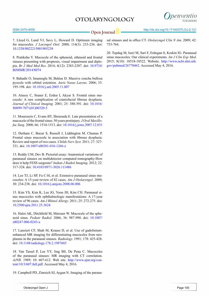
OTOLARYNGOLOGY
Open Journalhttp://dx.doi.org/10.17140/OTLOJ-2-121
Otolaryngol Open J
ISSN 2470-4059
7. Lloyd G, Lund VJ, Savy L, Howard D. Optimum imaging for mucoceles. J Laryngol Otol. 2000; 114(3): 233-236. doi: 10.1258/0022215001905238
8. Pratiksha Y. Mucocele of the sphenoid, ethmoid and frontal sinuses presenting with proptosis, visual impairment and diplo-pia. Br J Med Med Res. 2014; 4(12): 2383-2387. doi: 10.9734/BJMMR/2014/8074
9. Bahadir O, Imamoglu M, Bektas D. Massive concha bullosa pyocele with orbital extention. Auris Nasus Larynx. 2006; 33: 195-198. doi: 10.1016/j.anl.2005.11.007
10. Atasoy C, Stuner E, Erden I, Akyar S. Frontal sinus mu-cocele: A rare complication of craniofacial fibrous dysplasia. Journal of Clinical Imaging. 2001; 25: 388-391. doi: 10.1016/S0899-7071(01)00320-5
11. Mourouzis C, Evans BT, Shenouda E. Late presentation of a mucocele of the frontal sinus: 50 years postinjury. J Oral Maxillo-fac Surg. 2008; 66: 1510-1513. doi: 10.1016/j.joms.2007.12.031
12. Derham C, Bucur S, Russell J, Liddington M, Chumas P. Frontal sinus mucocele in association with fibrous dysplasia: Review and report of two cases. Childs Nerv Syst. 2011; 27: 327-331. doi: 10.1007/s00381-010-1266-z
13. Reddy UM, Dev B. Pictorial essay: Anatomical variations of paranasal sinuses on multidetector computed tomography-How does it help FESS surgeons? Indian J Radiol Imaging. 2012; 22: 317-324. doi: 10.4103/0971-3026.111486
14. Lee TJ, Li SP, Fu C-H, et al. Extensive paranasal sinus mu-coceles: A 15-year review of 82 cases. Am J Otolaryngol. 2009; 30: 234-238. doi: 10.1016/j.amjoto.2008.06.006
15. Kim YS, Kim K, Lee JG, Yoon JH, Kim CH. Paranasal si-nus mucoceles with ophthalmologic manifestations: A 17-year review of 96 cases. Am J Rhinol Allergy. 2011; 25: 272-275. doi: 10.2500/ajra.2011.25.3624
16. Haloi AK, Ditchfield M, Maixner W. Mucocele of the sphe-noid sinus. Pediatr Radiol. 2006; 36: 987-990. doi: 10.1007/s00247-006-0243-x
17. Lanzieri CF, Shah M, Krauss D, et al. Use of gadolinium-enhanced MR imaging for differentiating mucoceles from neo-plasms in the paranasal sinuses. Radiology. 1991; 178: 425-428. doi: 10.1148/radiology.178.2.1987603
18. Van Tassel P, Lee YY, Jing BS, De Pena C. Mucoceles of the paranasal sinuses: MR imaging with CT correlation. AJNR. 1989; 10: 607-612. Web site. http://www.ajnr.org/con-tent/10/3/607.full.pdf. Accessed May 4, 2016.
19. Campbell PD, Zinreich SJ, Aygun N. Imaging of the parana-
sal sinuses and in office CT. Otolaryngol Clin N Am. 2009; 42: 753-764.
20. Topdag M, Iseri M, Sari F, Erdogan S, Keskin IG. Paranasal sinus mucoceles: Our clinical experiments. Int J Clin Exp Med. 2015; 8(10): 18518-18522. Website. http://www.ncbi.nlm.nih.gov/pubmed/26770462. Accessed May 4, 2016.
Page 100