Otitis media - UCL Discovery · Otitis media (OM) or inflammation of the middle ear (consisting of...
48
Otitis media Anne G. M. Schilder 1,7 , Tasnee Chonmaitree 2 , Allan W. Cripps 3 , Richard M. Rosenfeld 4 , Margaretha L. Casselbrant 5 , Mark P. Haggard 6 and Roderick P. Venekamp 7 ABSTRACT Otitis media (OM) or middle ear inflammation is a spectrum of diseases including acute otitis media (AOM), otitis media with effusion (OME, ‘glue ear’) and chronic suppurative otitis media. OM is among the most common diseases in young children worldwide. Although OM may resolve spontaneously without complications, it can be associated with hearing loss and life- long sequelae. In developing countries, chronic suppurative OM is a leading cause of hearing loss. OM can be of viral or bacterial origin; during ‘colds’, viruses can ascent through the Eustachian tube to the middle ear and pave the way for bacterial otopathogens that reside in the nasopharynx. Diagnosis depends on typical signs and symptoms such as acute ear pain and bulging of the tympanic membrane (ear drum) for AOM and hearing loss for OME; diagnostic modalities include (pneumatic) otoscopy, tympanometry and audiometry. The use of antibiotics for AOM should be carefully considered given the self-limiting nature of the disease and the risk for adverse effects and antimicrobial resistance. Insertion of ventilation (tympanostomy) tubes and adenoidectomy are common operations for OM aimed at preventing AOM recurrences and restoring hearing; however, their effectiveness is still debated. The role of hearing aids to alleviate symptoms of hearing loss in the management of OME needs further study. Despite reports of a decline in OM incidence over the past decade, attributed to implementation of clinical guidelines promoting accurate diagnosis and judicious use of antibiotics and to pneumococcal conjugate vaccination, OM continues to be a leading cause for medical consultation and antibiotic prescription and surgery in high-income countries. 1 EvidENT, Ear Institute, University College London, Royal National Throat Nose and Ear Hospital, London, UK. 2 Division of Pediatric Infectious Diseases, Department of Pediatrics, University of Texas Medical Branch, Galveston, Texas, USA. 3 School of Medicine and Menzies Health Institute Queensland, Griffith University, Queensland, Australia. 4 Department of Otolaryngology, SUNY Downstate Medical Center, Brooklyn, New York, USA.
Transcript of Otitis media - UCL Discovery · Otitis media (OM) or inflammation of the middle ear (consisting of...
Otitis media Anne G. M. Schilder1,7, Tasnee Chonmaitree2, Allan W. Cripps3, Richard M. Rosenfeld4, Margaretha L. Casselbrant5, Mark P. Haggard6 and Roderick P. Venekamp7
ABSTRACT
Otitis media (OM) or middle ear inflammation is a spectrum of diseases including acute otitis
media (AOM), otitis media with effusion (OME, ‘glue ear’) and chronic suppurative otitis media.
OM is among the most common diseases in young children worldwide. Although OM may
resolve spontaneously without complications, it can be associated with hearing loss and life-
long sequelae. In developing countries, chronic suppurative OM is a leading cause of hearing
loss. OM can be of viral or bacterial origin; during ‘colds’, viruses can ascent through the
Eustachian tube to the middle ear and pave the way for bacterial otopathogens that reside in
the nasopharynx. Diagnosis depends on typical signs and symptoms such as acute ear pain and
bulging of the tympanic membrane (ear drum) for AOM and hearing loss for OME; diagnostic
modalities include (pneumatic) otoscopy, tympanometry and audiometry. The use of antibiotics
for AOM should be carefully considered given the self-limiting nature of the disease and the risk
for adverse effects and antimicrobial resistance. Insertion of ventilation (tympanostomy) tubes
and adenoidectomy are common operations for OM aimed at preventing AOM recurrences and
restoring hearing; however, their effectiveness is still debated. The role of hearing aids to
alleviate symptoms of hearing loss in the management of OME needs further study. Despite
reports of a decline in OM incidence over the past decade, attributed to implementation of
clinical guidelines promoting accurate diagnosis and judicious use of antibiotics and to
pneumococcal conjugate vaccination, OM continues to be a leading cause for medical
consultation and antibiotic prescription and surgery in high-income countries.
1 EvidENT, Ear Institute, University College London, Royal National Throat Nose and Ear Hospital, London, UK. 2 Division of Pediatric Infectious Diseases, Department of Pediatrics, University of Texas Medical Branch, Galveston, Texas, USA. 3 School of Medicine and Menzies Health Institute Queensland, Griffith University, Queensland, Australia. 4 Department of Otolaryngology, SUNY Downstate Medical Center, Brooklyn, New York, USA.
5 University of Pittsburgh School of Medicine, Pittsburgh, Pennsylvania, USA. 6 Department of Psychology, University of Cambridge, Cambridge, UK. 7 Julius Center for Health Sciences and Primary Care, University Medical Center Utrecht, Utrecht, The Netherlands Correspondence to: A.G.M.S. [email protected] [H1] INTRODUCTION
Otitis media (OM) or inflammation of the middle ear (consisting of the middle ear cavity and
ossicles, Figure 1) is an umbrella term that encapsulates acute OM (AOM), OM with effusion
(OME, ‘glue ear’) and chronic suppurative OM (CSOM) (Table 1).[1] These conditions are closely
related and can overlap. OM is one of the most common diseases in young children. In high-
income countries, it is also a leading cause for medical consultation and antibiotic prescription
and surgery.[2-4]
AOM is characterized by the presence of fluid in the middle ear (that is, middle ear effusion
(MEE)) together with signs and symptoms of an acute infection.[5] Many children occasionally
have AOM, but an important group of children have recurrent episodes of AOM (Table 1).[5]
Recurrent AOM episodes cause frequent episodes of acute ear pain, fever and general illness
and considerable distress to children and their parents. Suppurative (pus-forming)
complications of AOM, including acute mastoiditis, meningitis and brain abscesses, are rare
given the high incidence of OM but potentially serious. These complications pose a threat in
low-income countries in particular[6,7]; an estimated 21,000 people die from complications of
OM every year[2]. The global prevalence of hearing loss associated with OM is estimated at 30
(range 0.7-95.0) per 10,000 individuals.[2] Perforation of the tympanic membrane (ear drum)
can occur as a local sequela of AOM or as a complication associated with treatment with
ventilation tubes (tympanostomy tubes).
OME is characterized by the presence of MEE behind an intact tympanic membrane, but in
contrast to AOM, OME is not associated with signs and symptoms of an acute infection.[8] The
main symptom of OME is a conductive hearing loss caused by impaired transduction of sound
waves in the middle ear due to the presence of MEE. When this hearing loss persists or recurs
frequently, it may have negative impact on language, behaviour and progress at school.[9].
OME is very common with 80% of children having had ≥1 episode of OME by the age of 10
years. OME may occur as a new onset OME after a viral infection[10] or may occur after AOM,
when the inflammatory process subsides and MEE persists. In fact, after an AOM episode, all
children have OME for some time.[11,12] OME in itself is a risk factor for AOM, illustrating the
interrelatedness of these conditions.
CSOM is defined as chronic inflammation of the middle ear and mastoid cavity; persistent or
recurrent ear discharge through an tympanic membrane perforation or a ventilation tube is the
most prominent symptom.[13] CSOM causes a conductive hearing loss and might damage the
middle ear ossicles. It also increases the risk for permanent sensorineural hearing loss (hearing
loss owing to damage to the inner ear) and intracranial complications.[13] The of this condition
varies widely between countries, but it is most common in low- and middle-income
countries.[2]
Since publication of a landmark review on OM more than a decade ago[14], important
developments worldwide have been made, in particular regarding prevention of OM through
pneumococcal conjugate vaccination and treatment of OM following new guidelines focusing
on accurate diagnosis and judicious use of antibiotics. These events have modified the
epidemiology and clinical picture of OM worldwide. In this Primer, we provide a state-of-the art
review of OM epidemiology, its underlying pathophysiology, diagnosis, impact on children and
their families and preventative and treatment options. We also discuss promising future
directions of OM research that might guide clinicians and carers to optimize the health and
wellbeing of young children with OM.
[H1] Epidemiology
[H2] Incidence and prevalence
A recent systematic review on the global burden of OM estimated the average AOM incidence
rate at 10.8 new episodes per 100 people per year.[2] This rate ranges from an average of 3.6
for central Europe to an average of 43.4 for Sub-Saharan West Africa and central Africa,
reflecting that the burden of AOM varies with economic status (Figure 2A). The total annual
number of new AOM episodes is estimated at 709 million, with 51% occurring in children <5
years of age. Global AOM incidence rates are highest in children aged 1-4 years (61 new
episodes per 100 children per year) with a peak incidence in the first year of life (45.3 new
episodes per 100 children per year).[2]
Since OME is asymptomatic and may go undetected, its incidence and prevalence has been
difficult to establish accurately. The most reliable data on the epidemiology of OME come from
large cohort studies of children from developing countries, mostly performed in the 1980s and
1990s [15-18] showing a point prevalence of OME on screening tests of up to 20%.[19] The
peak incidence of OME is around 1 year of age; by 3 years, almost all children have experienced
at least one episode of OME.[18,20]
For CSOM the average global incidence rate is estimated at 4.8 new episodes per 1,000 people
(all ages) per year (Figure 2B).[2] The total annual number of new CSOM episodes was
estimated at 31 million, with 22% occurring in children <5 years of age. Global CSOM incidence
rates are highest in the first year of life (15.4 new cases per 1,000 children per year).[2]
Recent studies from Canada[21,22], the United States[23,24,25], Netherlands[26] and UK[27]
suggest a decline in OM incidence since the mid 1990s. This decline is attributed to the
introduction of clinical guidelines recommending stricter diagnostic criteria and judicious use of
antibiotics in OM as well as the introduction of pneumococcal conjugate vaccination. By
contrast, studies from developing countries and indigenous populations continue to
demonstrate a heavy burden of OM, particularly CSOM and its complications.[2,28-30]
[H2] Social and environmental risk factors
The risk of OM is significantly influenced by a number of host and environmental factors (Figure
3). Host factors increasing the risk of OM include: young age[31], male sex[32], race and
ethnicity[32], genetic factors and a family history of OM[33], craniofacial anomaly such as cleft
palate[34], atopy [33], immunodeficiency[35], upper respiratory tract infections (URTI) and
adenoid hypertrophy [33,36], and laryngopharyngeal reflux[37]. Environmental factors
increasing the risk of OM include: low socio-economic status, exposure to tobacco smoke[33],
having older siblings[38], day care attendance[31,38,39], and use of a pacifier[40,41]. Having
been breast-fed protects against OM.[42] In developing countries, malnutrition, contaminated
water, poor hygiene, overcrowding, HIV, tuberculosis, malaria and poor access to health care
increase the risk for chronicity and complications of OM.[2,43,44]
[H1] Mechanisms/pathophysiology
Despite the high disease burden, OM in developed countries is usually uncomplicated and self-
limiting and does not result in ongoing hearing problems or developmental delay.[6] However,
in high-risk populations in both developing and developed countries considerable hearing loss
does occur with life-long sequelae. In these populations, the progression of disease is a complex
aggregate continuum of exposures to numerous social, environmental and genetic risk factors.
OM pathogenesis starts with early and dense bacterial colonization of the nasopharynx, early
onset AOM, the establishment of an acute inflammatory cycle in the middle ear as a result of
continuing exposure to infective agents, including bacterial persistence in the middle ear
through biofilm formation, viral infections and finally severe chronic ear disease (Figure 4).
[H2] Eustachian tube anatomy
An anatomical and functioning Eustachian tube not only contributes to the protection of the
middle ear against the influx of bacterial otopathogens and respiratory viruses, but is also
essential for the drainage of secretions from the middle ear space and for pressure
equalization.[1] Indeed, the anatomy of the immature Eustachian tube in infants has a central
role in the susceptibility to infections of the middle ear (Figure 1). The Eustachian tube
epithelium is the frontline defense against the passage and colonization of otopathogens from
the nasopharynx. The Eustachian tube epithelium predominantly consists of ciliated respiratory
epithelial cells, which produce antimicrobial proteins (such as lysozyme), interspersed with
goblet cells, which produce both mucoid and serous mucus. The direction of mucociliary flow
from the middle ear through the Eustachian tube to the nasopharynx in combination with
epithelial secretion of antimicrobial proteins protects again bacterial colonization of the middle
ear.
Anatomically, the Eustachian tube is shorter, wider and more horizontal in infants and young
children (<1 year) than in adults, which facilitates otopathogen transmission through to the
middle ear and increases the risk of OM.[48] Frequent placement of infants in the supine
position can also exacerbate infection risk. As children grow, the skull base extends downward,
increasing the angle of the Eustachian tube gradually from approximately 10° at birth to 45° in
adults; concurrently, Eustachian tube length increases from 13mm to 35mm.[49] These
anatomical changes as well as functional maturation of the immune system might contribute to
a reduced risk of OM as children age, even in children at high-risk of OM.
[H2] Bacterial colonization and biofilms
Early colonization of the nasopharynx with bacterial otopathogens considerably increase the
risk of subsequent episodes of OM[50,51] Streptococcus pneumoniae (or pneumococcus),
nontypeable Haemophilus influenzae and Moraxella catarrhalis are the three dominant
bacterial otopathogens reported globally, but the individual species and strain dominance are
influenced by geographical location and pneumococcal conjugate vaccine use.[52,53] For
example, Indigenous Australian children aged 1-3 months are more likely to have ≥2
otopathogens isolated from their nasopharynx than non-indigenous Australian children. In
Indigenous Australian children, early carriage of nontypeable H. influenzae increases the risk of
OM whereas in non-Indigenous Australian children early carriage of M. catarrhalis was
associated with increased risk of OM. This difference between Indigenous and non-Indigenous
Australian children is most likely the result of different environmental risk factors. [54] Only a
few studies have examined the correlation of bacterial density or load in the nasopharynx with
OM and those have been focused on children who are at specific risk for developing OM.[57,58]
Nevertheless, these studies show that bacterial density in the nasopharynx is associated with
increased risk of OM.
Bacterial biofilms (colonization of bacteria embedded in extracellular matrix and adherent to a
surface), which are known to protect bacteria against antibiotic treatment[59,60] and the host’s
immune responses, have been demonstrated in middle ears of patients with CSOM[61,62],
persistent OME[61,63] and those with OM who have failed antibiotic treatment.[63] Biofilms
have been reported to occur in MEE[63], attached to the middle ear mucosa.[64] In animals,
immunization against nontypeable H. influenzae resulted in more rapid resolution of an
established biofilm infection, [65] suggesting that vaccination can induce immune responses
that are effective against pathogens residing in biofilms in the middle ear.
[H2] Viral infection
AOM is always preceded by viral infection of nasopharyngeal and Eustachian tube epithelium —
the so-called ‘common cold’ or viral URTI (Figure 5).[66] Bacterial otopathogens that are
colonized in the nasopharynx do not cause any harm until virus initiates the inflammatory
process in the nasopharynx. A wide variety of viruses that cause URTI symptoms can induce
AOM development. These include the following viruses in the order of importance: respiratory
syncytial virus (RSV), rhinovirus, adenovirus, coronavirus, bocavirus, influenza virus,
parainfluenza virus, enterovirus and human metapneumovirus.[10,66] Viral infection creates
changes in the nasopharyngeal mucosa by modifying host immune function[67], inducing
cytokine activity and inflammatory mediators[68] and increasing bacterial colonization and
adherence through up-regulation of host cell-surface antigens that serve as bacterial receptor
sites[69,70]. Viral infection also alters mucus properties and diminishes the normal mucociliary
clearance by mucosal cells of the Eustachian tube and nasopharynx. This causes tubal
dysfunction [69,71] leading to negative middle ear pressure, which occurs more severely in
children < 24 months, compared with children 25-47 months.[72,73] Negative middle ear
pressure facilitates influx of bacteria and/or viruses into the middle ear.[72] The risk for AOM
development after URTI depends on the colonized bacterial otopathogens; the risk is lowest
with no colonized bacteria and highest with colonization by all three pathogenic bacteria.[74]
The presence of live viruses in the middle ear, in addition to bacteria, is associated with
increased inflammatory mediators and cytokines such as histamine, leukotriene B4 and IL-8,
which can in turn interfere with antibiotic penetration into the middle ear.[75-78] Virus alone
can cause AOM, both in experimental animals and in children[66]. Approximately 5% of the
MEE isolated from children with AOM contain only viruses.[79] AOM following viral URTI often
only occurs when the infection is severe enough to cause URTI symptoms and associated
Eustachian tube dysfunction. Asymptomatic viral infection does not lead to AOM.[80] Viral
infection not only leads to AOM, but also new onset OME. In children at the peak age incidence
of OM (6-47 months), the rate of AOM and OME following URTI was 37% and 24%,
respectively.[10]
[H3] The innate immunity.
Both bacteria and viruses induce middle ear inflammation and MEE.[66] Innate immune
systems include physical barriers such mucocillary-generated flows of mucus and innate
defense molecules such as lysozyme, defensins, complement factors, cytokines and
chemokines[48,81]. These systems are responsible for initiating front-line responses to
pathogens within the nasopharynx, Eustachian tube and middle ear. Activation of pattern
recognition receptors, particularly Toll-like receptors by invading otopathogens, triggers the
release of several of the antimicrobial proteins and pro-inflammatory cytokines.[82,83] Up-
regulation of these innate mechanisms is critical for the rapid resolution of OM[84]. However,
these cytokines and antimicrobial proteins can also have a pathophysiological role[83,85]
characterized by persistent inflammation of the middle ear, as observed in CSOM.[86] The
predominant bacterial pathogens for CSOM — P. aeruginosa and S. aureus[86,87] — form
biofilms with other otopatogens and elicit an elevated innate inflammatory responses, which
might contribute to the chronicity of OM and progression to CSOM despite appropriate
intervention.[87] Evidence of the elevated inflammation includes high levels of IL-8 in the
middle ear fluid[88] and elevated mRNA and protein levels of TNFα, IL6, IL1β and INFγ in the
middle ear mucosa compared with patients with chronic OME.[89]
[H3] The role of adaptive immunity.
The middle ear is an effective immunocompetent site that maintains essentially a ‘sterile’
environment within the middle ear. Adaptive immune responses reflect aspects of both
mucosal and systemic immunity. Indeed, antigen-specific secretory IgA and IgG antibodies have
been detected in the middle ear fluid and IgA-producing cells have been detected in the middle
ear mucosa in response to infection. Research is only just commencing on the middle ear cell-
mediated responses to infection, but early data suggest that Treg cells may play a pivotal part in
controlling inflammation. The literature is unclear as to whether or not deficiencies in humoral
immunity contribute to susceptibility to OM. More research is required to explore for
aberrations in adaptive immune responses as potential risk factors for susceptibility to OM. [48]
[H2] Genetic factors
Estimates of heritability of AOM and OME range from 40% to 70%[90], with boys at slightly
higher risk than girls [32]. A range of genes regulating the innate immune response are
associated with predisposition to OM.[91] Some of the heritable risk for OM might result from
cytokine polymorphisms, that can be otopathogen specific. For example, polymorphisms in
IL10, IL6 and TNFA genes are predictive of OM coincident with RSV and rhinovirus infection[92]
whereas polymorphisms in a number of signal transduction pathways, such as TLR signalling,
have been associated with both risk and disease severity of OM in human studies and mouse
models.[48,83] Most polymorphisms described to date disrupt establishment of an effective
innate immune response, but TGFβ signalling pathway polymorphisms can be
pathophysiological through interference with moderation of pro-inflammatory
responses.[83,90] Although data with respect to deficiencies in specific antibody responses in
OM-prone children to otopathogens are conflicting, the role of possible cell-mediated
dysfunction is becoming clearer. The genetic contribution to these observations is unknown
and it is possible that pathogen-host-environment interactions might have a role. Further
research is needed to fully understand the role of these genetic factors in the pathogenesis of
OM.
[H1] Diagnosis, screening and prevention
[H2] Signs and symptoms
Signs and symptoms obtained from the history-taking (including ear-specific and non-specific
symptoms; Table 2) can raise suspicion for OM but are insufficient for accurate diagnosis. For
example, typical signs and symptoms of AOM might be absent or subtle.[5] OME, by definition,
does not have signs or symptoms of acute ear infection; children can be asymptomatic and
have less obvious signs, such as hearing problems, or subtle findings, such as ear rubbing,
clumsiness, disturbed sleep, language delay or poor school performance.[8]
Ear pain is the most consistent symptom of AOM, but only 50-60% of children with AOM
complain of ear pain.[93,94] In young preverbal children, ear pain may manifest with ear
manipulation (for example, tugging, rubbing or holding), excessive crying or with changes in the
child’s sleep and behaviour patterns.[5] These latter symptoms, however, are nonspecific and,
like fever and vomiting, do not differentiate children with AOM from those with URTI.[95]
MEE is required for diagnosing both AOM and OME and its absence precludes a diagnosis of
AOM or OME [5]. However, the difficulty of confirming MEE in primary care settings helps
explain why AOM is widely over-diagnosed.[96-98] By contrast, OME might be underdiagnosed
by paediatricians compared with otolaryngologists.[98] Ear discharge, or visible discharge in the
external ear canal, can be present in AOM (with acute tympanic membrane perforation or
draining ventilation tube), CSOM (with chronic tympanic membrane perforation and persistent
drainage), or acute otitis externa (inflammation of the external ear canal). Tympanic membrane
bulging visualized by otoscopy is a key diagnostic feature for AOM.[5]
[H2] Diagnostic modalities
AOM is diagnosed by otoscopy and can be further assessed using a symptom severity scale.
Pneumatic otoscopy is the primary diagnostic modality for OME, with tympanometry and
otomicroscopy as adjunct measures. Acoustic reflectometry can be used by parents to assess
MEE. Tympanic membrane perforation associated with CSOM may be diagnosed with otoscopy
or otomicroscopy, but may require removal of ear discharge by suctioning for adequate
visualization.
[H3] Symptom severity scales for AOM.
Several validated, parent-reported symptom scales have been developed to assess AOM
severity. The AOM Severity of Symptoms Scale (AOMSOS) is a 7-item scale with response
options of ‘no’, ‘a little’, or ‘a lot’ for the prevalence over the past 12 hours of ear pain, ear
tugging, irritability, difficulty sleeping, eating less, less playful, and fever.[99] The overall
AOMSOS score discriminates among children with and those without AOM, but all signs and
symptoms can be present to varying degrees in children with normal ears.[93] Another severity
measure, the AOM Faces Scale (AOM-FS), uses a scale with 7 choices ranging from 1 (not
present, not a problem) to 7 (extreme problem).[100]
[H3] Otoscopy.
Otoscopy is the mainstay of AOM diagnosis (Table 2; Figure 6). Obstructing cerumen (ear wax)
that prevents adequate visualization of the tympanic membrane must be removed to facilitate
accurate diagnosis.[101] When performing otoscopy, the clinician assesses and records ear
drum colour, opacity, position and integrity. A bulging ear drum, which is associated with a high
level of bacterial pathogens in the MEE[102] is the most consistent sign of AOM[94,103] (Figure
5), and is the most useful features for differentiating AOM from OME.[104] As the bulging
subsides, the tympanic membrane may have a cobblestoned appearance
(shagrination).[105,106] An opaque or cloudy tympanic membrane is highly predictive of MEE,
regardless of cause.[103] Several image-based scales exist to standardize recording and
interpretation of otoscopic findings.[106,107]
[H3] Pneumatic otoscopy.
Pneumatic otoscopy has been recommended as the primary diagnostic method for OME (Table
2) [8] because of its excellent diagnostic accuracy.[103,108] Otoscopy alone, without a
pneumatic bulb, might overlook OME because the tympanic membrane might appear normal
and ear-related symptoms can be minimal or absent. Conversely, pneumatic otoscopy can
avoid false-positive diagnoses of OME caused by surface abnormalities in the tympanic
membrane without MEE.[8] Distinctly impaired mobility of the tympanic membrane on
pneumatic otoscopy is highly predictive of OME[94,103] and improves diagnostic accuracy over
otoscopy alone.[109,110] However, the use of pneumatic otoscopy in clinical practice is
variable across the world; in the United States alone prevalence ranges from 7% to
33%.[111,112] Training medical residents in pneumatic otoscopy is challenging[5], but can be
enhanced with a structured, computerized curriculum with static and dynamic images of the
tympanic membrane.[97]
[H3] Otomicroscopy.
Otomicroscopy might help more than simple otoscopy in diagnosing OME (Table 2) [113], but
evidence is sparse and the need for special equipment and training often limits the examination
to secondary care. Otomicroscopy is most useful for assessing tympanic membrane
abnormalities (such as perforation, atrophy, tympanosclerosis, atelectasis and retraction
pockets) that may be associated with COME.[114]
[H3] Tympanometry.
Tympanometry objectively measures tympanic membrane mobility and middle ear function
(Table 2, Figure 6).[115] Compared with pneumatic otoscopy, tympanometry has comparable
sensitivity (range, 90-94%) but lower specificity (50-75% vs. 80% for tympanometry and
pneumatic otoscopy, respectively) for diagnosing OME[116] Barriers to tympanometry in
primary care settings include equipment cost and limited training, but tympanometry is easier
to perform and more useful in managing children with OM than pneumatic otoscopy.[117]
Tympanometry also estimates the equivalent ear canal volume, defined as the amount of air in
front of the probe, normally 0.3-0.9 ml in children.[118] A low equivalent volume (<0.3 ml)
could indicate an inaccurate reading because the ear canal is obstructed by cerumen or when
the probe is pressed against the canal wall; a high equivalent volume (1.0-5.5 ml) occurs when
the tympanic membrane is not intact because of a perforation or ventilation tube, and should
prompt further examination if neither was initially suspected. Tympanometry is generally
performed using a 226 Hz tone, but for children <6 months in age a 1,000 Hz probe tone is best
as the 226 Hz tone is insensitive to MEE.[119]
[H3] Acoustic reflectometry.
Acoustic reflectometry measures how much sound is reflected off the tympanic membrane,
with higher reflectivity indicating a greater probability of MEE (Table 2).[120] Advantages over
tympanometry include ease of use, no requirement for a hermetic seal, and availability of an
inexpensive consumer version, which can be used reliably by parents to monitor their child’s
middle ear status.[121] Reflectometry in some studies is less sensitive[122] and specific[123]
than tympanometry in detecting MEE, but its high specificity and negative predictive values
make reflectometry useful for ruling out MEE in children with upper respiratory tract
infections[124].
[H2] Screening
AOM is symptomatic and does not require screening. However, even screening of OME, which
is asymptomatic, has not been found useful because of the high incidence and recurrence in
young healthy children[8], the self-limited nature of most episodes[6], and the lack of
significant differences in developmental outcomes (language, behavioural problems, or
intelligence scores) between children not screened for OME and children with OME identified
by screening who have received expeditious ventilation tube insertion.[125] Current guidelines,
therefore, recommend against routine screening of otherwise healthy, asymptomatic children
for OME.[8]
Conversely, screening for OME is recommended at age 12-18 months for children with sensory,
physical, cognitive, or behavioural factors placing them at increased risk for developmental
comorbidities (Box 1). [8] OME accounts for about two-thirds of newborn hearing screen
failures.[126,127] Clinicians who manage these failures should know that only around 10% of
children with OME identified by hearing screening may also have the targeted concurrent
sensorineural hearing loss. This may interfere with detecting an underlying sensorineural
hearing loss because it may take several months after resolution of MEE for the extra impact of
an OME history on hearing ability to completely resolve.[128]
[H2] Prevention
Because OM is a multifactorial disease, a variety of strategies can be used for prevention. The
strategies mainly focus on reducing modifiable risk factors such as bacterial and viral infections
and environmental risks. Chemoprophylaxis using antibiotics and surgical interventions to
reduce the burden of OM to children are discussed in the Management section.
[H3] Vaccines directed against bacterial otopathogens.
The goal of the vaccines is to reduce or eliminate nasopharyngeal colonization of S.
pneumoniae, nontypeable H. influenzae and M. catarrhalis. The 7-valent pneumococcal
conjugate vaccine (PCV7), directed against 7 serotypes of the S. pneumonia, became available
in the United States and many European countries in 2000. The vaccine was added to the
primary series of universal vaccination at 2, 4 and 6 months, with a booster dose at 12-15
months. PCV7 was associated with 29% reduction in AOM caused by pneumococcal serotypes
contained in the vaccine, 6-7% reduction in overall AOM and 20% reduction in the use of
ventilation tube for chronic recurrent OM.[129-131] PCV13, available a decade later, has been
associated with further reduction of AOM, mastoiditis and ventilation tube insertions.[24]
The use of PCVs has led to replacement of serotypes of S. pneumoniae in the nasopharynx by
the serotypes that are not covered by the vaccine and nontypeable H. influenzae in vaccinated
children.[132,133] Nevertheless, the pneumococcal-associated AOM may continue to decrease
with PCV vaccination as the serotypes with greater capacity to cause AOM are replaced by less
otopathogenic serotypes.[134] There is now also growing evidence to support the hypothesis,
at least in developed countries, that the prevention of OM associated with the pneumococcal
serotypes present in the vaccine in young children results in a reduction of subsequent and
more-complex disease caused by non-vaccine serotypes and nontypeable H. influenzae.
Vaccination might, therefore, disrupt the continuum of evolution from pneumococcal-
associated OM towards chronic OM.[135,136] However, in communities in which there is early
and dense bacterial acquisition in the nasopharynx, and in some geographical regions such as
Oceania, nontypeable H. influenzae may be a primary otopathogen [53].
Importantly, PCVs do not prevent OM episodes if vaccination occurs after the first
episode.[137] The 10-valent pneumococcal vaccine with nontypeable H. influenzae protein D as
carrier protein (PD-PCV10) was designed to protect against both S. pneumoniae and
nontypeable H. influenzae and is available in Europe. Although effective for pneumococcal-
associated OM, PD-PCV10 may be less protective for nontypeable H. influenzae than originally
reported in a prototype vaccine study.[28,138-140] No other licensed vaccine against
nontypeable H. influenzae or M. catarrhalis exists, but numerous vaccines are in various stages
of development.
[H3] Vaccines directed against respiratory viruses.
As AOM is generally preceded by symptomatic viral URTI[10], prevention of viral URTI may
make an impact on AOM incidence. To date, the only available vaccines against viral respiratory
infection are for influenza virus. Trivalent flu vaccines (protecting against three influenza virus
strains), both inactivated influenza vaccines and live-attenuated influenza vaccines, have been
shown to reduce AOM during influenza seasons.[141-145] The vaccines work through
preventing influenza and influenza-associated AOM, which occurs in up to two-thirds of young
children with influenza.[141] The effectiveness in AOM prevention varies from year to year
depending on the level of influenza activity in the community and how well-matched the
vaccines are for the circulating strains. Recommendations for influenza vaccination in children
vary worldwide: influenza vaccines are recommended for children ≥6 months of age in the
United States[146], whereas the recommendation is restricted to children from the age of 2
years in the UK[147] and is restricted to children with significant medical comorbidities
including respiratory, cardiovascular, metabolic and renal disease in the Netherlands.[148]
[H3] Non-vaccine approaches to prevent viral URTI and AOM.
AOM occurs mostly on days 2-5 after URTI onset[10,149]; thus, early administration of antivirals
during uncomplicated URTI may prevent AOM. Studies have shown reduction in AOM
development by 43-85% in young children treated with oseltamivir within 12-48 hours of
influenza symptom onset.[150,151] However, a recent meta-analysis of both children and adult
data concluded that neither oseltamivir nor zanamivir significantly reduced OM risk.[152]
Echinacea, an immune-modulator and mild antiviral often used as a home remedy, has been
reported to reduce risks of recurrent respiratory infections, including virologically confirmed
cases, and OM.[153] Xylitol, a 5-carbon naturally occurring sugar alcohol with antibacterial
properties, has been shown to prevent recurrent AOM with some success.[154-156] However,
the successful dose regimens (that is, chewing gum or syrup given 5 times per day continuously
for 2-3 months) are not practical. Probiotics, mostly Lactobacillus and Bifidobacterium, have
been used to reduce risks of respiratory symptoms and OM and results have been encouraging
but warrant further investigation.[157-160]
[H3] Environmental risk factors.
Avoidance of well-known environmental risks such as daycare attendance, exposure to tobacco
smoke and use of pacifiers, especially during the OM peak age incidence (6-24 months), has
been associated with reduction of OM.[39,161-163] On the other hand, the benefit of
breastfeeding in preventing OM has long been known. Breastfeeding protects against OM for
the first 2 years and protection is greater for those who were exclusively breastfed and those
who were breastfed for a long duration (≥6 months).[25,42,161,162] Current guidance
recommends avoidance of tobacco smoke exposure, recommends exclusive breastfeeding for
≥6 months and discusses other lifestyles changes such as avoidance of supine bottle feeding,
reducing use of pacifier and consideration of alternative child care arrangements (for example,
with smaller groups or using a child minder).[5]
[H1] Management
[H2] AOM
Symptomatic management of ear pain and fever with analgesics at the appropriate age-
adjusted dose is the mainstay of AOM management.[5] Both oral paracetamol and ibuprofen
are effective in relieving ear pain.[164] Topical analgesics might provide additional brief benefit,
but current evidence on their effectiveness in relieving ear pain is limited.[165] An ongoing UK
trial is assessing the clinical and cost effectiveness of ear drops containing a combination of
benzocaine and phenazone as compared to placebo drops and no drops in children aged 6 to 10
years presenting in primary care with AOM.[166]
Oral antibiotics reduce the duration of AOM symptoms and consecutive MEE, but lead to
adverse effects like gastrointestinal symptoms and skin rash.[167] Their routine use in a
condition as common as AOM also enhances the risk of antimicrobial resistance, both on a
community as well as an individual level.[168] Because AOM runs a favourable natural course in
most otherwise healthy children, with symptoms settling within a few days and complications
being rare, the benefits and costs of antibiotic treatment need to be carefully weighed.[167]
The benefits are most prominent in children <2 years in age with bilateral AOM and in those of
any age presenting with acute ear discharge due to AOM.[169] Current guidance, therefore,
recommends considering immediate antibiotics in these children.[170] Immediate antibiotics
treatments is recommended in those with AOM who are <6 months in age,
immunocompromised or have craniofacial malformations, as well as those with severe illness
due to AOM.[5,170] In children with uncomplicated, non-severe AOM who are not at increased
risk of complications, watchful waiting or delayed antibiotic prescription (only filed when
symptoms of AOM persist for 48-72 hours) is recommended. Watchful waiting involves careful
monitoring of the disease course by the caregivers with specific instructions to return in case of
persistent symptoms or worsening of the child’s condition.[5,170] Limited evidence suggests
that amoxicillin (with or without clavulanic acid) is more effective than macrolides and
cephalosporin[171] and, therefore, first-line treatment with cefdinir, cefuroxime or
clarithromycin have been recommended as alternatives in patients with penicillin
allergy.[5,170] In choosing the appropriate antibiotic regimen, it is important that local
antimicrobial resistance patterns are taken into account.
Topical and oral decongestants, antihistamines and corticosteroids have either not been proven
effective or have shown conflicting results in resolving symptoms of AOM are, therefore, not
recommended.[172,173] Tympanocentesis or myringotomy, a small incision of the tympanic
membrane allowing the fluid to drain from the middle ear, may have a role in determining the
pathogens causing AOM, but is ineffective as a treatment modality for AOM.[174-176]
[H2] Recurrent AOM
Management of children with recurrent AOM focuses on the prevention of further AOM
episodes. Although immuniziation with pneumococcal conjugate vaccines in early infancy has
proved effective in reducing children’s risk of developing recurrent AOM, these vaccines are no
longer effective for children with established rAOM.[137] Antibiotic prophylaxis in children with
rAOM reduces the number of AOM recurrences by 1.5 per year (from 3 recurrences to
1.5).[177] However, their use is not recommended given the adverse effects associated with
prolonged antibiotic treatment and emerging antibiotic resistance.
The role of ventilation tubes in the management of children with rAOM has not been fully
established (Figure 7). Evidence on the benefits of ventilation tubes is mainly available for the
first 6 months after insertion: with approximately one AOM episode being prevented the
magnitude of its effect is modest.[178-180] Although not definitive, current evidence regarding
natural history and treatment benefits suggests that ventilation tubes are not helpful for rAOM
without persistent MEE but are an appropriate option for managing rAOM with persistent MEE
in one or both ears at the time of assessment for tube candidacy.[181]
The adenoids serve as a nasopharyngeal reservoir of respiratory pathogens and when enlarged
may cause obstruction of the nasal airway and impair Eustachian tube function. Surgical
removal of the adenoid, adenoidectomy, is practiced in children with rAOM to improve middle
ear function and thereby prevent further AOM episodes. A recent meta-analysis combining the
individual patient data of ten trials has shown that for recurrent AOM, adenoidectomy as a
stand-alone operation or as an adjunct to ventilation tube insertion is most beneficial in
children <2 years of age. The magnitude of the effect of this surgical intervention is, however,
modest so these benefits should be carefully balanced against any harms associated with this
surgical procedure.[182]
[H2] OME
The main sign or symptom of OME is hearing loss; management of OME is, therefore, primarily
aimed at alleviating or restoring hearing. OME settles spontaneously in many children within
several months[6] and medical treatments such as decongestants, antihistamines and
(intranasal) corticosteroids are either ineffective or may cause adverse effects[184-186].
Consequently, current guidelines recommend a 3-month period of watchful waiting in children
with OME who are not at particular risk for speech, language or learning problems.[181,183]
Ventilation tubes are an option in those still with documented hearing difficulties after 3
months.[181,183,187] Adenoidectomy as a standalone operation or as an adjunct to tube
insertion is most beneficial in children with OME aged ≥4 years.[182] In this subgroup of
children, adjuvant adenoidectomy has been shown to reduce the need for ventilation tube re-
insertions by around 10% as compared with tubes alone.[182] The role of hearing aids to
alleviate hearing loss in children with OME is unresolved[181] Hearing aids are currently
recommended for children with persistent bilateral OME in whom surgery is contraindicated or
not acceptable.[183] Recently, nasal balloon auto-inflation has been shown effective in clearing
MEE and improving ear symptoms at 3 months in school-aged children with recent onset of
OME presenting in primary care.[188] However, the effects observed were modest with a
number needed to treat to benefit of 9 patients, at a cost of GBP£132 per case resolved.[188]
Whether this approach reduces the need for ventilation tubes is yet to be answered. The same
applies to an ongoing UK trial assessing the clinical and cost effectiveness of a 7-day course of
oral corticosteroids in children aged 2-8 years with persistent bilateral OME and hearing
loss.[189] Balloon dilatation of the Eustachian tube has been proposed as a novel treatment for
children with persistent OME. There is however no evidence yet to support this management
option.[190]
[H2] Ventilation tube-associated ear discharge
Many children with ventilation tubes develop episodes of acute ear discharge; reported
incidence rates range from 26% to 75%.[191-193] These episodes may be accompanied by foul
odour, pain, and fever and can reduce the child's quality of life. They are thought to be the
result of AOM, whereby middle-ear fluid drains through the tube. Risk factors include young
age, rAOM as the indication for tubes, recent history of recurrent URTIs and the presence of
older siblings.[192] Bacterial biofilm formation on the ventilation tube may also play a role, in
particular when ear discharge recurs or becomes chronic.
Episodes of ear discharge can occur in the immediate postoperative period or at a later stage.
Management, therefore, focuses either on prevention at that early stage or treatment of
episodes occurring later. Many perioperative interventions have been tested and shown to be
of some benefit in preventing early postoperative ear discharge: saline washout of the middle
ear or application of antibiotic with or without corticosteroid ear drops during tube surgery and
the use of topical or systemic antibiotics during the early post-operative period.[194] The
largest effects of these interventions were found in studies where the risk of children
developing early postoperative ear discharge was high.[194] The bacterial otopathogens most
commonly found in acute ear discharge in children with ventilation tubes are H. influenzae, S.
aureus and P. aeruginosa and most infections are polymicrobial.[195] Most ototopical antibiotic
formulations cover these pathogens. Concerns, however, about their potential ototoxic side
effects when used in patients with a non-intact tympanic membrane, have prompted many
physicians to treat these children with systemic antibiotics. Quinolone (antibiotic) eardrops
have so far not shown ototoxicity and are recommended in the US over systemic
treatment.[181] Based upon a recent landmark trial showing that antibiotic and corticosteroid
ear drops are the most clinically and cost-effective management strategy in children developing
uncomplicated, acute ear discharge outside the immediate postoperative period[196,197],
current guidance recommends ototopical antibiotic drops as first-line treatment in these
children.[181] There is some evidence that eardrops containing a combination of antibiotic(s)
and a corticosteroid are superior over those containing antibiotic(s) alone.[198,199]
[H2] CSOM
Topical quinolone has been shown to be more effective than no drug treatment, topical
antiseptics and systemic antibiotics in clearing CSOM-related aural discharge in the short-term
(less than 4 weeks).[200,201] Current evidence assessing the effectiveness of quinolone versus
non-quinolone containing eardrops is inconclusive [201], with quinolones having the advantage
of being non-ototoxic.[202] Limited evidence suggests that treating patients with CSOM with a
combination of systemic and topical antibiotics is not more effective than topical antibiotics
alone.[200] Two recent reviews comparing two different autologous graft materials to repair
the tympanic membrane perforation (that is temporalis muscle fascia tympanoplasty with
cartilage tympanoplasty), found fewer post-operative tympanic membrane perforations with a
cartilage graft but no differences in terms of hearing.[203,204]
[H1] Quality of life
[H2] Measurement challenges
Traditionally, articles citing policy relevance of the impact of OM have focused on the economic
burden of the relevant healthcare, which is for example USD$5 billion annually in the United
States [205] As in other fields of medicine, formal measurement of quality of life (QoL) in OM
came late, dating mostly from the mid-1990s.[206] Most clinicians and researchers focus on
capturing the impact of OM and OM management with disease-specific symptoms, not impact
on QoL. Consequently, many instruments labelled ‘QoL’ are in fact OM symptom scores, and
mapping such scores to generic QoL changes the scale but not the level of generality of the
measure or its pattern of associations. Particular challenges of measuring generic QoL in OM,
are expected small effect sizes (OM being a common but often ‘mild’ disease), inaccuracies
owing to inevitable delay in documenting the essential parameter of persistence in an episodic
condition such as OM, and the need for proxy (parent or other carer) response [207].
[H2] Instruments
For OM, various validated QoL instruments are now available. There are short questionnaires
suitable for routine or audit use in a clinical setting and longer, more in-depth instruments for
more intensive QoL research. The OM-6 [208] has an efficient ‘any of the following’ item
format, which maximizes generality and ecological validity per item; it has a low burden for the
responder, but consequently leaves ambiguity about details of the presentation profile. More-
traditional instruments such as OM8-30 [206], its short form the OMQ-14 [209] and COMQ-12
for CSOM in adults [210], support from three to five scores. Brevity (few items) limits precision
and reliability, hence study power. Given the use of large sample sizes, brevity will still permit
‘positive findings’, that is, they will avoid false-negative error but may leave true scope and
effect sizes uncertain. Nevertheless, brevity encourages widespread routine adoption and the
advent of large-scale data registries creates an opportunity not to be missed. Routine use of
these questionnaires in the clinical setting can provide a useful link between research and
practice in general.
[H2] OME
The traditional picture of OME is semi-symptomatic and the major concern is with hearing loss
and consequent problems with speech, language, communication, social engagement,
schooling and behaviour, readily illustrated in descriptive studies, rather than on health
symptoms [211] These sequelae are largely generic, although not totally comprehensive for
generic QoL. The literature on QoL in OME has preferred these cognitive or academic
performance measures. For example, a large longitudinal cohort study found that a conjunction
(synergism) of OM history with poor socio-developmental environment gave the worst
outcomes on IQ.[212] Because of the importance of schooling for QoL, some knock-on effects
from academic problems to generic QoL would be expected, but no quantitative case-control
study has yet been done to demonstrate that link directly. Among children over age 5 years, , a
28-item (therefore, highly reliable) generic health questionnaire [213] showed consistent
deficits on most subscales of QoL, even in those without concurrently active OM. In a very small
uncontrolled study on OME management, pervasive QoL improvements measured by OM-6
were claimed after ventilation tubes insertion, compared to before the surgery [214], but
interpretation is unclear. This limited treatment literature suggests that effective interventions
do not necessarily result in measurable magnitudes of QoL improvement.[215]
[H2] Recurrent AOM
A large study among children with chronic or recurrent OM showed that children with rAOM or
a combination of rAOM and OME scored worse in 4 out of 6 domain items in the OM-6
(physical suffering, emotional distress, activity limitations and caregiver concerns) than children
with OME alone [217] In children with OM history diagnosed in primary and secondary care, the
number of AOM episodes was found to be a strong determinant of the child’s QoL [218]. A
primary care-based cohort study confirmed sleep disturbance as an important correlate of loss
of QoL in parents with rAOM.[219] In a clinical population of children with rAOM, the impact on
generic QoL equalled that of a comparison group of children with asthma [216], a useful anchor
on magnitude of impact. Two studies have addressed the impact of the fact that QOL
questionnaires are completed by of the proxy (caregiver), given that the QOL of the caregiver is
also affected by OM episodes of the patients [218,219]. A distinct dose/response effect
between the number of episodes and the reduction in QOL of the caregiver has been observed
[216]. Insertion of ventilation tubes in children with rAOM or combined rAOM and OME group
resulted in an important improvement on the OM-6 scale .[217] A recent trial showed that a
reduction of episodes by adenoidectomy did not lead to a measurable corresponding
improvement in QoL in young children with rAOM.[220] Vaccination improved specific but not
generic QOL outcomes.[221]
[H1] Outlook
A decline in OM incidence over the past decade has been reported, which might in part be
attributed to pneumococcal conjugate vaccination, implementation of clinical guidelines
emphasizing accurate diagnosis and more judicious use of antibiotics. Nevertheless, OM
continues to be among the most common diseases of infants and children and a prime
indication for antibiotic prescribing and surgery in children.[2-4] With growing concerns about
emerging antimicrobial resistance, further research should be designed to achieve further
reduction in antibiotic use in OM by improving its diagnosis and implementation of guidelines.
A better understanding of OM pathophysiology is also necessary to develop novel preventative
and therapeutic approaches.
[H2] Pathophysiology
Further studies are needed to examine the relationship between environmental risk factors,
bacterial density in the nasopharynx, bacterial biofilm formation, genetics and OM particularly
in respect to disease severity. Specific interactions between bacteria and viruses in the
nasopharynx may enhance AOM risk in children.[25,222] Future research should focus on the
interplay between viruses and bacteria. Better understanding of these complex mechanisms
could lead to new bacterial and viral vaccines that would help to reduce the burden of AOM.
Currently, research is ongoing focusing on the innate immune responses and interactions with
otopathogens to better understand the balance between the processes of effective recovery
from infection versus facilitation of chronic inflammation. Whereas a number of genetic
polymorphisms have been described in genes encoding for proteins involved in innate
immunity, their clinical relevance to risk and severity of disease is yet unknown in children.
Improved understanding of the interactions between host innate immune system and
otopathogens may lead to a wider range of treatment options.[223]
[H2] Diagnosis
AOM tends to be over-diagnosed (and thus over-treated), especially in the primary care setting,
owing to difficulties of confirming MEE.[96-98] Improving MEE diagnosis requires further work
to determine the optimal methods for teaching (pneumatic) otoscopy to trainees and clinicians
and to develop cost-effective methods to accurately detect MEE, such as handheld
(ultrasonography or tympanometry) devices. Multifrequency tympanometry and wideband
acoustic transfer functions are promising technologies for identifying middle ear disorders, but
limited evidence restricts conclusions on their diagnostic accuracy.[224] Future research is
needed to investigate whether these techniques provide any added value over current
diagnostic tests.
[H2] Biomarkers
Thus far, investigators have studied biomarkers in the serum and nasopharyngeal secretions
and correlated them with AOM diagnosis, types of bacteria or viruses, and outcome. High
serum Intercellular Adhesion Molecule 1 (ICAM1) was found in children with AOM compared to
healthy children.[225] At time of AOM onset, serum S100-A12 protein was elevated and this
returned to normal during recovery.[226] In children with AOM, high serum granulocyte-colony
stimulating factor (G-CSF) concentrations predicted RSV-induced AOM whereas high IL-13
concentrations predicted early clinical failure of antibiotic treatment.[227] Elevated serum IL-10
was associated with pneumococcal-induced AOM.[228] A serum biomarker risk score has been
developed to predict the presence and recovery from AOM caused by nontypeable H.
influenzae.[229] In nasopharyngeal secretions, IL-1B and lactate dehydrogenase concentrations
were associated with the risk for AOM development after viral URTI.[230,231] These data
together suggest that specific systemic and local biomarkers are helpful in predicting AOM
development, microbiology and clinical outcome. Further studies are required to explore other
biomarkers and to evaluate the usefulness of biomarker determination in clinical practice.
[H2] Prevention
Although vaccination against S. pneumoniae has been associated with a decline in OM
incidence, widespread use of PCVs has been associated with shifts in pneumococcal serotypes
and increased importance of nontypeable H. influenzae as a cause of AOM.[24,25,27,129-134]
There is a need for effective pneumococcal vaccines that cover more serotypes and effective
vaccines for H. influenzae and M. catarrhalis. In theory, protein-based vaccines would be
simpler and less costly to produce than conjugate vaccines. There are several protein vaccine
antigens of S. pneumoniae, H. influenzae, and M. catarrhalis at various stages of development;
licensing these vaccines will be an additional challenge.[232-37] So far, influenza virus
vaccination is the only viral vaccine that has been shown of some efficacy in OM. Future goals
to prevent OM by preventing viral URTI will need to include vaccines against other viruses.
Considerable efforts have been made in RSV vaccine development; numerous RSV vaccines are
in phases I and II clinical trials.[238] Further work is needed to establish whether other viral
vaccines are able to prevent OM.[235] Probiotics have been used in preventing OM with some
encouraging results [157-160], but further studies are required to identify the most promising
probiotic strains and to elucidate the mechanisms by which probiotics prevent OM. A recent
systematic review provided an overview of the global microbiology of AOM and OME between
1970 and 2014.[53] There are clear regional and temporal differences which have been
influenced by the introduction of pneumococcal conjugate vaccines. Hence it is important that
ongoing microbial surveillance be introduced to monitor shifts in causative otopathogens.
[H2] Treatment
Remarkably, most trials so far in OM have excluded the children that are most prone to the
condition: those with Down syndrome and craniofacial malformations like cleft palate. High-
quality studies evaluating the use of screening for OME and effectiveness of various
management strategies in these in at-risk children are a priority. Current approaches that need
further work include topical antibiotics for AOM with ear discharge due to a spontaneous
tympanic membrane perforation. Topical antibiotics approach has proven very effective in
children with ventilation tubes[196], but it is uncertain if these results are also applicable to
children without tubes.[239] In addition, ongoing research on transtympanic delivery of drugs
(that is without a tympanic membrane perforation or tube) is very promising. In a chinchilla
model, an application of an antibiotic-containing (ciprofloxacin) gel to the tympanic membrane
achieved antibiotic concentrations in middle ear fluid adequate for AOM treatment.[240,241]
Further work is needed to establish what methods of application are most practical and
effective in humans. The role of hearing aids and other acoustic approaches such as sound field
amplification in the management of children with OME is currently unresolved; there is an
urgent need for high quality evidence, particularly in at-risk children.[181] In CSOM, various
novel adjuvant treatments have been tested aimed at enhancing tympanic membrane
perforation repair, including biomolecules to stimulate growth of the perforation edges and
bioengineered scaffolds.[242,243] Further work is necessary to establish its role in clinical
practice.
Across all areas of epidemiology, prevention and treatment of OM, it is important that clinicians
and researchers agree on disease definitions, study methodologies and core outcome measures
so that results can be pooled or contrasted across future studies.[244-247] Recently, a
recommendation has been made for the outcomes that should be measured in studies of the
management of OME in children with cleft palate.[248] We encourage the development of core
outcome sets for all patient groups and all manifestations of OM including generic impact. We
strongly recommend that parents and children be systematically consulted, at an appropriate
level of detail, about the goals and that they are are involved in the planning process as well as
in all other stages of research in OM. By adding relevance to the children with OM and theirs
carers, high quality research with the statistical power and freedom from confounding can be
given additional ability to change practice for the better.
References
1. Bluestone CD. Definition, terminology, and classification. In: Rosenfeld RM, Bluestone CD (eds). Evidence-based Otitis Media, 2nd ed. Hamilton: BC Decker Inc, 2003;121.
2. Monasta L, Ronfani L, Marchetti F, Montico M, Vecchi Brumatti L, Bavcar A, et al. Burden of disease caused by otitis media: systematic review and global estimates. PLoS One 2012;7:e36226.
Comprehensive review providing global estimates on the burden of disease caused by OM.
3. Gulliford M, Latinovic R, Charlton J, Little P, van Staa T, Ashworth M. Selective decrease in
consultations and antibiotic prescribing for acute respiratory tract infections in UK primary care up to 2006. J Public Health 2009;31:512-20.
4. Cullen K, Hall M, Golosinskiy A. Ambulatory surgery in the United States, 2006. National health statistics reports, no. 11, revised. Hyattsville, MD: National Center for Health Statistics, 2009.
5. Lieberthal AS, Carroll AE, Chonmaitree T, Ganiats TG, Hoberman A, Jackson MA, et al. Clinical Practice Guideline The Diagnosis and Management of Acute Otitis Media. Pediatrics 2013;133(2):346.
6. Rosenfeld RM, Kay D. Natural history of untreated otitis media. Laryngoscope 2003;113:1645-57.
7. Thompson PL, Gilbert RE, Long PF, Saxena S, Sharland M, Wong IC. Effect of antibiotics for otitis media on mastoiditis in children: a retrospective cohort study using the United kingdom general practice research database. Pediatrics 2009;123:424-30.
8. Rosenfeld RM, Shin JJ, Schwartz SR, Coggins R, Gagnon L, Hackell JM, et al. Clinical practice guideline: otitis media with effusion (update). Otolaryngol Head Neck Surg 2016 (Suppl 1):S1-S41.
9. Bennett KE, Haggard MP, Silva PA, Stewart IA. Behaviour and development effects of otitis media with effusions into the teens. Arch Dis Child 2001;85:91–5.
10. Chonmaitree T, Revai K, Grady JJ, Clos A, Patel A, Nair S, et al. Viral upper respiratory tract infection and otitis media complication in young children. Clin Infect Dis 2008;46:815-23.
Large prospective study reports clear relationship between viral URTI and AOM and OME in children at the peak age incidence of otitis media.
11. Alho O, Oja H, Koivu M, Sorri M. Risk factors for chronic otitis media with effusion in infancy.
Each otitis media episode induces a high but transient risk. Arch Otolaryngol Head Neck Surg 1995;121:839-43.
12. Claessen JQ, Appelman CL, Touw-Otten FW, Hordijk GJ, de Melker RA. Persistence of middle ear dysfunction after recurrent acute otitis media. Clin Otolaryngol Allied Sci 1994;19:35-40.
13. Verhoeff M, van der Veen EL, Rovers MM, Sanders EAM, Schilder AGM. Chronic suppurative otitis media: a review. Int J Pediatr Otorhinolaryngol 2006;70:1-12.
14. Rovers MM, Schilder AG, Zielhuis GA, Rosenfeld RM. Otitis Media. Lancet 2004;363:465-73.
Landmark review on otitis media summarizing the state of knowledge, including advances in epidemiology, pathogenesis, diagnosis, clinical management, and prevention up to 2004.
15. Tos M. Epidemiology and natural history of secretory otitis. Am J Otol. 1984;5:459–62. 16. Zielhuis GA, Rach GH, Van den Broek P. The occurrence of otitis media with effusion in
Dutch pre-school children. Clin Otolaryngol Allied Sci. 1990;15:147-53. 17. Williamson IG, Dunleavey J, Bain J, et al. The natural history of otitis media with effusion--a
three-year study of the incidence and prevalence of abnormal tympanograms in four South West Hampshire infant and first schools. J Laryngol Otol. 1994;108:930–4.
18. Paradise JL, Rockette HE, Colborn K, Bernard BS, Smith CG, Kurs-Lasky M, et al. Otitis media in 2253 Pittsburgh-area infants: prevalence and risk factors during the first two years of life. Pediatrics 1997;99:318–33.
19. Casselbrant ML, Mandel EM. Epidemiology. In: Rosenfeld RM, Bluestone CD, eds. Evidence-based otitis media. Hamilton, ON: BC Decker; 1999:117–36.
20. Marchant CD, Shurin PA, Turczyk VA, Wasikowski DE, Tutihasi MA, Kinney SE. Course and outcome of otitis media in early infancy: a prospective study. J Pediatr 1984;104:826–31.
21. Wals P, Carbon M, Sévin E, Deceuninck G, Ouakki M. Reduced physician claims for otitis media after implementation of pneumococcal conjugate vaccine program in the province of Quebec, Canada. Pediatr Infect Dis J 2009;28:e271-4.
22. Thomas EM. Recent trends in upper respiratory infections, ear infections and asthma among young Canadian children. Health Reports 2010:21:1-6.
23. Grijalva CG, Nuorti JP, Griffin MR. Antibiotic prescription rates for 460 acute respiratory tract infections in US ambulatory settings. JAMA 2009;302:758–66.
24. Marom T, Tan A, Wilkinson GS, Pierson KS, Freeman JL, Chonmaitree T. Trends in Otits Media-related Health Care Utilization in the United States, 2001-2011. JAMA Pediatr 2014;168:68-75.
25. Chonmaitree T, Trujillo R, Jennings K, Alvarez-Fernandez P, Patel JA, Loeffelholz MJ, et al. Acute otitis media and other complications of viral respiratory infection. Pediatrics 2016;137(4). pii:e2015355.
26. Plasschaert AI, Rovers MM, Schilder AG, Verheij TJ, Hak E. Trends in doctor consultations, antibiotic perscription, and specialist referrals for otitis media in children: 1995-2003. Pediatrics 2006;117:1879-1986.
27. Lau WC, Murray M, El-Turki A, Saxena S, Ladhani S, Long P, Sharland M, Wong IC, Hsia Y. Impact of pneumococcal conjugate vaccines on childhood otitis media in the United Kingdom. Vaccine 2015;33:5072-9.
28. Leach, AJ, Wigger C, Andrews R, Chatfield M, Smith-Vaughn H, Morris PS. Otitis media in children vaccinated during consecutive 7-valent or 10-valent pneumococcal conjugate vaccination schedules. BMC Pediatrics 2014;14:200.
29. Dallaire F, Dewailly E, Vezina C, Bruneau S, Ayotte P. Portrait of outpatient visits and hospitalizations for acute infections in Nunavik preschool children. Can J Public Health 2006;97:362-368.
30. Morris PS, Leach AJ, Silberberg P, Mellon G, Wilson C, Hamilton E, et al. Otitis media in young Aboriginal children from remote communities in Northern and Central Australia: a cross-sectional survey. BMC Pediatr 2005;5:27.
31. Todberg T, Koch A, Andersson M, Olsen S, Lous J, Homoe P. Incidence of Otitis Media in a Contemporary Danish National Birth Cohort. PLoS One 2014;9:e111732.
32. Macintyre EA, Karr CJ, Koehoorn M, Demers P, Tamburic L, Lencar C, et al. Otits media incidence and risk factors in a population-based birth cohort. Paediatr Child Health 2010;15:437-42.
33. Zhang Y, Xu M, Zhang J, Zeng L, Wang Y, Zheng QY. Risk factors for chronic and recurrent otitis media - a meta-analysis. PLoS One 2014;9:e86397.
34. Austeng ME, Akre H, Overland B, Abdelnoor M, Falkenberg ES, Kvaerner KJ. Otitis media with effusion in children with Down syndrome. Int J Pediatr Otorhinolaryngol 2013;77:1329-32.
35. Wilson NW, Hogan MB. Otitis media as a presenting complaint in childhood immunodeficiency diseases. Curr Allergy Asthma Rep. 2008, 6:519-24.
36. Aydemir G, Ozkurt FE. Otitis media with effusion in primary schools in Princes’ Islands, Istanbul: prevalence and risk factors. J Int Med Res, 2011;39(3):866-72.
37. O’Reilly RC, Soundar S, Tonb D, Bolling L, Yoo E, Nadal T, et al. The role of gastric pepsin in the inflammatory cascade of pediatric otitis media. JAMA Otolaryngol Head Neck Surg 2015;141:350-7.
38. Brennan-Jones CG, Whitehouse AJO, Park J, Hegarty M, Jacques A, Eikelboom RH, et al. Prevalence and risk factors for parent-reported otitis media during early childhood in the Western Australian Pregnancy Cohort (Raine) Study. Journal of Paediatrics and Child Health 2015:51:403-9.
39. De Hoog MLA, Venekamp RP, van der Ent CK, Schilder A, Sanders EA, Damoiseaux RA, et al. Impact of early daycare on healthcare resource use related to upper respiratory tract infections during childhood: prospective WHISTLER cohort study. BMC Med 2014;12:107.
40. Salah M, Abdel-Aziz M, Al-Farok A, Jebrini A. Recurrent acute otitis media in infants: analysis of risk factors. Int J Pediatr Otorhinolaryngol 2013;77:1665-9.
41. Rovers MM, Numans ME, Langenbach E, Grobbee DE, Verheij TJ, Schilder AG. Is pacifier use a risk factor for acute otitis media? A dynamic cohort study. Fam Pract 2008;25:233-6.
42. Bowatte G, Tham R, Allen KJ, Tan DJ, Lau M, Dai X, et al. Breast feeding and childhood acute otitis media: a systematic review and meta-analysis. Acta Paediatr Suppl 2015;104:85-95.
43. Lasisi AO, Olaniyan FA, Muibi SA, Azeez IA, Abdulwasiu KG, Lasisi TJ, et al. Clinical and demographic risk factors associated with chronic suppurative otitis media. Int J Pediatr Otorhinolaryngol 2007;71:1549-54.
44. Taipale A, Pelkonen T, Paipale M, Bernardino L, Peltola H, Pitkäranta. Chronic suppurative otitis media in children of Luanda, Angola. Acta Paediatr 2011;100:e84-8.
45. Wiertsema SP, Leach AJ. Theories of otitis media pathogenesis, with a focus on Indigenous children. Med J Aust 2009;191:S50-4.
46. Vijayasekaran S, Coates H, Thornton RB, Wiertsema SP, Kirkham LA, Jamieson SE, et al. New findings in the pathogenesis of otitis media. Laryngoscope 2012;122:S61-2.
47. Lehmann D, Arumugaswamy A, Elsbury D, Finucane J, Stokes A, Monck R, et al. The Kalgoorlie Otitis Media Research Project: rationale, methods, population characteristics and ethical considerations. Paediatr Perinat Epidemiol 2008;22:60-71.
48. Massa HM, Lim DJ, Cripps AW. Middle ear and eustachian tube mucosal immunology. In: Russel MW, Lambrecht B (eds). Mucosal Immunology, 4th ed, 2015;1423-1942. A comprehensive review of the innate and acquired immunology of the middle ear and Eustachian tube.
49. Bluestone CD, Klein JO. Otitis media and eustachian tube dysfunction. In: Bluestone CD, Stoole SE, Alper CM (eds). Pediatric Otolaryngology, 4th ed, 2003;497-535.
50. Faden H, Duffy L, Wasielewski R, Wolf J, Krystofik D, Tung Y. Relationship between nasopharyngeal colonization and the development of otitis media in children. J Infect Dis 1997;175:1440-5.
51. Leach AJ, Boswell JB, Asche V, Nienhuys TG, Mathews JD. Bacterial colonization of the nasopharynx predicts very early onset and persistence of otitis media in Australian aboriginal infants. Pediatr Infect Dis J 1994;13:983-9.
52. Coker TR, Chan LS, Newberry SJ, Limbos MA, Suttorp MJ, Shekelle PG. Diagnosis, microbial epidemiology, and antibiotic treatment of acute otitis media in children: a systematic review. JAMA 2010;304:2161-9.
53. Ngo CC, Massa HM, Thornton RB, Cripps AW. Predominant bacteria isolated from the middle ear fluid of children experiencing otitis media: A systematic review. PLoS One 2016;11(3):e0150949.
Systematic review provides recent global estimates on the predominant bacterial pathogens involved in otitis media.
54. Sun W, Jacoby P, Riley TV, Bowman J, Leach AJ, Coates H, et al. Association between early
bacterial carriage and otitis media in Aboriginal and non-Aboriginal children in a semi-arid area of Western Australia: a cohort study. BMC Infect Dis 2012;12:366.
55. Darboe MK, Fulford AJC, Secka O, Prentice AM. The dynamics of nasopharyngeal streptococcus pneumoniae carriage among rural Gambian mother-infant pairs. BMC Infect Dis 2010;10:195.
56. Gratten M, Gratten H, Poli A, Carrad E, Raymer M, Koki G. Colonisation of Haemophilus influenzae and Streptococcus pneumoniae in the upper respiratory tract of neonates in Papua New Guinea: primary acquisition, duration of carriage, and relationship to carriage in mothers. Biol Neonate 1986;50:114-20.
57. Watson K, Carville K, Bowman T, Jacoby P, Riley TV, Leach AJ, et al. Upper respiratory tract bacterial carriage in Aboriginal and non-Aboriginal children in a semi-arid area of Western Australia. Pediatr Infect Dis J 2006;25:782-90.
58. Smith-Vaughan H, Byun R, Nadkarni M, Jacques NA, Hunter N, Halpin S, et al. Measuring nasal bacterial load and its association with otitis media. BMC Ear Nose Throat Disord 2006;6:10.
59. Slinger R, Chan F, Ferris W, Yeung SW, St Denis M, Gaboury I, et al. Multiple combination antibiotic susceptibility testing of nontypeable Haemophilus influenzae biofilms. Diagn Microbiol Infect Dis 2006;56:247-53.
60. Garcia-Cobos S, Moscoso M, Pumarola F, Arroyo M, Lara N, Pérez-Vázquez M, et al. Frequent carriage of resistance mechanisms to β-lactams and biofilm formation in Haemophilus influenzae causing treatment failure and recurrent otitis media in young children. J Antimicrob Chemother 2014;69:2394-9.
61. Lampikoski H, Aarnisalo AA, Jero J, Kinnari, TJ. Mastoid biofilm in chronic otitis media. Otol Neurotol 2012;33:785-8.
62. Gu X, Keyoumu Y, Long L, Zhang H. Detection of bacterial biofilms in different types of chronic otitis media. Eur Arch Otorhinolaryngol 2014;271:2877-83.

63. Van Hoecke H, De Paepe AS, Lambert E, Van Belleghem JD, Cools P, Van Simaey L, et al. Haemophilus influenzae biofilm formation in chronic otitis media with effusion. Eur Arch Otorhinolaryngol. 2016 Mar 5. [Epub ahead of print]
64. Hall-Stoodley L, Hu FZ, Gieseke A, Nistico L, Nguyen D, Hayes J, et al. Direct detection of bacterial biofilms on the middle-ear mucosa of children with chronic otitis media. JAMA 2006;296:202-11.
65. Novotny LA, Jurcisek JA, Ward MO Jr, Jordan ZB, Goodman SD, Bakaletz LO. Antibodies against the majority subunit of Type IV pili disperse nontypeable Haemophilus influenzae biofilms in a LuxS-dependent manner and confer therapeutic resolution of experimental otitis media. Mol Microbiol 2015;96:276-92.
66. Nokso-Koivisto J, Marom T, Chonmaitree T. Importance of viruses in acute otitis media. Curr Opin Pediatr 2015;27:110-5.
67. Abramson JS, Hudnor HR. Role of the sialophorin (CD43) receptor in mediating influenza A virus-induced polymorphonuclear leukocyte dysfunction. Blood 1995;85:1615-9.
68. Patel JA, Nair S, Revai K, Grady J, Chonmaitree T. Nasopharyngeal acute phase cytokines in viral upper respiratory infection: impact on acute otitis media in children. Pediatr Infect Dis J 2009;28:1002-7.
69. Bakaletz LO. Immunopathogenesis of polymicrobial otitis media. J Leukoc Biol. 2010;87:213-22.
Comprehensive review covering the host innate and acquired immune response involved in the pathogenesis of AOM, including information on viral-induced pathological changes in the nasopharynx and middle ear in animal models.
70. Avadhanula V, Rodriguez CA, Devincenzo JP, Wang Y, Webby RJ, Ulett GC, et al. Respiratory
viruses augment the adhesion of bacterial pathogens to respiratory epithelium in a viral species- and cell type-dependent manner. J Virol 2006;80:1629-36.
71. Pittet LA, Hall-Stoodley L, Rutkowski MR, Harmsen AG. Influenza virus infection decreases tracheal mucociliary velocity and clearance of Streptococcus pneumoniae. Am J Respir Cell Mol Biol 2010;42:450-60.
72. Buchman CA, Brinson GM. Viral otitis media. Curr Allergy Asthma Rep. 2003;3:335-40. 73. Revai K, Patel JA, Grady JJ, Chonmatree T. Tympanometric findings in young children during
upper respiratory tract infections with and without acute otitis media. Pediatr Infect Dis J 2008;27:292-5.
74. Revai K, Mamidi D, Chonmaitree T. Association of nasopharyngeal bacterial colonization during upper respiratory tract infection and the development of acute otitis media. Clin Infect Dis. 2008;46:e34-7.
75. Canafax DM, Yuan Z, Chonmaitree T, Deka K, Russlie HQ, Giebink GS. Amoxicillin middle ear fluid penetration and pharmacokinetics in children with acute otitis media. Pediatr Infect Dis J 1998;17:149-56.
76. Chonmaitree T, Patel JA, Lett-Brown MA, Uchida T, Garofalo R, Owen MJ. Virus and bacteria enhance histamine production in middle ear fluids of children with acute otitis media. J Infect Dis 1994;169:1265-70.
77. Chonmaitree T, Patel JA, Sim T, Garofalo R, Uchida T, Sim T, et al. Role of leukotriene B4 and interleukin-8 in acute bacterial and viral otitis media. Ann Otol Rhinol Laryngol 1996;105:968-74.
78. Jossart GH, Cranafax DM, Erdmann GR, Lovdahl MJ, Russlie HQ, Juhn SK, et al. Effect of Streptococcus pneumoniae and influenza A virus on middle ear antimicrobial pharmacokinetics in experimental otitis media. Pharm Res 1994;11:860-4.
79. Chonmaitree T, Ruohola A, Hendley JO. Presence of viral nucleic acids in the middle ear: Acute otitis media pathogen or bystander? Pediatr Infect Dis J 2012,31:325-330.
80. Chonmaitree T, Alvarez-Fernandez P, Jennings K, Trujillo R, Marom T, Loeffelholz MJ, et al. Symptomatic and asymptomatic respiratory viral infections in the first year of life: association with acute otitis media development. Clin Infect Dis 2015;60:1-9.
81. Murphy TF, Chonmaitree T, Barenkamp S, Kyd J, Nokso-Koivisto J, Patel JA, et al. Panel 5: Microbiology and immunology panel. Otolaryngol Head Neck Surg 2013;148:E64-89.
82. Trune DR, Kempton B, Hausman FA, Larrain BE, MacArthur CJ. Correlative mRNA and protein expression of middle and inner ear inflammatory cytokines during mouse acute otitis media. Hear Res 2015;326:49-58.
83. Leichtle A, Lai Y, Wollenberg B, Wasserman SI, Ryan AF. Innate signaling in otitis media: pathogenesis and recovery. Curr Allergy Asthma Rep 2011;11:78-84.
84. Kurabi A, Pak K, Ryan AF, Wasserman SI. Innate Immunity: Orchestrating Inflammation and Resolution of Otitis Media. Curr Allergy Asthma Rep 2016;16:6.
85. Mittal R, Kodiyan J, Gerring R, Mathee K, Li JD, Grati M, et al. Role of innate immunity in the pathogenesis of otitis media. Int J Infect Dis 2014;29:259-67.
86. Morris P. Chronic suppurative otitis media. BMJ Clin Evid 2012;pii:0507. 87. Mittal R, Lisi CV, Gerring R, Mittal J, Mathee K, Narasimhan G, et al. Current concepts in the
pathogenesis and treatment of chronic suppurative otitis media. J Med Microbiol 2015;64:1103-16.
88. Elmorsy S, Shabana YK, Raouf AA, Nagger ME, Bedir T, Taher S, et al. The role of IL8 in different types of otitis media and bacterialogical correlation. J Int Adv Otol 2010;6:269-73.
89. Si Y, Zhang ZG, Chen SJ, Zheng YQ, Chen YB, Liu Y, et al. Attenuated TLRs in middle ear mucosa contributes to susceptibility of chronic suppurative otitis media. Hum Immunol 2014;75:771-6.
90. Rye MS, Bhutta MF, Cheeseman MT, Burgner D, Blackwell JM, Brown SD, et al. Unraveling the genetics of otitis media: from mouse to human and back again. Mammalian Genome 2011;22:66-82.
91. Mittal R, Robalino G, Gerring R, Chan B, Yan D, Grati M, et al. Immunity genes and susceptibility to otitis media: a comprehensive review. J Genet Genomics 2014;41:567-81.
92. Alper CM, Winther B, Hendley JO, Doyle WJ. Cytokine polymorphisms predict the frequency of otitis media as a complication of rhinovirus and RSV infections in children. Eur Arch Otorhinolaryngol 2009;266:199-205.
93. Shaikh N, Hoberman A, Paradise JL, Rockette HE, Kurs-Lasky M, Colborn DK, et al. Responsiveness and construct validity of a symptom scale for acute otitis media. Pediatr Infect Dis J 2009;28:9–12.
94. Rothman R, Owens T, Simel DL. Does this child have acute otitis media? JAMA 2003;289:1633-40.
95. Laine MK, Tähtinen PA, Ruuskanen O, Huovinen P, Ruohola A. Symptoms or symptom-based scores cannot predict acute otitis media at otitis-prone age. Pediatrics 2010;125:1154-61.
96. Rosenfeld RM. Diagnostic certainty for acute otitis media. Int J Pediatr Otorhinolaryngol 2002;64:89-95.
97. Kaleida PH, Ploof DL, Kurs-Lasky M, Shaikh N, Colborn DK, Haralam MA, et al. Mastering diagnostic skills: enhancing proficiency in otitis media, a model for diagnostic skills training. Pediatrics 2009;124:e714-20.
98. Pichichero ME, Poole MD. Assessing diagnostic accuracy and tympanocentesis skills in the management of otitis media. Arch Pediatr Adolesc Med 2001;155:1137-42.
99. Shaikh N, Hoberman A, Paradise JL, Wald ER, Switze GE, Kurs-Lasky, et al. Development and preliminary evaluation of a parent-reported outcome instrument for clinical trials in acute otitis media. Pediatr Infect Dis J 2009; 28:5-8.
100. Friedman NR, McCormick DP, Pittman C, Chonmaitree T, Teichgraeber DC, Uchida T, et al. Development of a practical tool for assessing the severity of acute otitis media. Pediatr Infect Dis J 2006;25:101-7.
101. Roland PS, Smith TL, Schwartz SR, Rosenfeld RM, Ballachanda B, Earll JM, et al. Clinical practice guideline: cerumen impaction. Otolaryngol Head Neck Surg 2008;139:S1-S21.
102. McCormick DP, Lim-Melia E, Saeed K, Baldwin CD, Chonmaitree T. Otitis media: can clinical findings predict bacterial or viral etiology? Pediatr Infect Dis J 2000;19:256–258.
103. Karma PH, Penttilä MA, Sipilä, Kataja MJ. Otoscopic diagnosis of middle ear effusion in acute and non-acute otitis media. I. The value of different otoscopic findings. Int J Pediatr Otorhinolaryngol 1989;17:37-49.
104. Shaikh N, Hoberman A, Kaleida PH, Rockette HE, Kurs-Lasky M, Hoover H, et al. Otoscopic signs of otitis media. Pediatr Infect Dis J 2011;30:822-6.
105. Armengol CE, Hendley JO. Shagrination during acute otitis media. J Pediatr 2014;164:870.
106. Lundberg T, Hellström S, Sandström H. Development and validation of a new grading scale for otitis media. Pediatr Infect Dis J 2013;32:341-5.
107. McCormick DP, Saeed KA, Pittman C, Baldwin CD, Friedman N, Teichgraeber DC, et al. Bullous myringitis: a case-control study. Pediatrics 2003;112:982-86.
108. P Shekelle, G Takata, LS Chan, R Mangione-Smith, PM Corley, T Morphew, and S Morton. Diagnosis, Natural History and Late Effects of Otitis Media with Effusion: Evidence Report/Technology Assessment No. 55. Rockville, MD: Agency for Healthcare Research and Quality; 2003. AHRQ publication 03-E023.
109. Jones WS, Kaleida PH. How helpful is pneumatic otoscopy in improving diagnostic accuracy? Pediatrics 2003112:510-513.
110. American Academy of Pediatrics. Section on Infectious Diseases. A view through the otoscope: distinguishing acute otitis media from otitis media with effusion [video]. Available at: http://www2.aap.org/sections/infectdis/video.cfm. Accessed January 5, 2016.
111. Forrest CB, Fiks AG, Bailey LC, Localio R, Grundmeier RW, Richards T, et al. Improving adherence to otitis media guidelines with clinical decision support and physician feedback. Pediatrics 2013;131:e1071-81.
112. Lannon C, Peterson LE, Goudie A. Quality measures for the care of children with otitis media with effusion. Pediatrics 2011;127:e1490-7.
113. Lee DH. How to improve the accuracy of diagnosing otitis media with effusion in a pediatric population. Int J Pediatr Otorhinolaryngol 2010;74:151-3.
114. Schilder AG, Zielhuis GA, Haggard MP, van den Broek P. Long-term effects of otitis media with effusion: otomicroscopic findings. Am J Otol 1995;16:365-72.
115. Onusko E. Tympanometry. Am Fam Physician 2004;70:1713-1720. 116. Takata GS, Chan LS, Morphew T, Mangione-Smith R, Morton SC, Shekelle P. Evidence
assessment of the accuracy of methods of diagnosing middle ear effusion in children with otitis media with effusion. Pediatrics 2003;112:1379-87.
117. Abbott P, Rosenkranz S, Hu W, Gunsekera H, Reath J. The effect and acceptability of tympanometry and pneumatic otoscopy in general practitioner diagnosis and management of childhood ear disease. BMC Fam Pract 2014;15:181-90
118. American Speech-Language-Hearing Association. Guideline for screening infants and children for outer and middle ear disorders, birth through 18 years. In: Guidelines for Audiologic Screening. Rockville, MD: American Speech-Language-Hearing Association; 1997:15-22.
119. Hunter LL, Preve BA, Kei J, Sanford CA. Pediatric application of wideband acoustic immittance measures. Ear Hear 2013; 34(Suppl 1):36S-42S.
120. Combs JT, Combs MK. Acoustic reflectometry: spectral analysis and the conductive hearing loss of otitis media. Pediatr Infect Dis J 1996; 15:683-6.
121. Erkkola-Anttinen N, Laine MK, Tähtinen PA, Ruohola A. Parental role in the diagnostics of otitis media: can layman parents use spectral gradient acoustic reflectometry reliably? Int J Pediatr Otorhinolaryngol 2015; 79:1516-21.
122. Chianese J, Hoberman Paradise JL, Colborn DK, Kearney D, Rockette HE, et al. Spectral gradient acoustic reflectometry compared with tympanometry in diagnosing middle ear effusion in children aged 6 to 24 months. Arch Pediatr Adolesc Med 2007; 161:884-8.
123. Muderris T, Yazici A, Bercin S, Yalciner G, Sevil E, Kiris M. Consumer acoustic reflectometry: accuracy in diagnosis of otitis media with effusion in children. Int J Pediatr Otorhinolaryngol 2013; 77:1771-4.
124. Puhakka T, Pulkkinen JU, Silvennoinen H, Heikkinen T. Comparison of spectral gradient acoustic reflectometry and tympanometry for detection of middle-ear effusion in children. Pediatr Infect Dis J 2014;33:e183-6.
125. Simpson SA, Thomas CL, van der Linden M, MacMillan H, van der Wouden JC, Butler CC. Identification of children in the first four years of life for early treatment for otitis media with effusion. Cochrane Database of Systematic Reviews 2007, Issue 1. Art. No.: CD004163. DOI: 10.1002/14651858.CD004163.pub2.
126. Boone RT, Bower CM, Martin PF. Failed newborn hearing screens as presentation for otitis media with effusion in the newborn population. Int J Pediatr Otorhinolaryngol 2005;69:393-397.
127. Holster IL, Hoeve LJ, Wieringa MH, Willis-Lorrier RMS, de Gier HHW. Evaluation of hearing loss after failed neonatal hearing screening. J Pediatr 2009;155:646-50.
128. Boudewyns A, Declau F, Van den Ende J, Van Kerschaver E, Dirckx S, Hofkens-Van den Brandt A, , et al. Otitis media with effusion: an underestimated cause of hearing loss in infants. Otol Neurotol 2011;32:799-804.
129. Fortanier AC, Venekamp RP, Boonacker CWB, Hak E, Schilder AGM, Sanders EAM, et al. Pneumococcal conjugate vaccines for preventing otitis media. Cochrane Database of Systematic Reviews 2014, Issue 4. Art. No.: CD001480. DOI: 10.1002/14651858.CD001480.pub4.
130. Eskola J, Kilpi T, Palmu A, Jokinen J, Haapakoski J, Herva E, et al. Efficacy of a pneumococcal conjugate vaccine against acute otitis media. N Engl J Med 2001;344:403-9.
131. Black S, Shinefield H, Fireman B, Lewis E, Ray P, Hansen JR, et al. Efficacy, safety and immunogenicity of heptavalent pneumococcal conjugate vaccine in children. Pediatr Infect Dis J 2000;19:187-95.
132. Casey JR, Adlowitz DG, Pichichero ME. New patterns in the otopathogens causing acute otitis media six to eight years after introduction of pneumococcal conjugate vaccine. Pediatr Infect Dis J 2010;29:304-9.
133. Casey JR, Kaur R, Friedel VC, Pichichero ME. Acute otitis media otopathogens during 2008 to 2010 in Rochester, New York. Pediatr Infect Dis J 2013;32:805-9.
134. Shea KM, Weycker D, Stevenson AE, Strutton DR, Pelton SI. Modeling the decline in pneumococcal acute otitis media following the introduction of pneumococcal conjugate vaccines in the US. Vaccine 2011;29:8042-8.
135. Dagan R, Pelton S, Bakaletz L, Cohen R. Prevention of early episodes of otitis media by pneumococcal vaccines might reduce progression to complex disease. Lancet Infect Dis. 2016;16:480-92.
Review providing evidence to support the hypothesis that prevention of vaccine-serotype pneumococcal otitis media in early life lead to a reduction of subsequent and more complex disease caused by non-vaccine serotypes and nontypeable H. influenzae.
136. Ben-Shimol S, Givon-Lavi N, Leibovitz E, Raiz S, Greenberg D, Dagan R. Near-elimination of otitis
media caused by 13-valent pneumococcal conjugate vaccine (PCV) serotypes in southern Israel shortly after sequential introduction of 7-valent/13-valent PCV. Clin Infect Dis. 2014;59:1724-32.
137. Veenhoven R, Bogaert D, Uiterwaal C, Brouwer C, Kiezebrink H, Bruin J, et al. Effect of conjugate pneumococcal vaccine followed by polysaccharide pneumococcal vaccine on recurrent acute otitis media: a randomised study. Lancet 2003;361:2189-95.
138. Prymula R, Peeters P, Chrobok V, Kriz P, Novakova E, Kaliskova E, et al. Pneumococcal capsular polysaccharides conjugated to protein D for prevention of acute otitis media caused by both Streptococcus pneumoniae and non-typable Haemophilus influenzae: a randomised double-blind efficacy study. Lancet 2006;367:740-8.
139. van den Bergh MR, Spijkerman J, Swinnen KM, François NA, Pascal TG, Borys D, et al. Effects of the 10-valent pneumococcal nontypeable Haemophilus influenzae protein D-conjugate vaccine on nasopharyngeal bacterial colonization in young children: a randomized controlled trial. Clin Infect Dis 2013;56:e30-9.
140. Tregnaghi MW, Sáez-Llorens X, López P, Abate H, Smith E, Pósleman A, et al. Efficacy of pneumococcal nontypable Haemophilus influenzae protein D conjugate vaccine (PHiD-CV) in young Latin American children: A double-blind randomized controlled trial. PLoS Med 2014;11:e1001657.
141. Heikkinen T, Ruuskanen O, Waris M, Ziegler T, Arola M, Halonen P. Influenza vaccination in the prevention of acute otitis media in children. Am J Dis Child 1991;145:445-8.
142. Clements DA, Langdon L, Bland C, Walter E. Influenza A vaccine decreases the incidence of otitis media in 6- to 30-month old children in day care. Arch Pediatr Adolesc Med 1995;149:113-7.
143. Belshe RB, Edwards KM, Vesikari T, Black SV, Walker RE, Hultquist M, et al. Live attenuated versus inactivated influenza vaccine in infants and young children. N Engl J Med 2007;356:685-96.
144. Block SL, Heikkinen T, Toback SL, Zheng W, Ambrose CS. The efficacy of live attenuated influenza vaccine against influenza associated acute otitis media in children. Pediatr Infect Dis J 2011;30:203-7.
145. Norhayati MN, Ho JJ, Azman MY. Influenza vaccines for preventing acute otitis media in infants and children. Cochrane Database of Systematic Reviews 2015, Issue 3. Art. No.: CD010089. DOI: 10.1002/14651858.CD010089.pub2.
146. Committee on Infectious Diseases, American Academy of Pediatrics. Recommendations for prevention and control of influenza in children, 2015–2016. Pediatrics 2015;136:792-808.
147. http://www.nhs.uk/conditions/vaccinations/pages/which-children-are-eligible-for-the-flu-vaccine.aspx
148. Rijksinstituut voor Volksegezondheid en Milieu/LCI. Richtlijn influenza (griep). 2003 p. 1–19.
149. Koivunen P, Kontiokari T, Neimelä M, Pokka T, Uhari M. Time to development of acute otitis media during an upper respiratory tract infection in children. Pediatr Infect Dis J 1999;18:303-5.
150. Winther B, Block SL, Reisinger K, Dutkowski R. Impact of oseltamivir treatment on the incidence and course of acute otitis media in children with influenza. Int J Pediatr Otorhinolaryngol 2010;74:684-8.
151. Heinonen S, Silvennoinen H, Lehtinen P, Vainionpää R, Vahlberg T, Ziegler T, et al. Early oseltamivir treatment of influenza in children 1-3 years of age: a randomized controlled trial. Clin Infect Dis 2010;51:887-94.
152. Jefferson T, Jones MA, Doshi P, Del Mar CB, Hama R, Thompson MJ, et al. Neuraminidase inhibitors for preventing and treating influenza in adults and children. Cochrane Database of Systematic Reviews 2014, Issue 4. Art. No.: CD008965. DOI: 10.1002/14651858.CD008965.pub4.
153. Schapowal A, Klein P, Johnston SL. Echinacea reduces the risk of recurrent respiratory tract infections and complications: A meta-analysis of randomized controlled trials. Adv Ther 2015;32:187-200.
154. Uhari M, Kontiokari T, Koskela M, Niemelä M. Xylitol chewing gum in prevention of acute otitis media: double blind randomised trial. BMJ 1996;9:1180-4.
155. Azarpazhooh A, Limeback H, Lawrence HP, Shah PS. Xylitol for preventing acute otitis media in children up to 12 years of age. Cochrane Database of Systematic Reviews 2011, Issue 11. Art. No.: CD007095. DOI: 10.1002/14651858.CD007095.pub2.
156. Vernacchio L, Corwin MJ, Vezina RM, Pelton SI, Feldman HA, Coyne-Beasley T, et al. Xylitol syrup for the prevention of acute otitis media. Pediatrics 2014;133:289-95.
157. Niittynen L, Pitkäranta A, Korpela R. Probiotics and otitis media in children. Int J Pediatr Otorhinolaryngol 2012;76:465-70.
158. Kumpu M, Lehtoranta L, Rovainen M, Rönkkö E, Ziegler T, Söderlund-Venermo M, et al. The use of the probiotic Lactobacillus rhamnosus GG and viral findings in the nasopharynx of children attending day care. J Med Virol 2013;85:1632-8.
159. Luoto R, Ruuskanen O, Waris M, Kalliomäki M, Salminen S, Isolauri E, et al. Prebiotic and probiotic supplementation prevents rhinovirus infections in preterm infants: a randomized, placebo-controlled trial. J Allergy Clin Immunol 2014;133:405-13.
160. Liu S, Hu P, Du X, Zhou T, Pei X. Lactobacillus rhamnosus GG supplementation for preventing respiratory infections in children: a meta-analysis of randomized, placebo-controlled trials. Indian Pediatr 2013;50:377-81.
161. Uhari M, Mäntysaari K, Niemelä M. A meta-analytic review of the risk factors for acute otitis media. Clin Infect Dis 1996;22:1079-83.
162. Daly KA, Giebink GS. Clinical epidemiology of otitis media. Pediatr Infect Dis J 2000;19(Suppl):S31-6.
163. Nelson AM. A comprehensive review of evidence and current recommendations related to pacifier usage. J Pediatr Nurs 2012;27:690-9.
164. Bertin L, Pons G, d’Athis P, Duhamel JF, Maudelonde C, Lasfargues G, et al. A randomized, double-blind, multicentre controlled trial of ibuprofen versus acetaminophen and placebo for symptoms of acute otitis media in children. Fundam Clin Pharmacol 1996;10(4):387-92.
165. Foxlee R, Johansson A, Wejfalk J, Dawkins J, Dooley L, Del Mar C. Topical analgesia for acute otitis media. Cochrane Database of Systematic Reviews 2006, Issue 3. Art. No.: CD005657. DOI: 10.1002/14651858.CD005657.pub2.
166. http://www.isrctn.com/ISRCTN09599764 167. Venekamp RP, Sanders S, Glasziou PP, Del Mar CB, Rovers MM. Antibiotics for acute
otitis media in children. Cochrane Database of Systematic Reviews 2015, Issue 6. Art. No.: CD000219. DOI: 10.1002/14651858.CD000219.pub4.
168. Costelloe C, Metcalfe C, Lovering A, Mant D, Hay D. Effect of antibiotic prescribing in primary care on antimicrobial resistance in individual patients: systematic review and meta-analysis. BMJ 2010;340:c2096.
169. Rovers MM, Glasziou P, Appelman CL, Burke P, McCormick DP, Damoiseaux RA, et al. Antibiotics for acute otitis media: a meta-analysis with individual patient data. Lancet 2006;368:1429-35.
Meta-analysis of individual patient data providing important information on subgroups of children with AOM (children <2 years in age with bilateral AOM and in those of any age presenting with acute ear discharge due to AOM) that do benefit most from oral antibiotics.
170. Centre for Clinical Practice at NICE (UK). Respiratory Tract Infections - Antibiotic
Prescribing: Prescribing of Antibiotics for Self-Limiting Respiratory Tract Infections in Adults and Children in Primary Care. London: National Institute for Health and Clinical Excellence (UK); 2008 Jul. (NICE Clinical Guidelines, No. 69.) Available from: http://www.ncbi.nlm.nih.gov/books/NBK53632/
171. Venekamp RP, Damoiseaux RA, Schilder AG. Acute otitis media in children. Clin Evid (Online) 2014; pii: 0301.
172. Coleman C, Moore M. Decongestants and antihistamines for acute otitis media in children. Cochrane Database of Systematic Reviews 2008, Issue 3. Art. No.: CD001727. DOI: 10.1002/14651858.CD001727.pub4.
173. Chonmaitree T, Saeed K, Uchida T, Heikkinen T, Baldwin CD, Freeman DH Jr, et al. A randomized, placebo-controlled trial of the effect of antihistamine or corticosteroid treatment in acute otitis media. Journal of Pediatrics 2003;143:377-85.
174. van Buchem FL, Dunk JH, van't Hof MA. Therapy of acute otitis media: myringotomy, antibiotics, or neither? A double blind study in children. Lancet 1981;318:883–887.
Landmark RCT providing evidence that tympanocentesis is not effective in children with AOM thereby changing first-line management recommendations of this condition across the world.
175. Engelhard D, Cohen D, Strauss N, et al. Randomised study of myringotomy,
amoxicillin/clavulanate, or both for acute otitis media in infants. Lancet 1989;2:141–143. 176. Kaleida PH, Casselbrant ML, Rockette HE, et al. Amoxicillin or myringotomy or both for
acute otitis media: results of a randomised clinical trial. Pediatrics 1991;87:466–474. 177. Leach AJ, Morris PS. Antibiotics for the prevention of acute and chronic suppurative
otitis media in children. Cochrane Database of Systematic Reviews 2006, Issue 4. Art. No.: CD004401. DOI: 10.1002/14651858.CD004401.pub2.
178. McDonald S, Langton Hewer CD, Nunez DA. Grommets (ventilation tubes) for recurrent acute otitis media in children. Cochrane Database of Systematic Reviews 2008, Issue 4. Art. No.: CD004741. DOI: 10.1002/14651858.CD004741.pub2.
179. Hellström S, Groth A, Jörgensen F, Pettersson A, Ryding M, Uhlén I, et al. Ventilation tube treatment: a systematic review of the literature. Otolaryngol Head Neck Surg 2011;145:383-95.
180. Lous J, Ryborg CT, Thomsen JL. A systematic review of the effect of tympanostomy tubes in children with recurrent acute otitis media. Int J Pediatr Otorhinolaryngol 2011;75:1058-61.
181. Rosenfeld RM, Schwartz SR, Pynnonen MA, Tunkel DE, Hussey HM, Fichera JS, et al. Clinical practice guideline: Tympanostomy tubes in children. Otolaryngol Head Neck Surg 2013;149(1 Suppl):S1-35.
182. Boonacker CW, Rovers MM, Browning GG, Hoes AW, Schilder AG, Burton MJ. Adenoidectomy with or without grommets for children with otitis media: an individual patient data meta-analysis. Health Technol Assess 2014;18(5):1-118.
183. National Institute for Health and Clinical Excellence. Surgical Management of Otitis Media with Effusion in Children. NICE Clinical Guideline 60. Developed by the National Collaborating Centre for Women’s and Children’s Health 2008. http://www.nice.org.uk/nicemedia/pdf/CG60NICEguideline.pdf.
184. Griffin G, Flynn CA. Antihistamines and/or decongestants for otitis media with effusion (OME) in children. Cochrane Database of Systematic Reviews 2011, Issue 9. Art. No.: CD003423. DOI: 10.1002/14651858.CD003423.pub3.
185. Simpson SA, Lewis R, van der Voort J, Butler CC. Oral or topical nasal steroids for hearing loss associated with otitis media with effusion in children. Cochrane Database of Systematic Reviews 2011, Issue 5. Art. No.: CD001935. DOI: 10.1002/14651858.CD001935.pub3.
186. Venekamp RP, Burton MJ, van Dongen TMA, van der Heijden GJ, van Zon A, Schilder AGM. Antibiotics for otitis media with effusion in children. Cochrane Database of Systematic Reviews 2016, Issue 6. Art. No.: CD009163. DOI: 10.1002/14651858.CD009163.pub4.
187. Rovers MM, Black N, Browning GG, Maw R, Zielhuis GA, Haggard MP. Grommets in otitis media with effusion: an individual patient data meta-analysis. Arch Dis Child 2005;90(5):480-5.
188. Williamson I, Vennik J, Harnden A, et al. An open randomised study of autoinflation in 4- to 11-year-old school children with otitis media with effusion in primary care. Health Technol Assess 2015;19(72).
189. http://www.isrctn.com/ISRCTN49798431 190. Miller BJ, Elhassan HA. Balloon dilatation of the Eustachian tube: an evidence-based
review of case series for those considering its use. Clin Otolaryngol 2013;38(6):525-532. 191. Kay DJ, Nelson M, Rosenfeld RM. Meta-analysis of tympanostomy tube sequelae.
Otolaryngol Head Neck Surg 2001;124:374-80. 192. van Dongen TM, van der Heijden GJ, Freling HG, Venekamp RP, Schilder AG. Parent-
reported otorrhea in children with tympanostomy tubes: incidence and predictors. PLoS One 2013;8(7):e69062.
193. Ah-Tye C, Paradise JL, Colborn DK. Otorrhea in young children after tympanostomy-tube placement for persistent middle-ear effusion: prevalence, incidence, and duration. Pediatrics 2001;107:1251-8.
194. Syed MI, Suller S, Browning GG, Akeroyd MA. Interventions for the prevention of postoperative ear discharge after insertion of ventilation tubes (grommets) in children. Cochrane Database of Systematic Reviews 2013, Issue 4. Art. No.: CD008512. DOI: 10.1002/14651858.CD008512.pub2.
195. Van Dongen TM, Venekamp RP, Wensing AM, Bogaert D, Sanders EA, Schilder AG. Acute otorrhea in children with tympanostomy tubes: prevalence of bacteria and viruses in the post-pneumococcal conjugate vaccine era. Pediatric Infect Dis J 2015;34:355-60.
196. van Dongen TM, van der Heijden GJ, Venekamp RP, Rovers MM, Schilder AG. A trial of treatment for acute otorrhea in children with tympanostomy tubes. N Engl J Med 2014;370(8):723-33.
Landmark RCT providing evidence that topical antibiotics are more effective than oral antibiotics and initial observation in children with ventilation tubes who develop acute ear discharge beyond the immediate postoperative period.
197. van Dongen TM, Schilder AG, Venekamp RP, de Wit GA, van der Heijden GJ. Cost-
effectiveness of treatment of acute otorrhea in children with tympanostomy tubes. Pediatrics 2015;135(5):e1182-9.
198. Roland PS, Kreisler LS, Reese B, Anon JB, Lanier B, Conroy PJ, et al. Topical ciprofloxacin/dexamethasone otic suspension is superior to ofloxacin otic solution in the treatment of children with acute otitis media with otorrhea through tympanostomy tubes. Pediatrics 2004;113(1 Pt 1):e40-6.
199. Roland PS, Anon JB, Moe RD, Conroy PJ, Wall GM, Dupre SJ. Topical ciprofloxacin/dexamethasone is superior to ciprofloxacin alone in pediatric patients with
acute otitis media and otorrhea through tympanostomy tubes. Laryngoscope 2003;113(12):2116-22.
200. Macfadyen CA, Acuin JM, Gamble CL. Systemic antibiotics versus topical treatments for chronically discharging ears with underlying eardrum perforations. Cochrane Database of Systematic Reviews 2006, Issue 1. Art. No.: CD005608. DOI: 10.1002/14651858.CD005608.
201. Macfadyen CA, Acuin JM, Gamble CL. Topical antibiotics without steroids for chronically discharging ears with underlying eardrum perforations. Cochrane Database of Systematic Reviews 2005, Issue 4. Art. No.: CD004618. DOI: 10.1002/14651858.CD004618.pub2.
202. Pappas S, Nikolopoulos TP, Korres S, Papacharalampous G, Tzangalurakis A, Ferekidis E. Topical antibiotic ear drops: are they safe? Int J Clin Pract 2006;60(9):115-9.
203. Iacovou E, Vlastarakos PV, Papacharalampous G, Kyrodimos E, Nikolopoulos TP. Is cartilage better than temporalis muscle fascia in type I tympanoplasty? Implications for current surgical practice. Eur Arch Otorhinolaryngol. 2013;270(11):2803-2813.
204. Mohamad SH, Khan I, Hussain SS. Is cartilage tympanoplasty more effective than fascia tympanoplasty? A systematic review. Otol Neurotol. 2012;33(5):699-705.
205. Klein JO. The burden of otitis media. Vaccine 2001;19 Suppl 1:S2-8. 206. Dakin H, Petrou S, Haggard M, Benge S, Williamson I. Mapping analyses to estimate
health utilities based on responses to the OM8-30 otitis media questionnaire. Quality of Life Research 2010;19:65-80.
207. Sneeuw KC, Sprangers MA, Aaronson NK, The role of health care providers and significant others in evaluating the quality of life of patients with chronic disease. J Clin Epidemiol 2002;55:1130-43.
208. Rosenfeld RM, Goldsmith AJ, Tetlus L, Balzano A. Quality of life for children with otitis media. Arch Otolaryngol Head Neck Surg 1997;123:1049-54.
209. Milovanovic, J., Filipovic, S. A., Marchisio, P., et al. Precision-scored parental report questions and HL-scaled tympanometry as informative measures of hearing in otitis media 1: Large-sample evidence on determinants and complementarity to pure-tone audiometry. International journal of pediatric otorhinolaryngology,2016; 83, 113-131.
210. Phillips, J. S., Haggard, M., & Yung, M. (2014). A new health-related quality of life measure for active chronic otitis media (COMQ-12): development and initial validation. Otology & Neurotology : Official Publication of the American Otological Society, American Neurotology Society [and] European Academy of Otology and Neurotology, 35(3), 454–8
211. Bellussi L, Mandala M, Passali FM, Passali GC, Lauriello M, Passali D. Quality of life and psycho-social development in children with otitis media with effusion. Acta Otorhinolaryngol Ital 2005;25:359.
212. Hall AJ, Maw R, Midgley E, Golding J, Steer C. Glue ear, hearing loss and IQ: An association moderated by the child’s home environment. PLoS ONE 2014;9:e87021.
213. Goldberg, D. P., & Hillier, V. F. (1979). A scaled version of the General Health Questionnaire. Psychological Medicine 1979;, 9, 139–145.http://doi.org/10.1017/S0033291700021644
214. Chow Y, Wabnitz DA, Ling J. Quality of life outcomes after ventilating tube insertion for otitis media in an Australian population. Int J Pediatr Otorhinolaryngol 2007;71:543-7.
215. Streiner DL, Norman GR, Cairney J. Health measurement scales: a practical guide to their development and use. 2014; Oxford University Press, USA.
216. Heidemann, CH, Godballe C, Kjeldsen AD, Johansen ECJ, Faber CE, Lauridsen HH. The Otitis Media-6 questionnaire: psychometric properties with emphasis on factor structure and interpretability. Health Qual Life Outcomes 2013;11:201.
217. Boruk M, Lee P, Faynzilbert Y, Rosenfeld R. Caregiver well-being and child quality of life. Otolaryngol Head Neck Surg 2007;136:159-68.
218. Ryborg CT, Søndergaard J, Lous J, Munck A, Larsen PV, Thomsen JL. Quality of life in children with otitis media--a cohort study. Fam Pract 2014;31:30-7.
219. Brouwer CNM, Rovers MM, Maille AR, Veenhoven RH, Grobbee DE, Sanders, EAM, et al. The impact of recurrent acute otitis media on the quality of life of children and their caregivers. Clin Otolaryngol 2005;30:258-265.
220. Kujala T, Alho OP, Kristo A, Uhari M, Renko M, Pokka T, et al. Quality of life after surgery for recurrent otitis media in a randomized controlled trial. Pediatr Infect Dis J 2014;33:715-9.
221. Brouwer CN, Maillé AR, Rovers MM, Veenhoven RH, Grobbee, DE, Sanders EA, et al. Effect of pneumococcal vaccination on quality of life in children with recurrent acute otitis media: a randomized, controlled trial. Pediatrics 2005;115:273-9.
222. Pettigrew MM, Gent JF, Pyles RB, Miller AL, Nokso-Koivisto J, Chonmaitree T. Viral-bacterial interactions and risk of acute otitis media complicating upper respiratory tract infection. J Clin Microbiol, 2011, 49: 3750-5.
223. Marom T, Marchisio P, Tamir SO, Torretta S, Gavriel H, Esposito S. Complementary and alternative medicine treatment options for otitis media: a systematic review. Medicine (Baltimore). 2016;95:e2695.
224. Sanford CA, Schooling T, Frymark T. Determining the presence of absence of middle ear disorders: an evidence-based systematic review on the diagnostic accuracy of selected assessment instruments. Am J Audiol 2012; 21:251-68.
225. Liu K, Casey J, Pichichero M. Serum intercellular adhesion molecule 1 Variations in young children with acute otitis media. Clin Vaccine Immunol. 2010;17:1909-16.
226. Liu K, Pichichero ME. Clinical significance of serum S100A12 in acute otitis media in young children. Pediatr Infect Dis J 2012;31:e56-8.
227. Patel JA, Nair S, Grady JJ, Revai R, Victor S, Brasier AR, et al. Systemic cytokine response profiles associated with respiratory virus-induced acute otitis media. Pediatr Infect Dis J 2009,28:407-411.
228. Liu K, Kaur R, Almudevar A, Pichichero ME. Higher serum levels of interleukin 10 occur at onset of acute otitis media caused by Streptococcus pneumoniae compared to Haemophilus influenzae and Moraxella catarrhalis. Laryngoscope 2013;123:1500-5.
229. Pichichero ME, Almudevar A. Serum cytokine biomarkers accurately predict presence of acute otitis media infection and recovery caused by Haemophilus influenzae. Int J Pediatr Otorhinolaryngol 2016;83:200-4.
230. Patel JA, Nair S, Revai K, Grady JJ, Chonmaitree T. Nasopharyngeal acute phase cytokines in viral upper respiratory infection: impact on acute otitis media in children. Pediatr Infect Dis J 2009,28:1002-7.
231. Ede LC, O’Brien J, Chonmaitree T, Han Y, Patel JA. Lactate dehydrogenase as a marker of nasopharyngeal inflammatory injury during viral upper respiratory infection: implications for acute otitis media. Pediatr Res. 2013;73:349-54.
232. Pettigrew MM, et al. Panel 6: Vaccines. Otolaryngol Head Neck Surg 2016; Accepted for publication - In press.
233. Daniels CC, Rogers PD, Shelton CM. A Review of Pneumococcal Vaccines: Current Polysaccharide Vaccine Recommendations and Future Protein Antigens. J Pediatr Pharmacol Ther. 2016;21:27-35.
234. Murphy TF. Vaccines for nontypeable Haemophilus influenzae: the future Is now. Clin Vaccine Immunol. 2015;22:459-66.
235. Cripps AW, Otczyk DC. Prospects for a vaccine against otitis media. Expert Rev Vaccines 2006;5:517-34.
236. Murphy TF, Bakaletz LO, Kyd JM, Watson B, Klein DL. Vaccines for otitis media: proposals for overcoming obstacles to progress. Vaccine 2005;23:2696-702.
237. Murphy TF Vaccine development for non-typeable Haemophilus influenzae and Moraxella catarrhalis: progress and challenges. Expert Rev Vaccines 2005;4:843-53.
238. Mazur NI, Martinón-Torres F, Baraldi E, Fauroux B, Greenough A, Heikkinen T, et al. Lower respiratory tract infection caused by respiratory syncytial virus: current management and new therapeutics. Lancet Respir Med 2015;11:888-900.
239. Venekamp RP, Prasad V, Hay AD. Are topical antibiotics an alternative to oral antibiotics for children with acute otitis media and ear discharge? BMJ 2016;352:i308.
240. Khoo X, Simons EJ, Chiang HH, Hickey JM, Sabharwal V, Pelton SI, et al. Formulations for trans-tympanic antibiotic delivery. Biomaterials 2013;34(4):1281-8.
241. Media ISfO. 18th International Symposium on Recent Advances in Otitis Media. In: Rosenfeld R, ed. 18th International Symposium on Recent Advances in Otitis Media. National Harbor, MD, USA, 2015:p390. http://www.otitismediasociety.org/2015-symposium.html
242. Hong P, Bance M, Gratzer PF. Repair of tympanic membrane perforation using novel adjuvant therapies: a contemporary review of experimental and tissue engineering studies. Int J Pediatr Otorhinolaryngol. 2013;77(1):3-12.
243. Kanemaru S, Umeda H, Kitani Y, Nakamura T, Hirano S, Ito J. Regenerative treatment for tympanic membrane perforation. Otol Neurotol. 2011;32(8):1218-23.
244. http://www.comet-initiative.org 245. http://www.ichom.org 246. http://www.ideal-collaboration.net 247. http://www.invo.org.uk 248. Harman NL, Bruce IA, Kirkham JJ, Tierney S, Callery P, O’Brien K, et al. The Importance of
Integration of Stakeholder Views in Core Outcome Set Development: Otitis Media with Effusion in Children with Cleft Palate. PLoS One 2015;10(6):e0129514.
249. Leibovitz E, Asher E, Piglansky L, Givon-Lavi N, Satran R, Raiz S, et al. Is bilateral acute otitis media clinically different than unilateral acute otitis media? Pediatr Infect Dis J 2007;26:589-92.
250. McCormick DP, Chandler SM, Chonmaitree T. Laterality of acute otitis media: different clinical and microbiological characteristics. Pediatr Infect Dis J 2007;26:583-8.
251. Uitti JM, Laine MK, Tähtinen PA, Ruuskanen O, Ruohola A. Symptoms and otoscopic signs in bilateral and unilateral acute otitis media. Pediatrics 2013; 131:e398-405.
Acknowledgements A.G.M.S. leads the evidENT team at UCL, United Kingdom which is supported by a National Institute for Health Research (NIHR), Research Professorship Award. T.C. is supported, in part, by grants R01 DC005841 and UL1 TR001439 from the National Institutes of Health, USA.
Author contributions
Introduction (A.G.M.S. and R.P.V.); Epidemiology (M.L.C.); Mechanisms/pathophysiology
(A.W.C. and T.C.); Diagnosis, screening and prevention (R.M.R. and T.C.); Management
(A.G.M.S. and R.P.V.); Quality of life (M.P.H.); Outlook (All); overview of Primer (A.G.M.S.).
Competing interests All other authors declare no competing financial or non-financial interests.
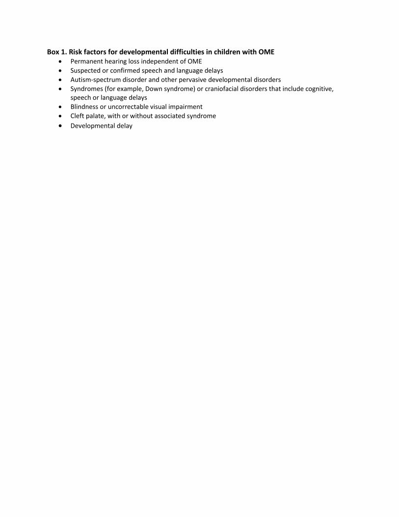
Box 1. Risk factors for developmental difficulties in children with OME Permanent hearing loss independent of OME
Suspected or confirmed speech and language delays
Autism-spectrum disorder and other pervasive developmental disorders
Syndromes (for example, Down syndrome) or craniofacial disorders that include cognitive, speech or language delays
Blindness or uncorrectable visual impairment
Cleft palate, with or without associated syndrome
Developmental delay
Figure 1. Anatomy of the human ear The ear can be divided into three parts: the outer, middle and inner ear. The outer ear consists of the auricle (or pinna) and the ear canal. The tympanic membrane (ear drum), a thin cone-shaped membrane, separates the outer from the middle ear. The middle ear consists of the middle ear cavity and the ossicles (malleus, incus, stapes) which are attached to the tympanic membrane. The oval window connects the middle ear with the inner ear which includes the semicircular ducts and the cochlea. The middle ear cavity is connected to the nasopharynx by the Eustachian tube. Figure 2. Global AOM and CSOM incidence. A | AOM incidence. Incidence rate estimates (per 100 people) in 2005 based on data from 39 papers conducted in six WHO regions. b | CSOM incidence. Incidence rate estimates (per 100 people) in 2005 based on data from 65 papers worldwide. (permission: Monasta L, Ronfani L, Marchetti F, Montico M, Vecchi Brumatti L, Bavcar A, et al. Burden of disease caused by otitis media: systematic review and global estimates. PLoS One 2012;7:e36226.) [2] Figure 3. Causal pathways for otitis media. Otitis media is a multifactorial disease. Specific host and environmental factors put children at risk for otitis media through various mechanisms as illustrated in this diagram. Reducing the burden of OM will therefore require attention to more than a single risk factor. Given the complex causal pathways for OM, public health interventions may need to be prioritized differently for various at risk populations and geographical regions. Diagram based on data obtained from [47]. Figure 4. Steps in the pathogenesis of virus-induced acute otitis media. The child might have a pre-existing nasopharyngeal bacterial colonization, which does not cause symptoms. When the child contracts a common cold, the viral infection initiates inflammation of the nasopharynx and the Eustachian tube, leading to increased adherence and colonization of bacteria and other activating mechanisms. Eustachian tube dysfunction follows, leading to negative middle ear pressure, allowing bacteria and/or virus in the nasopharynx into the middle ear causing infection and/or inflammation. Figure 5. Otoscopical images. A | normal tympanic membrane. B | Red and bulging tympanic membrane indicative for acute otitis media. C | Otitis media with effusion. D | Presence of a ventilation tube in the tympanic membrane. Figure 6. Tympanogram. The tympanometric curve, or tracing, is categorized as type A, B, or C based on middle ear pressure and the presence or absence of a discernable peak. A | Type A tympanogram. The type A tympanogram curve has a sharp peak and normal middle ear pressure and therefore a low probability of middle ear effusion. b | Type B tympanograms. The type B tympanogram curve has a flattened shape with no discernible peak pressure and has a high probability of middle ear effusion. A flat tympanogram with a normal equivalent ear canal volume usually indicates middle ear effusion. A flat tympanogram associated with a low equivalent ear canal volume indicates probe obstruction by cerumen or contact with the ear canal. A flat tympanogram with a high volume indicates a patent ventilation tube or a tympanic membrane perforation. C |Type C tympanogram curves (intermediate probability of effusion) have negative middle ear pressure with a sharp (C1) or rounded (C2) peak.
Figure 7 | Ventilation tubes. Ventilation tubes (tympanostomy tubes) are tiny plastic tubes put into the eardrum (tympanic membrane) during a short operation under general anaesthesia. The tubes usually stay in place for 6-12 months and fall out themselves. Ventilation tubes can prevent further AOM episodes by draining the fluid from the ear and improving its ventilation. In addition, by providing access to the middle ear, they may allow for local antibiotic treatment of AOM rather than systemic.
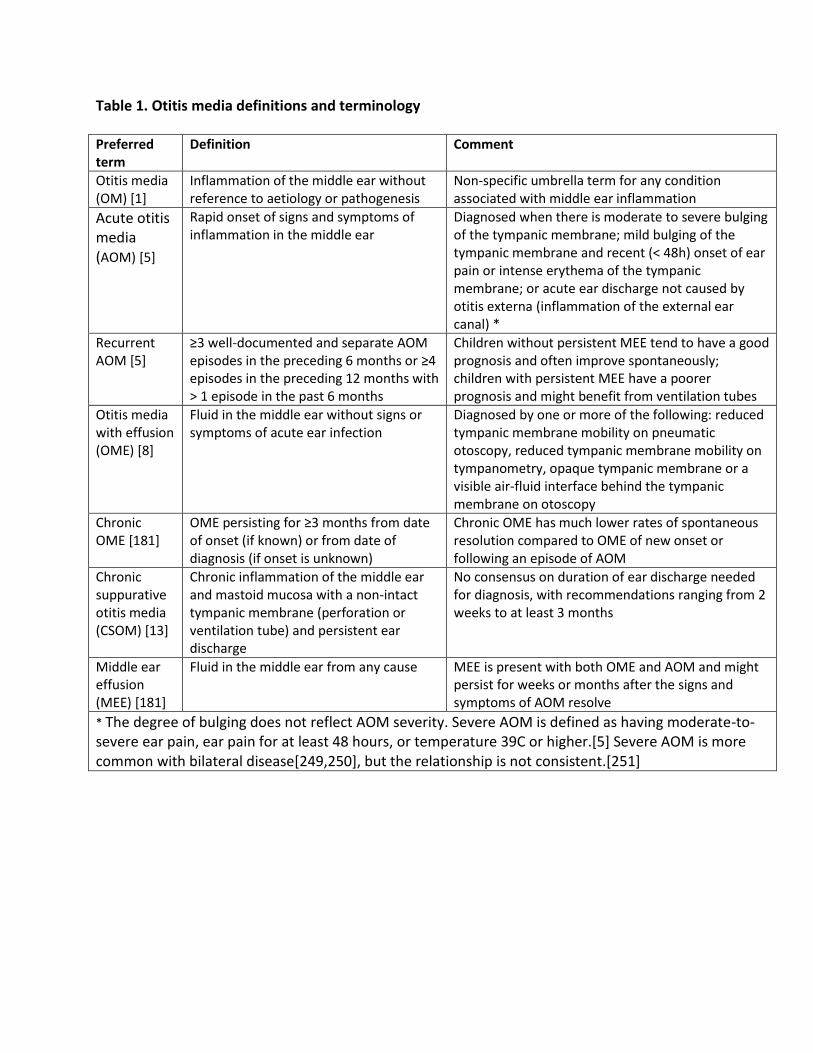
Table 1. Otitis media definitions and terminology Preferred term
Definition Comment
Otitis media (OM) [1]
Inflammation of the middle ear without reference to aetiology or pathogenesis
Non-specific umbrella term for any condition associated with middle ear inflammation
Acute otitis media (AOM) [5]
Rapid onset of signs and symptoms of inflammation in the middle ear
Diagnosed when there is moderate to severe bulging of the tympanic membrane; mild bulging of the tympanic membrane and recent (< 48h) onset of ear pain or intense erythema of the tympanic membrane; or acute ear discharge not caused by otitis externa (inflammation of the external ear canal) *
Recurrent AOM [5]
≥3 well-documented and separate AOM episodes in the preceding 6 months or ≥4 episodes in the preceding 12 months with > 1 episode in the past 6 months
Children without persistent MEE tend to have a good prognosis and often improve spontaneously; children with persistent MEE have a poorer prognosis and might benefit from ventilation tubes
Otitis media with effusion (OME) [8]
Fluid in the middle ear without signs or symptoms of acute ear infection
Diagnosed by one or more of the following: reduced tympanic membrane mobility on pneumatic otoscopy, reduced tympanic membrane mobility on tympanometry, opaque tympanic membrane or a visible air-fluid interface behind the tympanic membrane on otoscopy
Chronic OME [181]
OME persisting for ≥3 months from date of onset (if known) or from date of diagnosis (if onset is unknown)
Chronic OME has much lower rates of spontaneous resolution compared to OME of new onset or following an episode of AOM
Chronic suppurative otitis media (CSOM) [13]
Chronic inflammation of the middle ear and mastoid mucosa with a non-intact tympanic membrane (perforation or ventilation tube) and persistent ear discharge
No consensus on duration of ear discharge needed for diagnosis, with recommendations ranging from 2 weeks to at least 3 months
Middle ear effusion (MEE) [181]
Fluid in the middle ear from any cause MEE is present with both OME and AOM and might persist for weeks or months after the signs and symptoms of AOM resolve
* The degree of bulging does not reflect AOM severity. Severe AOM is defined as having moderate-to-severe ear pain, ear pain for at least 48 hours, or temperature 39C or higher.[5] Severe AOM is more common with bilateral disease[249,250], but the relationship is not consistent.[251]
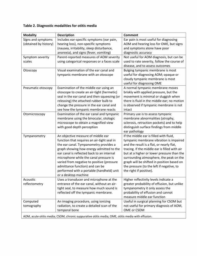
Table 2. Diagnostic modalities for otitis media Modality Description Comment
Signs and symptoms (obtained by history)
Includes ear-specific symptoms (ear pain, hearing loss), non-specific symptoms (nausea, irritability, sleep disturbance, anorexia), and signs (fever, vomiting)
Ear pain is most useful for diagnosing AOM and hearing loss for OME, but signs and symptoms alone have poor diagnostic accuracy
Symptom severity scales
Parent-reported measures of AOM severity using categorical responses or a faces scale
Not useful for AOM diagnosis, but can be used to rate severity, follow the course of disease, and to assess outcomes
Otoscopy Visual examination of the ear canal and tympanic membrane with an otoscope
Bulging tympanic membrane is most useful for diagnosing AOM; opaque or cloudy tympanic membrane is most useful for diagnosing OME
Pneumatic otoscopy Examination of the middle ear using an otoscope to create an air-tight (hermetic) seal in the ear canal and then squeezing (or releasing) the attached rubber bulb to change the pressure in the ear canal and see how the tympanic membrane reacts
A normal tympanic membrane moves briskly with applied pressure, but the movement is minimal or sluggish when there is fluid in the middle ear; no motion is observed if tympanic membrane is not intact
Otomicroscopy Examination of the ear canal and tympanic membrane using the binocular, otologic microscope to obtain a magnified view with good depth perception
Primary use is to assess tympanic membrane abnormalities (atrophy, sclerosis, retraction pockets) and to help distinguish surface findings from middle ear pathology
Tympanometry An objective measure of middle ear function that requires an air-tight seal in the ear canal. Tympanometry provides a graph showing how energy admitted to the ear canal is reflected back to an internal microphone while the canal pressure is varied from negative to positive (pressure admittance function) and can be performed with a portable (handheld) unit or a desktop machine
If the middle ear is filled with fluid, tympanic membrane vibration is impaired and the result is a flat, or nearly flat, tracing. If the middle ear is filled with air but at a higher or lower pressure than the surrounding atmosphere, the peak on the graph will be shifted in position based on the pressure (to the left if negative, to the right if positive).
Acoustic reflectometry
Uses a transducer and microphone at the entrance of the ear canal, without an air-tight seal, to measure how much sound is reflected off the tympanic membrane.
Higher reflectivity levels indicate a greater probability of effusion, but unlike tympanometry it only assess the probability of effusion and cannot measure middle ear function
Computed tomography
An imaging procedure, using ionizing radiation, to create a detailed scan of the temporal bone
Useful in surgical planning for CSOM but not useful for primary diagnosis of AOM, OME or CSOM
AOM, acute otitis media; CSOM, chronic suppurative otitis media; OME, otitis media with effusion.