Other Blood Group Systemsnybloodcenter.org/media/filer_public/2017/04/14/burgos_other_blood... ·...
57
1 Other Blood Group Systems Anna Burgos, MT(ASCP)SBB Senior Immunohematologist Laboratory of Immunohematology and Genomics April 18, 2017
Transcript of Other Blood Group Systemsnybloodcenter.org/media/filer_public/2017/04/14/burgos_other_blood... ·...

1
Other Blood Group Systems
Anna Burgos, MT(ASCP)SBB Senior Immunohematologist
Laboratory of Immunohematology and Genomics
April 18, 2017

2
Introduction to Immunohematology
I. Blood Group Immunology/ Pre-transfusion Testing/ABO/Rh
II. Other Blood Group Systems
III. Antibody Identification I&II

3
Other Blood Group Systems: points to consider
•Most commonly encountered antigens and their respective antibodies
•Which antibodies are clinically significant?
•Impact on the Blood Bank

4
Blood Groups: Discovery and Elucidation
•1900s-1950s: serology/family studies
•1950-1980s: biochemical analysis
•Late 1980s: molecular genetics
•A blood group antigen is defined serologically by antibodies made by a human
• In order to be assigned a number by the ISBT Terminology Working Party the antigen must be shown to be inherited

5
Today: 36 blood group systems; 300+ antigens 2015
2012 2013 2014
VEL
CD59
AUG
Blood Group System
Growth spurt thanks to new technologies Some favorite “old” antigens (that were detected many years ago) have now become systems

6
RBC Membrane Components & 35 blood group systems
CD59
Figure adapted from: Blood Group Antigen FactsBook; 3rd ed Reid, Lomas-Francis & Olsson
VEL All 36 blood group genes have been cloned and sequenced

7
ISBT Working Party on Red Cell Immunogenetics and Blood Group Terminology 36 Blood group systems (001 through 036) A blood group system consists of one or more antigens
controlled at a single gene locus, or by two or more very closely linked homologous genes
Blood group collections: antigens are related serologically, biochemically or genetically, but do not
fit the criteria required for system status (Cost, Er) 700 series: of low incidence antigens that are not part of
a blood group system or collection; incidence of <1% in most population tested (e.g., Bi, Kg)
901 series : of high incidence antigens (> 90%) in most population tested that are not part of a blood group system or collection (e.g., MAM, AnWj)

8
ISBT Working Party on Terminology for Red Cell Surface Antigens
Number System name ISBT gene name 001 ABO ABO 002 MNS MNS 003 P P1 004 Rh RHD, RHCE 005 Lutheran LU 006 Kell KEL 007 Lewis LE 008 Duffy FY 009 Kidd JK 010 Diego DI 011 Yt ACHE 012 Xg XG 013 Scianna SC 014 Dombrock DO 015 Colton CO 016 Landsteiner-Wiener LW 017 Chido/Rodgers C4A, C4B 018 Hh H 019 Kx XK 020 Gerbich GE 021 Cromer CROM 022 Knops KN 023 Indian IN 024 Ok OK 025 Raph RAPH 026 JMH JMH 027 I I 028 GLOB P 029 GILL GIL 030 RHAG RHAG 031 FORS FORS 032 Jr JR 033 Lan LAN
Criteria for the establishment of new blood group systems: For an antigen to form a new blood group system it must be: • Defined by a human alloantibody • Inherited character • Gene encoding it must have
been identified and sequenced • Known chromosomal location • Gene must be different from,
and not a closely-linked homologue of, all other genes encoding antigens of existing blood group systems.
Number System name ISBT gene name 034 Vel SMIM1 035 CD59 CD59 036 Augustine ENT1

9
Blood group antigens that are sugars
•The antigens of the P1PK (formerly P) and Lewis systems are sugars that are produced by a series of reactions in which enzymes (glycosyltransferases) catalyze the transfer of sugar units to the carrier protein in the RBC membrane
•A person’s DNA determines the type of enzyme and therefore, the immunodominant sugar (and antigen) on the RBCs
10
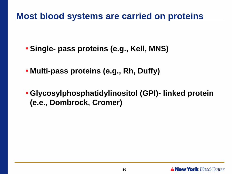
• Single- pass proteins (e.g., Kell, MNS)
• Multi-pass proteins (e.g., Rh, Duffy)
• Glycosylphosphatidylinositol (GPI)- linked protein (e.e., Dombrock, Cromer)
Most blood systems are carried on proteins

11
Blood Group Systems and their Chromosomes
Courtesy of Dr. Marion Reid Note: # antigens reflect those identified as of 2009

12
Other Blood Group Systems: Review of Key Features
•Distinguishing characteristics
– Structure/function/disease associations
•Antigen Prevalence/ISBT number
•Antibodies
– Reactivity
– Clinical significance

13
Points to consider for RBC transfusion
• Is the antibody identified clinically significant?
• What is the antigen prevalence in the donor population or How difficult is it to find compatible blood for the patient?

14
“Other” blood group systems (BGS): Non-ABO/D
•P1PK (formerly P) •Lewis
•Other Rh antigens •MNS •Kell •Duffy •Kidd
Rh-hr Kell Kidd Duffy Lewis MNSs P
cell D C E c e K k Jka Jkb Fya Fyb Lea Leb M N S s P1 37C AHG
I + + 0 0 + + + + 0 0 + + 0 + + 0 + +
II + 0 + + 0 0 + 0 + + 0 0 + 0 + + 0 0
III 0 0 0 + + 0 + + 0 + + 0 + + 0 0 + +
15
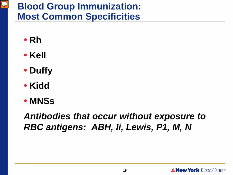
Blood Group Immunization: Most Common Specificities
• Rh • Kell • Duffy • Kidd • MNSs Antibodies that occur without exposure to RBC antigens: ABH, Ii, Lewis, P1, M, N
Presenter
Presentation Notes
RhD is the most immunogenic (quoted at approx. 50%), then K (5%), followed by other antigens within the Rh system (e.g. c (2%), E (1.7%) Why only 5 systems commonly encountered - frequency distribution of Ags “Naturally-occurring” Abs result of sugar-determined Ags - ubiquitous in nature; i.e.; plants, bacteria
16
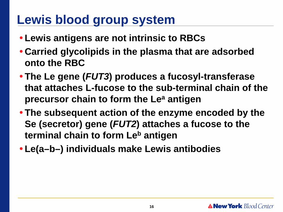
Lewis blood group system • Lewis antigens are not intrinsic to RBCs • Carried glycolipids in the plasma that are adsorbed
onto the RBC • The Le gene (FUT3) produces a fucosyl-transferase
that attaches L-fucose to the sub-terminal chain of the precursor chain to form the Lea antigen
• The subsequent action of the enzyme encoded by the Se (secretor) gene (FUT2) attaches a fucose to the terminal chain to form Leb antigen
• Le(a–b–) individuals make Lewis antibodies

17
Lewis blood group system (continuation)
• Antibodies are frequently found but are usually NOT clinically significant
• Rare examples of hemolytic anti-Lea and even rarer examples of anti-Leb have been found
• Mostly not necessary to type donor blood Lewis antigens prior to transfusion or crossmatching
– Reactions obtained in the crossmatch provide a good index of transfusion safety
– If agglutination and/or hemolysis are observed at 37C or IAT, then the blood should not be given and antigen-negative blood should be used

18
P1PK Blood Group system (formerly P system)
• P1 antigen formed on cellular paragloboside with Type II chains
• Immunodominant sugar =D-galactose • No L-fucose added to subterminal sugar • P1-positive phenotype = P1 • P1-negative phenotype = P2 • Shares common precursor with P (globoside) • Anti-P1 NOT clinically significant • Anti-P1 is mostly IgM, it does not cross the placenta
and has not been reported to cause HDFN – P1 antigen is poorly expressed on fetal cells
Presenter
Presentation Notes
Expression dependent on globoside but interaction between 2 structures not understood P = receptor for B19 parvovirus

19
Rh blood group system
•The most polymorphic BGS in humans • 56 antigens to date and counting! • 2nd most important system after ABO • Antigens are highly immunogenic • Usually clinically significant: can cause transfusion
reactions and HDFN •Rh antibodies rarely, if ever, bind complement
– RBC destruction is mediated almost exclusively via macrophages in the spleen
-

20
Single antigen prevalence (calculated)
•D 85% Caucasians, 93% Blacks, 99% Asians – Therefore HDFN due to anti-D very rare in Asian populations
•C 70% Caucasians, 27% Blacks, 93% Asians •E 30% Caucasians, 22% Blacks, 39 % Asians •c 80% Caucasians, 96% Blacks, 47% Asians •e 98% Caucasians, 98% Blacks, 96% Asians

21
MNS blood group system • 48 antigens
• Carried on sialoglycoproteins: – glycophorin A (GPA) and glycophorin B (GPB)
• Encoded by 2 genes: GYPA, GYPB
M or N; S or s antigens
• Inherited as a haplotype : MS, Ms, NS or Ns
• Disease associations – GPA is a pathogen receptor (E. coli; influenza virus)
– GPA deficient RBCS are resistant to P. falciparum invasion

22
MNS Blood Group • Many enzyme cleavage sites along both
molecules; useful in antibody studies
• Multiple low incidence antigens caused by point mutations
• Various hybrid molecules define novel antigens
Null phenotypes:
En(a–) M–N–; cells lack GPA
U negative S–s–; cells lack GPB or have aberrant molecule [Uvar (S–s–U+W)]
Mk Cells lack both GPA and GPB
Presenter
Presentation Notes
Various hybrid molecules… due to unequal crossing over or gene conversion En(a-) represents several different molecules GPC and GPD, products of single gene, carry Ge GPC links to protein 4.1 in cell’s skeleton and is essential for normal cell shape Cells that lack GPC (also Gerbich null) are elliptocytes GPA and GPB are not essential for normal morphology; U neg and other nulls have normal shape and survival GPA associates with Band 3 and Wrb Ag formed by both proteins Alternative translation initiation sites
23
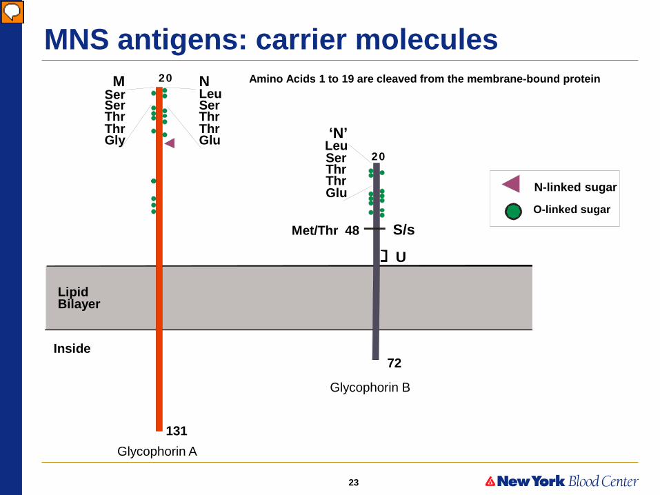
MNS antigens: carrier molecules Amino Acids 1 to 19 are cleaved from the membrane-bound protein
Blood Center
Glycophorin B
Glycophorin A
72
131
S/s Met/Thr 48
U
Inside
Lipid Bilayer
20
20
‘N’ Leu Ser Thr Thr Glu
M Ser Ser Thr Thr Gly
N Leu Ser Thr Thr Glu
N-linked sugar
O-linked sugar
Presenter
Presentation Notes
Glycophorins are single-pass proteins Genes located close to each other and duplication event resulted in 2 homologous proteins GPA similar in structure but larger than GPB Both carry sialic acid residues responsible for cell’s negative charge MN on GPA determined by substitutions at AA positions 1and 5 Ss on GPB determined by substitutions at AA position 29 AAs 1-5 on GPB identical to those on GPA in N+ person (‘N’) Mutation prevents transcription of exon 3

24
MNS System: Phenotypes and Prevalence
Pheno- M N type Whites Blacks
+ 0 M+N– 28 26
+ + M+N+ 50 44
0 + M–N+ 22 30
Phenotype Prevalence (%)
Reactions with Anti-
Adapted from AABB Technical Manual

25
Phenotypes and Prevalence in the MNS System
Pheno- S s U type Whites Blacks
+ 0 + S+s–U+ 11 3
+ + + S+s+U+ 44 28
0 + + S–s+U+ 45 69
0 0 0 S–s–U– 0 < 1
Phenotype Prevalence (%)
Reactions with Anti-
Adapted from AABB Technical Manual
Presenter
Presentation Notes
U neg only in Blacks

26
MNS Antibodies: anti-M Anti-M •IgG (cold reactive; many direct agglutinins) and IgM
–React at 24ºC (RT) or 4C; rarely also reactive by IAT –M antigen: large quantity (up to 1 million copies) on RBCs so that agglutination in saline test may occur even the antibody is wholly IgG –Anti-M demonstrates dosage
•Generally not clinically significant –Rare examples have caused transfusion reactions or HDFN
• If reactivity is at 37C the anti-M should be considered potentially significant
Presenter
Presentation Notes
MN “naturally-occurring” Anti-M more common and more potent than anti-N, perhaps due to immune tolerance induced by ‘N’ some antibodies require glycosylation to react, but specificity determined by AA sequence

27
MNS antibodies: anti-N
Anti-N • IgM and IgG (some direct agglutinins)
– typically behave like weakly reactive cold agglutinins – Rarely reactive at IAT
- Usually considered clinically insignificant (although some powerful and potentially significant IgG examples have been observed) • Antibodies showing dosage are rarely encountered • Rare N–S–s–U– people make an antibody that
reacts with N on GPA and GPB and may be clinically significant

28
MNS Antibodies: anti-S, -s, -U Anti-S and anti-s •Usually IgG; react by IAT but some anti-S and anti-s are IgM
•Anti-S may be “naturally-occurring” without known RBC stimulation
•RBC units for transfusion must be antigen negative and crossmatch compatible
Anti-U • IgG; reacts by IAT; reacts with enzyme treated RBCs as U antigen is resistant to enzyme treatment
•May cause HDFN; can be difficult to manage be U– blood is rare
Presenter
Presentation Notes
MN “naturally-occurring” Anti-M more common and more potent than anti-N, perhaps due to immune tolerance induced by ‘N’ some antibodies require glycosylation to react, but specificity determined by AA sequence

29
Proteolytic Enzymes
•Useful tools for investigating complex antibody problems
•Papain, ficin, bromelin •Modify RBC membrane/remove negatively charged molecules
•Enzymes destroy M, N, S antigens – however, s antigen may or may not be denatured
by enzyme treatment
30
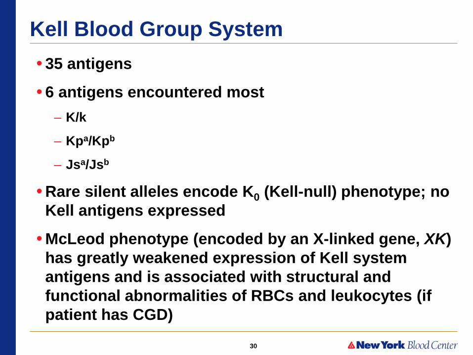
Kell Blood Group System • 35 antigens
• 6 antigens encountered most – K/k
– Kpa/Kpb
– Jsa/Jsb
• Rare silent alleles encode K0 (Kell-null) phenotype; no Kell antigens expressed
• McLeod phenotype (encoded by an X-linked gene, XK) has greatly weakened expression of Kell system antigens and is associated with structural and functional abnormalities of RBCs and leukocytes (if patient has CGD)

31
Kell Glycoprotein • Member of Neprilysin (M13)
family of zinc endo-peptidases
• Kell cleaves big endothelin-3 to release ET-3, a potent vaso constrictor
• Kell antigen expression greatly reduced when Kx protein (encoded by XK gene) is absent (McLeod phenotype) NH2
COOH
Out1
2
3 4 5347
72
In
Kell
C
NH2
COOH
K12
K19
TOU
Weak k
K22
K23
K11/K17
K/k
HELLH
K14/K24
CC
C
CC
C
C
C
C CC
C
C
CCC
C
CC
CCC
C
C
CCCC
C
C
C C
K18
XK
VLANRAZ
Kp /Kp /Kpa b c
Ula
Js /Jsa b
Courtesy C. Lomas-Francis, modified

32
Kell System: Phenotypes and Prevalence
Pheno- K k type Whites Blacks
+ 0 K+k– 0.2 rare
+ + K+k+ 8.8 2
0 + K–k+ 91 98
Prevalence (%) Reactions with Anti-
Adapted from AABB Technical Manual

33
Kell System: Phenotypes and Prevalence
Pheno- Kpa Kpb type Whites Blacks
+ 0 Kp(a+b–) rare 0
+ + Kp(a+b+) 2.3 rare
0 + Kp(a–b+) 97.7 100
Prevalence (%) Reactions with Anti-
Adapted from AABB Technical Manual
34
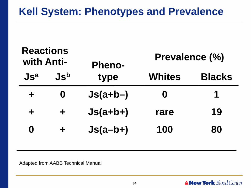
Kell System: Phenotypes and Prevalence
Pheno- Jsa Jsb type Whites Blacks
+ 0 Js(a+b–) 0 1
+ + Js(a+b+) rare 19
0 + Js(a–b+) 100 80
Prevalence (%) Reactions with Anti-
Adapted from AABB Technical Manual
35
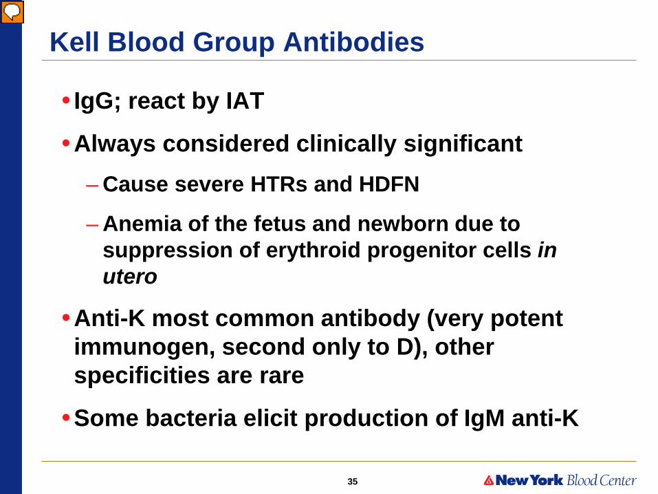
Kell Blood Group Antibodies
• IgG; react by IAT
•Always considered clinically significant – Cause severe HTRs and HDFN
– Anemia of the fetus and newborn due to suppression of erythroid progenitor cells in utero
•Anti-K most common antibody (very potent immunogen, second only to D), other specificities are rare
•Some bacteria elicit production of IgM anti-K
Presenter
Presentation Notes
HDN - 10% of severe cases due to anti-K1 severe anemia not due to hemolysis alone Ab appears to depress erythropoiesis at progenitor cell level titer not good predictor of anemia in infant infants have lower bilirubin levels and lower retic counts than those affected by Rh disease

36
HDFN due to Anti-D and to Anti-K
Anti-D Anti-K
Pictures courtesy of Dr. Greg Denomme
Hydropic Hydropic and anemic

37
Duffy Blood Group
•5 antigens: Fya, Fyb, Fy3, Fy5 and Fy6 •Most common are Fya and Fyb
•The Duffy gene encodes a glycoprotein that is expressed in other tissues, including brain, kidney, spleen, heart and lung
• In Fy(a–b–) individuals, transcription in the bone marrow is prevented and Duffy protein is absent from the red cell
•Duffy protein is expressed normally in non-erythroid cells of these Fy(a–b–) persons

38
RBC lipid bilayer
NH2
COOH
42nd a.a.residue
Antigen Nucleotide Amino acidVariation Variation
Fyb ‘’ A ‘’ Asp
Fya 125th G 42nd Gly
Molecular Basis of Duffy (Fya & Fyb) Antigens
Presenter
Presentation Notes
2 principle antigens determined by one AA substitution
39
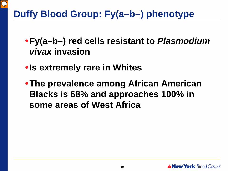
Duffy Blood Group: Fy(a–b–) phenotype
•Fy(a–b–) red cells resistant to Plasmodium vivax invasion
• Is extremely rare in Whites
•The prevalence among African American Blacks is 68% and approaches 100% in some areas of West Africa
Presenter
Presentation Notes
…resistant to P. vivax invasion common phenotype in people of African descent P. falciparum uses another receptor - NANA on GYP and CR1 …multiple tissues kidney, spleen, lung, Purkinje cells in cerebellum, endothelial cells, but not liver …disrupts erythroid expression now know that Blacks whose RBCs lack Fy Ags not really Fy null mutation in promoter region of Fyb allele disrupts erythroid-specific transcription adaptive response to resist malaria; RBCs otherwise normal express Duffy on other tissues; explains absence of anti-Fyb …extremely rare found in a few Whites; due to various mutations; make Abs …DARC aka promiscuous chemokine receptor or sink for excess chemokine

40
Duffy System: Phenotypes and Prevalence
Pheno-- Fya Fyb type Whites Blacks
+ 0 Fy(a+b–) 20 10
+ + Fy(a+b+) 48 3
0 + Fy(a–b+) 32 20
0 0 Fy(a–b–) 0 67
Reactions with Anti-
Prevalence)
Adapted from AABB Technical Manual
Presenter
Presentation Notes
Only 2 Ags

41
Duffy Blood Group Antibodies
• IgG; react by IAT; clinically significant
•Anti-Fya stronger and more common than anti-Fyb
•Anti-Fya and -Fyb are non-reactive with enzyme-treated cells
•Anti-Fy3, sometimes made by Fy(a–b–) people
– The Fy3 antigen is resistant to enzyme treatment
Presenter
Presentation Notes
Anti-Fy3 is potent Ab made by true nulls

42
Kidd Blood Group System • ISBT symbol JK, ISBT number 009
•3 Antigens Jka/Jkb Jk3
•Glycoprotein with 10 membrane spanning domains
•Jka/Jkb polymorphisms on the 4th extracellular loop
•Function = urea transport
•Jk(a–b–) individuals are rare – are unable to maximally concentrate urine

43
Kidd Gene and Protein
1 2 3
211
Stop
280
389N
4 5 6
30 kb
7 8 9 10 11
93 64 157 172 190 129 193 148 135 50 551
Lucien, , 1998;273:12973J.Biol.Chem.
ATG G838AAsp AsnJk Jk
→→a b
Presenter
Presentation Notes
Urea transport protein HUTII - tell story Delay in lysis of Kidd null cells due to slow entry of urea Kidd null RBCs have no other abnormalities Null individuals sometimes have a diminished capacity to concentrate urine 2 transport proteins, which may compensate for lack of the other protein in Jk(a-b-) people

44
Kidd System: Phenotypes and Prevalence
Pheno- Jka Jkb type Whites Blacks
+ 0 Jk(a+b–) 28 57
+ + Jk(a+b+) 49 34
0 + Jk(a–b+) 23 9
0 0 Jk(a–b–) Exceedingly rare
Reactions with Anti- Prevalence (%)
Adapted from AABB Technical Manual
Presenter
Presentation Notes
Only 2 Ags
45
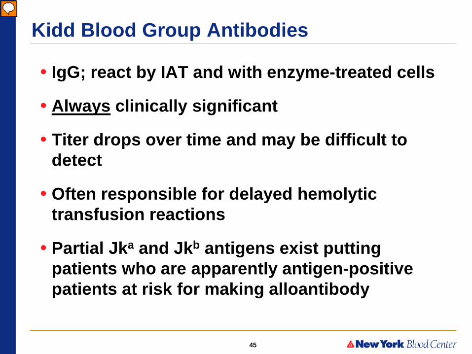
Kidd Blood Group Antibodies
• IgG; react by IAT and with enzyme-treated cells
• Always clinically significant
• Titer drops over time and may be difficult to detect
• Often responsible for delayed hemolytic transfusion reactions
• Partial Jka and Jkb antigens exist putting patients who are apparently antigen-positive patients at risk for making alloantibody
Presenter
Presentation Notes
Minor IgM component found in some examples Responsible for hemolysis? Does not usually cause severe HDN, so is the IgG particularly significant? Null individuals make anti-Jk3

46
Common vs Uncommonly Encountered Specificities
Common Uncommon
Specificities Rh, MNS, Kell, Fy, Jk
Di, Cr, Do, Yt, Lu, Ch/Rg, Kn
FDA licensed typing reagents available?
Yes No
RBCs on commercial panels routinely phenotyped?
Always Usually not
Antibody easily identified by hospital BB?
Yes No
47
Some other blood group systems
•010 Diego •011 Yt •014 Dombrock •015 Colton •020 Gerbich •021 Cromer

48
Structure and Function of Blood Group Antigens
•Membrane transporters
•Receptors and adhesion molecules
•Complement regulatory glycoproteins
•Structural components
•Enzymes

49

50
Antibody Detection: 3-cell screen
Rh-hr Kell
Kidd
Duffy
Lewis
MNSs
P
cell
D
C
E
c
e
K
k
Jka
Jkb
Fya
Fyb
Lea
Leb
M
N
S
s
P1
37ºC
AHG
I
+
+
0
0
+
+
+
+
0
0
+
+
0
+
+
0
+
+ 0 0
II
+
0
+
+
0
0
+
0
+
+
0
0
+
0
+
+
0
0 0 2+
III
0
0
0
+
+
0
+
+
0
+
+
0
+
+
+
0
+
+ 0 0
Indicates an antibody is present but must test further to identify!
51
Multiple alloantibodies: points to consider
•What antibodies are identified? •How many units will I need to screen to find compatible blood?
•Will I find them in my inventory or need to place an order with Blood Center?

52
Phenotype Prevalence
•Multiply the individual frequencies (incidence of an antigen negative), since phenotypes are independent of one another
•This number will be the % negative for that particular combination

53
What is the incidence (or phenotype frequency) of c- K- Jk(a-) unit? c neg = .20 K neg = .91 Jk(a-) = .23 (.20 x .91 x .23 = .04) Therefore 4% or 4/100 units would be c- K- Jk(a-) If the question reads, how many units would you need to screen to find 2 antigen neg units for surgery, proceed with a further calculation: 4 = 2 100 x 4x=200 and x = 50 Answer: 50 units need to be screened to find the 2 units ordered
Phenotype Prevalence Example

54
Blood Bank Challenges

55
Serological Challenges
•Multiple alloantibodies – Which phase and by which method do the
antibodies react? – Selected cell panels – Other helpful techniques?
•POS DAT/warm autoantibodies – Unable to RBC phenotype – Underlying alloantibodies?
•ABO discrepancies •Delayed transfusion reactions
– RBC phenotype unreliable
56
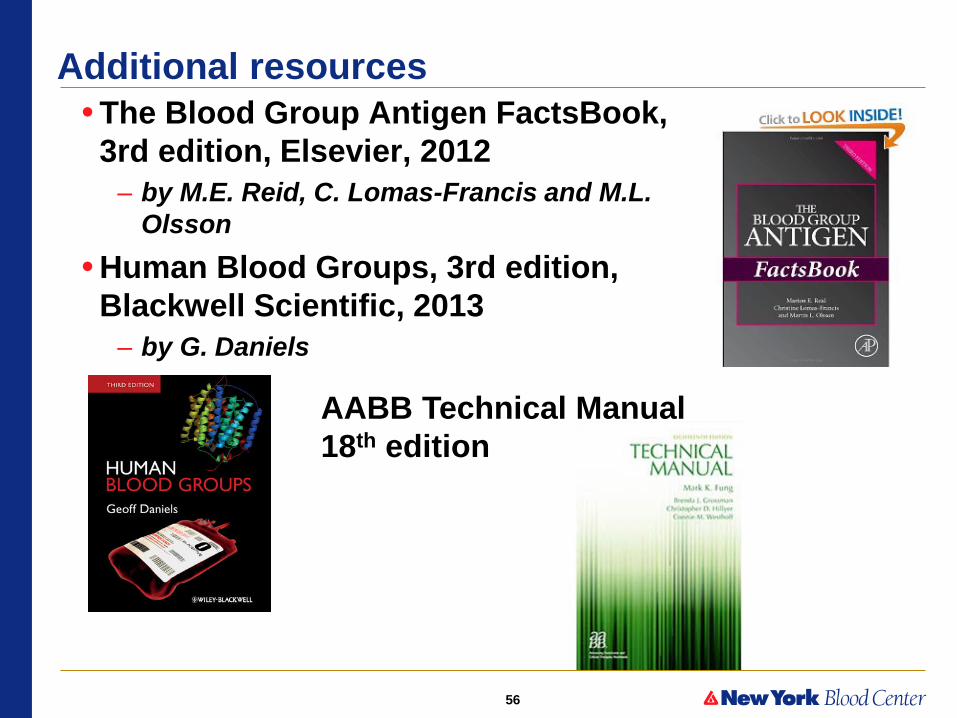
Additional resources • The Blood Group Antigen FactsBook,
3rd edition, Elsevier, 2012 – by M.E. Reid, C. Lomas-Francis and M.L.
Olsson • Human Blood Groups, 3rd edition,
Blackwell Scientific, 2013 – by G. Daniels
AABB Technical Manual 18th edition
57
Questions?