Osteoporosis Stephanie Wetmore, PT PED 596: Adv. Cardiac Rehab Wayne State College.
46
Osteoporosis Stephanie Wetmore, PT PED 596: Adv. Cardiac Rehab Wayne State College
-
Upload
elijah-butler -
Category
Documents
-
view
219 -
download
4
Transcript of Osteoporosis Stephanie Wetmore, PT PED 596: Adv. Cardiac Rehab Wayne State College.
Osteoporosis
Stephanie Wetmore, PT
PED 596: Adv. Cardiac Rehab
Wayne State College
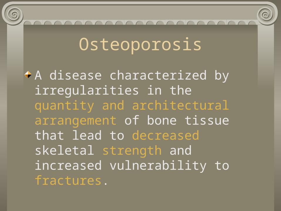
Osteoporosis
A disease characterized by irregularities in the quantity and architectural arrangement of bone tissue that lead to decreased skeletal strength and increased vulnerability to fractures.
Normal Physiology
FunctionsProvides support to body
Protects vital organs
Assists in movement via leverage
Hematopoiesis (blood cell production)
Storage area for Ca++
Cell TypesOsteoblasts
Synthesize boneRemodeling and repair
OsteoclastsResponsible for bone resorptionRemodeling and repair
OsteocytesPrimary cells of mature boneOsteoblasts surrounded by matrix during bone formationMaintenance and resorption
Bone Formation & Growth
Intermembranous ossificationBone forms directly in the embryonic connective tissue
Endochondral ossificationA “scale model” of hyaline cartilage is replaced by bone
Process of formation for most bones

A Closer Look at Endochondral Ossification and Growth
1. Formation of cartilage skeleton in embryo (6-12 wks gestation)
2. Ossification and growth occur in subsequent months
3. When ossification completed, growth in length occurs at epiphyseal plates
4. Widen by multiplication of cartilage cells and cancellous bone replaces the dying cartilage5. Growth in width occurs by depositing of compact bone beneath the periosteum (outer surface) and enlargement of the marrow cavity by bone resorption6. Growth ceases when epiphyseal plate is replaced by bone.

Endochondral Ossification & Growth
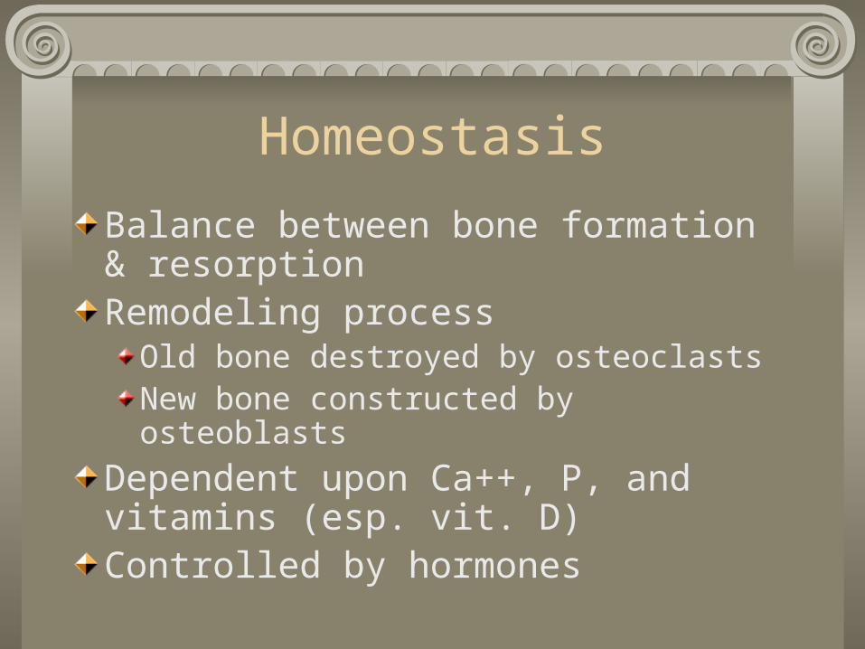
Homeostasis
Balance between bone formation & resorptionRemodeling process
Old bone destroyed by osteoclastsNew bone constructed by osteoblasts
Dependent upon Ca++, P, and vitamins (esp. vit. D)Controlled by hormones
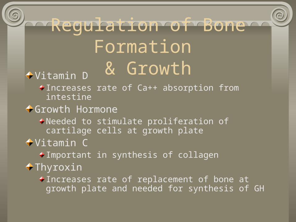
Regulation of Bone Formation & Growth
Vitamin DIncreases rate of Ca++ absorption from intestine
Growth HormoneNeeded to stimulate proliferation of cartilage cells at growth plate
Vitamin CImportant in synthesis of collagen
ThyroxinIncreases rate of replacement of bone at growth plate and needed for synthesis of GH
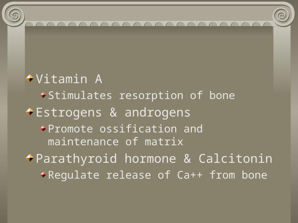
Vitamin AStimulates resorption of bone
Estrogens & androgensPromote ossification and maintenance of matrix
Parathyroid hormone & CalcitoninRegulate release of Ca++ from bone

Parathyroid hormone & calcitonin
When blood Ca++ levels are low, PTH is released.Release of PTH increases rate of bone resorption, which increases the concentration of Ca++ in the blood.When blood Ca++ levels are high, calcitonin is released, which inhibits resorption.
Pathophysiology
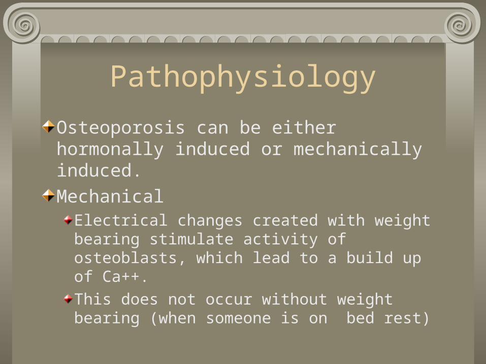
Osteoporosis can be either hormonally induced or mechanically induced.
MechanicalElectrical changes created with weight bearing stimulate activity of osteoblasts, which lead to a build up of Ca++.
This does not occur without weight bearing (when someone is on bed rest)
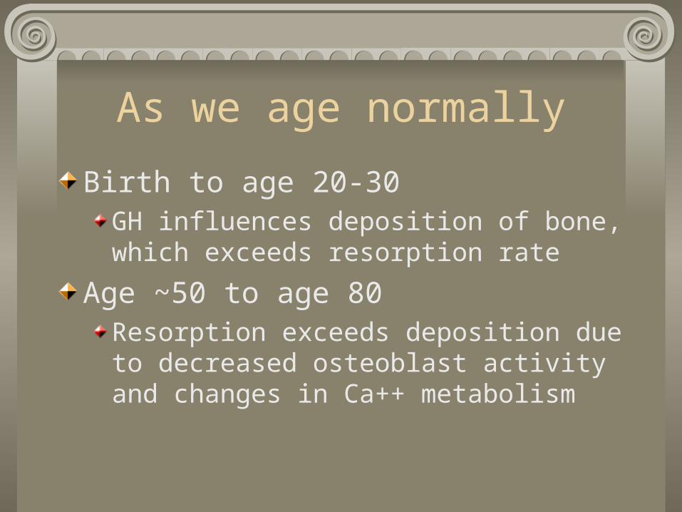
As we age normally
Birth to age 20-30GH influences deposition of bone, which exceeds resorption rate
Age ~50 to age 80Resorption exceeds deposition due to decreased osteoblast activity and changes in Ca++ metabolism
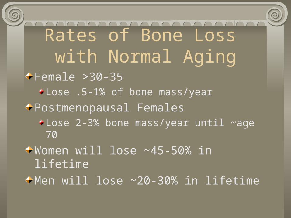
Rates of Bone Loss with Normal Aging
Female >30-35Lose .5-1% of bone mass/year
Postmenopausal FemalesLose 2-3% bone mass/year until ~age 70
Women will lose ~45-50% in lifetime
Men will lose ~20-30% in lifetime
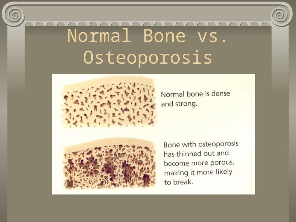
Normal Bone vs. Osteoporosis

Normal vs. Ostoeporosis
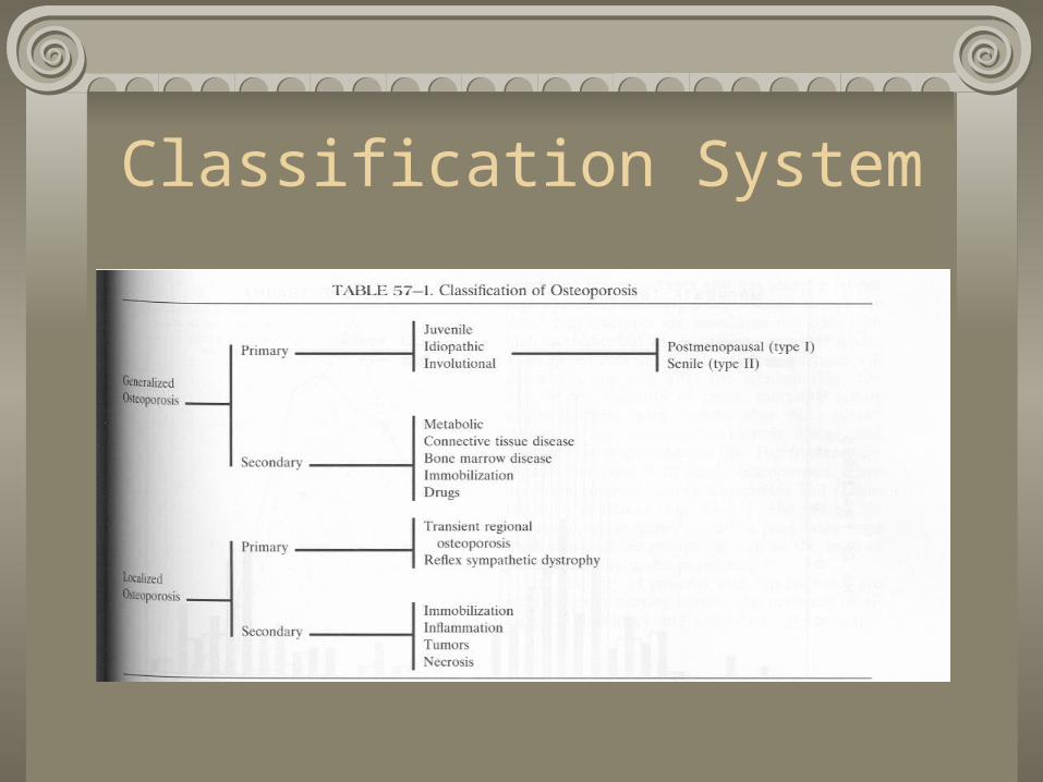
Classification System
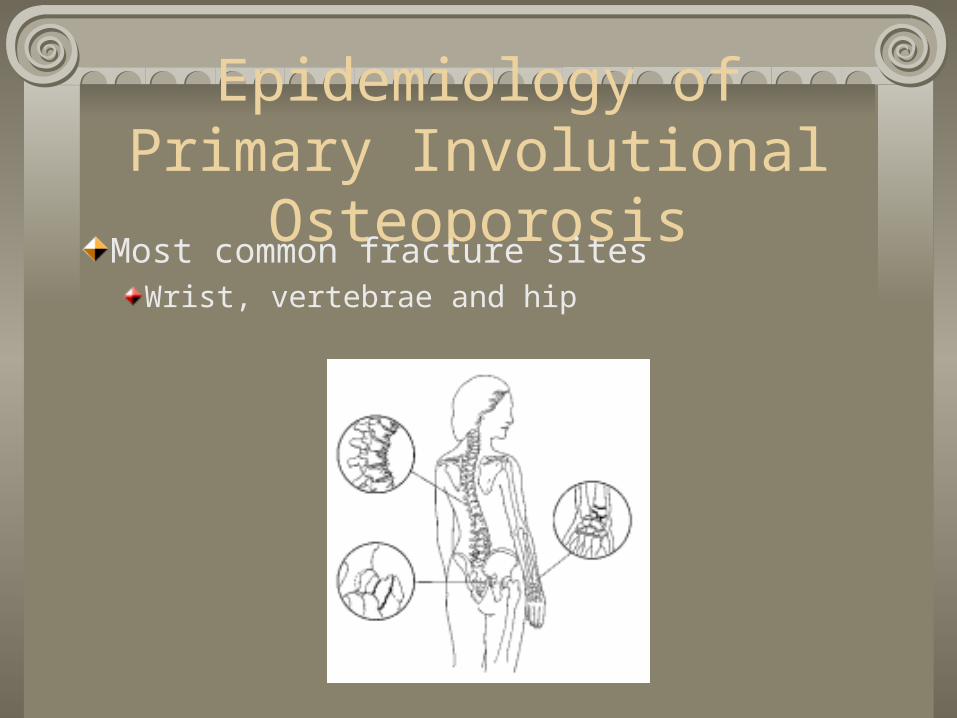
Epidemiology of Primary Involutional Osteoporosis
Most common fracture sitesWrist, vertebrae and hip
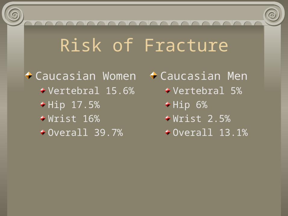
Risk of Fracture
Caucasian WomenVertebral 15.6%
Hip 17.5%
Wrist 16%
Overall 39.7%
Caucasian MenVertebral 5%
Hip 6%
Wrist 2.5%
Overall 13.1%
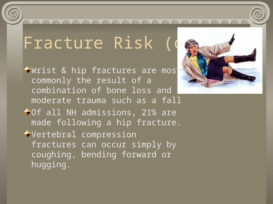
Fracture Risk (cont.)
Wrist & hip fractures are most commonly the result of a combination of bone loss and moderate trauma such as a fall
Of all NH admissions, 21% are made following a hip fracture.
Vertebral compression fractures can occur simply by coughing, bending forward or hugging.

Risk Factors
Advancing age – 1.4 to 1.8 fold increase with each decade
Gender – women > men
Family or personal hx of fx as an adult
Repeated fx’s, severe stooped posture

Risk factors (cont.)
Race – Caucasian & Asian > African American or Hispanic
Bone Structure and Body Weight – small-boned and thin women (<127#) are at greater risk
Menopause/Menstrual historyNormal, premature (<45 y/o) or surgical
Late onset menarche (>15 y/o) or prolonged amenorrhea – anorexia nervosa, bulimia, excessively low body fat
Risk Factors (cont.)
Lifestyle/NutritionCigarette smoking – inhibits estrogen
Alcoholism
Inadequate intake of Ca++
Sedentary lifestyle
High caffeine consumption and phosphoric acid intake (cola drinks)
Eating disorders

Are you getting enough Calcium?

What is adequate Ca++ intake?
Age 1-3 years 500 mg/day
Age 4-8 years 800 mg/day
Age 9-18 years 1300 mg/day
Age 19-50 years 1000 mg/day
Age >50 years 1200 mg/day
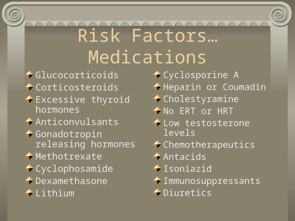
Risk Factors…Medications
GlucocorticoidsCorticosteroidsExcessive thyroid hormonesAnticonvulsantsGonadotropin releasing hormonesMethotrexateCyclophosamideDexamethasoneLithium
Cyclosporine AHeparin or CoumadinCholestyramineNo ERT or HRTLow testosterone levelsChemotherapeuticsAntacidsIsoniazidImmunosuppressantsDiuretics
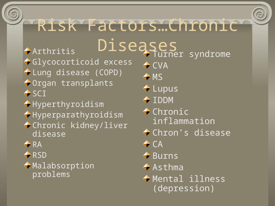
Risk Factors…Chronic DiseasesArthritisGlycocorticoid excessLung disease (COPD)Organ transplantsSCIHyperthyroidismHyperparathyroidismChronic kidney/liver diseaseRARSDMalabsorption problems
Turner syndromeCVAMSLupusIDDMChronic inflammationChron’s diseaseCABurnsAsthmaMental illness (depression)

Bone Mineral Density Testing
Painless, non-invasive test, which identifies osteoporosis, determines fx risk and monitors response to treatment.
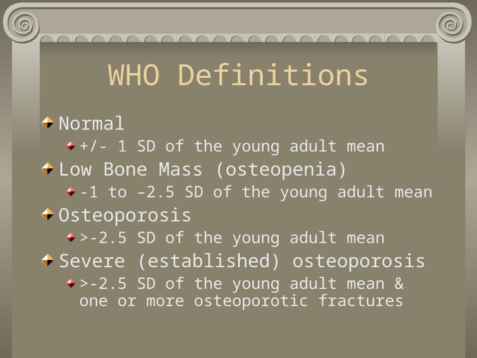
WHO Definitions
Normal+/- 1 SD of the young adult mean
Low Bone Mass (osteopenia)-1 to –2.5 SD of the young adult mean
Osteoporosis>-2.5 SD of the young adult mean
Severe (established) osteoporosis>-2.5 SD of the young adult mean & one or more osteoporotic fractures
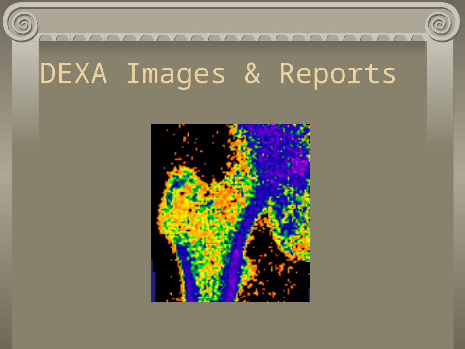
DEXA Images & Reports
Pharmacology
Estrogen Replacement Therapy/Hormone Replacement Therapy
Reduces bone loss, increases bone density, reduces risk of fx in postmenopausal women
Increase risk of uterine and breast CA, increased risk of thromboembolism
Biphosphonates
Alendronate Sodium (Fosamax)Reduces bone loss, increases bone density, reduces risk of spine and hip fracturesSide effects include bone, muscle and/or joint pain and headache
Risedronate Sodium (Actonel)Slows bone loss, increases bone density and decreases spine and hip fracturesAlso approved for men & women to prevent and/or treat steroid-induced osteoporosis
SERMs family
Selective estrogen receptor modulators
Raloxifene (Evista)Prevent bone loss, increase bone mass and decrease risk of vertebrae fracture
Side effects: DVT, leg cramps, syncope, arthralgia, tendon disorder and vertigo – chest pain, myalgia and arthritis possibly (<placebo).
Calcitonin (Miacalcin)
Naturally occurring hormone involved in Ca++ regulation and bone metabolism
Slows bone loss, increases bone density and relieves pain associated with vertebral fractures
Exercise Testing Modification/Exercise Limitations/Capacity
Weight-bearing exercise and resistance training recommended with precautions
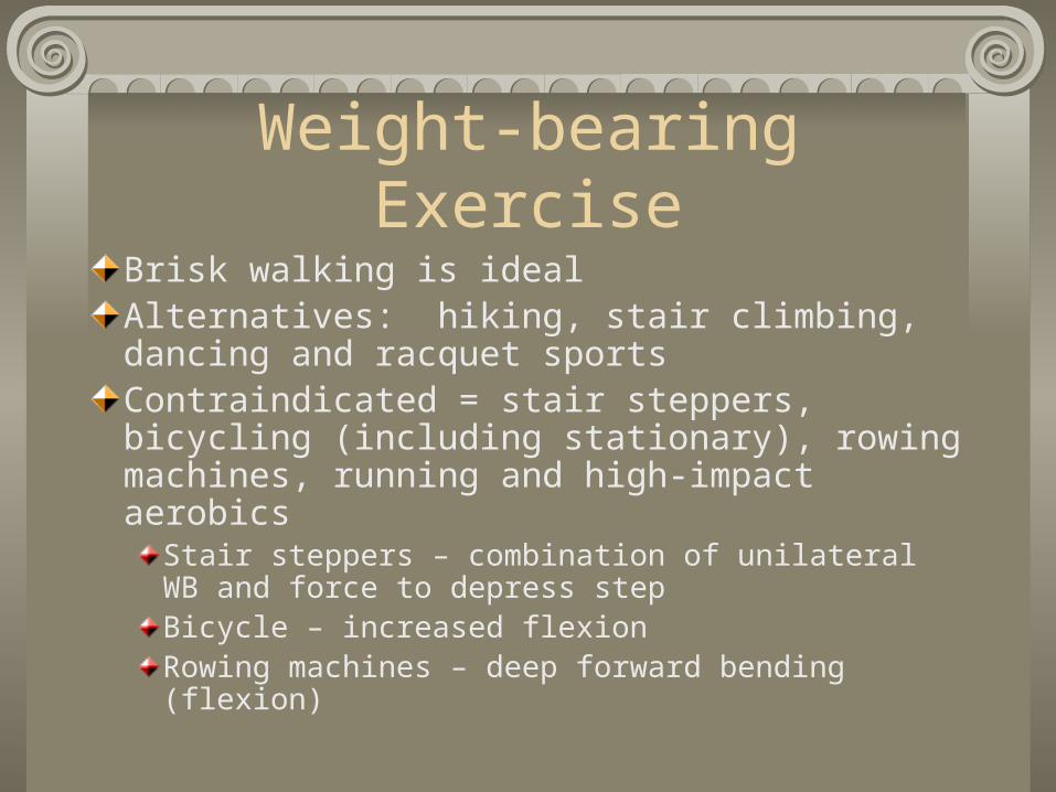
Weight-bearing Exercise
Brisk walking is idealAlternatives: hiking, stair climbing, dancing and racquet sportsContraindicated = stair steppers, bicycling (including stationary), rowing machines, running and high-impact aerobics
Stair steppers – combination of unilateral WB and force to depress stepBicycle – increased flexionRowing machines – deep forward bending (flexion)

Testing Contraindications
Sub maximal cycle ergometer
Step-tests
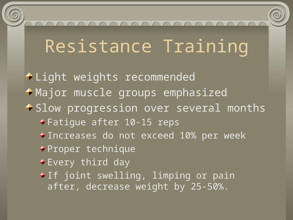
Resistance Training
Light weights recommended
Major muscle groups emphasized
Slow progression over several monthsFatigue after 10-15 reps
Increases do not exceed 10% per week
Proper technique
Every third day
If joint swelling, limping or pain after, decrease weight by 25-50%.
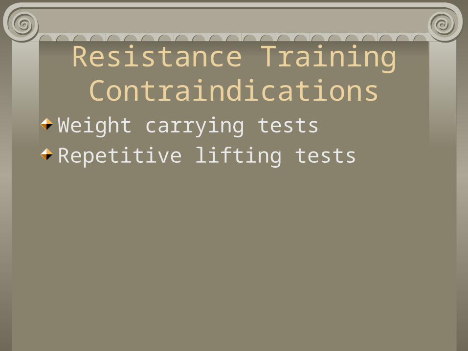
Resistance TrainingContraindications
Weight carrying tests
Repetitive lifting tests
Flexibility Exercises
Flexion exercises contraindicated if vertebral bone density decreased or risk of compression fx
Avoid knee to chestForward bendingTouching the toesPartial sit-up
Okay if thoracic spine stabilized and do not lift head and chest above T-6 level.
Flexibility ExercisesContraindications
Sit-and-reach test
Curl-up muscular endurance test

Other exercise
HR, BP, ECG, ventilation frequency, tidal volume, oxygen saturation and expired oxygen and carbon dioxide should not be affected by osteoporosis medications.
Increasing kyphosis of the thoracic spine will make it more difficult to expand the lungs fully during inspiration
Sample Exercise PrescriptionBrisk walking 15-20 minutes 3-4x/wk
Begin with 5-minute walks and increase by one minute every other session
Flexibility program – body extender, shoulder pinches, chin tucks, elbow backs, arm reaches and back arches dailySinaki & Mikkelsen study
Flexion programs – 86% fx rateExtension programs – 16% fx rateControl group – 67% fx rateFlex/Ext programs – 57% fx rate
Sample Exercise (cont.)
Resistance TrainingEvery third dayMajor muscle groups especially those integral to fall prevention
Hip extensors, flexors, adductors, abductors, quadriceps, ankle dorsiflexors & plantar flexors and trunk extensors & stabilizers
One set 10-15 repsIncrease no greater than 10% per week for amount of weight

Resources
National Osteoporosis Foundationhttp://www.nof.org
American Academy of Orthopedic Surgeons http://www.aaos.org
Lewis, C.B. (1990), Aging: The Healthcare Challenge (2nd ed.)
Sinaki & Mikkelsen (1988)
Katz & Sherman (1998)