Osteopathy in the Oncology Field - acoi.org · Christopher McCord Stephenson, DO American College...
96
Christopher McCord Stephenson, DO American College of Osteopathic Internists October 8, 2017 Osteopathy in the Oncology Field
Transcript of Osteopathy in the Oncology Field - acoi.org · Christopher McCord Stephenson, DO American College...

Christopher McCord Stephenson, DO
American College of Osteopathic Internists
October 8, 2017
Osteopathy in the Oncology Field

Big Picture
• Who created Calculus?
• Why?
• What led Dr. Still to discover
Osteopathy?
• Why are we different?
2

Andrew T. Still : Autobiography
“I decided then that God was not a guessing
God, but a God of truth. And all His works,
spiritual and material, are harmonious… So a
wise God had certainly placed the remedy within
the material house in which the spirit of life
dwells.”
3© 2017 Rising Tide

What is the Root of Osteopathy?
“So God created mankind in his
own image, in the image of
God he created them; male and
female he created them.”
Genesis 1:27
© 2017 Rising Tide 4

Introduction to Osteopathy in The Oncology Field
• Breast Cancer
• Head and Neck Cancer
• Colon Cancer
“An osteopath reasons from his knowledge of anatomy. He compares the work of the abnormal body with the normal body.”
A. T. Still MD, DO, Osteopathy Research and Practice
5© 2017 Rising Tide

Andrew T. Still : Autobiography
“Soon I saw the green islands of health all over
the sea of reason... all the remedies necessary to
health exist in the human body… Man should
study and use the drugs compounded in his own
body.”
6© 2017 Rising Tide

A 57-year old woman undergoes follow-up evaluation. The patient underwent
bilateral breast reduction surgery 3 months ago. The initial pathology report
noted bilateral atypical ductal hyperplasia. Examination of additional pathology
specimens showed no evidence of carcinoma. A mammogram obtained 2
months prior to the breast reduction surgery was normal.
The patient has been taking continuous conjugated estrogen and
medroxyprogesterone hormone replacement therapy (HRT) since menopause at
age 50 years. HRT has been tapered since the diagnosis of atypical ductal
hyperplasia, and plans are to discontinue therapy in 1 month. There is no family
history of breast or ovarian cancer.
On physical examination, vital signs are normal. Well-healed mastopexy
incisions with mild induration are present. There are no breast masses. The
remainder of the examination is unremarkable.
7
Breast cancer scenario #1
© 2017 Rising Tide

Which of the following is the most appropriate breast cancer
prevention strategy?
A. Begin antiestrogen chemoprevention therapy
B. Begin vitamin D supplementation
C. Bilateral prophylactic mastectomy
D. Continue hormone replacement therapy
Patients with newly diagnosed atypical ductal hyperplasia should be
offered breast cancer chemoprophylaxis; exemestane (Aromasin) is
associated with the greatest reduction in breast cancer risk.
8
Breast cancer scenario #1 (continued):
© 2017 Rising Tide
Breast cancer scenario #2
A 32-year old woman undergoes postoperative follow-up evaluation. The
patient was diagnosed with a 1.9-cm, stage II, estrogen receptor-positive,
progesterone receptor-positive, HER2-positive grade 3 invasive ductal carcinoma
of the left breast, with 2/6 positive lymph nodes. She underwent breast excision
2 weeks ago. Medical history is otherwise noncontributory. She and her husband
have one child and wish to have additional children.
On physical examination, vital signs are normal. There is a healing left breast
incision. There is no lymphadenopathy. The remainder of the examination is
unremarkable.
Laboratory studies are normal.
9© 2017 Rising Tide
Which of the following is the most appropriate next step in the management of this patient?
A. Begin adjuvant chemotherapy with out trastuzumab
B. Delay chemotherapy until after further childbearing
C. Recommend embryo cryopreservation before chemotherapy
D. Advise against further pregnancies
Women being treated for breast cancer who wish to preserve fertility should be referred to a fertility specialist to discuss embryo cryopreservation or other fertility preservations methods before adjuvant chemotherapy is initiated.
10
Breast cancer scenario #2
© 2017 Rising Tide
Breast cancer can begin in different areas of the breast — the ducts, the lobules, or in some cases, the tissue in between. In this section, you can learn about the different types of breast cancer, including non-invasive, invasive, recurrent, and metastatic breast cancers, as well as the intrinsic or molecular subtypes of breast cancer.
• DCIS (Ductal Carcinoma in Situ)
• IDC (Invasive Ductal Carcinoma)
• IDC Type ((Tubular Carcinoma of the Breast)
• IDC Type (Medullary Carcinoma of the Breast)
• IDC Type (Mucinous Carcinoma of the Breast)
• IDC Type (Papillary Carcinoma of the Breast)
• IDC Type (Cribriform Carcinoma of the Breast)
• ILC (Invasive Lobular Carcinoma)
• Inflammatory Breast Cancer
• LCIS (Lobular Carcinoma in Situ)
• Male Breast Cancer
• Molecular Subtypes of Breast Cancer
• Phyllodes Tumors of the Breast
• Recurrent & Metastatic Breast Cancer
11
Types of Breast Cancer
Source: Breastcancer.org
© 2017 Rising Tide

Two Most Common types of Breast Cancer
• Ductal carcinoma
These cancers start in the cells lining the milk ducts and make up
the majority of breast cancers.– Ductal carcinoma in situ (DCIS) - This cancer is only located in the duct.
– Invasive or infiltrating ductal carcinoma - This is cancer that has spread outside of the duct
• Lobular carcinoma
This is cancer that starts in the lobules.– Lobular carcinoma in situ (LCIS) Only located in the lobules. LCIS is not considered cancer.
However, LCIS is a risk factor for developing invasive breast cancer in both breasts
12
Source: Cancer.net (ASCO Patient Information Website)
© 2017 Rising Tide

Breast Cancer Risk Factors
• General Risk factors:
– Aging
– Gender
• Genetics
– Family history
– Inherited factors
• Body
– Obesity
– Not having children
– High breast density
– Certain breast changes
– Menstrual history
13
• Lifestyle:
– A sedentary lifestyle
– Heavy drinking
• Previous treatments
– Birth control pills
– post-menopausal hormone
therapy (PHT)
– Radiation
© 2017 Rising Tide
Source: Breastcancer.org
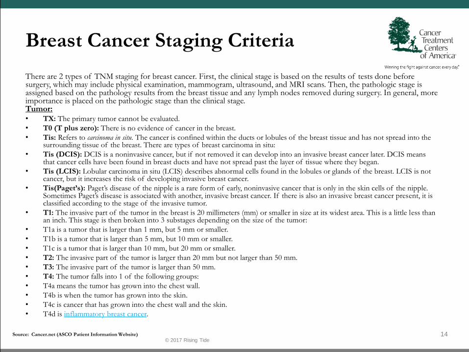
Breast Cancer Staging Criteria
There are 2 types of TNM staging for breast cancer. First, the clinical stage is based on the results of tests done before surgery, which may include physical examination, mammogram, ultrasound, and MRI scans. Then, the pathologic stage is assigned based on the pathology results from the breast tissue and any lymph nodes removed during surgery. In general, more importance is placed on the pathologic stage than the clinical stage.Tumor:• TX: The primary tumor cannot be evaluated.
• T0 (T plus zero): There is no evidence of cancer in the breast.
• Tis: Refers to carcinoma in situ. The cancer is confined within the ducts or lobules of the breast tissue and has not spread into the surrounding tissue of the breast. There are types of breast carcinoma in situ:
• Tis (DCIS): DCIS is a noninvasive cancer, but if not removed it can develop into an invasive breast cancer later. DCIS means that cancer cells have been found in breast ducts and have not spread past the layer of tissue where they began.
• Tis (LCIS): Lobular carcinoma in situ (LCIS) describes abnormal cells found in the lobules or glands of the breast. LCIS is not cancer, but it increases the risk of developing invasive breast cancer.
• Tis(Paget’s): Paget’s disease of the nipple is a rare form of early, noninvasive cancer that is only in the skin cells of the nipple. Sometimes Paget’s disease is associated with another, invasive breast cancer. If there is also an invasive breast cancer present, it is classified according to the stage of the invasive tumor.
• T1: The invasive part of the tumor in the breast is 20 millimeters (mm) or smaller in size at its widest area. This is a little less than an inch. This stage is then broken into 3 substages depending on the size of the tumor:
• T1a is a tumor that is larger than 1 mm, but 5 mm or smaller.
• T1b is a tumor that is larger than 5 mm, but 10 mm or smaller.
• T1c is a tumor that is larger than 10 mm, but 20 mm or smaller.
• T2: The invasive part of the tumor is larger than 20 mm but not larger than 50 mm.
• T3: The invasive part of the tumor is larger than 50 mm.
• T4: The tumor falls into 1 of the following groups:
• T4a means the tumor has grown into the chest wall.
• T4b is when the tumor has grown into the skin.
• T4c is cancer that has grown into the chest wall and the skin.
• T4d is inflammatory breast cancer.
14Source: Cancer.net (ASCO Patient Information Website)© 2017 Rising Tide
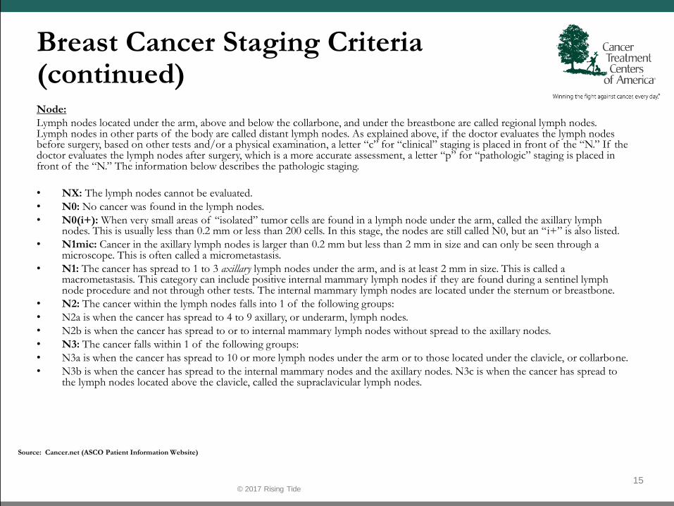
Breast Cancer Staging Criteria (continued)Node:
Lymph nodes located under the arm, above and below the collarbone, and under the breastbone are called regional lymph nodes. Lymph nodes in other parts of the body are called distant lymph nodes. As explained above, if the doctor evaluates the lymph nodes before surgery, based on other tests and/or a physical examination, a letter “c” for “clinical” staging is placed in front of the “N.” If the doctor evaluates the lymph nodes after surgery, which is a more accurate assessment, a letter “p” for “pathologic” staging is placed in front of the “N.” The information below describes the pathologic staging.
• NX: The lymph nodes cannot be evaluated.
• N0: No cancer was found in the lymph nodes.
• N0(i+): When very small areas of “isolated” tumor cells are found in a lymph node under the arm, called the axillary lymph nodes. This is usually less than 0.2 mm or less than 200 cells. In this stage, the nodes are still called N0, but an “i+” is also listed.
• N1mic: Cancer in the axillary lymph nodes is larger than 0.2 mm but less than 2 mm in size and can only be seen through a microscope. This is often called a micrometastasis.
• N1: The cancer has spread to 1 to 3 axillary lymph nodes under the arm, and is at least 2 mm in size. This is called a macrometastasis. This category can include positive internal mammary lymph nodes if they are found during a sentinel lymph node procedure and not through other tests. The internal mammary lymph nodes are located under the sternum or breastbone.
• N2: The cancer within the lymph nodes falls into 1 of the following groups:
• N2a is when the cancer has spread to 4 to 9 axillary, or underarm, lymph nodes.
• N2b is when the cancer has spread to or to internal mammary lymph nodes without spread to the axillary nodes.
• N3: The cancer falls within 1 of the following groups:
• N3a is when the cancer has spread to 10 or more lymph nodes under the arm or to those located under the clavicle, or collarbone.
• N3b is when the cancer has spread to the internal mammary nodes and the axillary nodes. N3c is when the cancer has spread to the lymph nodes located above the clavicle, called the supraclavicular lymph nodes.
15
Source: Cancer.net (ASCO Patient Information Website)
© 2017 Rising Tide
Breast Cancer Staging Criteria(continued)
Metastasis (M)
Evidence of metastatic cancer means this is no longer considered earl-stage or locally advanced cancer.
• MX: Distant spread cannot be evaluated.
• M0: The disease has not metastasized.
• M0 (i+): There is no clinical or radiographic evidence of distant metastases. Microscopic evidence of tumor
cells is found in the blood, bone marrow, or other lymph nodes that are no larger than 0.2 mm.
• M1: There is evidence of metastasis to another part of the body, meaning there are breast cancer cells growing
in other organs.
16
Source: Cancer.net (ASCO Patient Information Website)
© 2017 Rising Tide
Post Mastectomy Chest Wall Pain Syndrome
First Reported in 1978 Wood
Foley 1987
Granek 1984
© 2017 Rising Tide17

Incidence
Stevens, Carpenter et al 1998 ͌ 20%
Fassoulaki/Maunsell/Tasmuth/Wallace 50% or more
© 2017 Rising Tide18
British Journal of Cancer2008 Aug 19; 99(4):604-610
Epidemiology
Risk Factors:
1. Prior breast surgery
2. Tumor located in the upper lateral quadrant
3. Young age; African Americans and Latinas >> Whites
4. Sectioning of the intercostobrachial nerve
5. Dissection of the axillary lymph node
6. Phantom breast pain
7. Neuroma/ scar pain
© 2017 Rising Tide19

Complaints of Patients
• “Tolerable during the day.”
• “I can’t reach over my head.”
• “My shoulders are pulled forward.”
• “I keep thinking to flex the chest muscles and make them
stronger, I stretch, I massage, I work out. I can’t seem to
get it right.”
• “I can’t sleep at night. I wake up to readjust myself.”
20© 2017 Rising Tide
Chronic Post-Surgical Pain
1. Pain develops after a surgical operation.
2. Pain lasts for at least two months.
3. Other causes of pain (infection/ cancer) are not present.
4. The pain is not pain continuing from the patient’s
original condition.
21© 2017 Rising Tide
Phantom Pain
• Scar pain – nearly a third of mastectomy patients
• More patients, whose reconstruction involved implants,
have pain than those who did not have implants.
22© 2017 Rising Tide

Prevention
• Venlafacine (Effexor) the night before surgery and post
operatively for two weeks.
Decreased pain with movement of the chest wall and
arm pain at 6 months.
• Early physical therapy may help to prevent the functional
limitations in the affected arm in cases of frozen
shoulder.
• Fat Transfer23
© 2017 Rising Tide

Tried Treatments for Chest Wall Pain
• Rest
• Cold and Warm Compresses
• Deep Breathing Exercises – Incentive Spirometry
• Narcotic Medication – Moderate to Severe Breast Pain
• NSAID: Ibuprofen/Naproxen/Celecoxib
• Muscle Relaxants
• Physical Therapy
• Nerve Block
• Acupuncture, Massage, Psychotherapy
• Clonidine
• Neurostimulation
• Intraspinal Therapies
• Neuropathic Pain – Anticonvulsants, Trigyclic Antidepressants, Opioids,
Gabapentin
• Official Side Sleeper PillowTM
• Topical Capsaicin24
© 2017 Rising Tide

Intercostal Nerves
25© 2017 Rising Tide
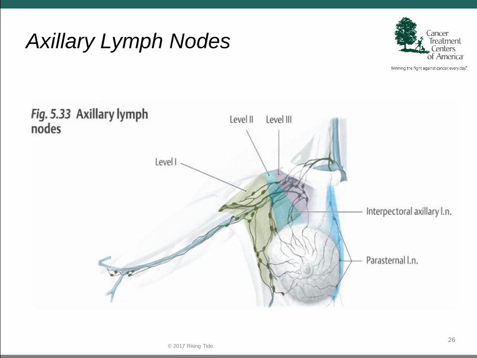
Axillary Lymph Nodes
26© 2017 Rising Tide

Axillary Lymph Nodes: Anterior View
27© 2017 Rising Tide

Anterior Thoracic Wall
28© 2017 Rising Tide
Chest Muscles
• Pectoralis Major
• Pectoralis Minor
• Sternalis (Inconstant)
• Subclavicular Muscle
29
• Serratus Anterior
• Latissimus Dorsi
• External Intercostal Muscles
• Internal Intercostal Muscles
• Transversus Thoracis
• External Oblique Starting at the 5th Rib
© 2017 Rising Tide

Neck Muscles
• Sternocleidomastoid
• Sternothyroid
• Sternohyoid
• Omohyoid
© 2016 Rising Tide30

Nerves
• Lateral Cutaneous Branches of Intercostal Nerves
• Intercostobrachial Nerve (Axilla)
• Long Thoracic Nerve
• Brachial Plexus
31© 2017 Rising Tide
Fascia
• Fascia intertwines all muscles, bones, ligaments,
arteries, veins, nerves and lymphatics
32© 2017 Rising Tide
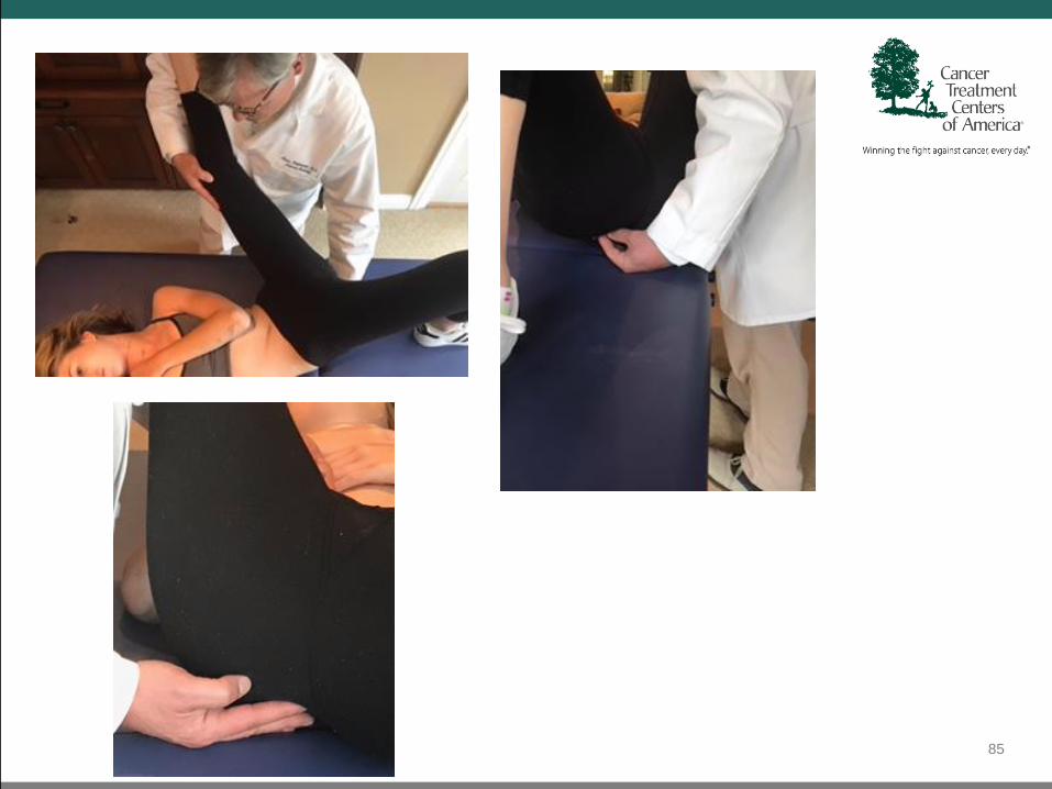
Proposed Mechanism of InjuryPositioning of the Patient
1. Supine, intubated and paralyzed
2. One or both arm(s) raised over the head or to 135
3. Surgery lasts 2 – 4.5 hours
4. Wedge under the scapula may be used at times to
induce further thoracic convexity
5. Ribs may “lock” into an inhaled position
33© 2017 Rising Tide

What are the steps of the counterstrain
basic treatment sequence?
1. Identify tender point.
2.Establish tenderness scale.
3.Monitor and retest.
4.Position to eliminate tenderness.
5.Maintain position 90 seconds.
6.Slowly return to starting position.
7.Recheck tender point.34
© 2017 Rising Tide

Mastectomy
35© 2017 Rising Tide

Sternal Tender Points
36© 2017 Rising Tide

Midclavicular Tender PointsRibs 2 & 3
37© 2017 Rising Tide
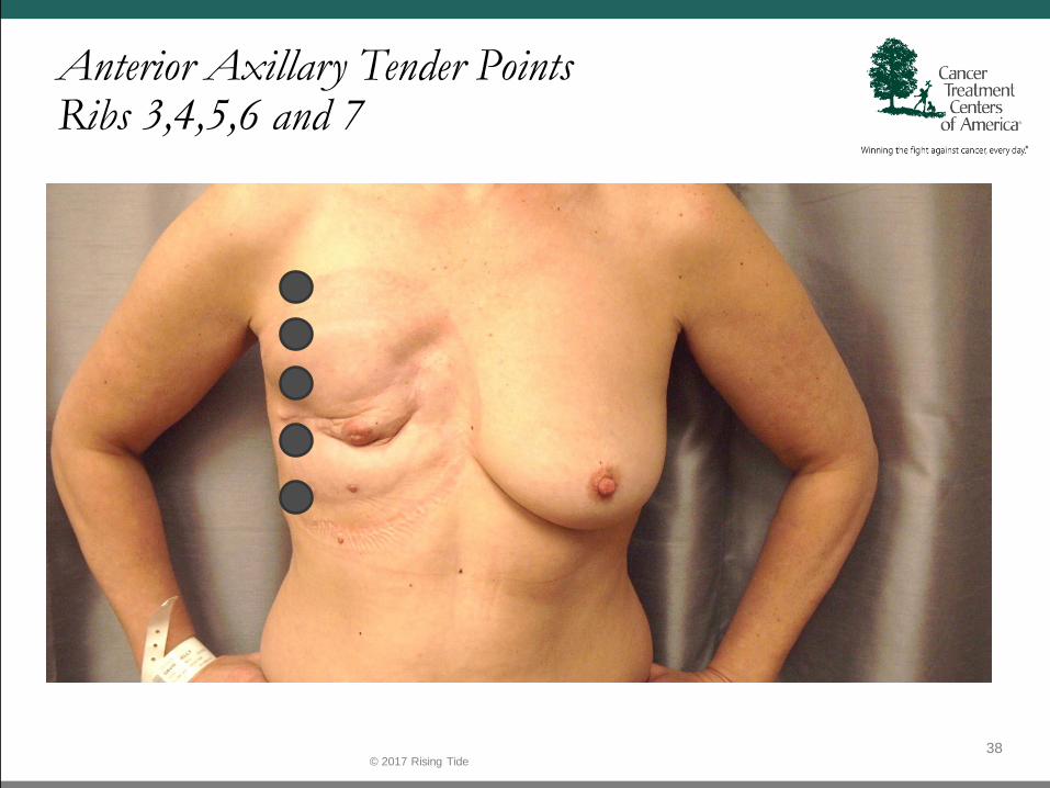
Anterior Axillary Tender PointsRibs 3,4,5,6 and 7
38© 2017 Rising Tide
What are some relative contraindications for counterstrain?
• Patient can’t voluntarily relax
• Patient is severely ill
• Vertebral artery disease
• Severe osteoporosis
39© 2017 Rising Tide

What do we do?
• Instruct the patient to breathe deeply in
through their nose, exhale through their
mouth.
• Place the patient supine in order to evaluate
the chest wall, with knees comfortably bent.
40© 2017 Rising Tide

41© 2017 Rising Tide

42

43

Breast Cancer Lesions
44© 2017 Rising Tide

Staging Breast Cancer Criteria
• Stage 0 (non-invasive carcinoma in situ) breast cancer
– In stage 0, there is no evidence of cancer cells breaking out of the part of the breast in
which they started, or of getting through to or invading neighboring normal tissue.
• Stage I breast cancer
– In stage I, the tumor measures up to two centimeters and no lymph nodes are involved.
• Stage II breast cancer
– In stage II, the tumor measures between two to five centimeters, or the cancer has spread to
the lymph nodes under the arm on the same side as the breast cancer.
• Stage III locally advanced breast cancer– In stage III, the tumor in the breast is more than two inches in diameter across and the cancer is
extensive in the underarm lymph nodes, or has spread to other lymph nodes or tissues near the breast.
• Stage IV (metastatic) breast cancer– In stage IV, the cancer has spread beyond the breast, underarm and internal mammary
lymph nodes to other parts of the body near to or distant from the breast.
• Recurrent breast cancer
– In recurrent breast cancer, the disease has returned in spite of the initial treatment.
45© 2017 Rising Tide
Source: Cancer Treatment Centers of America

46© 2017 Rising Tide

Common types of Head and Neck Cancer
o Hypopharyngeal cancer
o Nasal cavity and paranasal sinus cancer
o Nasopharyngeal cancer
o Salivary gland cancer
o Oral cancer
o Oropharyngeal cancer
47© 2017 Rising Tide
Picture courtesy of: European School of Oncology, published in: Health & Medicine 9.12.11
Get more neck cancer
types.
• Tobacco
• Alcohol
• Gender
• Age
• Certain illnesses: (Epstein-Barr virus (EBV), human papillomavirus
(HPV) and two inherited genetic syndromes—Fanconi anemia and
dyskeratosis congenita—have been linked to head and neck)
• Sun exposure
• Radiation therapy
• Nutrition
• Vaping
48
Head & Neck Risk factors for Cancer
© 2017 Rising Tide
Source: Cancer.net (ASCO Patient Information Website)
Head and neck cancer scenario #1
A 63-year old woman is evaluated for a 1-month history of a
painful lump underneath the tongue. She has a 45-pack-year
smoking history and continues to smoke.
On physical examination, vital signs are normal. An ulcerated
lesion measuring approximately 1 cm is seen on the anterior floor
of the mouth.
The lesion is resected. Pathology specimens identify poorly
differentiated squamous cell carcinoma with negative margins.
The patient is encouraged to stop smoking. Following
discussion of the benefits and risks, she is enrolled in a lung cancer
screening program utilizing low-dose CT scanning
49© 2017 Rising Tide

Head and neck cancer scenario #1 (continued):
Which of the following surveillance tests should also be recommended for this patient?
A. CT scans
B. PET/CT scan
C. Oral examinations and direct laryngoscopy
D. No additional follow-up
Posttreatment surveillance of patients with head and neck cancer should be directed toward development of both locally recurrent cancer and a second primary cancer at a more distant site.
50© 2017 Rising Tide
Philosophy and Mechanical Principles of Osteopathy by AT Still
“What bone would you pull when treating
whooping-cough?”
My answer would be: “The bones that hold by
attachment the muscles of the hyoid system in
such irritable condition, beginning with the
atlas and terminating with the sacrum.”
51© 2017 Rising Tide
52© 2017 Rising Tide

The Rule of the Artery is Supreme
• When blood and lymphatics flow freely, the tissues can
perform their physiologic functions without impedance.
• With the occurrence of trauma (physical or emotional), the
tissues contract, twist and compress. The fluid flow
becomes obstructed.
• Micro-climates of underperfusion result and are considered
to be a significant contributor to the onset of disease.
• Releasing fascial obstructions allows for the unrestricted
reprofusion and restoration of chronic injuries.
53© 2017 Rising Tide
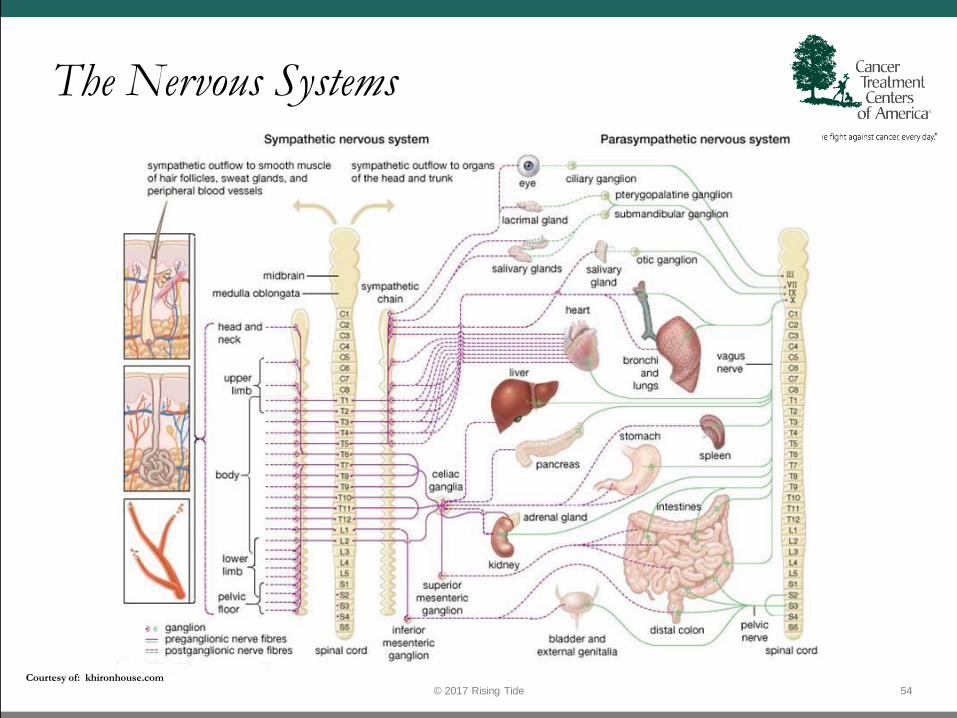
The Nervous Systems
54© 2017 Rising Tide
Courtesy of: khironhouse.com

Sympathetic Nervous System“Fight or Flight”
55© 2017 Rising Tide
Courtesy of: khironhouse.com
Parasympathetic Nervous System“Rest and Digest”
56© 2017 Rising Tide
Courtesy of: khironhouse.com
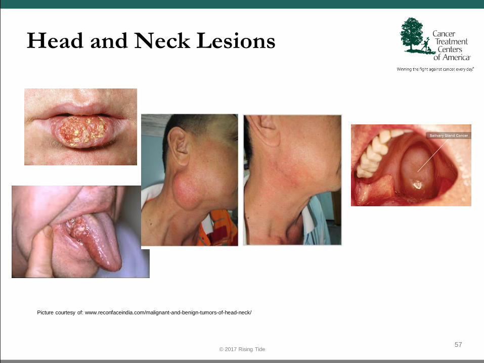
Head and Neck Lesions
57© 2017 Rising Tide
Picture courtesy of: www.reconfaceindia.com/malignant-and-benign-tumors-of-head-neck/
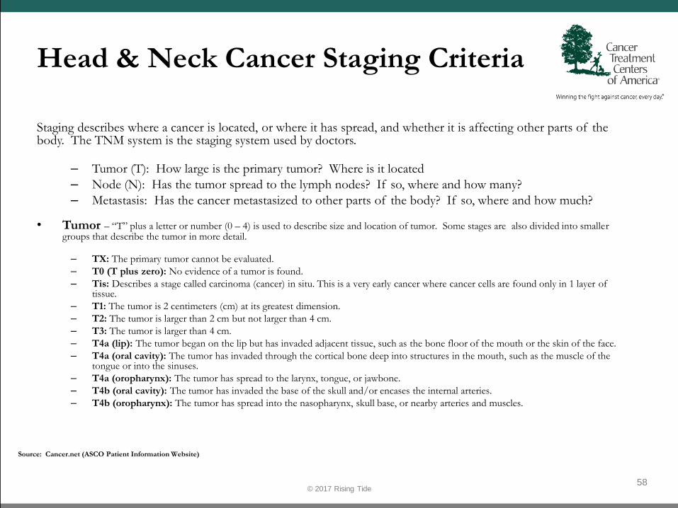
Head & Neck Cancer Staging Criteria
58© 2017 Rising Tide
Staging describes where a cancer is located, or where it has spread, and whether it is affecting other parts of the body. The TNM system is the staging system used by doctors.
– Tumor (T): How large is the primary tumor? Where is it located
– Node (N): Has the tumor spread to the lymph nodes? If so, where and how many?
– Metastasis: Has the cancer metastasized to other parts of the body? If so, where and how much?
• Tumor – “T” plus a letter or number (0 – 4) is used to describe size and location of tumor. Some stages are also divided into smaller groups that describe the tumor in more detail.
– TX: The primary tumor cannot be evaluated.
– T0 (T plus zero): No evidence of a tumor is found.
– Tis: Describes a stage called carcinoma (cancer) in situ. This is a very early cancer where cancer cells are found only in 1 layer oftissue.
– T1: The tumor is 2 centimeters (cm) at its greatest dimension.
– T2: The tumor is larger than 2 cm but not larger than 4 cm.
– T3: The tumor is larger than 4 cm.
– T4a (lip): The tumor began on the lip but has invaded adjacent tissue, such as the bone floor of the mouth or the skin of the face.
– T4a (oral cavity): The tumor has invaded through the cortical bone deep into structures in the mouth, such as the muscle of the tongue or into the sinuses.
– T4a (oropharynx): The tumor has spread to the larynx, tongue, or jawbone.
– T4b (oral cavity): The tumor has invaded the base of the skull and/or encases the internal arteries.
– T4b (oropharynx): The tumor has spread into the nasopharynx, skull base, or nearby arteries and muscles.
Source: Cancer.net (ASCO Patient Information Website)
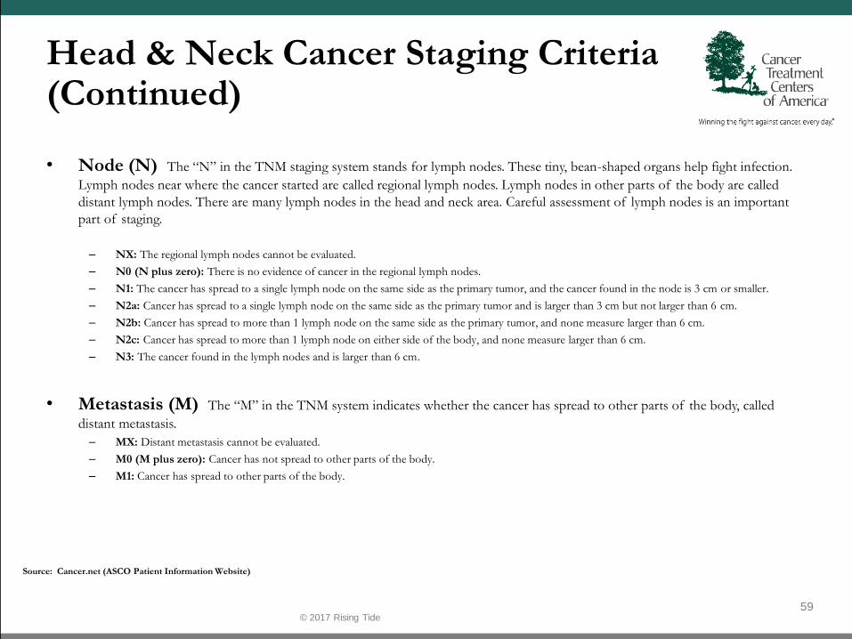
• Node (N) The “N” in the TNM staging system stands for lymph nodes. These tiny, bean-shaped organs help fight infection.
Lymph nodes near where the cancer started are called regional lymph nodes. Lymph nodes in other parts of the body are called
distant lymph nodes. There are many lymph nodes in the head and neck area. Careful assessment of lymph nodes is an important
part of staging.
– NX: The regional lymph nodes cannot be evaluated.
– N0 (N plus zero): There is no evidence of cancer in the regional lymph nodes.
– N1: The cancer has spread to a single lymph node on the same side as the primary tumor, and the cancer found in the node is 3 cm or smaller.
– N2a: Cancer has spread to a single lymph node on the same side as the primary tumor and is larger than 3 cm but not larger than 6 cm.
– N2b: Cancer has spread to more than 1 lymph node on the same side as the primary tumor, and none measure larger than 6 cm.
– N2c: Cancer has spread to more than 1 lymph node on either side of the body, and none measure larger than 6 cm.
– N3: The cancer found in the lymph nodes and is larger than 6 cm.
• Metastasis (M) The “M” in the TNM system indicates whether the cancer has spread to other parts of the body, called
distant metastasis.
– MX: Distant metastasis cannot be evaluated.
– M0 (M plus zero): Cancer has not spread to other parts of the body.
– M1: Cancer has spread to other parts of the body.
59
Head & Neck Cancer Staging Criteria(Continued)
Source: Cancer.net (ASCO Patient Information Website)
© 2017 Rising Tide

Do you want to keep this instead of previous slides?
60
• Stage 0 head and neck cancer
– At stage 0, the tumor is only growing in the part of the head and neck where it started. No cancer cells are present in deeper layers of tissue, nearby structures, lymph nodes or distant sites (e.g., Tis, N0, M0 Carcinoma in situ).
• Stage I head and neck cancer
– At stage 1, the primary tumor is 2 cm across or smaller, and no cancer cells are present in nearby structures, lymph nodes or distant sites (e.g., T1, N0, M0).
• Stage II head and neck cancer
– At stage II, the head and neck tumor measures 2-4 cm across, and no cancer cells are present in nearby structures, lymph nodes or distant sites (e.g., T2, N0, M0).
• Stage III head and neck cancer
– At stage III, the tumor fits one of the following criteria:
– It is larger than 4 cm across, and no cancer cells are present in nearby structures, lymph nodes or distant sites (e.g., T3, N0, M0).
– It is any size but has not grown into nearby structures or distant sites. However, cancer cells are present in one lymph node, which is located on the same side of the head or neck as the primary tumor and is smaller than 3 cm across (e.g., T1-3, N1, M0).
• Stage IV head and neck cancer
– Stage IVA: One of the following applies:
• T4a, N0 or N1, M0: The head and neck cancer tumor is any size and is growing into nearby structures.
– T1-4a, N2, M0: The tumor is any size and may or may not have invaded nearby structures. It has not spread to distant sites
Source: Cancer.net (ASCO Patient Information Website)
© 2017 Rising Tide
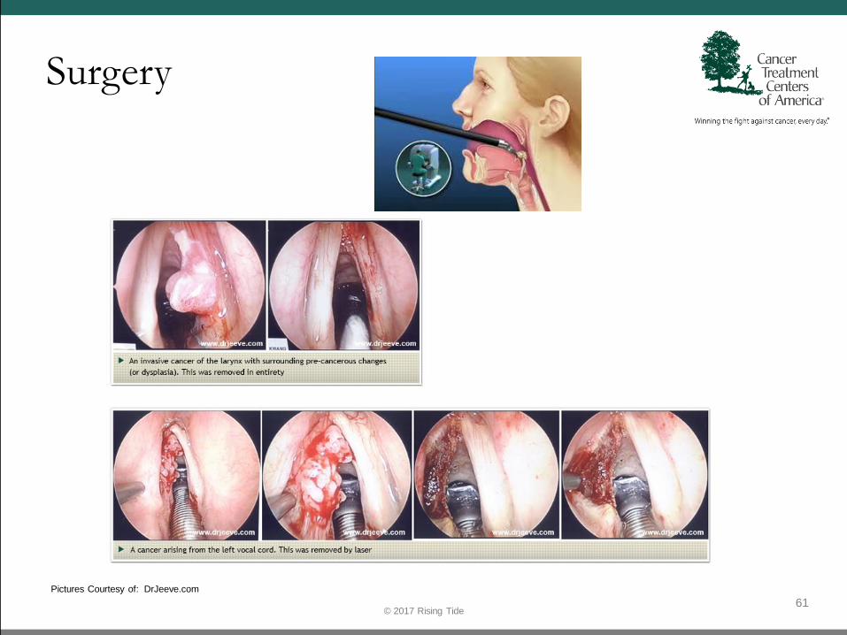
Surgery
61© 2017 Rising Tide
Pictures Courtesy of: DrJeeve.com

Pain in the Throat following Surgical Trauma
S/P Intubation – Hyoid Bone Dysfunction
S/P Thyroidectomy – Recurrent Laryngeal Nerve Dysfunction
62© 2017 Rising Tide
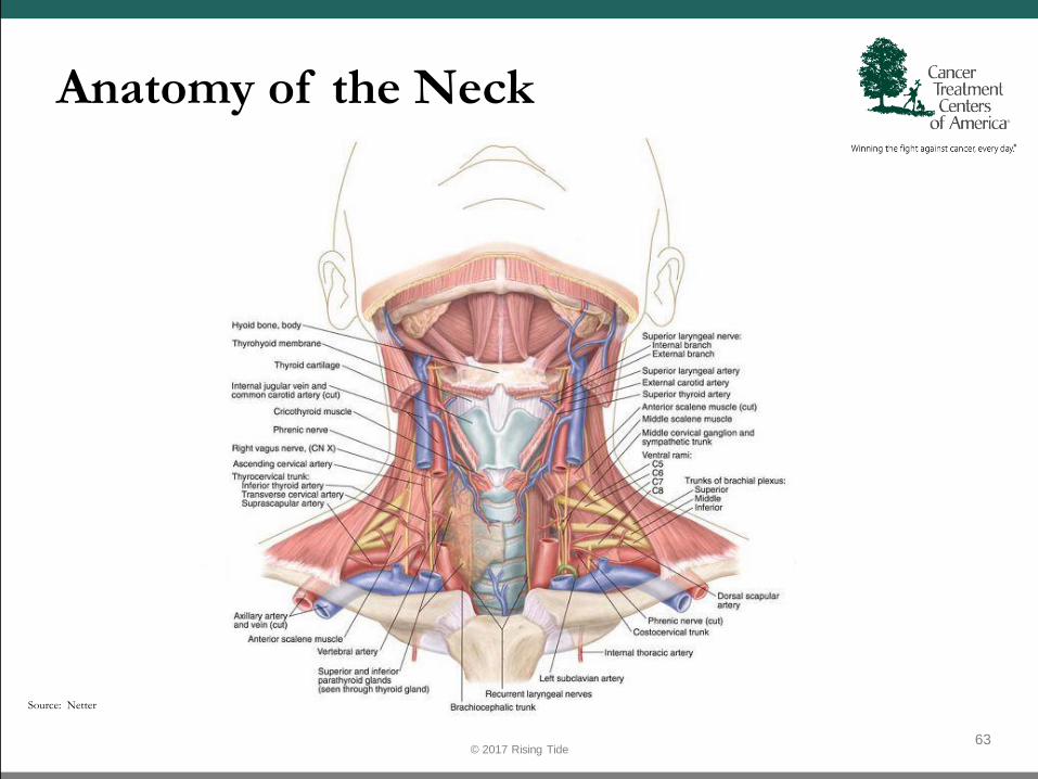
Anatomy of the Neck
63© 2017 Rising Tide
Source: Netter
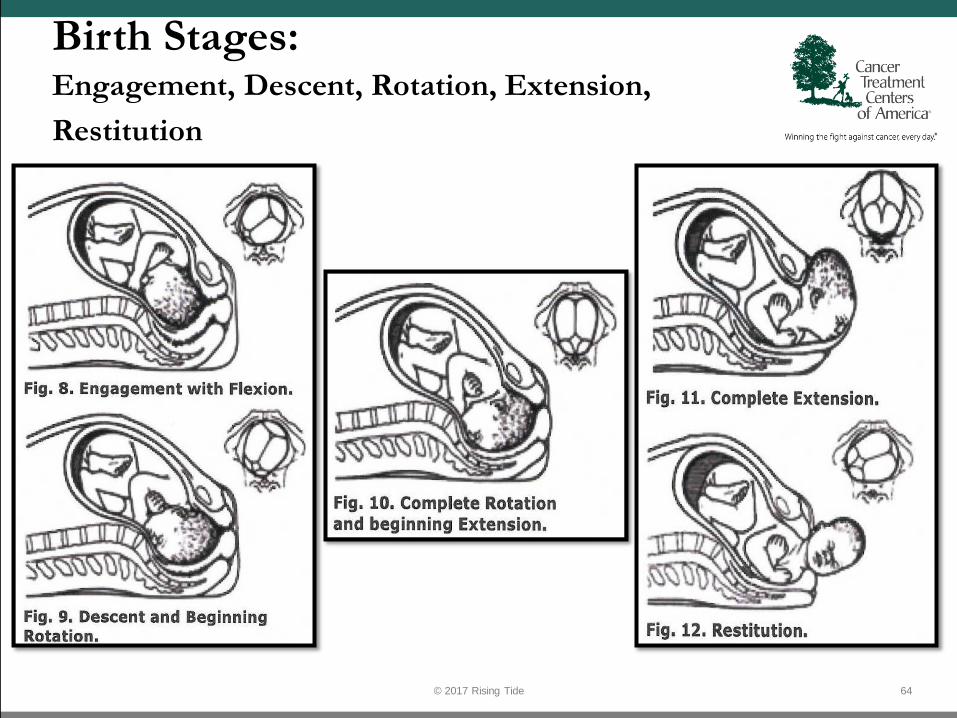
Birth Stages:Engagement, Descent, Rotation, Extension,
Restitution
64© 2017 Rising Tide

65
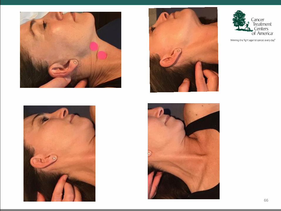
66

67
Eval vocal chords

68© 2017 Rising Tide

Radical Neck Resection
69© 2017 Rising Tide
Source: Netter
Contra-indications
• What to do if?
• What not to do if?
70© 2017 Rising Tide
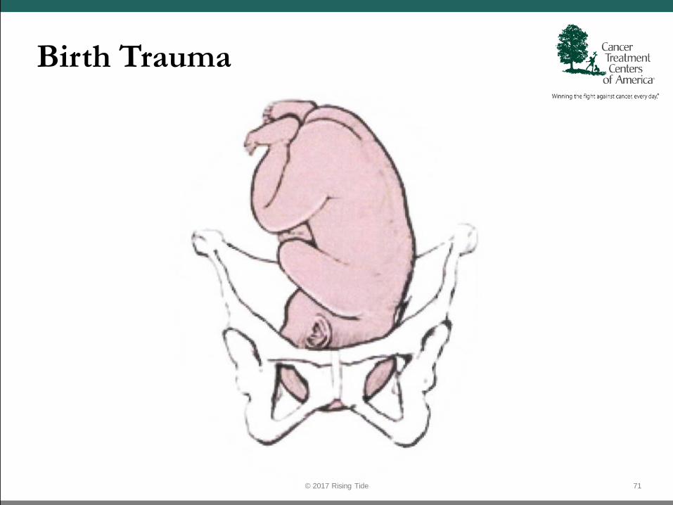
Birth Trauma
71© 2017 Rising Tide
Colon Cancer
72© 2017 Rising Tide
• Types of Colon Cancer:– Colorectal Adenocarcinoma
• "Adeno" is a prefix meaning glands. "Carcinoma" is a type of cancer that grows in epithelial cells that line the surfaces inside and outside the body. Colorectal adenocarcinomas develop in the lining of the colon or rectum, which make up the large intestine. They tend to start in the inner lining and then spread to other layers.
– Gastrointestinal Carcinoid Tumors• Carcinoid tumors develop in nerve cells called neuroendocrine cells, which help regulate hormone
production. These tumors are among a group of cancers called neuroendocrine tumors (NETs). Carcinoid tumors cells are slow-growing and may develop in the lungs and/or gastrointestinal tract. They account for 1 percent of all colorectal cancers and half of all cancers found in the small intestine.
– Metastatic Colorectal Cancer• Cancer cells may break away from a tumor in the colon or rectum and spread to other parts of the body
through the bloodstream or lymphatic system. These cells may settle and form new tumors on a different organ.
• The most common site of metastases for colon or rectal cancer is the liver. Colorectal cancer cells may also spread to the lungs, bones, brain or spinal cord.
– Other Colorectal Cancers:• Primary colorectal lymphomas
• Gastrointestinal stromal tumors
• Leiomyosarcomas
• Melanomas
Source: Cancer.net (ASCO Patient Information Website)

Colon cancer scenario #1
A 61-year old woman undergoes routine follow-up evaluation.
Stage II colon cancer was diagnosed 3 years ago and was treated
with surgical resection. The patient now feels well. She works full
time and exercises regularly. Medical history is otherwise
unremarkable, and she takes no medications.
Findings on physical examination, including vital signs, are
normal.
Routine surveillance CT scans of the chest and abdomen show
three new hypodense lesions in the right lobe of the liver, ranging
in size from 1 to 3 cm. No other abnormalities are seen.
73© 2017 Rising Tide
Colon cancer scenario #1(continued):
Which of the following is the most appropriate management?
A. CT-guided needle biopsy of a liver lesion
B. Hepatic artery embolization
C. Palliative systemic chemotherapy
D. Radiation therapy to the liver
E. Right hepatectomy
The development of oligometastatic disease (usually to the liver or lung) in a patient who previously was treated for colorectal cancer is potentially curable by surgical resection.
74© 2017 Rising Tide

Colon cancer scenario #2
A 77-year old woman is evaluated for the new-onset
fatigue and anemia. She otherwise feels well. Medical
history is unremarkable, and she takes no medications.
Physical examination findings, including vital signs, are
normal. BMI is 22.
Colonoscopy identifies a 7-cm mass in the transverse
colon. Biopsy of the mass shows poorly differentiated
adenocarcinoma. Contrast-enhanced CT scans of the
chest, abdomen, and pelvis show the mass, but no other
abnormalities are identified.
75© 2017 Rising Tide

Colon cancer scenario #2
Which of the following is likely to be the most important factor in
determining this patient’s prognosis?
A. Degree of differentiation of the tumor
B. Patient’s performance status
C. Size of the tumor
D. Stage of the tumor
Tumor stage is usually the most important prognostic factor in
determining outcome in a patient with newly diagnosed cancer.
76© 2017 Rising Tide

Colon Cancer Lesions
77© 2017 Rising Tide
Picture Courtesy of: Amiscus Visual Solutions
Staging Colon Cancer Criteria
78© 2017 Rising Tide
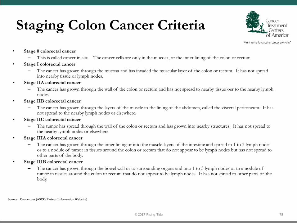
• Stage 0 colorectal cancer
– This is called cancer in situ. The cancer cells are only in the mucosa, or the inner lining of the colon or rectum
• Stage I colorectal cancer
– The cancer has grown through the mucosa and has invaded the muscular layer of the colon or rectum. It has not spread into nearby tissue or lymph nodes.
• Stage IIA colorectal cancer
– The cancer has grown through the wall of the colon or rectum and has not spread to nearby tissue oer to the nearby lymph nodes.
• Stage IIB colorectal cancer
– The cancer has grown through the layers of the muscle to the lining of the abdomen, called the visceral peritoneum. It has not spread to the nearby lymph nodes or elsewhere.
• Stage IIC colorectal cancer
– The tumor has spread through the wall of the colon or rectum and has grown into nearby structures. It has not spread to the nearby lymph nodes or elsewhere.
• Stage IIIA colorectal cancer
– The cancer has grown through the inner lining or into the muscle layers of the intestine and spread to 1 to 3 lymph nodes or to a nodule of tumor in tissues around the colon or rectum that do not appear to be lymph nodes but has not spread to other parts of the body.
• Stage IIIB colorectal cancer
– The cancer has grown through the bowel wall or to surrounding organs and into 1 to 3 lymph nodes or to a nodule of tumor in tissues around the colon or rectum that do not appear to be lymph nodes. It has not spread to other parts of the body.
Source: Cancer.net (ASCO Patient Information Website)

Pain in the Abdomen following Surgical Trauma
S/P Cholycystectomy – Hepatic Capsule Pain – Right Shoulder Pain
S/P Appendectomy
S/P Laparotomy
S/P Splenectomy
S/P Heartbreak?
79© 2017 Rising Tide
For patients with metastatic colorectal cancer, the sidedness of the primary tumor within the colon appears to affect both survival and the effectiveness of commonly used biological agents Avastin (bevacizumab) and Erbitux (cetuximab), which are designed to interfere with the formation of blood vessels that feed a tumor, and with growth factor receptor signaling. Patients with left-sided disease enjoyed a median overall survival of 33 months compared with a 19.4 months in right-sided disease. And a cxomparison of Avastin and Erbitux shoed that Erbitux might actually be harmful to patients with right-sided tumors.
80
Source: Curetoday.com

Colon Resection
81© 2017 Rising Tide
Picture Courtesy of: Amiscus Visual Solutions

82

83
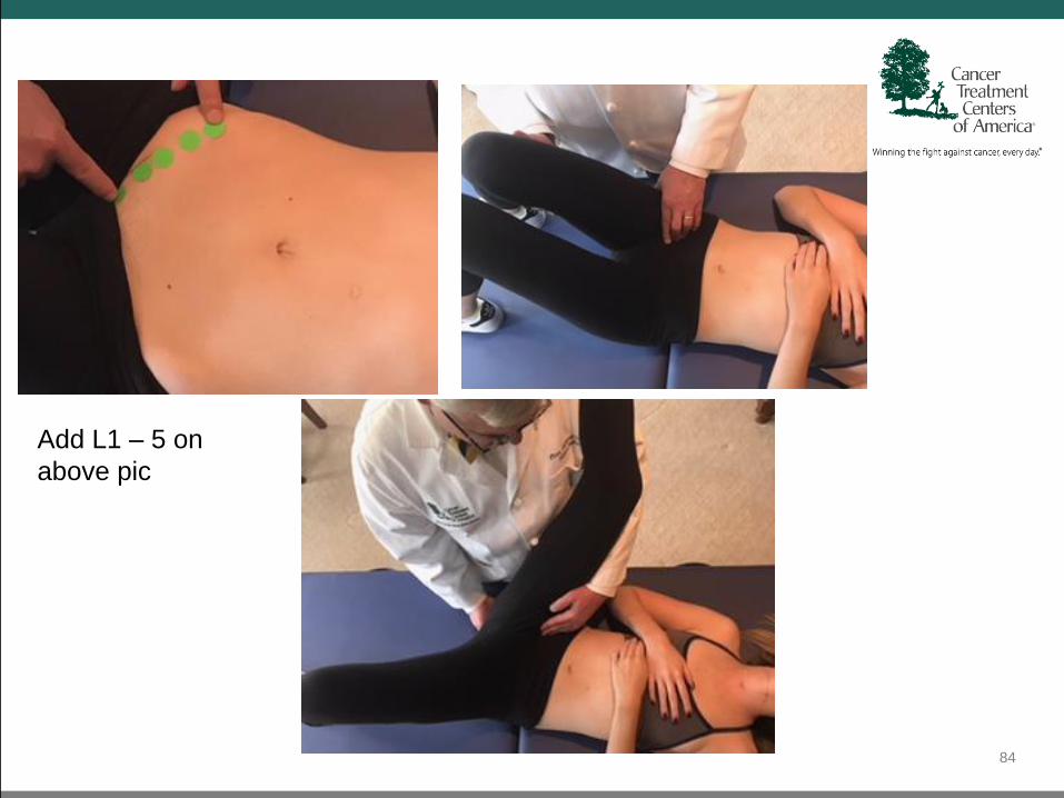
84
Add L1 – 5 on
above pic
85

86
Philosophy and Mechanical Principles of Osteopathy by AT Still
“We cure these diseases by subduing the cause that has
produced such alarming effects as angry ulcers, cancers,
tumors, and all diseases that assail woman. We must apply
our mental and physical energies to the place in the spine
controlling the blood-supply sustaining the life and health
of the womb, the bladder, kidneys, liver, spleen, pancreas,
lymphatics, and all parts of abdominal life.” (Ch.VIII, The Abdomen)
87© 2017 Rising Tide

Contra-indications
• What to do if?
• What not to do if?
88© 2017 Rising Tide

Abdominal Visceral Anatomy
89© 2017 Rising Tide
Courtesy of: anatomyclass123.com
Become the champion of the patient’s social history
– they will love you for it.
Traditional: Smoking/ETOH/Illicits etc.
Have fun – 10 minutes will change their lives. It lets them know that you care.
“Tell me about yourself.”
• Favorite color?
• Favorite food?
• Religion?
• Children’s name?
• Spouse’s name and how they met?
• How do you like to be addressed?
• Hobbies?
• What is your career?
• What are you famous for?
• Do you have a fondest or funniest memory?
• Imagine your perfect day: Where would you be and who would you be with?
90© 2017 Rising Tide

Philosophy and Mechanical Principles of Osteopathy by AT Still
The soul of man,
with all the streams of pure living water,
seems to dwell in the fascia of his body.
91© 2016 Rising Tide

Philosophy and Mechanical Principles of Osteopathy by AT Still
“Life is in danger,
and can be saved by skill,
not by force and ignorance.” (Ch.VI, The Thorax)
92© 2017 Rising Tide

Philosophy and Mechanical Principles of Osteopathy by AT Still
The osteopathic surgeon uses “the knife of
blood” to keep out “the knife of steel,” and
saves life by saving the injured or diseased limbs
and organs from the body by reduction, in
place of removing them.
93© 2017 Rising Tide

Great Books
By Andrew Taylor Still
Autobiography of A. T. Still (1897)
Philosophy of Osteopathy (1899)
The Philosophy and Mechanical Principles of Osteopathy (1902)
Osteopathy, Research and Practice (1910)
By John Lewis
A. T. Still: From the Dry Bone to the Living Man (2014)
94© 2017 Rising Tide

Current Good Reads
• Rodney Yee - Moving Towards Balance
• Kelly Turner, PhD - Radical Remission
• David R. Hawkins, PhD - Power vs. Force
• Robert Fulford, D.O., FAAO - Touch of Life: The healing Power of the
Natural Life Force
95© 2017 Rising Tide

Osteopathy is…
“Osteopathy is a therapeutic gold
mine. Many veins of high grade ore
have been found, and are being
worked; but others just as valuable are
yet to be discovered.”C.B. Rowlingson, DO, Editor The Western Osteopath
96© 2017 Rising Tide