Osteogenic differentiation of mesenchymal stem cells using PAMAM dendrimers as gene delivery vectors
8
Osteogenic differentiation of mesenchymal stem cells using PAMAM dendrimers as gene delivery vectors José Luís Santos a , Elena Oramas a , Ana Paula Pêgo b,c , Pedro Lopes Granja b,c , Helena Tomás a, ⁎ a Centro de Química da Madeira, Departamento de Química, Universidade da Madeira, Campus Universitário da Penteada, 9000-390 Funchal, Portugal b INEB — Instituto de Engenharia Biomédica, Divisão de Biomateriais, Rua do Campo Alegre, 823, 4150-180 Porto, Portugal c Universidade do Porto, Faculdade de Engenharia, Dep. Eng. Metalúrgica e Materiais, Rua Dr. Roberto Frias s/n, 4200-465 Porto, Portugal abstract article info Article history: Received 27 July 2008 Accepted 11 November 2008 Available online 24 November 2008 Keywords: PAMAM dendrimers Bone regeneration Nonviral gene delivery Mesenchymal stem cells Bone morphogenetic protein This paper reports the use of different generations of polyamidoamine (PAMAM) dendrimers for the in vitro transfection of mesenchymal stem cells (MSCs). A systematic study was carried out on the transfection efficiency achieved by the PAMAM dendrimers using a β-galactosidase reporter gene system. Transfection results were shown to be dependent upon the generation of dendrimers, the amine to phosphate group ratio and the cell passage number. In all cases, the transfection efficiency was very low. Nevertheless, it was hypothesized that a low transfection level could be sufficient to promote the in vitro differentiation of MSCs towards the osteoblastic lineage. To address this possibility, dendrimers carrying the human bone morphogenetic protein-2 (hBMP-2) gene-containing plasmid were used. All quantitative (alkaline phosphatase activity, osteocalcin secretion and calcium deposition) and qualitative (von Kossa staining) osteogenic markers were significantly stronger in transfected cells when compared to non-transfected ones. This study not only clearly demonstrates that a low transfection level can be sufficient for inducing in vitro differentiation of MSCs to the osteoblast phenotype but also highlights the importance of focusing research on the development of gene delivery vectors in the concrete application. © 2008 Elsevier B.V. All rights reserved. 1. Introduction Stem cell-mediated gene therapy is currently one of the most attractive strategies under investigation for bone regeneration. In this approach, an osteoinductive factor is secreted by the cells that have been genetically modified to express the desired gene [1–7]. Reports on bone regeneration by genetically modified mesenchymal stem cells (MSCs) mainly employ adenovirus or retrovirus vectors carrying bone morpho- genetic proteins (BMPs) as therapeutic genes [8–12]. This can be attributed to the high efficiency in gene transfer by the viral vectors, especially in slowly dividing or non-dividing cells. However, the risk of mutagenesis and/or oncogenesis associated with viral systems, as well as their potential immunogenicity, provides a strong motivation for the search of alternative methods. In this scope, several investigations are currently focused on developing high efficiency nonviral vectors, covering a wide range of systems, such as the ones based on cationic polymers, liposomes and calcium phosphates [13–15]. In general, these systems have been shown to be effective in well-established cell lines but have limitations when using primary cell cultures, namely bone marrow-derived MSCs [16,17]. Also, authors often base their conclusions on reporter gene assays and disregard the concrete applications, not exploring the delivery of genes encoding for bioactive agents [18,19]. Dendrimers are spherical macromolecules that consist of a core moiety from which branches radiate. A typical dendrimer consists of a multifunctional central core, branched units (organised in layers called “generations”, G) and surface groups. Polycationic dendrimers entail a particularly interesting system for nuclei acid delivery. These molecules display a well-defined architecture as well as a high multivalent surface moiety (amino termini) to molecular volume ratio [20–22]. Polyami- doamine (PAMAM) dendrimers are amongst the earliest synthesized and commercialized dendritic systems. Since the pioneer work of Haensler and Szoka in 1993 [23], these molecules have been tested and used as gene transfer agents in unmodified and modified forms [24,25]. Systems based on the PAMAM dendrimers have been shown to transfect a series of cell types, most of which include cell lines [26]. Gebhart et al. compared the performance of several dendrimeric systems and found that the transfection efficiency strongly varied not only with the kind of vector molecule but also with the cell-type [27]. In the present work, the transfection efficiency achieved by different generations of PAMAM dendrimers was evaluated. MSCs were employed here due to their relevance in bone regeneration applications. To our knowledge, this is the first report on the use of polyamidoamine dendrimers for in vitro transfection of MSCs. A β-galactosidase reporter gene system was used and several experimental conditions were assayed. The transfection efficiency obtained for this system was always very low but it was however hypothesized that such low transfection levels could still be sufficient for the envisaged application, i.e., to Journal of Controlled Release 134 (2009) 141–148 ⁎ Corresponding author. Tel.: +351 291705100; fax: +351 291705149. E-mail address: [email protected] (H. Tomás). GENE DELIVERY 0168-3659/$ – see front matter © 2008 Elsevier B.V. All rights reserved. doi:10.1016/j.jconrel.2008.11.007 Contents lists available at ScienceDirect Journal of Controlled Release journal homepage: www.elsevier.com/locate/jconrel
-
Upload
jose-luis-santos -
Category
Documents
-
view
213 -
download
0
Transcript of Osteogenic differentiation of mesenchymal stem cells using PAMAM dendrimers as gene delivery vectors

Journal of Controlled Release 134 (2009) 141–148
GENEDELIVERY
Contents lists available at ScienceDirect
Journal of Controlled Release
j ourna l homepage: www.e lsev ie r.com/ locate / jconre l
Osteogenic differentiation of mesenchymal stem cells using PAMAM dendrimers asgene delivery vectors
José Luís Santos a, Elena Oramas a, Ana Paula Pêgo b,c, Pedro Lopes Granja b,c, Helena Tomás a,⁎a Centro de Química da Madeira, Departamento de Química, Universidade da Madeira, Campus Universitário da Penteada, 9000-390 Funchal, Portugalb INEB — Instituto de Engenharia Biomédica, Divisão de Biomateriais, Rua do Campo Alegre, 823, 4150-180 Porto, Portugalc Universidade do Porto, Faculdade de Engenharia, Dep. Eng. Metalúrgica e Materiais, Rua Dr. Roberto Frias s/n, 4200-465 Porto, Portugal
⁎ Corresponding author. Tel.: +351 291705100; fax: +3E-mail address: [email protected] (H. Tomás).
0168-3659/$ – see front matter © 2008 Elsevier B.V. Aldoi:10.1016/j.jconrel.2008.11.007
a b s t r a c t
a r t i c l e i n f oArticle history:
This paper reports the use o Received 27 July 2008Accepted 11 November 2008Available online 24 November 2008Keywords:PAMAM dendrimersBone regenerationNonviral gene deliveryMesenchymal stem cellsBone morphogenetic protein
f different generations of polyamidoamine (PAMAM) dendrimers for the in vitrotransfection of mesenchymal stem cells (MSCs). A systematic study was carried out on the transfectionefficiency achieved by the PAMAM dendrimers using a β-galactosidase reporter gene system. Transfectionresults were shown to be dependent upon the generation of dendrimers, the amine to phosphate group ratioand the cell passage number. In all cases, the transfection efficiency was very low. Nevertheless, it washypothesized that a low transfection level could be sufficient to promote the in vitro differentiation of MSCstowards the osteoblastic lineage. To address this possibility, dendrimers carrying the human bonemorphogenetic protein-2 (hBMP-2) gene-containing plasmid were used. All quantitative (alkalinephosphatase activity, osteocalcin secretion and calcium deposition) and qualitative (von Kossa staining)osteogenic markers were significantly stronger in transfected cells when compared to non-transfected ones.This study not only clearly demonstrates that a low transfection level can be sufficient for inducing in vitrodifferentiation of MSCs to the osteoblast phenotype but also highlights the importance of focusing researchon the development of gene delivery vectors in the concrete application.
© 2008 Elsevier B.V. All rights reserved.
1. Introduction
Stem cell-mediated gene therapy is currently one of the mostattractive strategies under investigation for bone regeneration. In thisapproach, anosteoinductive factor is secreted by the cells that have beengenetically modified to express the desired gene [1–7]. Reports on boneregeneration by genetically modified mesenchymal stem cells (MSCs)mainly employ adenovirus or retrovirus vectors carrying bone morpho-genetic proteins (BMPs) as therapeutic genes [8–12]. This can beattributed to the high efficiency in gene transfer by the viral vectors,especially in slowly dividing or non-dividing cells. However, the risk ofmutagenesis and/or oncogenesis associated with viral systems, as wellas their potential immunogenicity, provides a strong motivation for thesearch of alternative methods. In this scope, several investigations arecurrently focused on developing high efficiency nonviral vectors,covering a wide range of systems, such as the ones based on cationicpolymers, liposomes and calcium phosphates [13–15]. In general, thesesystems have been shown to be effective in well-established cell linesbut have limitations when using primary cell cultures, namely bonemarrow-derivedMSCs [16,17]. Also, authors often base their conclusionson reporter gene assays and disregard the concrete applications, notexploring the delivery of genes encoding for bioactive agents [18,19].
51 291705149.
l rights reserved.
Dendrimers are spherical macromolecules that consist of a coremoiety from which branches radiate. A typical dendrimer consists of amultifunctional central core, branched units (organised in layers called“generations”, G) and surface groups. Polycationic dendrimers entail aparticularly interesting system for nuclei acid delivery. Thesemoleculesdisplay awell-defined architecture as well as a high multivalent surfacemoiety (amino termini) to molecular volume ratio [20–22]. Polyami-doamine (PAMAM) dendrimers are amongst the earliest synthesizedand commercialized dendritic systems. Since the pioneer work ofHaensler and Szoka in 1993 [23], these molecules have been tested andused as gene transfer agents in unmodified and modified forms [24,25].Systemsbased on the PAMAMdendrimers have been shown to transfecta series of cell types, most of which include cell lines [26]. Gebhart et al.compared the performance of several dendrimeric systems and foundthat the transfection efficiency strongly varied not only with the kind ofvector molecule but also with the cell-type [27].
In the presentwork, the transfection efficiency achieved by differentgenerations of PAMAM dendrimers was evaluated. MSCs wereemployed here due to their relevance in bone regeneration applications.To our knowledge, this is the first report on the use of polyamidoaminedendrimers for in vitro transfection of MSCs. A β-galactosidase reportergene system was used and several experimental conditions wereassayed. The transfection efficiency obtained for this systemwas alwaysvery low but it was however hypothesized that such low transfectionlevels could still be sufficient for the envisaged application, i.e., to

Table 1Molecular weight and number of amine surface groups for the three generations ofPAMAM dendrimers used
Generation Molecular weight Number of amine surface groups
G5 28,826 128G6 58,048 256G7 116,493 512
Table 2Concentration of dendrimers in solution for the N:P charge ratios used in thetransfection experiments (the concentration of DNA was always kept constant at avalue of 1 µg/cm2 of plate surface)
N:Pratio
Concentration of dendrimers (nM)
G5 G6 G7
5:1 376 188 948:1 602 301 15010:1 752 376 188
GENEDELIVERY
142 J.L. Santos et al. / Journal of Controlled Release 134 (2009) 141–148
improve the in vitro differentiation of MSCs towards the osteoblasticlineage. To confirm this possibility, MSCswere genetically engineered toexpress human BMP-2 (hBMP-2) using PAMAM dendrimers. Thedifferentiation of the MSCs was studied in the absence of otherosteoinductive factors (e.g. dexamethasone). This was achieved throughthe analysis of established markers of the osteoblastic phenotype,including alkaline phosphatase (ALP) activity, osteocalcin, and deposi-tion of a calcified matrix. The results clearly indicate that the PAMAM/hBMP-2 system strongly induces in vitro differentiation of MSCs to theosteoblast phenotype.
2. Materials and methods
2.1. Materials and reagents
Generations 5, 6 and 7 of the PAMAM dendrimers, possessingethylenediamine cores and amine termini, were purchased in watersolution from Dendritech Inc. The chemical structures, molecularweights and number of amine surface groups presented by thedendrimers are shown in Table 1. Tissue culture plates were fromNUNC. All other materials and reagents were obtained from SIGMA-ALDRICH unless otherwise stated.
2.2. Isolation and culture of bone marrow-derived MSCs
Rat bonemarrow-derivedMSCswere harvested from themarrow ofyoung adult male (6 weeks) Wistar rats (Charles River Laboratories,Spain) according to a previously described method [28,29]. Briefly, ratswere euthanized using 1ml of 20% pentobarbital. The femurs and tibiaswere aseptically excised from the hind limbs. External soft tissue wasdiscarded and the bones were then washed in α-Minimum EssentialMedium (α-MEM) containing 5% antibiotic–antimycotic (AbAm) solu-tion. Epiphyses were removed and the bone marrow was flushed outwithα-MEMsupplementedwith10% Foetal Bovine Serum (FBS, GIBCO),1% AbAm and 50 µg/ml ascorbic acid. This was done using a 21-gaugeneedle syringe. The cell suspension was seeded into six T-75 flasks andcultured at 37 °C and 5% carbon dioxide atmosphere.
In order to remove nonadherent hematopoietic cells, the mediumwas changed on day 4 of culture, and every 3 days thereafter. Betweendays 7 and 10 the cells were trypsinized using trypsin-EDTA (GIBCO)and subcultured at a density of 2×10⁴ cells/cm2 for expansion. Whenthey reached 90% confluency the cells were detached and subse-quently used.
2.3. Culture of human embryonic kidney cells
Human embryonic kidney cells (HEK 293T) were cultured inDulbecco's Modified Eagle's Medium (DMEM, GIBCO) with high glucosecontent and GlutaMAX-I containing 10% FBS and 1% AbAm. Fortransfection experiments, HEK 293T cells were subcultured at a densityof 2×10⁴ cells/cm2.
2.4. Plasmid propagation and isolation
Plasmid DNA (pCMV SPORT-βgal, 7.8 kb, Invitrogen) encoding β-galactosidase under the control of the cytomegalovirus (CMV)
promoter was used as a reporter gene to evaluate the transfection ofMSCs by conventional measurement of β-galactosidase activity. ForBMP-2 gene delivery, a pcDNA3.1/Hist©/hBMP2 vector/plasmid(8.5 kb) (kindly donated by Prof. Yasuhiko Tabata, University ofKyoto, Japan) encoding a human BMP-2 sequence inserted down-stream the CMV promoter was used. The plasmids were amplified inEscherichia coli host strain, DH5α, grown overnight in Luria-BrothBase medium containing ampicillin. Afterwards, the plasmids wereisolated and purified using a Qiagen Plasmid Maxi Kit (Qiagen). Theisolated plasmids were dissolved in a small amount of ultra-purewater and the concentration of plasmid DNA in solution wasdetermined from the UV absorbance at 260 nm. For evaluation ofplasmid purity the absorbance ratio at 260 and 280 nmwas measuredand was always found to be in the range of 1.7 and 1.9. The plasmidintegrity was assessed through agarose gel electrophoresis.
2.5. Cytotoxicity studies
The cytotoxicity of the dendrimers and polyplexes was evaluatedby the MTT assay. MSCs were seeded on 96-well plates at a densityof 1.5×10⁴ cells/cm2 and cultivated in 100 µl of media. After 24 hthe medium was removed, the cells were washed once withPhosphate Buffered Saline solution (PBS) and 85 µl of fresh mediumcontaining 5% FBS was added. Then, 15 µl of the dendrimers andpolyplexes solutions were added to each well and incubated furtherfor 4 h. The β-galactosidase reporter gene carrying plasmid wasused in these cytotoxicity experiments and was kept constant at aconcentration of 1 µg/cm2 of plate surface. The polyplexes solutionswere prepared by mixing 50 µl of α-MEM containing the adequateconcentration of dendrimer with an equal volume of α-MEMcontaining the adequate concentration of plasmid DNA. Themixture was gently vortexed and incubated for 20 min at roomtemperature prior addition to the wells. The final concentration ofdendrimers in polyplexes solutions, in the wells, ranged from 0 to1000 nM. As the number of amine groups in the periphery of thedendrimers double in each generation, these conditions correspondto a range of N:P ratios tested of 0–13.3, 0–26.6 and 0–53.2 for theG5, G6 and G7 dendrimers, respectively.
The polyplexes solutions were removed from the wells after 4 h andreplacedwith freshmedium. The cellswere incubated for 44 h, followedby the addition of 10 μl of MTT stock solution (5 mg/ml) per well. Afterfurther incubation for 3 h at 37 °C the mediumwas removed and 100 μlof DMSO was added to dissolve the formazan crystals formed bymetabolically active cells. Absorbance wasmeasured at 550 nm and thecellular metabolic activity was expressed as a percentage of the valueobtained for the control (untreated cells).
2.6. Transfection assays using a β-galactosidase reporter gene
For transfection assays using a β-galactosidase reporter genecarrying plasmid, MSCs were seeded (1.5×10⁴ cells/cm2) into 24-wellplates andgrown in1mlof culturemedium for 24hprior to transfection.At the time of transfection, themediumwas replaced by 500 µlα-MEM

Fig.1. Effect of dendrimer concentration on the percentage ofmetabolic activity of MSCsin relation to the control (0 nM): (a) for dendrimers and (b) for polyplexes — this rangeincludes dendrimer concentrations used in later transfection assays (Table 2). Valuesrepresent means from six replicates±s.e.m.
GENEDELIVERY
143J.L. Santos et al. / Journal of Controlled Release 134 (2009) 141–148
containing 5% FBS. Polyplexes solutions were prepared as describedin the last section for three N:P charge ratios (see Table 2). 50 µl ofα-MEM containing the adequate concentration of dendrimer wereadded to an equal volume of α-MEM containing the adequateconcentration of plasmid DNA. The mixtures were gently vortexedand incubated for 20 min at room temperature and then added tothe wells. The polyplexes were incubated with the cells for 4 h andthen removed and replaced with α-MEM containing 10% FBS andascorbic acid. After 48 h, transfected and non-transfected cells(control) were washed with PBS, lysed in lysis buffer (0.25 M Tris,pH=8) and either analyzed immediately or stored at −20 °C for lateranalysis. The β-galactosidase activity was detected spectrophoto-metrically (405 nm) using o-nitrophenol galactoside as the substrateand purified β-galactosidase enzyme as the primary referencestandard. The β-galactosidase activity was normalized to the proteinconcentration in the lysed cell samples, which was measured by theBCA protein assay method. Each experiment was carried out twotimes independently.
Cytochemical X-Gal staining was performed to visualize the β-galactosidase expression in transfected cells. The cells were incubatedwith a glutaraldehyde–formaldehyde fixing solution and then with asolution containing 5-bromo-4-chloro-3-indolyl-β-D-galactopyranoside(X-Gal, BDH-PROLABO). In transfected cells,β-galactosidase cleavesX-Galto produce a blue stain.
2.7. Transfection assays using a plasmid carrying the hBMP-2 gene
For dendrimer-mediatedhBMP-2 genedelivery,MSCs (1.5×10⁴ cells/cm2) were plated in 12- or 24-well plates at 37 °C in culturemedium forqualitative and quantitative tests, respectively. After 24 h, the cells weretransfected as described before, but now using the hBMP-2 plasmid(final dose of 1 µg/cm2). After 4 h themediumwas removed and the cellswerewashedwith PBS. The cells (transfected and non-transfected)werecultured in complete medium containing 10% FBS, 50 µg/ml ascorbicacid and 5mMof β-glycerophosphate. The mediumwas replaced every2 days. At different time points, the cultures were characterized forosteogenesis evaluation.
2.8. Expression of the BMP-2 protein
Three days after transfection, the expression level of BMP-2 genewas measured. 100 µl of the supernatant was collected and theamount of BMP-2 protein was determined by a human BMP-2 ELISAKit (Human BMP-2 Super X-ELISA, Antigenix America INC., USA)according to the manufacturer's instructions.
2.9. Alkaline phosphatase activity assay
The intracellular alkaline phosphatase activity was quantified byusing a colorimetric endpoint assay that involves the hydrolysis of p-nitrophenyl phosphate to p-nitrophenol. A standard curve wasgenerated by using serial dilutions of p-nitrophenol, and the ALPvalues were normalized to the total protein in cell lysates. Cells wereharvested after 7, 14 and 21 days in culture. Each experiment wascarried out two times independently.
2.10. Osteocalcin (OC) secretion assay
The amount of OC secreted to the culture medium was measuredby using a commercially available sandwich ELISA assay (BT-490),specific for rat osteocalcin (BTI, Stoughton, MA). The ELISA assay isbased on two goat OC antibodies, recognizing both the carboxylated(C-terminal protein) and the decarboxylated (N-terminal protein) ratosteocalcin. Media samples were collected at 14, 21 and 28 days of theculture period and stored at −20 °C for later analysis. These sampleswere thawed on an ice bath, vortexed for 10 s and analysed according
to the manufacture's recommendations. The lowest detectable level ofOC with this assay kit is 0.5 ng/ml. Samples were run in triplicates andcompared against rat osteocalcin standards.
2.11. Calcium deposition assay
Deposited calcium on the cell layers was measured by using ortho-cresolphthalein complexon method (Wako, Calcium C test). Briefly,non-transfected and transfectedMSCswere cultured during 21 days incomplete medium. At this time point, the cells were washed twicewith PBS and the calcium content was quantified by dissolving the cellmatrix in 0.25 ml of 0.6 N HCl at room temperature for 24 h, withgentle shaking. The calcium content was quantified according to themanufacture's recommendations and the values normalized to thetotal protein concentration.
2.12. Von Kossa staining
Mineralization was assessed qualitatively by the von Kossastaining. For that purpose, cells were first fixed using the sameprocedure used for ALP staining. Cells were then incubated with2.5% (wt/v) silver nitrate solution under ultra-violet light for 45 min.Then, the plates were rinsed with deionised water followed bytreatment with 5% sodium thiosulfate for 3–5 min.
2.13. Statistical analysis
Data were analyzed by two-way analysis of variance, and thendifferences between means were analyzed using the Fisher's
GENEDELIVERY
144 J.L. Santos et al. / Journal of Controlled Release 134 (2009) 141–148
protected least significant difference multiple comparison test.Calculations were performed using SPSS® version 15.0.
3. Results
3.1. Cytotoxicity studies
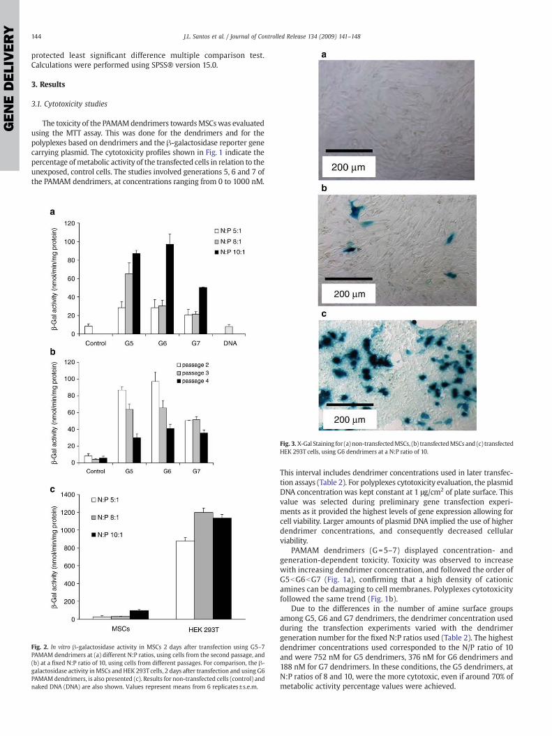
The toxicity of the PAMAMdendrimers towardsMSCswas evaluatedusing the MTT assay. This was done for the dendrimers and for thepolyplexes based on dendrimers and the β-galactosidase reporter genecarrying plasmid. The cytotoxicity profiles shown in Fig. 1 indicate thepercentage ofmetabolic activity of the transfected cells in relation to theunexposed, control cells. The studies involved generations 5, 6 and 7 ofthe PAMAM dendrimers, at concentrations ranging from 0 to 1000 nM.
Fig. 2. In vitro β-galactosidase activity in MSCs 2 days after transfection using G5–7PAMAM dendrimers at (a) different N:P ratios, using cells from the second passage, and(b) at a fixed N:P ratio of 10, using cells from different passages. For comparison, the β-galactosidase activity inMSCs and HEK 293Tcells, 2 days after transfection and using G6PAMAM dendrimers, is also presented (c). Results for non-transfected cells (control) andnaked DNA (DNA) are also shown. Values represent means from 6 replicates±s.e.m.
Fig. 3. X-Gal Staining for (a) non-transfectedMSCs, (b) transfectedMSCs and (c) transfectedHEK 293T cells, using G6 dendrimers at a N:P ratio of 10.
This interval includes dendrimer concentrations used in later transfec-tion assays (Table 2). For polyplexes cytotoxicity evaluation, the plasmidDNA concentration was kept constant at 1 μg/cm2 of plate surface. Thisvalue was selected during preliminary gene transfection experi-ments as it provided the highest levels of gene expression allowing forcell viability. Larger amounts of plasmid DNA implied the use of higherdendrimer concentrations, and consequently decreased cellularviability.
PAMAM dendrimers (G=5–7) displayed concentration- andgeneration-dependent toxicity. Toxicity was observed to increasewith increasing dendrimer concentration, and followed the order ofG5bG6bG7 (Fig. 1a), confirming that a high density of cationicamines can be damaging to cell membranes. Polyplexes cytotoxicityfollowed the same trend (Fig. 1b).
Due to the differences in the number of amine surface groupsamong G5, G6 and G7 dendrimers, the dendrimer concentration usedduring the transfection experiments varied with the dendrimergeneration number for the fixed N:P ratios used (Table 2). The highestdendrimer concentrations used corresponded to the N/P ratio of 10and were 752 nM for G5 dendrimers, 376 nM for G6 dendrimers and188 nM for G7 dendrimers. In these conditions, the G5 dendrimers, atN:P ratios of 8 and 10, were the more cytotoxic, even if around 70% ofmetabolic activity percentage values were achieved.

Fig. 5. Time course of ALP activity presented by MSCs transfected with a plasmid DNAcontaining the hBMP-2 gene using generations 5, 6 and 7 of PAMAM dendrimers atvarious N:P charge ratios. The values represent means from six replicates±s.e.m.
GENEDELIVERY
145J.L. Santos et al. / Journal of Controlled Release 134 (2009) 141–148
3.2. Transfection assays using a β-galactosidase reporter gene
The ability of the PAMAM dendrimers to transfect MSCs in vitrowas assayed using generation 5, 6 and 7 dendrimers and a plasmidencoding the β-galactosidase reporter gene. Assays were done atdifferent N:P ratios (Fig. 2a). Two factor ANOVA revealed that thelevel of gene expression was always significantly higher at thehighest N:P ratio used (10:1) for all the dendrimer generationstested (pb0.05, power=1). At this N:P ratio, results obtained for G5and G6 dendrimers were significantly higher than those obtainedfor G7 (pb0.001, power=1). Higher transfection efficiencies werethen obtained in conditions of higher cytotoxicity. Transfection withlower generation PAMAM dendrimers (data not shown) or withnaked DNA did not result in any significant increase in β-galactosidase activity when compared to control cultures. More-over, the extent of transfection was affected by the passage numberof the cells (Fig. 2b). Increasing the cell passage number from 2 to 4resulted in a marked decrease in gene expression levels in MSCstransfected with either G5 or G6 dendrimers (pb0.05, power=1).Based on these results, MSCs from passage 2 were always used inthe transfection experiments involving the hBMP-2 encodingplasmid.
Similar transfection experiments were performed using humanembryonic kidney 293T cells. The results obtained here clearlyindicate that the transfection levels attained using MSCs are verylow (pb0.001, power=1). This is exemplified by the quantitativecomparison of β-galactosidase activity in both cell types aftertransfection using generation 6 PAMAM dendrimers at a N:P ratio of10 and under the same experimental conditions (Fig. 2c). A qualitativecomparison based on X-Gal staining (Fig. 3), a substrate that turns bluewhen hydrolyzed by β-galactosidase, revealed a high number of bluecells in HEK 293T transfected cultures, and a scarce number of bluecells in MSCs transfected cultures.
3.3. Transfection assays using a plasmid carrying the hBMP-2 gene
The first set of experiments involving the use of the β-galactosidasereporter gene revealed very low levels of transfection efficiency. Theresults of BMP-2 expression 3 days after transfection (Fig. 4) showed apattern similar to the results obtainedwith the β-galactosidase reportergene. BMP-2 expression was higher for the N:P ratio of 10. At this ratio,results obtained for G5 and G6 dendrimers were significantly higherthan those obtained for G7 (pb0.5, power=1).
Anyway we decided to determine whether the low levels of geneexpression obtained using the hBMP-2 containing plasmid would stillbe sufficient for the in vitro differentiation of MSCs towards theosteoblastic lineage. In order to assess this, established markers of theosteogenic phenotype were evaluated: alkaline phosphatase (ALP)
Fig. 4. In vitro BMP-2 expression by MSCs 3 days after transfection. Values representmeans from three replicates±s.e.m.
activity, an early marker of osteogenesis; osteocalcin (OC) secretion, anon-collagenous matrix protein that is only synthesized by matureosteoblasts, and is not present in the early stages of calcification, beingexpressed only after calcified deposits start to form; and extracellularmatrix mineralization, the ultimate step in osteogenic differentiation.
Fig. 5 shows the time course of the ALP activity (normalized to thetotal protein content) presented by the transfectedMSCs. Significantlyhigher levels of ALP activity were always observed for cells harvestedat day 7 compared to those harvested on days 14 and 21 (pb0.05,power=0.99). From this it is clear that the expression of ALP by cells isa transient event with a peak at the beginning of the osteogenesisprocess. At days 7 and 14, the ALP activity values for the transfectedMSCs were always significantly higher (pb0.05, power=1) than thoseobtained for the non-transfected cells. However, the effects of thedifferent dendrimer generations and N:P ratios tested were notdistinguishable.
The OC content secreted into the culture media was determined atdays 14, 21 and 28, and is presented in Fig. 6. A peak in the levels ofsecreted OC was always observed on day 21, revealing the late natureof this marker of osteogenesis. The results at this time-point clearlyhighlight that the transfected MSCs secreted significant higheramounts of OC than the non-transfected cells (pb0.001, power=1).In fact, the OC content in culture was observed to increase with anincreasing N:P ratio (significant differences were detected for the G6and G7 dendrimers). As for ALP activity, the effects of the differentgenerations of dendrimers were not distinguishable.
Fig. 6. Time course of osteocalcin content secreted by MSCs transfected with a plasmidcontaining the hBMP-2 gene using generations 5, 6 and 7 of PAMAM dendrimers atvarious N:P charge ratios. The values represent means from three replicates±s.e.m.

Fig. 7. Calcium deposition on extracellular matrix. The cells were transfected with aplasmid DNA containing the hBMP-2 gene using generations 5, 6 and 7 of PAMAMdendrimers, and cultured for 21 days. The values represent means from threereplicates±s.e.m.
GENEDELIVERY
146 J.L. Santos et al. / Journal of Controlled Release 134 (2009) 141–148
To further examine cell differentiation in the osteoblastic lineage,the calcium content on the extracellularmatrix was assessed at day 21.The calcium deposition assay data (Fig. 7) indicated that thetransfected cells had accumulated significantly higher levels ofcalcium when compared with the control group (non-transfectedcells—pb0.001, power=1). The highest levels of deposited calciumwere observed for the cells transfected with the G5 and G6dendrimers, using a N:P ratio of 10. For G5, it was clear that anincrease in the N:P ratio resulted in a progressive increase inmineralizedmatrix deposition (pb0.001, power=0.998). The presenceof a mineralized matrix on day 21 was also confirmed by von Kossastaining. The expression of BMP-2 by the transfected cells dramaticallyenhanced matrix mineralization (Fig. 8). This was detectable by thetypical black colour of the cultures and was particularly noticeablewhen using the G5 and G6 dendrimers.
Fig. 8. von Kossa staining showing matrix mineralization. (a) Non-transfected MSCs were coPAMAM dendrimers of generations (b) 5, (c) 6 and (d) 7 at a N:P charge ratio of 10. Cells w
To confirm if this enhanced osteogenic activity was specific to thehBMP-2 gene, as earlier suggested, the response of the osteogenesismarkers in theMSC cultures after transfectionwith polyplexes formedby dendrimers and the β-galactosidase reporter gene containingplasmid was also evaluated. The results obtained here did not differfrom the control.
4. Discussion
The reported successful works [23–25] on the use of cationicdendrimers as vectors for gene delivery encouraged the present authorsto assess their efficacy on MSCs with a view on applications related tobone regeneration and tissue engineering. To our knowledge, thepresent study is the first of its kind in this field.
The efficacy of dendrimers in gene delivery can be explained by thecharge-based interactions between these molecules and DNA. ThePAMAM dendrimers bear positively charged primary amine groups ontheir surface, while having tertiary amine groups inside. The primaryamine groups participate in DNA binding, compact DNA intonanoscale particles and promote the cellular uptake of DNA, whilethe buried tertiary amino groups act as a proton sponge in endosomesand enhance the release of DNA into the cytoplasm [21,30]. From thebeginning, investigators have searched for a correlation betweentransfection and the molecular size and shape of the PAMAMdendrimers. Haensler and Szoka found that the generation 6PAMAM dendrimers, with a diameter of 68 nm and resembling thehistone core in chromatin around which DNA is wrapped, wereoptimal in mediating transfection [23]. Another study using a varietyof cell lines showed that the best performing generation (from G5 toG10) for cell transfectionwas strongly dependent on the cell type, andthat the DNA binding ability increased with the generation number[26,31]. However, while some authors describe PAMAMdendrimers asconducting to high levels of transfection, others still search for ways ofmodifying them to improve their efficiency [24]. In this respect, it is
mpared with MSCs transfected with a plasmid DNA containing the hBMP-2 gene usingere cultured for 21 days.

GENEDELIVERY
147J.L. Santos et al. / Journal of Controlled Release 134 (2009) 141–148
worth mentioning that partially degraded PAMAM dendrimers wereshown to be 100-fold more active as gene delivery vectors [21].
In order to determine their behaviour as gene transfer agentstowards MSCs, three different generations of PAMAM dendrimers (G5,G6 and G7), at different N:P charge ratios, were analysed in the first setof the present results. Plasmid DNA encoding the β-galactosidasereporter gene was employed here. From the data it was found that theMSCs could be transfectedwith the PAMAMdendrimers. However, theobserved transfection levels were very low. Several investigators havefaced the same problemwhen using other non-viral gene vehicles andMSCs, thereby indicating that this cell type, like other primary cells, istypically refractory to transfection [32]. Nevertheless, even with lowlevels of transfection, it was possible to distinguish different levels oftransfection efficacy amongst the different dendrimer generations andthe different N:P charge ratios tested. The best results were obtainedwith the G5 and G6 dendrimers, using a N:P ratio of 10. These resultshowever, corresponded to higher cytotoxicity conditions. This was tobe expected, since the cytotoxicity of cationic polymers has beenreported to be a function of the interaction between the polymer andthe cell membrane and/or of cellular uptake efficiency, two factorsthat are also important for the efficacy of gene delivery agents [33,34].In practice, the increase in transfection efficiency was accompanied byan increase in the polymer concentration. Interestingly, the cytotoxi-city shown by the polyplexes was not very different from thatpresented by the dendrimers. This is in contrast to studies which haveshown that complexation with DNA tends to reduce toxicity [20]. Thepresent study also reinforces the conclusions reached by other authorsthat transfection may strongly vary with the type of cells used. Here,the PAMAM dendrimers were shown to be efficient towards the HEK293Tcell line, whichwas used as a positive control in the experiments.Additionally, transfection varied drastically with the cell passagenumber, showing that not only the cell type but also the cellproliferation state, are important parameters to take into account.Indeed, the transfection efficiency has been shown to be related withthe cell cycle activity and cell growth [35,36].
It has been reported that while MSCs possess intrinsic osteogenicactivity, their level of BMP production is extremely low [37]. Combinedtherapy with recombinant BMP has been shown to promote osteogenicdifferentiation in vitro [38–41] and to induce bone formation in vivo[42,43]. The high concentrations of BMP-2 protein required to achieve abiological effect, as well as its rapid degradation after introduction intothe body, has however rendered its use cost ineffective. Recently, severalstudies on bone regeneration have reported successful results usingadenoviruses, retroviruses and baculoviruses vectors to carry BMP-2cDNA [8,10–12,43,44]. Althoughwork in this field is expanding, the dataavailable on nonviral gene delivery for bone regeneration and tissueengineering applications is still scarce [6,45].
In the second set of the present results, using a DNA plasmidencoding the hBMP-2 gene, it was aimed to evaluate whether or not alow transfection level would be sufficient for the envisaged applica-tion, i.e., to improve the in vitro differentiation of MSCs towards theosteoblastic lineage. All quantitative assays involving the osteogenicmarkers (ALP activity, osteocalcin and mineralization) respondedpositively to this hypothesis. Taken together, the results con-firmed optimal transfection efficacies for the G5 and G6 PAMAMdendrimers at a N:P charge ratio of 10 (as for the assays employing theβ-galactosidase reporter gene). The von Kossa staining performedhere was in agreement with the quantitative data.
In conclusion, a coherent set of resultswasobtained indicatinga clearcorrelation between the data obtained with the hBMP-2 and the β-galactosidase reporter gene containing plasmids. Furthermore, the lowlevels of transfection achieved were shown to be sufficient to promotethe osteogenic differentiation of MSCs. A recent in vivo study, usingC3H10T1/2-derived C9 MSCs expressing BMP-2 under control of thedoxycycline (Dox)-repressible promoter, revealed that the short-termexpression of this proteinaceous growth factor was sufficient to
irreversibly induce bone formation. From this it is clear that a stablegenetic modification of MSCs is not required for stem cell-based boneengineering [46]. The fact that nonviral gene delivery vectors, such aspolycations, only mediate the transient expression of the transferredgenes, opens the door to the use of such vectors in this kind ofapplication. In addition, the present in vitro results suggest that a lowlevel of gene expressionmayalsobe enough for the successful treatmentof bone defects or bone-related diseases. This findingmay be relevant inthe clinical scenario, where the delivery of high or uncontrollable dosesof BMP-2 is a cause of concern and thus should be avoided. The presentresults also point out the relevance of focusing research on thedevelopment of nonviral gene delivery vectors on concrete applications,instead of focusing on the improvement of the transfection efficiency.
Acknowledgments
The Portuguese Foundation for Science and Technology (FCT) isacknowledged for funding through the Project DENDRALGENE— PTDC/SAU-BEB/71161/2006 and for the scholarship attributed to J. L. Santos(SFRH/BD/19450/2004). Authors are also grateful to Prof. Dr. YasuhikoTabata for kindly providing the hBMP-2 plasmid DNA.
References
[1] J. Dai, A.B.M. Rabie, U. Hagg, R.A. Xu, Alternative gene therapy strategies for therepair of craniofacial bone defects, Curr. Gene Ther. 4 (4) (2004) 469–485.
[2] C.A. Gersbach, J.E. Phillips, A.J. Garcia, Genetic engineering for skeletal regenerativemedicine, Annu. Rev. Biomed. Eng. 9 (2007) 87–119.
[3] M.D. Kofron, C.T. Laurencin, Orthopaedic applications of gene therapy, Curr. GeneTher. 5 (1) (2005) 37–61.
[4] M.D. Kofron, C.T. Laurencin, Bone tissue engineering by gene delivery, Adv. DrugDeliv. Rev. 58 (4) (2006) 555–576.
[5] J. Luo,M.H. Sun, Q. Kang, Y. Peng,W. Jiang, H.H. Luu, Q. Luo, J.Y. Park, Y. Li, R.C. Haydon,T.C. He, Gene therapy for bone regeneration, Curr. Gene Ther. 5 (2) (2005) 167–179.
[6] K.A. Partridge, R.O. Oreffo, Gene delivery in bone tissue engineering: progress andprospects using viral and nonviral strategies, Tissue Eng. 10 (1–2) (2004) 295–307.
[7] G. Turgeman, H. Aslan, Z. Gazit, D. Gazit, Cell-mediated gene therapy for boneformation and regeneration, Curr. Opin. Mol. Ther. 4 (4) (2002) 390–394.
[8] Y. Chen, K.M.C. Cheung, H.F. Kung, J.C.Y. Leong, W.W. Lu, K.D.K. Luk, In vivo newbone formation by direct transfer of adenoviral-mediated bone morphogeneticprotein-4 gene, Biochem. Biophys. Res. Commun. 298 (1) (2002) 121–127.
[9] J.R. Lieberman, A. Daluiski, S. Stevenson, L. Wu, P. McAllister, Y.P. Lee, J.M. Kabo, G.A.Finerman, A.J. Berk, O.N. Witte, The effect of regional gene therapy with bonemorphogenetic protein-2-producing bone-marrow cells on the repair of seg-mental femoral defects in rats, J. Bone Joint Surg. Am. 81 (7) (1999) 905–917.
[10] J. Park, J. Ries, K. Gelse, F. Kloss, K. von der Mark, J. Wiltfang, F.W. Neukam, H.Schneider, Bone regeneration in critical size defects by cell-mediated BMP-2 genetransfer: a comparison of adenoviral vectors and liposomes, Gene Ther. 10 (13)(2003) 1089–1098.
[11] K.D. Riew, N.M. Wright, S.L. Cheng, L.V. Avioli, J. Lou, Induction of bone formationusing a recombinant adenoviral vector carrying the human BMP-2 gene in a rabbitspinal fusion model, Calcif. Tissue Int. 63 (4) (1998) 357–360.
[12] H. Tsuda, T. Wada, Y. Ito, H. Uchida, H. Dehari, K. Nakamura, K. Sasaki, M. Kobune, T.Yamashita, H. Hamada, Efficient BMP2 gene transfer and bone formation ofmesenchymal stem cells by a fiber-mutant adenoviral vector, Mol. Ther. 7 (3)(2003) 354–365.
[13] L. De Laporte, J. Cruz Rea, L.D. Shea, Design of modular non-viral gene therapyvectors, Biomaterials 27 (7) (2006) 947–954.
[14] H. Eliyahu, Y. Barenholz, A.J. Domb, Polymers for DNA delivery, Molecules 10 (1)(2005) 34–64.
[15] C.M. Roth, S. Sundaram, Engineering synthetic vectors for improved DNA delivery:insights from intracellular pathways, Annu. Rev. Biomed. Eng. 6 (2004) 397–426.
[16] K. Corsi, F. Chellat, L. Yahia, J.C. Fernandes, Mesenchymal stem cells, MG63 andHEK293 transfection using chitosan-DNA nanoparticles, Biomaterials 24 (7) (2003)1255–1264.
[17] A. Hamm, N. Krott, I. Breibach, R. Blindt, A.K. Bosserhoff, Efficient transfectionmethod for primary cells, Tissue Eng. 8 (2) (2002) 235–245.
[18] U. Lakshmipathy, B. Pelacho, K. Sudo, J.L. Linehan, E. Coucouvanis, D.S. Kaufman, C.M.Verfaillie, Efficient transfection of embryonic and adult stem cells, Stem Cells 22 (4)(2004) 531–543.
[19] S.W. Kim, T. Ogawa, Y. Tabata, I. Nishimura, Efficacy and cytotoxicity of cationic-agent-mediated nonviral gene transfer into osteoblasts, J. Biomed. Mater. Res. A 71(2) (2004) 308–315.
[20] C. Dufes, I.F. Uchegbu, A.G. Schatzlein, Dendrimers in gene delivery, Adv. DrugDeliv. Rev. 57 (15) (2005) 2177–2202.
[21] M. Guillot-Nieckowski, S. Eisler, F. Diederich, Dendritic vectors for gene transfection,New J. Chem. 31 (7) (2007) 1111–1127.
[22] S. Svenson, D.A. Tomalia, Dendrimers in biomedical applications—reflections onthe field, Adv. Drug Deliv. Rev. 57 (15) (2005) 2106–2129.

GENEDELIVERY
148 J.L. Santos et al. / Journal of Controlled Release 134 (2009) 141–148
[23] J. Haensler, F.C. Szoka, Polyamidoamine cascade polymers mediate efficienttransfection of cells in culture, Bioconjug. Chem. 4 (5) (1993) 372–379.
[24] J.S. Choi, K. Nam, J. Park, J.B. Kim, J.K. Lee, J. Park, Enhanced transfection efficiencyof PAMAM dendrimer by surface modification with L-arginine, J. Control. Release99 (3) (2004) 445–456.
[25] K. Kono, H. Akiyama, T. Takahashi, T. Takagishi, A. Harada, Transfection activity ofpolyamidoamine dendrimers having hydrophobic amino acid residues in theperiphery, Bioconjug. Chem. 16 (1) (2005) 208–214.
[26] J.F. KukowskaLatallo, A.U. Bielinska, J. Johnson, R. Spindler, D.A. Tomalia, J.R. Baker,Efficient transfer of genetic material into mammalian cells using Starburstpolyamidoamine dendrimers, Proc. Natl. Acad. Sci. U. S. A. 93 (10) (1996) 4897–4902.
[27] C.L. Gebhart, A.V. Kabanov, Evaluation of polyplexes as gene transfer agents,J. Control. Release 73 (2) (2001) 401–416.
[28] J.E. Aubin, Osteoprogenitor cell frequency in rat bone marrow stromal popula-tions: role for heterotypic cell–cell interactions in osteoblast differentiation, J. Cell.Biochem. 72 (3) (1999) 396–410.
[29] D.P. Lennon, S.E. Haynesworth, R.G. Young, J.E. Dennis, A.I. Caplan, A chemically-defined medium supports in-vitro proliferation and maintains the osteochondralpotential of rat marrow-derived mesenchymal stem-cells, Exp. Cell Res. 219 (1)(1995) 211–222.
[30] A.U. Bielinska, C.L. Chen, J. Johnson, J.R. Baker, DNAcomplexingwith polyamidoaminedendrimers: implications for transfection, Bioconjug. Chem. 10 (5) (1999) 843–850.
[31] J.D. Eichman, A.U. Bielinska, J.F. Kukowska-Latallo, J.R. Baker Jr., The use of PAMAMdendrimers in the efficient transfer of genetic material into cells, Pharm. Sci.Technol. Today 7 (2000) 232–245.
[32] H. Haleem-Smith, A. Derfoul, C. Okafor, R. Tuli, D. Olsen, D.J. Hall, R.S. Tuan,Optimization of high-efficiency transfection of adult human mesenchymal stemcells in vitro, Mol. Biotechnol. 30 (1) (2005) 9–19.
[33] D. Fischer, Y.X. Li, B. Ahlemeyer, J. Krieglstein, T. Kissel, In vitro cytotoxicity testingof polycations: influence of polymer structure on cell viability and hemolysis,Biomaterials 24 (7) (2003) 1121–1131.
[34] R. Jevprasesphant, J. Penny, R. Jalal, D. Attwood, N.B. McKeown, A. D'Emanuele, Theinfluence of surface modification on the cytotoxicity of PAMAM dendrimers, Int. J.Pharm. 252 (1–2) (2003) 263–266.
[35] M. Wilke, E. Fortunati, M. vandenBroek, A.T. Hoogeveen, B.J. Scholte, Efficacy of apeptide-based gene delivery system depends on mitotic activity, Gene Ther. 3 (12)(1996) 1133–1142.
[36] Y.B. Xie, S.T. Yang, D.A. Kniss, Three-dimensional cell-scaffold constructs promoteefficient gene transfection: implications for cell-based gene therapy, Tissue Eng. 7(5) (2001) 585–598.
[37] H. Yamagiwa, N. Endo, K. Tokunaga, T. Hayami, H. Hatano, H.E. Takahashi, In vivobone-forming capacity of human bone marrow-derived stromal cells is stimulatedby recombinant human bone morphogenetic protein-2, J. Bone Miner. Metab. 19(1) (2001) 20–28.
[38] A. Chaudhari, E. Ron, M.P. Rethman, Recombinant human bone morphogeneticprotein-2 stimulates differentiation in primary cultures of fetal rat calvarialosteoblasts, Mol. Cell. Biochem. 167 (1–2) (1997) 31–39.
[39] E. Hay, M. Hott, A.M. Graulet, A. Lomri, P.J. Marie, Effects of bone morphogeneticprotein-2 on human neonatal calvaria cell differentiation, J. Cell. Biochem. 72 (1)(1999) 81–93.
[40] A. Yamaguchi, Regulation of differentiation pathway of skeletal mesenchymal cellsin cell-lines by transforming growth-factor-beta superfamily, Semin. Cell Biol. 6 (3)(1995) 165–173.
[41] A. Yamaguchi, T. Ishizuya, N. Kintou, Y. Wada, T. Katagiri, J.M. Wozney, V. Rosen,S. Yoshiki, Effects of BMP-2, BMP-4, and BMP-6 on osteoblastic differentiation ofbone marrow-derived stromal cell lines, ST2 and MC3T3-G2/PA6, Biochem.Biophys. Res. Commun. 220 (2) (1996) 366–371.
[42] A.W.A. Baltzer, C. Lattermann, J.D. Whalen, P. Wooley, K. Weiss, M. Grimm,S.C. Ghivizzani, P.D. Robbins, C.H. Evans, Genetic enhancement of fracture repair:healing of an experimental segmental defect by adenoviral transfer of the BMP-2gene, Gene Ther. 7 (9) (2000) 734–739.
[43] E.A. Wang, V. Rosen, J.S. Dalessandro, M. Bauduy, P. Cordes, T. Harada, D.I. Israel,R.M. Hewick, K.M. Kerns, P. Lapan, D.P. Luxenberg, D. Mcquaid, I.K. Moutsatsos, J.Nove, J.M. Wozney, Recombinant human bone morphogenetic protein inducesbone-formation, Proc. Natl. Acad. Sci. U. S. A. 87 (6) (1990) 2220–2224.
[44] C.K. Chuang, L.Y. Sung, S.M. Hwang, W.H. Lo, H.C. Chen, Y.C. Hu, Baculovirus as anew gene delivery vector for stem cell engineering and bone tissue engineering,Gene Ther. 14 (19) (2007) 1417–1424.
[45] H. Aslan, Y. Zilberman, V. Arbeli, D. Sheyn, Y. Matan, M. Liebergall, J.Z. Li, G.A. Helm,D. Gazit, Z. Gazit, Nucleofection-based ex vivo nonviral gene delivery to humanstem cells as a platform for tissue regeneration, Tissue Eng. 12 (4) (2006) 877–889.
[46] D. Noel, D. Gazit, C. Bouquet, F. Apparailly, C. Bony, P. Plence, V. Millet, G. Turgeman,M. Perricaudet, J. Sany, C. Jorgensen, Short-term BMP-2 expression is sufficient forin vivo osteochondral differentiation of mesenchymal stem cells, Stem Cells 22 (1)(2003) 74–85.