Osteogenic activity and antibacterial effects on titanium ... · Osteogenic activity and...
12
Osteogenic activity and antibacterial effects on titanium surfaces modified with Zn-incorporated nanotube arrays Kaifu Huo a, b, ** , Xuming Zhang b , Hairong Wang a , Lingzhou Zhao c, *** , Xuanyong Liu d , Paul K. Chu b, * a Wuhan National Laboratory for Optoelectronics (WNLO), Huazhong University of Science and Technology (HUST), Wuhan 430074, China b Department of Physics and Materials Science, City University of Hong Kong, Tat Chee Avenue, Kowloon, Hong Kong, China c Department of Periodontology and Oral Medicine, School of Stomatology, The Fourth Military Medical University, No.145 West Changle Road, Xi’an 710032, China d State Key Laboratory of High Performance Ceramics and Superfine Microstructure, Shanghai Institute of Ceramics, Chinese Academy of Sciences, Shanghai 200050, China article info Article history: Received 30 December 2012 Accepted 18 January 2013 Available online 8 February 2013 Keywords: Mesenchymal stem cells Titania nanotubes Zinc Antibacterial property Osteogenic differentiation abstract Titanium implants having enhanced osteogenic activity and antibacterial property are highly desirable for the prevention of implant associated infection and promotion of osseointegration. In this study, coatings containing titania nanotubes (NTs) incorporated with zinc (NT-Zn) are produced on Ti implants by anodization and hydrothermal treatment in Zn containing solutions. The amount of incorporated Zn can be adjusted by varying the structural parameters such as the nanotube diameter and length as well as hydrothermal treatment time. The suitable NT-Zn coatings with good intrinsic antibacterial properties can prevent post-operation infection. Excellent osteogenesis inducing ability in the absence of extraneous osteogenic supplements is demonstrated and the ERK1/2 signaling is found to be involved. The NT-Zn structure which is simple, stable, and easy to produce and scale up has immense potential in bone implant applications. Ó 2013 Elsevier Ltd. All rights reserved. 1. Introduction Although titanium (Ti)-based dental and orthopedic implants are widely used clinically, failures still occur mainly due to deficient osseointegration and implant associated infections [1,2]. To com- plicate matters, implant therapies may not be implemented for patients who suffer from bone healing problem due to factors such as osteoporosis, aging, and diabetes. Alternative implant materials that possess both osteogenesis inducing ability and antibacterial effects are thus demanded by the orthopedic community and in this respect, some researchers have attempted to graft the titanium surface with anti-adhesive polymers or antibacterial agents to inhibit bacterial colonization. Bioactive molecules such as RGD, bone morphogenetic protein-2, and vascular endothelial growth factor have also been introduced into Ti implant surface to restore and improve cell functionalities [3e6]. Nonetheless, these coatings tend to be unstable and the fabrication process is usually time consuming and costly. Development of simple and stable implants having both enhanced osteogenic activity and antibacterial prop- erty is of scientific and clinical significance. Our previous studies suggest that the proper topographies, especially a nanostructured surface, can promote osseointegration [7,8]. In particular, titania nanotubes (NTs) fabricated by electro- chemical anodization have been demonstrated to promote bone cell functionalities in vitro [7] and bone implant osseointegration in vivo [8]. In addition, these NTs constitute an excellent drug loading and delivering platform, particularly for inorganic bioactive elements such as silver and trace elements which are stable and functional at low doses [2,9]. These NT coatings not only facilitate loading of these agents due to the hollow-core structures, but also provide a means to control the loading capacity and release rate by varying the structural parameter such as tube diameter and length. We have fabricated silver (Ag) loaded NTs with long-lasting anti- bacterial ability but some cytotoxicity is observed [10]. Similar to Ag, zinc (Zn) possesses antibacterial ability [11e 13]. In addition, Zn shows anabolic effects on bone metabolism by stimulating osteo- blast proliferation and mineralization [14e19], promotes osteoblast marker gene expressions as well as calcium deposition on mesen- chymal stem cells (MSCs) [13,20], inhibits bone resorption by reducing osteoclast formation and adsorbing ability [21,22], and has anti-inflammation properties [23,24]. Zn has thus attracted the interest of the biomedical community and has been incorporated into bioglasses and bioceramics [17e19,23e25]. * Corresponding author. Tel.: þ852 27887724; fax: þ852 27889549. ** Corresponding author. Wuhan National Laboratory for Optoelectronics (WNLO), Huazhong University of Science and Technology (HUST), Wuhan 430074, China *** Corresponding author. Department of Periodontology and Oral Medicine, School of Stomatology, The Fourth Military Medical University, No. 145 West Changle Road, Xi’an 710032, China E-mail addresses: [email protected] (K. Huo), [email protected] (L. Zhao), [email protected] (P.K. Chu). Contents lists available at SciVerse ScienceDirect Biomaterials journal homepage: www.elsevier.com/locate/biomaterials 0142-9612/$ e see front matter Ó 2013 Elsevier Ltd. All rights reserved. http://dx.doi.org/10.1016/j.biomaterials.2013.01.071 Biomaterials 34 (2013) 3467e3478
-
Upload
dinhkhuong -
Category
Documents
-
view
223 -
download
0
Transcript of Osteogenic activity and antibacterial effects on titanium ... · Osteogenic activity and...
Osteogenic activity and antibacterial effects on titanium surfacesmodified with Zn-incorporated nanotube arrays
Kaifu Huo a,b,**, Xuming Zhang b, Hairong Wang a, Lingzhou Zhao c,***, Xuanyong Liu d, Paul K. Chu b,*
aWuhan National Laboratory for Optoelectronics (WNLO), Huazhong University of Science and Technology (HUST), Wuhan 430074, ChinabDepartment of Physics and Materials Science, City University of Hong Kong, Tat Chee Avenue, Kowloon, Hong Kong, ChinacDepartment of Periodontology and Oral Medicine, School of Stomatology, The Fourth Military Medical University, No. 145 West Changle Road, Xi’an 710032, Chinad State Key Laboratory of High Performance Ceramics and Superfine Microstructure, Shanghai Institute of Ceramics, Chinese Academy of Sciences, Shanghai 200050, China
a r t i c l e i n f o
Article history:Received 30 December 2012Accepted 18 January 2013Available online 8 February 2013
Keywords:Mesenchymal stem cellsTitania nanotubesZincAntibacterial propertyOsteogenic differentiation
a b s t r a c t
Titanium implants having enhanced osteogenic activity and antibacterial property are highly desirablefor the prevention of implant associated infection and promotion of osseointegration. In this study,coatings containing titania nanotubes (NTs) incorporated with zinc (NT-Zn) are produced on Ti implantsby anodization and hydrothermal treatment in Zn containing solutions. The amount of incorporated Zncan be adjusted by varying the structural parameters such as the nanotube diameter and length as wellas hydrothermal treatment time. The suitable NT-Zn coatings with good intrinsic antibacterial propertiescan prevent post-operation infection. Excellent osteogenesis inducing ability in the absence ofextraneous osteogenic supplements is demonstrated and the ERK1/2 signaling is found to be involved.The NT-Zn structure which is simple, stable, and easy to produce and scale up has immense potential inbone implant applications.
� 2013 Elsevier Ltd. All rights reserved.
1. Introduction
Although titanium (Ti)-based dental and orthopedic implantsare widely used clinically, failures still occur mainly due to deficientosseointegration and implant associated infections [1,2]. To com-plicate matters, implant therapies may not be implemented forpatients who suffer from bone healing problem due to factors suchas osteoporosis, aging, and diabetes. Alternative implant materialsthat possess both osteogenesis inducing ability and antibacterialeffects are thus demanded by the orthopedic community and inthis respect, some researchers have attempted to graft the titaniumsurface with anti-adhesive polymers or antibacterial agents toinhibit bacterial colonization. Bioactive molecules such as RGD,bone morphogenetic protein-2, and vascular endothelial growthfactor have also been introduced into Ti implant surface to restoreand improve cell functionalities [3e6]. Nonetheless, these coatingstend to be unstable and the fabrication process is usually time
consuming and costly. Development of simple and stable implantshaving both enhanced osteogenic activity and antibacterial prop-erty is of scientific and clinical significance.
Our previous studies suggest that the proper topographies,especially a nanostructured surface, can promote osseointegration[7,8]. In particular, titania nanotubes (NTs) fabricated by electro-chemical anodization have been demonstrated to promote bonecell functionalities in vitro [7] and bone implant osseointegrationin vivo [8]. In addition, these NTs constitute an excellent drugloading and delivering platform, particularly for inorganic bioactiveelements such as silver and trace elements which are stable andfunctional at low doses [2,9]. These NT coatings not only facilitateloading of these agents due to the hollow-core structures, but alsoprovide a means to control the loading capacity and release rate byvarying the structural parameter such as tube diameter and length.We have fabricated silver (Ag) loaded NTs with long-lasting anti-bacterial ability but some cytotoxicity is observed [10]. Similar toAg, zinc (Zn) possesses antibacterial ability [11e13]. In addition, Znshows anabolic effects on bone metabolism by stimulating osteo-blast proliferation andmineralization [14e19], promotes osteoblastmarker gene expressions as well as calcium deposition on mesen-chymal stem cells (MSCs) [13,20], inhibits bone resorption byreducing osteoclast formation and adsorbing ability [21,22], andhas anti-inflammation properties [23,24]. Zn has thus attracted theinterest of the biomedical community and has been incorporatedinto bioglasses and bioceramics [17e19,23e25].
* Corresponding author. Tel.: þ852 27887724; fax: þ852 27889549.** Corresponding author. Wuhan National Laboratory for Optoelectronics (WNLO),Huazhong University of Science and Technology (HUST), Wuhan 430074, China*** Corresponding author. Department of Periodontology and Oral Medicine,School of Stomatology, The Fourth Military Medical University, No. 145 WestChangle Road, Xi’an 710032, China
E-mail addresses: [email protected] (K. Huo), [email protected](L. Zhao), [email protected] (P.K. Chu).
Contents lists available at SciVerse ScienceDirect
Biomaterials
journal homepage: www.elsevier .com/locate/biomater ia ls
0142-9612/$ e see front matter � 2013 Elsevier Ltd. All rights reserved.http://dx.doi.org/10.1016/j.biomaterials.2013.01.071
Biomaterials 34 (2013) 3467e3478
In thework reported in this paper, Zn incorporatedNTstructures(NT-Zn) are fabricated on Ti implants to achieve both osteogenesisinducing ability and antibacterial effects. Since the effect of Zn isdose dependent with low doses enhancing bone formation butultrahigh doses inducing toxicity [17], the proper NT-Zn structurewith optimal Zn release is required. Series of NT and NT-Zn sampleswith different diameters and lengths as well as different Zn loadingand release capacities are fabricated. The antibacterial ability andosteogenic activity in the absence of exogenous osteogenic factors(OS) are systemically investigated. The molecular mechanismunderlying the osteogenic ability of NT-Zn is discussed.
2. Materials and methods
2.1. Sample fabrication
Ti foils (99.7% pure, Aldrich) 10 � 10 � 1 mm3 in size were ground and ultra-sonically cleaned in acetone, ethanol, and deionized water. The NTs were fabricatedusing a two electrode configuration in an ethylene glycol solution comprising 0.5wt%NH4F, 5 vol%CH3OHand5vol%H2Oat10V for1hand40V for40min, respectivelyanddesignated as NT10 and NT40, respectively. During anodization, the electrolyte wascontinuously agitated with a magnetic stirrer. After anodization, the samples wererinsedwith distilledwater and dried in air. The as-anodizedNTswere hydrothermallytreated in 40 mL of 0.1 M zinc acetate at 200 �C for 1 and 3 h to produce the ZnincorporatedNTs (NT-Zn) samples designated as NT10-Zn1, NT10-Zn3, NT40-Zn1, andNT40-Zn3. All the samples were annealed at 450 �C for 3 h in air to promote samplecrystallization and strengthen adhesion between the coating and Ti substrate. Beforecell culturing, all the samples were sterilized by ultraviolet irradiation of 30 min.
2.2. Surface characterization
Field-emission scanning electron microscopy (FE-SEM, HITACHI S-4800) wasutilized to examine the surface topography of the specimens. The crystallinestructure of the samples was determined by X-ray diffraction (XRD, Philips X0 PertPro) and the chemical composition of the coatings and Zn depth profiles weredetermined by X-ray photoelectron spectroscopy (XPS, ESCALAB MK-II).
2.3. Zn loading capacity and release
The amount of Zn released from NT-Zn was investigated in the phosphate buf-fered saline (PBS). The samples were immersed in 5 mL PBS for 1 d, and thenretrieved to be immersed in fresh PBS of 5 mL. This process was repeated for a total 1month. The amounts of Zn leached to the PBS solutions were measured byinductively-coupled plasma atomic emission spectrometry (ICP-AES, IRIS AdvantageER/S). To determine the total amount of Zn incorporated into the NT-Zn samples, theNT-Zn samples were dissolved in HF and HNO3 and the Zn contents were deter-mined by the ICP-AES.
2.4. Antibacterial assay
Staphylococcus aureus (S. aureus)was chosen to assess the antibacterial ability ofNT-Zn. S. aureus was cultured in beef extract-peptone (BEP) at 37 �C for 12 h andadjusted to a concentration of 105 CFU/mL. The Zn-NT samples were incubated in1 mL of the bacterial suspension for 1 d and the culture medium was collected todetermine the viable counts of planktonic bacteria. After removing the looselybonded bacteria from the samples, the adherent ones were ultrasonically (40 W)detached in 1mL BEP to count the viable adherent bacteria. Complete detachment ofthe adherent bacteria from the samples was verified by staining and fluorescencemicroscopy (Olympus). The samples were then ultrasonically cleaned, dried, ster-ilized, and re-inoculated as described above. This process was repeated daily fora total of 14 d. The viable bacteria in the suspensions were evaluated by the spreadplate method. The antibacterial rates with regard to planktonic bacteria in the cul-ture medium (Rp) and the antibacterial rates for adhered bacteria on the specimens(Ra) were calculated based on previously described formulas [10].
2.5. Protein absorption assay
A droplet (1 mL) of the a minimal essential medium (a-MEM, Gibco) containing10% fetal calf serum (FCS, Gibco) was pipetted onto the sample and maintained at37 �C for 2 h. The samples were transferred to new 24 well plates and washed withPBS, and then the adsorbed proteins on the samples were detached into 500 mL of 1%sodium dodecyl sulfate (SDS) solution by shaking for 1 h. The protein amounts in theSDS solutions were measured via a MicroBCA kit (Pierce). To observe the proteindistribution, the samples were incubated in 10% FCS in a-MEM for 2 d. Afterwards,the samples with absorbed proteins were dehydrated, freeze-dried, sputter coatedwith gold, and observed by FE-SEM.
2.6. Rat bone MSC acquisition and culture
The boneMSCs were harvested fromyoung Sprague Dawley rats and cultured aspreviously described [7]. In addition, the green fluorescence protein (GFP) labeledones were obtained from GFP transgenic rats using the same method. The animalexperiments were approved by the University Research Ethics Committee of TheFourth Military Medical University. The cells were incubated in a-MEMwith 10% FCSat 37 �C with the medium changed every 3 d. The cells at passages 2 to 4 were usedin the experiments. The samples were placed in 24 well plates and tissue cultureplates served as the control (C).
2.7. Lactate dehydrogenase activity assay
As a marker of cytotoxicity, the lactate dehydrogenase (LDH) released by thecells into the culture media was measured. The MSCs were seeded at a density of2 � 104 cells/well and incubated for 3 d. The culture media were collected andcentrifuged and the activity of LDH in the supernatant was determined.
2.8. Initial cell adhesion
The MSCs were inoculated at a density of 4 � 104 cells/well and at 0.5, 1, and 2 h,the attached cells on the samples were fixed, stained with 40 ,60-diamidino-2-phenylindole (DAPI, Sigma), and counted from five random 5� fields on eachsample using a fluorescence microscope (Leica).
2.9. Cell morphology
The cells were seeded at a density of 2 � 104 cells/well. After culturing for 2 d,the cells on the samples were washed in PBS, fixed with 3% glutaraldehyde,dehydrated in the graded ethanol series, freeze dried, coated with gold, andexamined by FE-SEM. The GFP labeled MSCs were inoculated at a density of 1 �104
cells/well and after 8 d, the images were captured on a fluorescence microscope.
2.10. Alkaline phosphatase staining
The cells were seeded at a density of 2 � 104 cells/well. After culturing for 5 and10 d, the culture was washed with PBS and fixed and alkaline phosphatase (ALP) wasstained with the BCIP/NBT kit (Beyotime).
2.11. Extracellular matrix mineralization
Extracellular matrix (ECM) mineralization by the cells was evaluated by AlizarinRed staining. Thecellswere inoculatedat adensityof 2�104 cells/well and culturedonthe samples for 2 weeks. After rinsing by PBS and fixation, mineralization was eval-uated by 40 mM Alizarin Red (pH 4.2, Solarbio). The unbound dyes were completelyeliminated by thorough washing with distilled water before the images were taken.
2.12. Western blot assay
The MSCs were seeded at a density of 1 �105 cells/well. After culturing for 48 hculturing, the cells were lysed in the RIPA buffer (10 mM TriseHCL, 1 mM EDTA, 1%sodium dodecyl sulfate, 1% Nonidet P-40, 1:100 proteinase inhibitor cocktail, 50 mM
b-glycerophosphate, and 50 mM sodium fluoride) together with the completeprotease inhibitors cocktail tablets (Roche). The protein concentration in theextracted lysates was determined using a BCA protein assay kit (Beyotime). Thesamples were separated by SDS-PAGE and transferred onto a PVDF membrane(Bio-Rad). The membranewas blocked with 5% BSA (Gibco) in the TTBS solution andincubated with anti-ERK1/2, anti-phosphated ERK1/2 (pERK1/2) (1:1000 dilution;R&D), and anti-b-actin primary antibody (1:1000 dilution; Abcam) and then withHRP-conjugated antibody (Boshide). The bands were monitored on a Western-LightChemiluminescent Detection System (Peiqing).
2.13. Statistical analysis
The experiments were conducted in triplicate and the data were expressedas means � standard deviations. The one-way ANOVA combined with the Student-Newman-Keuls (SNK) post hoc test was applied to determine the level of signifi-cance. Significance and high significance was considered for p value < 0.05 and 0.01,respectively.
3. Results
3.1. Sample surface characterization
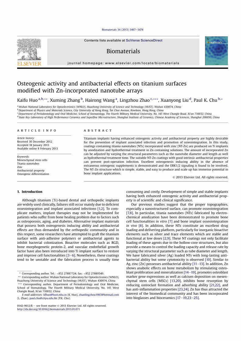
The morphology of the samples is observed by the FE-SEM(Fig. 1A) and NT10 and NT40 have diameters of 30 and 80 nm,respectively. Fig. 1A clearly indicates that NT10 forms bundles of
K. Huo et al. / Biomaterials 34 (2013) 3467e34783468
100e400 nm in size and there is an even distribution of gaps about80 nm wide between the nanotube bundles. The top ends of thenanotube wall on NT40 are not smooth and have flakes. After thehydrothermal treatment, the nanotubular architecture is preserved.The diameter of the nanotubes on NT-Zn becomes smaller and thenanotube wall is thicker after the hydrothermal treatment. Incomparison with NT-10, the gap between the nanotube bundles onNT10-Zn is larger. With regard to NT40-Zn, the top nanotube wallsbecome nanonodular with an even distribution of nanonoduleswith diameters of about 10e30 nm. In NT40-Zn3, the intertubulargap areas are filled with nanoscale nodules which have apparentboundaries with the walls of the nanotubes.
The XRD spectra in Fig. 1B disclose that only Ti and anatase TiO2peaks are found from the NTs, but peaks from ZnTiO3 emerge afterthe hydrothermal treatment which forms NT-Zn. The ZnTiO3 peaksbecome more intense while the TiO2 peaks weaken with hydro-thermal reaction time. The XPS spectra in Fig. 1C acquired from theNT-Zn samples reveal Ti, O, Zn peaks. The C signals should arisefrom surface contamination. Figs. 1DeG display the XPS depthprofiles of Ti, O, and Zn of the NT-Zn samples. In NT10-Zn1 andNT40-Zn1, the Zn distribution is very shallow and about 40 and120 nm deep, respectively. A longer hydrothermal treatment in-corporates more Zn and Zn is distributed along the entire length ofthe nanotubes on NT10-Zn3 and NT40-Zn3. The Zn concentrations
Fig. 1. (A) Surface morphology of the samples observed by the FE-SEM with the insets showing the higher-magnification pictures. (B) XRD spectra. (C) XPS survey spectra of thesamples. XPS depth profiles of (D) NT10-Zn1, (E) NT10-Zn3, (F) NT40-Zn1, and (G) NT40-Zn3.
K. Huo et al. / Biomaterials 34 (2013) 3467e3478 3469
are larger on the outer layers on NT10-Zn3 and NT40-Zn3 anddecrease quickly with depth in the top 40 and 100 nm, respectively.
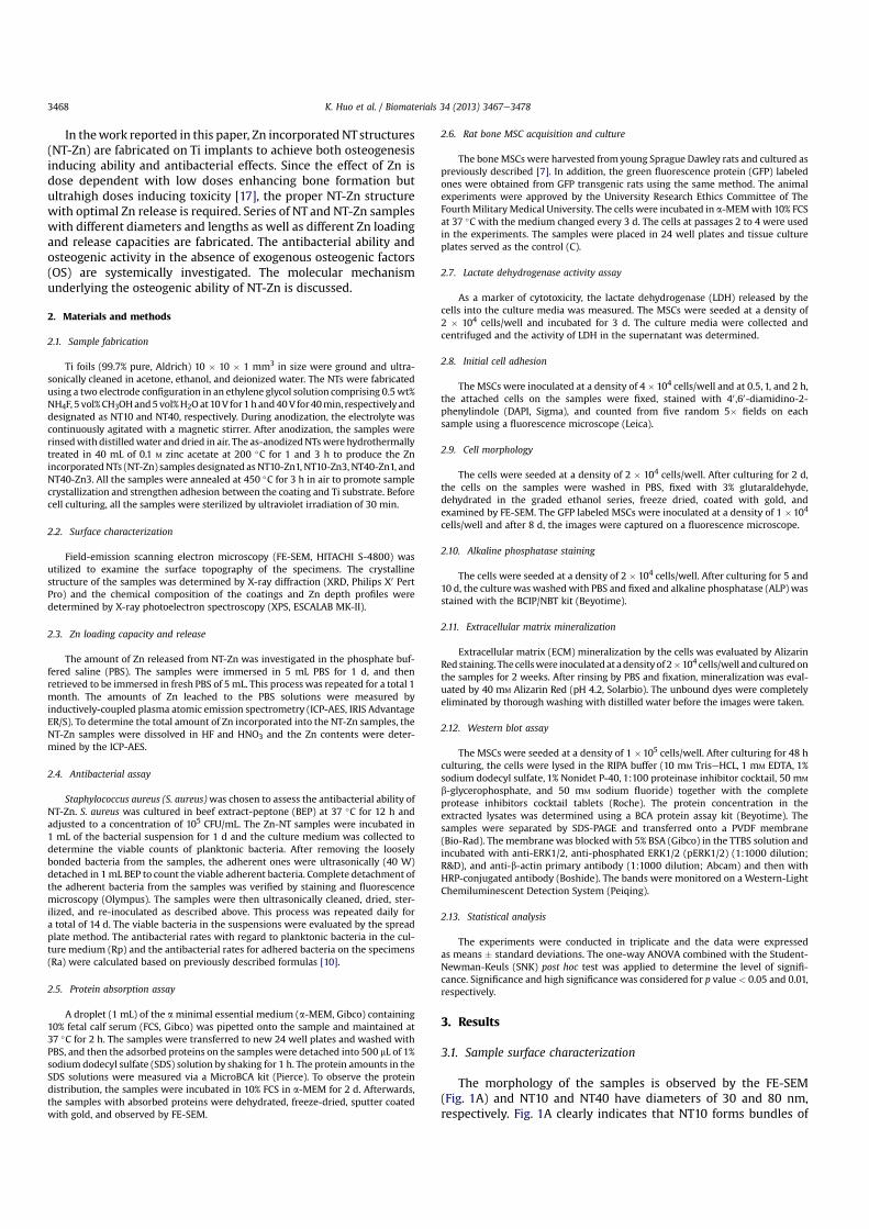
3.2. Zn loading capacity and release
For the 1 cm2 coatings, the total Zn contents in NT10-Zn1,NT10-Zn3, NT40-Zn1 and NT40-Zn3 are 1.2, 11.4, 3.4 and 60.2 mgrespectively, as shown in Fig. 2A. The Zn release kinetics is assessedby immersing the samples in 5 mL PBS for a total month, andas shown in Fig. 2B, NT10-Zn1 and NT40-Zn1 show small amountsof released Zn. At the first day, about 0.01 ppm Zn is detected. Theamounts of Zn released decline with time and one monthlater, nearly no Zn can be detected by ICP-AES. In comparison,NT10-Zn3 delivers more Zn and NT40-Zn3 generates the highestdelivered amount. The amounts of Zn released from NT40-Zn3 andNT10-Zn3 are 0.06 and 0.05 ppm in the first day, respectively anddecrease quickly with time. After 20 d, about 0.01 ppm Zn isreleased.
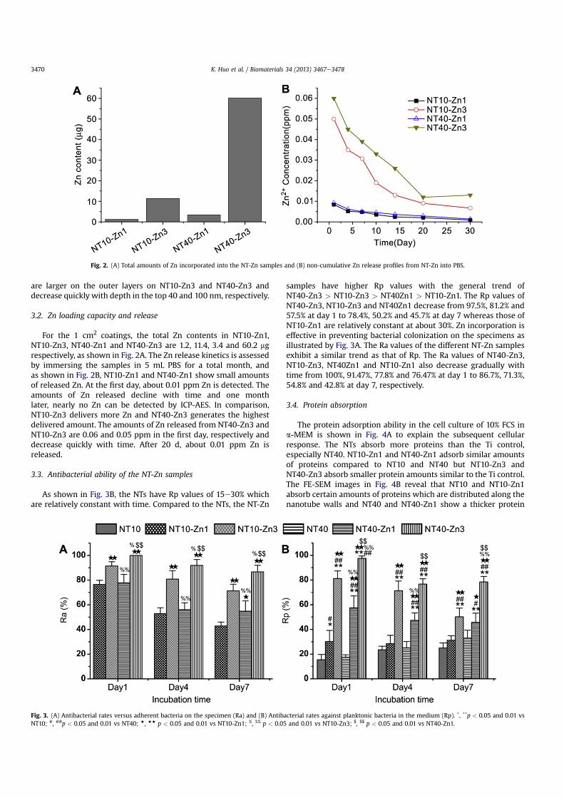
3.3. Antibacterial ability of the NT-Zn samples
As shown in Fig. 3B, the NTs have Rp values of 15e30% whichare relatively constant with time. Compared to the NTs, the NT-Zn
samples have higher Rp values with the general trend ofNT40-Zn3 > NT10-Zn3 > NT40Zn1 > NT10-Zn1. The Rp values ofNT40-Zn3, NT10-Zn3 and NT40Zn1 decrease from 97.5%, 81.2% and57.5% at day 1 to 78.4%, 50.2% and 45.7% at day 7 whereas those ofNT10-Zn1 are relatively constant at about 30%. Zn incorporation iseffective in preventing bacterial colonization on the specimens asillustrated by Fig. 3A. The Ra values of the different NT-Zn samplesexhibit a similar trend as that of Rp. The Ra values of NT40-Zn3,NT10-Zn3, NT40Zn1 and NT10-Zn1 also decrease gradually withtime from 100%, 91.47%, 77.8% and 76.47% at day 1 to 86.7%, 71.3%,54.8% and 42.8% at day 7, respectively.
3.4. Protein absorption
The protein adsorption ability in the cell culture of 10% FCS ina-MEM is shown in Fig. 4A to explain the subsequent cellularresponse. The NTs absorb more proteins than the Ti control,especially NT40. NT10-Zn1 and NT40-Zn1 adsorb similar amountsof proteins compared to NT10 and NT40 but NT10-Zn3 andNT40-Zn3 absorb smaller protein amounts similar to the Ti control.The FE-SEM images in Fig. 4B reveal that NT10 and NT10-Zn1absorb certain amounts of proteins which are distributed along thenanotube walls and NT40 and NT40-Zn1 show a thicker protein
Fig. 2. (A) Total amounts of Zn incorporated into the NT-Zn samples and (B) non-cumulative Zn release profiles from NT-Zn into PBS.
Fig. 3. (A) Antibacterial rates versus adherent bacteria on the specimen (Ra) and (B) Antibacterial rates against planktonic bacteria in the medium (Rp). *, **p < 0.05 and 0.01 vsNT10; #, ##p < 0.05 and 0.01 vs NT40; +, ++ p < 0.05 and 0.01 vs NT10-Zn1; %, %% p < 0.05 and 0.01 vs NT10-Zn3; $, $$ p < 0.05 and 0.01 vs NT40-Zn1.
K. Huo et al. / Biomaterials 34 (2013) 3467e34783470

layer forming high protein pillars. The absorbed protein layers onNT10-Zn3 and NT40-Zn3 are thinner.
3.5. Initial cell adhesion
The initial cell adhesion assayed during the first 2 h of cell/biomaterial interaction is illustrated in Fig. 5. The adherentcell numbers increase with incubation time. The NTs do notshow obvious difference in the initial adherent cell numberscompared to the Ti control. At 0.5 h, the adherent cell numberson NT-Zn are the same as those on NTs and Ti control, but at 1and 2 h, the NT-Zn samples show larger adherent cell numbers atall the time points although not all of them are statisticallysignificant.
3.6. Cytotoxicity
As the effect of Zn is highly dose dependent and overdose of Znwill lead to cytotoxicity, the LDH released from cells cultured on the
samples is measured to assess the cytotoxicity (Fig. 6). No obviouscytotoxicity is observed from all the NTs and NT-Zn samplescompared to the Ti control.
3.7. Cell morphology
The cell morphology is observed by FE-SEM 2 d after culture andby fluorescence microscopy after 8 d of culturing. The SEM pictures(Fig. 7) show that the MSCs on the flat Ti spread relatively poorlywith a spindle shape. NT10 dramatically promotes cell extensionforming the typical osteoblastic shape, whereas NT40 just slightlyenhances cell spreading. NT10-Zn1, NT10-Zn3 and NT40-Zn3 fur-ther enhance cell spreading in contrast to NT10 and NT40, whileNT40-Zn1 show a cell spreading area similar to NT40. According tothe high magnification SEM pictures, the cell bodies attach closelyon NT10, NT10-Zn1, NT10-Zn3, and NT40-Zn3 but they bridge overthe protein pillars on NT40 and NT40-Zn1. The fluorescence images(Fig. 8) demonstrate a similar trend of cell spreading after a longerculture period of 8 d.
Fig. 4. (A) Protein absorption on the samples after 2 h of immersion 10% FCS in a-MEM. *, **p < 0.05, 0.01 vs Ti, ##p < 0.01 vs NT10, and %%p < 0.01 vs NT40; (B) Protein absorptionpattern after incubation for 2 d in 10% FCS in a-MEM.
K. Huo et al. / Biomaterials 34 (2013) 3467e3478 3471
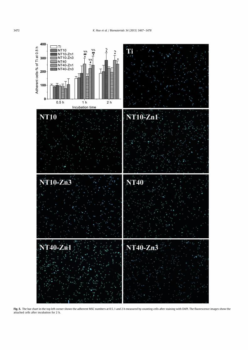
Fig. 5. The bar chart in the top left corner shows the adherent MSC numbers at 0.5, 1 and 2 h measured by counting cells after staining with DAPI. The fluorescence images show theattached cells after incubation for 2 h.
K. Huo et al. / Biomaterials 34 (2013) 3467e34783472

3.8. ALP activity
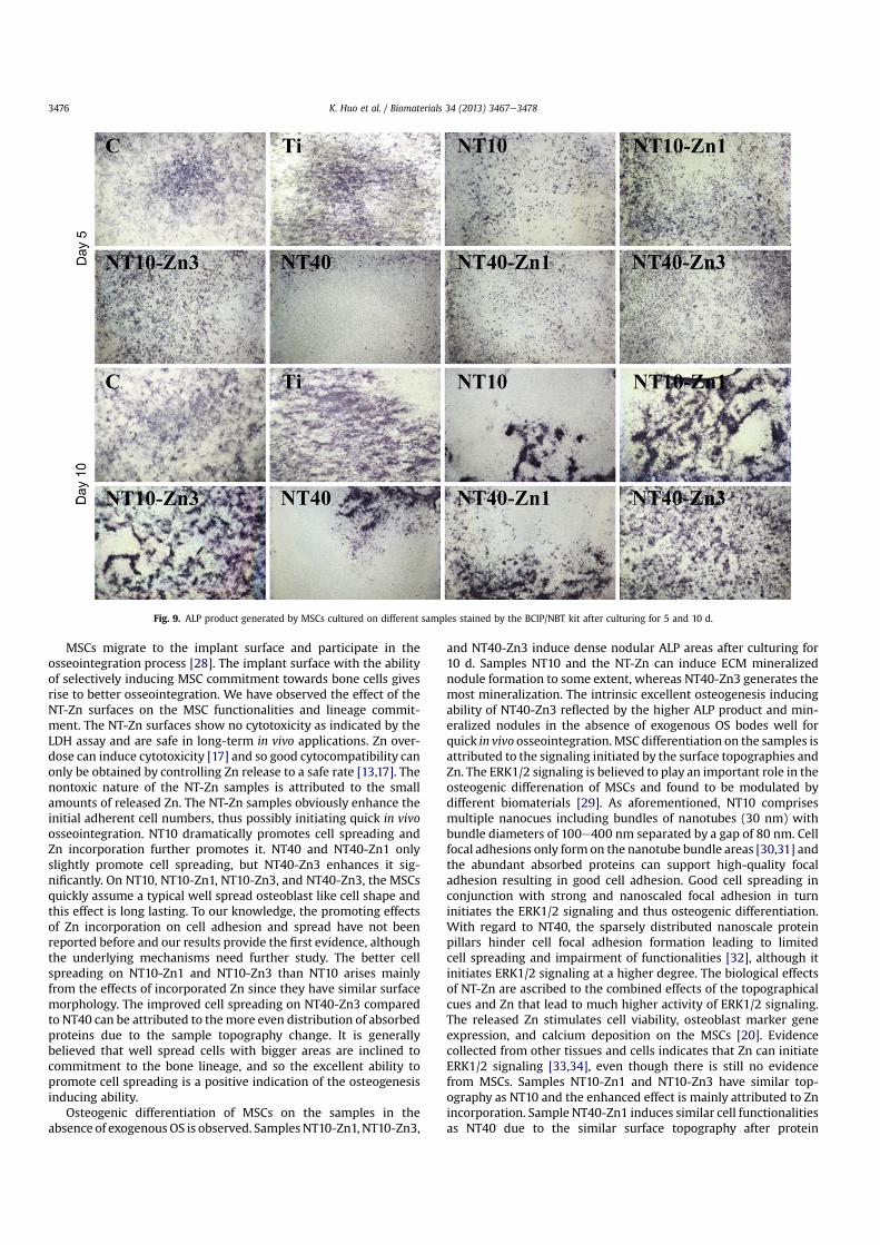
ALP is a marker for early osteogenic differentiation (Fig. 9). Asearly as day 5 after culturing, the cells already produce a certainamount of ALP and more is produced after 10 d. Zn incorporationsignificantly enhances the ALP product. At day 5, NT10-Zn1, NT10-Zn3 and NT40-Zn3 generate obviouslymore ALP product than NT10and NT40, but no obvious difference can be observed amongthemselves. At day 10, large and dense nodular ALP positive areasoccur on NT10-Zn1, NT10-Zn3, and NT40-Zn3.
3.9. ECM mineralized nodule formation
ECMmineralization assayed by Alizarin Red staining is shown inFig. 10. On the flat Ti control and NT40, there is no obvious ECMmineralized nodule formation. On the other surfaces, there arevisible mineralized nodules as indicated by the red arrows. Par-ticularly, NT10-Zn3 generates the biggest mineralized nodules andNT40-Zn3 generates the most in number.
3.10. ERK1/2 signaling activity
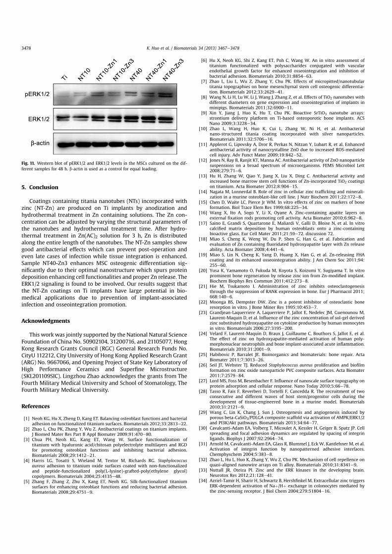
The activity of the ERK1/2 signaling is assessed by western blotanalysis after seeding for 48 h. As shown in Fig. 11, the pERK1/2protein level, which represents the ERK activation status, is sig-nificantly enhanced by the NT structure, especially NT40. ThepERK1/2 protein level is further increased by Zn incorporation andNT40-Zn3 induces the highest pERK1/2 protein level.
4. Discussion
Next-generation orthopedic implants possessing both osteoin-ductivity and antibacterial ability are needed for better clinicalperformance. Attempts have been made but the structure is typi-cally very complicated and fabrication tends to be time consumingand costly. For example, anti-adhesive or antibacterial polymershave been grafted onto Ti to inhibit bacterial colonization andbioactive molecules are immobilized subsequently to restore cellfunctionalities [3e6]. In addition, although in vitro experimentsdemonstrate the efficiency to some extent, the long term in vivostability and effects of these coatings are debatable and not wellunderstood. In comparison, the NT-Zn structure reported here is
simple and stable and can be fabricated by a simple, economical,and upscalable hydrothermal process in a Zn containing solution.After 3 h of hydrothermal treatment, Zn is incorporated along theentire length of the NTs and the amount of Zn released can bealtered by the nanotube diameter and hydrothermal treatmenttime. In particular, NT40-Zn3 has a larger Zn loading capacity andreleases more Zn showing excellent osteogenesis inducing activitywithout cytotoxicity as well as good antibacterial effects. Ag dopedNTs produced in our laboratory also show good long-term anti-bacterial ability, but disappointingly they induce cytotoxicity [10].In comparison, the NT40-Zn3 structure is a better balance betweenbone promotion and antibacterial ability.
The NT-Zn samples exhibit different antibacterial ability. Forinstance, NT40-Zn3 and NT10-Zn3 have much higher antibacterialability than NT40-Zn1 and NT10-Zn1, due to the larger amount ofloaded and released Zn. The dose dependent antibacterial effects ofZn have been reported [13,26]. The antibacterial ability of the NT-Znsamples decreases gradually with time and the tendency is con-sistent with the Zn time release profiles. Initially, larger amounts ofZn are released but they decrease gradually with immersion time. Itis noted that the antibacterial effectiveness against surroundingplanktonic bacteria diminishes more quickly while that againstadherent bacteria on the samples decline more slowly. This isbecause the former is ascribed to the effect of the released Zn [26],whereas the latter is due to a synergistic effect of both released andsurface incorporated Zn. The incorporated Zn may inhibit bacterialcolonization by generating reactive oxygen species (ROS) [13].These antibacterial properties are crucial to clinical applications forthe following reasons. A potent antibacterial capability is needed inthe initial phase of after implant insertion, since the implant in thisstage is most vulnerable to infection. The larger initial Zn releasefrom NT40-Zn3 and NT10-Zn3 effectively kill adherent bacteria aswell as surrounding planktonic bacteria in this early stage to pre-vent perioperative infection and foster normalwound healing. Afterthis stage, osseointegration is gradually established accompaniedby the host defense. Prohibition of biofilm formation by preventionof bacterial colonization on the implant is expected to be sufficientto prevent further infection with the help of the host defense [2].The long-term ability of NT40-Zn3 and NT10-Zn3 to prohibit bac-terial colonization and decline in Zn release with time is the idealscenario in clinical practice to harness the desirable defects whileavoiding potential side effects associated with Zn overdose.
The absorbed proteins convey the effect of topographical cues tothe attached cells/tissues and thus are important to the biologicalperformance of biomaterials [27]. NT10 and NT10-Zn1 absorbabundant proteins which are distributed along the nanotube walls.NT40 and NT40-Zn1 induce even thicker protein layers with tallprotein pillars. On the other hand, the absorbed protein layers onNT10-Zn3 and NT40-Zn3 are much thinner. High protein absorp-tion on NT10, NT10-Zn1, NT40, and NT40-Zn1 can be attributed tothe nanoscale tube walls that provide effective nucleation sites forprotein absorption. In addition, since they are relatively thin, theeffects to attract protein deposition are long lasting. On the con-trary, the thicker tube walls on NT10-Zn3 and NT40-Zn3 are lesseffective in protein accumulation and more importantly, theyquickly become smooth after protein deposition thereby losing theprotein attraction effect. The tall protein pillars with small topdimensions of <50 nm away from each other on NT40 andNT40-Zn1 can be explained by the unsmooth top ends of the thinnanotube wall with many flakes that constitute excellent localnucleation sites for protein aggregation. Since cells interact withthe protein layer absorbed on biomaterials instead of the primitivebiomaterial surface [27], the cell functionalities on the samples aremore related to the topographies after protein adsorption ratherthan the primitive implant topographies.
Fig. 6. Amounts of LDH released by cells to the culture medium in the first 3 d ofculturing.
K. Huo et al. / Biomaterials 34 (2013) 3467e3478 3473
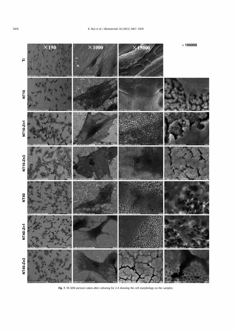
Fig. 7. FE-SEM pictures taken after culturing for 2 d showing the cell morphology on the samples.
K. Huo et al. / Biomaterials 34 (2013) 3467e34783474

Fig. 8. Fluorescence images showing the cell morphology on the samples after culturing for 8 d.
K. Huo et al. / Biomaterials 34 (2013) 3467e3478 3475
MSCs migrate to the implant surface and participate in theosseointegration process [28]. The implant surface with the abilityof selectively inducing MSC commitment towards bone cells givesrise to better osseointegration. We have observed the effect of theNT-Zn surfaces on the MSC functionalities and lineage commit-ment. The NT-Zn surfaces show no cytotoxicity as indicated by theLDH assay and are safe in long-term in vivo applications. Zn over-dose can induce cytotoxicity [17] and so good cytocompatibility canonly be obtained by controlling Zn release to a safe rate [13,17]. Thenontoxic nature of the NT-Zn samples is attributed to the smallamounts of released Zn. The NT-Zn samples obviously enhance theinitial adherent cell numbers, thus possibly initiating quick in vivoosseointegration. NT10 dramatically promotes cell spreading andZn incorporation further promotes it. NT40 and NT40-Zn1 onlyslightly promote cell spreading, but NT40-Zn3 enhances it sig-nificantly. On NT10, NT10-Zn1, NT10-Zn3, and NT40-Zn3, the MSCsquickly assume a typical well spread osteoblast like cell shape andthis effect is long lasting. To our knowledge, the promoting effectsof Zn incorporation on cell adhesion and spread have not beenreported before and our results provide the first evidence, althoughthe underlying mechanisms need further study. The better cellspreading on NT10-Zn1 and NT10-Zn3 than NT10 arises mainlyfrom the effects of incorporated Zn since they have similar surfacemorphology. The improved cell spreading on NT40-Zn3 comparedto NT40 can be attributed to themore even distribution of absorbedproteins due to the sample topography change. It is generallybelieved that well spread cells with bigger areas are inclined tocommitment to the bone lineage, and so the excellent ability topromote cell spreading is a positive indication of the osteogenesisinducing ability.
Osteogenic differentiation of MSCs on the samples in theabsence of exogenousOS is observed. Samples NT10-Zn1, NT10-Zn3,
and NT40-Zn3 induce dense nodular ALP areas after culturing for10 d. Samples NT10 and the NT-Zn can induce ECM mineralizednodule formation to some extent, whereas NT40-Zn3 generates themost mineralization. The intrinsic excellent osteogenesis inducingability of NT40-Zn3 reflected by the higher ALP product and min-eralized nodules in the absence of exogenous OS bodes well forquick in vivo osseointegration.MSC differentiation on the samples isattributed to the signaling initiated by the surface topographies andZn. The ERK1/2 signaling is believed to play an important role in theosteogenic differenation of MSCs and found to be modulated bydifferent biomaterials [29]. As aforementioned, NT10 comprisesmultiple nanocues including bundles of nanotubes (30 nm) withbundle diameters of 100e400 nm separated by a gap of 80 nm. Cellfocal adhesions only form on the nanotube bundle areas [30,31] andthe abundant absorbed proteins can support high-quality focaladhesion resulting in good cell adhesion. Good cell spreading inconjunction with strong and nanoscaled focal adhesion in turninitiates the ERK1/2 signaling and thus osteogenic differentiation.With regard to NT40, the sparsely distributed nanoscale proteinpillars hinder cell focal adhesion formation leading to limitedcell spreading and impairment of functionalities [32], although itinitiates ERK1/2 signaling at a higher degree. The biological effectsof NT-Zn are ascribed to the combined effects of the topographicalcues and Zn that lead to much higher activity of ERK1/2 signaling.The released Zn stimulates cell viability, osteoblast marker geneexpression, and calcium deposition on the MSCs [20]. Evidencecollected from other tissues and cells indicates that Zn can initiateERK1/2 signaling [33,34], even though there is still no evidencefrom MSCs. Samples NT10-Zn1 and NT10-Zn3 have similar top-ography as NT10 and the enhanced effect is mainly attributed to Znincorporation. Sample NT40-Zn1 induces similar cell functionalitiesas NT40 due to the similar surface topography after protein
Fig. 9. ALP product generated by MSCs cultured on different samples stained by the BCIP/NBT kit after culturing for 5 and 10 d.
K. Huo et al. / Biomaterials 34 (2013) 3467e34783476

absorption. In comparison, NT40-Zn3 induces more even proteinabsorption and obviously enhances cell spreading in turn leading tothe highest ERK1/2 signaling activity and excellent cell osteogenicdifferentiation with the aid of the released Zn.
The suitable NT-Zn sample such as NT40-Zn3 boasts bothantibacterial and osteogenesis inducing ability and has immensepotential in orthopedics and other biomedical applications. Their
simple and inorganic structure in conjunction with easy and eco-nomical production bode well for clinical applications. Zn also hasother merits such as inhibiting effects on bone resorption byreducing osteoclast formation and adsorbing ability [21,22] andinhibiting effect on the implant-associated inflammation [23,24].Further work is being conducted to evaluate the in vivo perfor-mance of the NT-Zn materials.
Fig. 10. ECM mineralized nodules generated by MSCs on different samples after culturing for 2 weeks.
K. Huo et al. / Biomaterials 34 (2013) 3467e3478 3477
5. Conclusion
Coatings containing titania nanotubes (NTs) incorporated withzinc (NT-Zn) are produced on Ti implants by anodization andhydrothermal treatment in Zn containing solutions. The Zn con-centration can be adjusted by varying the structural parameters ofthe nanotubes and hydrothermal treatment time. After hydro-thermal treatment in Zn(AC)2 solution for 3 h, Zn is distributedalong the entire length of the nanotubes. The NT-Zn samples showgood antibacterial effects which can prevent post-operation andeven late cases of infection while tissue integration is enhanced.Sample NT40-Zn3 enhances MSC osteogenic differentiation sig-nificantly due to their optimal nanostructure which spurs proteindeposition enhancing cell functionalities and proper Zn release. TheERK1/2 signaling is found to be involved. Our results suggest thatthe NT-Zn coatings on Ti implants have large potential in bio-medical applications due to prevention of implant-associatedinfection and osseointegration promotion.
Acknowledgments
This work was jointly supported by the National Natural ScienceFoundation of China No. 50902104, 31200716, and 21105077, HongKong Research Grants Council (RGC) General Research Funds No.CityU 112212, City University of Hong Kong Applied Research Grant(ARG) No. 9667066, and Opening Project of State Key Laboratory ofHigh Performance Ceramics and Superfine Microstructure(SKL201109SIC). Lingzhou Zhao acknowledges the grants from TheFourth Military Medical University and School of Stomatology, TheFourth Military Medical University.
References
[1] Neoh KG, Hu X, Zheng D, Kang ET. Balancing osteoblast functions and bacterialadhesion on functionalized titanium surfaces. Biomaterials 2012;33:2813e22.
[2] Zhao L, Chu PK, Zhang Y, Wu Z. Antibacterial coatings on titanium implants.J Biomed Mater Res Part B Appl Biomater 2009;91:470e80.
[3] Chua PH, Neoh KG, Kang ET, Wang W. Surface functionalization oftitanium with hyaluronic acid/chitosan polyelectrolyte multilayers and RGDfor promoting osteoblast functions and inhibiting bacterial adhesion.Biomaterials 2008;29:1412e21.
[4] Harris LG, Tosatti S, Wieland M, Textor M, Richards RG. Staphylococcusaureus adhesion to titanium oxide surfaces coated with non-functionalizedand peptide-functionalized poly(L-lysine)-grafted-poly(ethylene glycol)copolymers. Biomaterials 2004;25:4135e48.
[5] Zhang F, Zhang Z, Zhu X, Kang ET, Neoh KG. Silk-functionalized titaniumsurfaces for enhancing osteoblast functions and reducing bacterial adhesion.Biomaterials 2008;29:4751e9.
[6] Hu X, Neoh KG, Shi Z, Kang ET, Poh C, Wang W. An in vitro assessment oftitanium functionalized with polysaccharides conjugated with vascularendothelial growth factor for enhanced osseointegration and inhibition ofbacterial adhesion. Biomaterials 2010;31:8854e63.
[7] Zhao L, Liu L, Wu Z, Zhang Y, Chu PK. Effects of micropitted/nanotubulartitania topographies on bone mesenchymal stem cell osteogenic differentia-tion. Biomaterials 2012;33:2629e41.
[8] Wang N, Li H, Lu W, Li J, Wang J, Zhang Z, et al. Effects of TiO2 nanotubes withdifferent diameters on gene expression and osseointegration of implants inminipigs. Biomaterials 2011;32:6900e11.
[9] Xin Y, Jiang J, Huo K, Hu T, Chu PK. Bioactive SrTiO3 nanotube arrays:strontium delivery platform on Ti-based osteoporotic bone implants. ACSNano 2009;3:3228e34.
[10] Zhao L, Wang H, Huo K, Cui L, Zhang W, Ni H, et al. Antibacterialnano-structured titania coating incorporated with silver nanoparticles.Biomaterials 2011;32:5706e16.
[11] Applerot G, Lipovsky A, Dror R, Perkas N, Nitzan Y, Lubart R, et al. Enhancedantibacterial activity of nanocrystalline ZnO due to increased ROS-mediatedcell injury. Adv Funct Mater 2009;19:842e52.
[12] Jones N, Ray B, Ranjit KT, Manna AC. Antibacterial activity of ZnO nanoparticlesuspensions on a broad spectrum of microorganisms. FEMS Microbiol Lett2008;279:71e6.
[13] Hu H, Zhang W, Qiao Y, Jiang X, Liu X, Ding C. Antibacterial activity andincreased bone marrow stem cell functions of Zn-incorporated TiO2 coatingson titanium. Acta Biomater 2012;8:904e15.
[14] Nagata M, Lonnerdal B. Role of zinc in cellular zinc trafficking and minerali-zation in a murine osteoblast-like cell line. J Nutr Biochem 2011;22:172e8.
[15] Chen D, Waite LC, Pierce Jr WM. In vitro effects of zinc on markers of boneformation. Biol Trace Elem Res 1999;68:225e34.
[16] Wang X, Ito A, Sogo Y, Li X, Oyane A. Zinc-containing apatite layers onexternal fixation rods promoting cell activity. Acta Biomater 2010;6:962e8.
[17] Saino E, Grandi S, Quartarone E, Maliardi V, Galli D, Bloise N, et al. In vitrocalcified matrix deposition by human osteoblasts onto a zinc-containingbioactive glass. Eur Cell Mater 2011;21:59e72. discussion 72.
[18] Miao S, Cheng K, Weng W, Du P, Shen G, Han G, et al. Fabrication andevaluation of Zn containing fluoridated hydroxyapatite layer with Zn releaseability. Acta Biomater 2008;4:441e6.
[19] Miao S, Lin N, Cheng K, Yang D, Huang X, Han G, et al. Zn-releasing FHAcoating and its enhanced osseointegration ability. J Am Chem Soc 2011;94:255e60.
[20] Yusa K, Yamamoto O, Fukuda M, Koyota S, Koizumi Y, Sugiyama T. In vitroprominent bone regeneration by release zinc ion from Zn-modified implant.Biochem Biophys Res Commun 2011;412:273e8.
[21] Hie M, Tsukamoto I. Administration of zinc inhibits osteoclastogenesisthrough the suppression of RANK expression in bone. Eur J Pharmacol 2011;668:140e6.
[22] Moonga BS, Dempster DW. Zinc is a potent inhibitor of osteoclastic boneresorption in vitro. J Bone Miner Res 1995;10:453e7.
[23] Grandjean-Laquerriere A, Laquerriere P, Jallot E, Nedelec JM, Guenounou M,Laurent-Maquin D, et al. Influence of the zinc concentration of sol-gel derivedzinc substituted hydroxyapatite on cytokine production by human monocytesin vitro. Biomaterials 2006;27:3195e200.
[24] Velard F, Laurent-Maquin D, Braux J, Guillaume C, Bouthors S, Jallot E, et al.The effect of zinc on hydroxyapatite-mediated activation of human poly-morphonuclear neutrophils and bone implant-associated acute inflammation.Biomaterials 2010;31:2001e9.
[25] Habibovic P, Barralet JE. Bioinorganics and biomaterials: bone repair. ActaBiomater 2011;7:3013e26.
[26] Seil JT, Webster TJ. Reduced Staphylococcus aureus proliferation and biofilmformation on zinc oxide nanoparticle PVC composite surfaces. Acta Biomater2011;7:2579e84.
[27] Lord MS, Foss M, Besenbacher F. Influence of nanoscale surface topography onprotein adsorption and cellular response. Nano Today 2010;5:66e78.
[28] Tasso R, Fais F, Reverberi D, Tortelli F, Cancedda R. The recruitment of twoconsecutive and different waves of host stem/progenitor cells during thedevelopment of tissue-engineered bone in a murine model. Biomaterials2010;31:2121e9.
[29] Wang C, Lin K, Chang J, Sun J. Osteogenesis and angiogenesis induced byporous beta-CaSiO3/PDLGA composite scaffold via activation of AMPK/ERK1/2and PI3K/Akt pathways. Biomaterials 2013;34:64e77.
[30] Cavalcanti-Adam EA, Volberg T, Micoulet A, Kessler H, Geiger B, Spatz JP. Cellspreading and focal adhesion dynamics are regulated by spacing of integrinligands. Biophys J 2007;92:2964e74.
[31] Arnold M, Cavalcanti-Adam EA, Glass R, Blummel J, EckW, Kantlehner M, et al.Activation of integrin function by nanopatterned adhesive interfaces.Chemphyschem 2004;5:383e8.
[32] Zhao L, Hu L, Huo K, Zhang Y, Wu Z, Chu PK. Mechanism of cell repellence onquasi-aligned nanowire arrays on Ti alloy. Biomaterials 2010;31:8341e9.
[33] Nuttall JR, Oteiza PI. Zinc and the ERK kinases in the developing brain.Neurotox Res 2012;21:128e41.
[34] Azriel-Tamir H, Sharir H, Schwartz B, Hershfinkel M. Extracellular zinc triggersERK-dependent activation of Naþ/Hþ exchange in colonocytes mediated bythe zinc-sensing receptor. J Biol Chem 2004;279:51804e16.
Fig. 11. Western blot of pERK1/2 and ERK1/2 levels in the MSCs cultured on the dif-ferent samples for 48 h. b-actin is used as a control for equal loading.
K. Huo et al. / Biomaterials 34 (2013) 3467e34783478