Osman CogBrRes2005
5
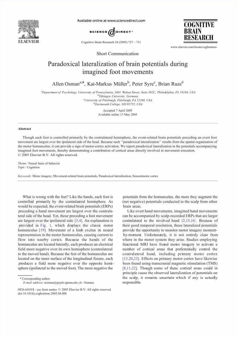
Short Communication Paradoxical lateralization of brain potentials during imagined foot movements Allen Osman a, * , Kai-Markus Mu ¨ ller b , Peter Syre c , Brian Russ d a Department of Psychol ogy , Universi ty of Pennsylv ania, 3401 Walnut Street, Suite 302C, Philad elphia, P A 19104, USA b Tu ¨ bingen University, Germany c University of Pittsbu rgh, Pittsbur gh, PA 15260, USA d Dartmouth College, NH 03755, USA Accepted 7 April 2005 Available online 13 May 2005 Abstract Though each foot is controlled primarily by the contralateral hemisphere, the event-related brain potentials preceding an overt foot movement are largest over the ipsilateral side of the head. Because such ‘‘paradoxical lateralization’’ results from the spatial organization of the motor homunculus, it can provide a sign of motor-cortex activation. We report paradoxical lateralization in the potentials accompanying imagined foot movements, thereby demonstrating a contribution of cortical areas directly involved in movement execution. D 2005 Elsevier B.V. All rights reserved. Theme: Neural basis of behavior Topic: Cognition Keywords: Motor imagery; Movement-related brain potentials; Paradoxical lateralization; Sensorimotor cortex What is wrong with the feet? Like the hands, each foot is controlle d primarily by the contralateral hemi sphere . As would be expected, the event-related brain potentials (ERPs) preceding a hand movement are largest over the contrala- teral side of the head. Yet, those preceding a foot movement are largest over the ipsilateral side [3,4]. An explanation is pr ovided in Fi g. 1, whi ch dis pla ys the cla ssi c motor homunculus [19]. Movement of a limb exc ite s it s neural representation in the motor homunculus, causing current to flow into nearby cortex. Because the hands of the homunculus are located laterally, each produces an electrical field more negative over its own hemisphere (contralateral to the moved hand). Because the feet of the homunculus are located on the inner surface of the longitudinal fissure, each pr oduces a fie ld more neg ati ve over the opposi te hemi- sphere (ipsilateral to the moved foot). The more negative the potentials from the homunculus, the more they augment the (net negative) potentials conducted to the scalp from other brain areas. Like overt hand movements, imagined hand movements can be accompanied by scalp-recorded ERPs that are larger contr alate ral to the invo lved hand [2,15,16]. Bec aus e of their good temporal resolution, these lateralized potentials provide the opportunity to monitor motor imagery moment- by-mo men t. Unf ort una tel y , it is not ent ire ly cle ar fr om where in the motor system they arise. Studies employing fun cti onal MRI have found mot or ima ger y to act iva te a number of cor tical areas tha t pre fer ent ial ly control the contralateral hand, including pri mary motor cortex [13,20,23]. Effects on primary motor cortex have likewise been found using transcranial magnetic stimulation (TMS) [8,11,22] . Tho ugh some of the se cor tic al areas cou ld in principle cause the observed lateralization of potentials on the sca lp, it remain s unc ert ain which if any is act ual ly responsible. 0926-6410/$ - see front matter D 2005 Elsevier B.V. All rights reserved. doi:10.1016 /j.cogbrainre s.2005.04.00 4 * Corresponding author. E-mail address: aosman@psy ch.upenn .edu (A. Osman). Cognitive Brain Research 24 (2005) 727 – 731 www.elsevier.com/locate/cogbrainres
-
Upload
kai-markus-mueller -
Category
Documents
-
view
234 -
download
0
Transcript of Osman CogBrRes2005
8/6/2019 Osman CogBrRes2005
http://slidepdf.com/reader/full/osman-cogbrres2005 1/5
Short Communication
Paradoxical lateralization of brain potentials duringimagined foot movements
Allen Osman a,*, Kai-Markus Muller b, Peter Syre c, Brian Russ d
a Department of Psychology, University of Pennsylvania, 3401 Walnut Street, Suite 302C, Philadelphia, PA 19104, USA bTubingen University, Germany
cUniversity of Pittsburgh, Pittsburgh, PA 15260, USAd Dartmouth College, NH 03755, USA
Accepted 7 April 2005Available online 13 May 2005
Abstract
Though each foot is controlled primarily by the contralateral hemisphere, the event-related brain potentials preceding an overt foot movement are largest over the ipsilateral side of the head. Because such ‘‘paradoxical lateralization’’ results from the spatial organization of the motor homunculus, it can provide a sign of motor-cortex activation. We report paradoxical lateralization in the potentials accompanyingimagined foot movements, thereby demonstrating a contribution of cortical areas directly involved in movement execution.D 2005 Elsevier B.V. All rights reserved.
Theme: Neural basis of behavior Topic: Cognition
Keywords: Motor imagery; Movement-related brain potentials; Paradoxical lateralization; Sensorimotor cortex
What is wrong with the feet? Like the hands, each foot iscontrolled primarily by the contralateral hemisphere. Aswould be expected, the event-related brain potentials (ERPs) preceding a hand movement are largest over the contrala-teral side of the head. Yet, those preceding a foot movement are largest over the ipsilateral side [3,4] . An explanation is provided in Fig. 1, which displays the classic motor homunculus [19]. Movement of a limb excites its neural
representation in the motor homunculus, causing current toflow into nearby cortex. Because the hands of thehomunculus are located laterally, each produces an electricalfield more negative over its own hemisphere (contralateralto the moved hand). Because the feet of the homunculus arelocated on the inner surface of the longitudinal fissure, each produces a field more negative over the opposite hemi-sphere (ipsilateral to the moved foot). The more negative the
potentials from the homunculus, the more they augment the(net negative) potentials conducted to the scalp from other brain areas.
Like overt hand movements, imagined hand movementscan be accompanied by scalp-recorded ERPs that are larger contralateral to the involved hand [2,15,16] . Because of their good temporal resolution, these lateralized potentials provide the opportunity to monitor motor imagery moment-
by-moment. Unfortunately, it is not entirely clear fromwhere in the motor system they arise. Studies employingfunctional MRI have found motor imagery to activate anumber of cortical areas that preferentially control thecontralateral hand, including primary motor cortex[13,20,23] . Effects on primary motor cortex have likewise been found using transcranial magnetic stimulation (TMS)[8,11,22] . Though some of these cortical areas could in principle cause the observed lateralization of potentials onthe scalp, it remains uncertain which if any is actuallyresponsible.
0926-6410/$ - see front matter D 2005 Elsevier B.V. All rights reserved.doi:10.1016/j.cogbrainres.2005.04.004
* Corresponding author. E-mail address: [email protected] (A. Osman).
Cognitive Brain Research 24 (2005) 727 – 731
www.elsevier.com/locate/cogbrainres
8/6/2019 Osman CogBrRes2005
http://slidepdf.com/reader/full/osman-cogbrres2005 2/5
To help resolve this ambiguity, we wanted to see if paradoxical lateralization would occur when movement of afoot is merely imagined. If so, it would implicate either primary motor or somatosensory (jointly termed sensorimo-tor) cortex, since only these areas contain homunculi withthe requisite spatial arrangement. In contrast, suppose for example that lateral premotor cortex (an area less directlyinvolved in movement execution than sensorimotor cortex)is solely responsible for ERP lateralization during motor imagery. Because it is located entirely on the outer aspectsof the hemispheres, the same non-paradoxical pattern shouldthen be observed for the feet as for the hands. (For a similar analysis of ERPs during advance preparation for a to-be-signaled movement, see [12].)
Nineteen participants providing informed consent weretested in a protocol approved by the University of Pennsylvania Institutional Review Board. Participants alter-nated between two types of 72-trial blocks. On ‘‘hand blocks’’ they made or imagined taps with their left or right index fingers, and on ‘‘foot blocks’’ they made or imaginedtaps with their left or right big toes. A single tap wasrequired on each trial. Each block consisted of four trialtypes defined by whether the tap was overt or imagined and
whether it involved the left or right limb. These occurredequally often and in a random order. There were two test sessions, the first of which served as practice. The reportedresults are based on five hand and five foot blocks from each participant’s second session.
To help control the time at which the imagined move-ments occurred, both overt and imagined movements weresynchronized to the temporally predictable occurrence of a brief visual signal (synch signal). Each trial began with theappearance of a fixation cross in the middle of a computer monitor. After 500 ms, the fixation cross was temporarilyreplaced for 250 ms by a warning signal (‘‘E’’ or ‘‘I’’) that indicated whether the response on that trial should be
overtly executed or imagined. One second later, the fixationcross was replaced for 250 ms by a second warning signal(‘‘L’’ or ‘‘R’’) that indicated whether the response shouldinvolve the left or right limb. Finally, after another second,the fixation cross was replaced for 50 ms by the synchsignal (‘‘X’’). The trial ended 700 ms later, with the fixationcross vanishing from the screen.
After each block, participants received feedback about their performance and filled out a brief questionnaire.Feedback concerned their overt taps on the preceding block,including the deviation from synchrony with the synchsignals and number (if any) on trials requiring imaginedtaps. The questionnaire asked participants to rate their imagined taps during the preceding block on severaldimensions, including sensation of movement, sense of intention or effort, vividness, and absence of muscleactivity. Its main purpose was to encourage participants tocultivate their images along these lines. The eight questionscomprising the questionnaires are shown in Table 1 .
Participants sat facing a monitor across a table with their heads supported by a chinrest, forearms resting on the table,and shoeless feet flat on the floor below. On hand blocks,
Fig. 1. Motor homunculus (adapted from [19] ). Arrows indicate general left– right direction of current flow caused by movement of a hand or foot. Electrodesshow scalp locations where normal lateralization during hand movements and paradoxical lateralization during foot movements can be recorded.
Table 1Questions on the imagery questionnaires
(1) How strong was the ‘‘sensation of movement’’ in the imaginedmovements?
(2) How strong was the sense of ‘‘intention’’ or ‘‘effort’’ in the imaginedmovements?
(3) How ‘‘concrete’’ or ‘‘vivid’’ were the imagined movements?(4) How often did you imagine the signaled movement on imagery trials?(5) How accurate was the timing of your imagined movements compared
with that of your executed movements?(6) How accurate were your initial choices of response finger or toe for
imagined movements compared with those for executed movements?(7) How often did you move your muscles while imagining movements?(8) How difficult was it for you to imagine movements?
A. Osman et al. / Cognitive Brain Research 24 (2005) 727–731728
8/6/2019 Osman CogBrRes2005
http://slidepdf.com/reader/full/osman-cogbrres2005 3/5
their index fingers rested on two adjacent response keysdirectly in front of them. On foot blocks, their big toesrested on two adjacent response keys under the table. Eachkey was immobile and attached snugly to a finger or toewith a velcro strap. Responses were overt and imagined
isometric flexions of the left and right index fingers or bigtoes. Stimuli were individual characters presented on themonitor against a dark background at the participant’smidline.
Continuous recordings were made of key pressure,electroencephalographic activity (EEG), electro-ocular activity (EOG), and electromyographic activity (EMG).Pressure was measured by a stress gauge attached to eachkey. EEG was recorded from 57 standard sites across thescalp [1] and referenced to the left mastoid (though themeasures of ERP lateralization are reference free). Verticaland horizontal EOG were recorded bipolarly from sitesabove and below the right eye and external to the outer
canthus of each eye. EMG was recorded bipolarly withsurface electrodes from the Flexor Digitorum Superficialis of each arm and Flexor Hallucis Longus of each leg. EEG andEOG were filtered on-line with a band pass of 0.03 to 30 Hz.EMG was filtered on-line with a band pass of 0.1 to 500 Hzand then RMS-converted to a DC signal. All signals weredigitized at 100 Hz and low-pass filtered offline at 4 Hz.
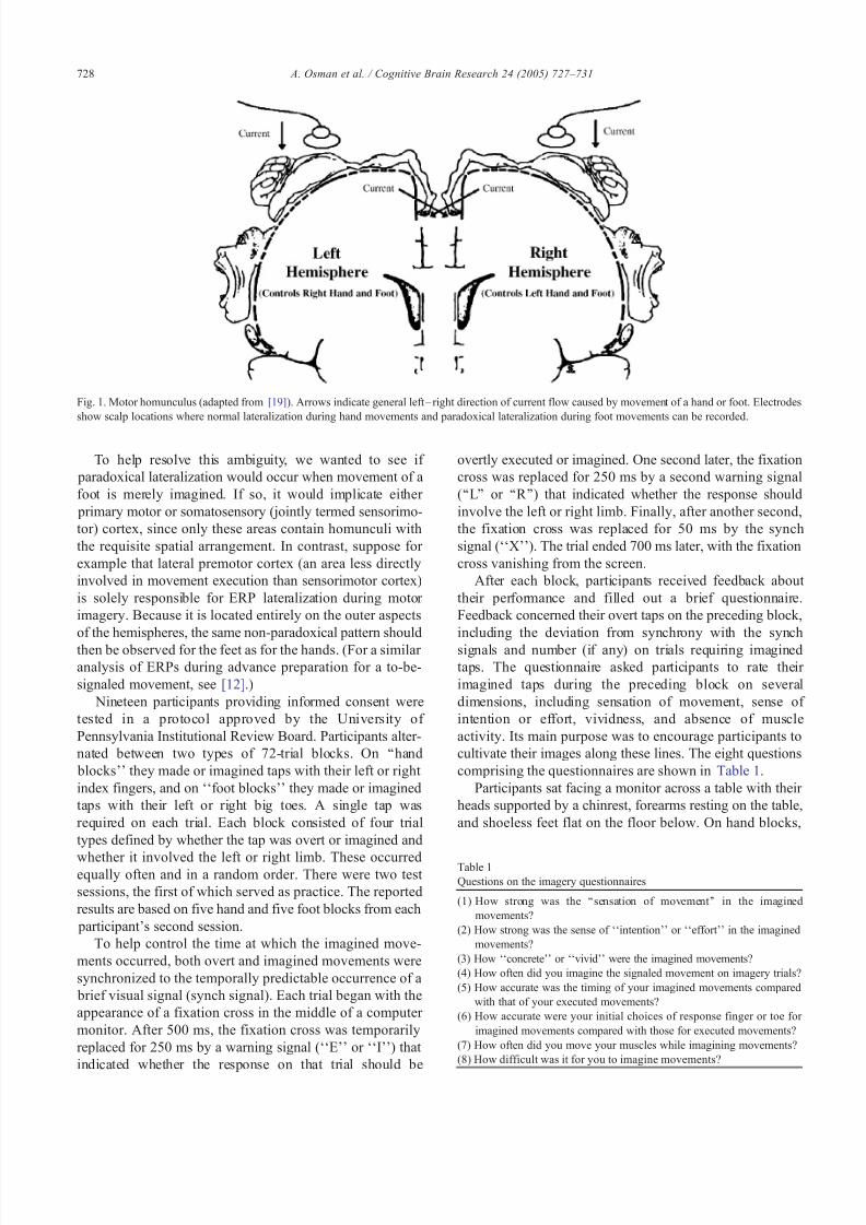
The most important results concern left– right differ-ences in movement-related activity at the muscular (EMG)and cortical (ERP) levels. Differences between EMGactivity in the sig naled and unsignaled limb are shown inthe top panel of Fig. 2. This difference, reflecting muscleactivity that depends on which response side is signaled,was significant at synch signal onset for both overt finger taps [t (18) = 7.67, P (one-tailed) < 0.001] and overt toe taps[t (18) = 6.93, P (one-tailed) < 0.001]. In contrast, it wasvirtually absent during motor imagery, as reflected in itssize for imagined taps relative to that for overt taps at synch
signal onset (fingers: v = 0.9%, SE = 0.4%; toes: v = 3.2%,SE = 2.5%).Analogous differences between ERPs recorded over the
signaled and unsignaled hemispheres (contralateral andipsilateral to the signaled limb) are shown in the bottom
Fig. 2. EMG and ERPs during overt or imagined flexions of a finger or toe.Top panel: difference between rectified EMG recorded from the flexor muscles of the signaled (left or right) and unsignaled limb. S2 = stimulussignaling the finger or toe with which to make (or imagine) the flexion. S3 =signal to which the flexion (overt or imagined) was synchronized. Bottom panel: difference be tween voltage over the hand areas of primary motor cortex (C3/4 sites, [1]) contralateral and ipsilateral to the signaled limb. Negative and positive values indicate respectively larger ERPs contralateral(normal lateralization) and ipsilateral (paradoxical lateralization).
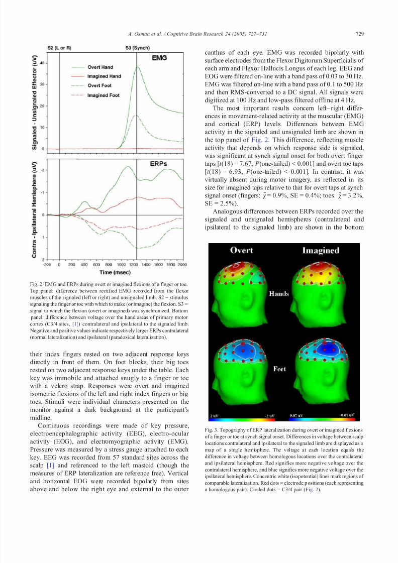
Fig. 3. Topography of ERP lateralization during overt or imagined flexionsof a finger or toe at synch signal onset. Differences in voltage between scalplocations contralateral and ipsilateral to the signaled limb are displayed as amap of a single hemisphere. The voltage at each location equals thedifference in voltage between homologous locations over the contralateraland ipsilateral hemisphere. Red signifies more negative voltage over thecontralateral hemisphere, and blue signifies more negative voltage over theipsilateral hemisphere. Concentric white (isopotential) lines mark regions of comparable lateralization. Red dots = electrode p ositions (each representinga homologous pair). Circled dots = C3/4 pair ( Fig. 2).
A. Osman et al. / Cognitive Brain Research 24 (2005) 727–731 729

8/6/2019 Osman CogBrRes2005
http://slidepdf.com/reader/full/osman-cogbrres2005 4/5
panel of Fig. 2. These were likewise tested statistically at synch signal onset. As expected, a normal pattern of lateralization was observed during overt finger taps [ t (18) =6.45, P (one-tailed) < 0.001] and a paradoxical pattern wasobserved during overt toe taps [ t (18) = 5.41; P (one-tailed) <0.001]. ERP lateralization occurred also during imaginedtaps, despite the lack of EMG. As with overt move-ments, the pattern was normal for the fingers [ t (18) =3.48, P (one-tailed) < 0.002] and paradoxical for the toes[t (18) = 3.35, P (one-tailed) < 0.002]. A more complete picture of ER P later alization at synch signal onset is presented in Fig. 3, which displays its topographicdistribution. It can be seen here that lateralization duringovert and imagined movements had a similar overalltopography, which was normal for the hands and paradoxical for the feet.
In summary, paradoxical lateralization was observed inthe ERPs accompanying imagined foot movemen ts without
EMG. This finding a grees with functional MRI [13,20,23]and TMS [8,11,22] evidence that motor imagery canactivate the primary motor cortex without also producingmuscle activity. Motor imagery has been hypothesized toevoke cognitive processes similar to those that prepare andcontrol overt movement [5,10,17] . But, because earlier studies had failed to detect its effects on the primary motor cortex [6,9,18,21] , motor imagery was generally believed aslate as the mid- 1990s t o comprise only the more abstract motor functions [5,6,18] . Since then, however, evidence hascontinued to accumulate that it can engage, not only portions of the motor system involved in action planning, but also those more directly involved in movement execution (but see [7]).
What is new about the present findings is a demonstrationthat the electrical fields responsible for ERP lateralizationduring motor imagery do in fact arise from sensorimotor cortex. The time course of this lateralization can thus providea window on the dynamic state of sensorimotor cortex asmotor imagery unfolds. The temporal structure of an overt act is thought to be preserved when it is imagined[5,10,17,18] . Does this temporal isomorphism extend tothe sensorimotor cortex? Because of their good temporalresolution, ERPs can help answer this question [15]. Our findings may be relevant as well to situations besides motor
imagery where people drive their motor systems without producing overt movement. Simple non-invasive measureslike those examined here might, for example, provide strokevictims with feedback, enabling them to activate spared portions of their motor cortices at an earlier stage in theneural reorganization following injury [14].
Acknowledgments
We thank John Kounios and two anonymous reviewersfor helpful comments. This work was supported by NINDSgrant NS37528.
References
[1] American Electroencephalographic Society, Guidelines for standardelectrode position nomenclature, J. Clin. Neurophysiol. 8 (1991)200–201.
[2] R. Beisteiner, P. Hollinger, G. Lindinger, W. Lang, A. Berthoz, Mentalrepresentations of movements: brain potentials associated with
imagination of hand movements, Electroencephalogr. Clin. Neuro- physiol. 96 (1995) 183–193.
[3] J. Boschert, L. Deeke, Cerebral potentials preceding voluntary toe,knee, and hip movements and their vectors in human precentral gyrus,Brain Res. 376 (1986) 175–179.
[4] C.H.M. Brunia, W.E.J. Van Den Bosch, Movement-related slow potentials: I. A contrast between finger and foot movements in right-handed subjects, Electroencephalogr. Clin. Neurophysiol. 57 (1984)515–527.
[5] J. Decety, Do imagined and executed actions share the same neuralsubstrate? Cogn. Brain Res. 3 (1996) 87 –93.
[6] J. Decety, D. Perani, M. Jeannerod, V. Bettinardi, B. Tadary,R. Woods, J.C. Mazziotta, F. Fazio, Mapping motor represen-tations with positron emission tomography, Nature 371 (1994)600–602.
[7] P. Dechent, K. Merboldt, J. Frahm, Is the human primary motor cortex involved in motor imagery? Cogn. Brain Res. 19 (2004)138–144.
[8] R. Hashimoto, J.C. Rothwell, Dynamic changes in corticospinalexcitability during motor imagery, Exp. Brain Res. 125 (1999)75–81.
[9] D.H. Ingvar, L. Philipson, Distribution of cerebral blood flow in thedominant hemisphere during motor ideation and motor performance,Ann. Neurol. 2 (1977) 230–237.
[10] M. Jeannerod, The representing brain: neural correlates of motor intention and imagery, Behav. Brain Sci. 17 (1994)187–245.
[11] T. Kasai, S. Kawai, M. Kawanishi, S. Yahagi, Evidence of facilitationof motor evoked potentials (MEPs) induced by motor imagery, BrainRes. 744 (1997) 147–150.
[12] H. Leuthold, I. Jentzsch, Distinguishing neural sources of movement preparation and execution: an electrophysiological analysis, Biol.Psychol. 60 (2002) 173–198.
[13] M. Lotze, P. Montoya, M. Erb, E. Hulsmann, H. Flor, U. Klose, N.Birbaumer, W. Grodd, Activation of cortical and cerebellar motor areas during executed and imagined hand movements: an fMRI study,J. Cogn. Neurosci. 11 (1999) 491–501.
[14] R.J. Nudo, B.M. Wise, F. SiFuentes, G.W. Milliken, Neural substratesfor the effects of rehabilitative training on motor recovery after ischemic infarct, Science 272 (1996) 1791–1794.
[15] A. Osman, R. Albert, K.R. Ridderinkhof, G. Band, M. van der Molen,The beat goes on: rhythmic modulation of cortical potentials byimagined tapping (submitted for publication).
[16] L. Parra, C. Alvino, A. Tang, B. Pearlmutter, N. Young, A. Osman, P.Sajda, Linear spatial integration for single trial detection in encepha-lography, NeuroImage 17 (2002) 223–230.
[17] L.M. Parsons, Imagined spatial transformations of one’s hands andfeet, Cognit. Psychol. 19 (1987) 178–241.
[18] L.M. Parsons, P.T. Fox, J.H. Downs, T. Glass, T.B. Hirsch, C.C.Martin, P.A. Jerabek, J.L. Lancaster, Use of implicit motor imagery for visual shape discrimination as revealed by PET, Nature 375 (1995)54–58.
[19] W. Penfield, T. Rasmussen, The cerebral cortex of man, A ClinicalStudy of Localization Function, Macmillan, New York, 1950.
[20] C.A. Porro, M.P. Francescato, V. Cettolo, M.E. Diamond, P. Baraldi,C. Zuiani, M. Bazzocchi, D.E. di Prompero, Primary motor andsensory cortex activation during motor performance and motor imagery: a functional magnetic resonance imaging study, J. Neurosci.16 (1996) 7688– 7698.
[21] P.E. Roland, E. Skinhoj, N.A. Lassen, B. Larsen, Different cortical
A. Osman et al. / Cognitive Brain Research 24 (2005) 727–731730

8/6/2019 Osman CogBrRes2005
http://slidepdf.com/reader/full/osman-cogbrres2005 5/5
areas in man in organization of voluntary movements in extrapersonalspace, J. Neurophysiol. 43 (1980) 137–150.
[22] P.M. Rossini, S. Rossi, P. Pasqualetti, F. Tecchio, Corticospinalexcitability modulation to hand muscles during movement imagery,Cereb. Cortex 9 (1999) 161–167.
[23] M. Roth, J. Decety, M. Raybaudi, R. Massarelli, C. Delon-Martin, C.Segebarth, S. Morand, A. Gemignani, M. Decorps, M. Jeannerod,Possible involvement of primary motor cortex in mentally simulatedmovement: a functional magnetic resonance imaging study, Neuro-Report 7 (1996) 1280–1284.
A. Osman et al. / Cognitive Brain Research 24 (2005) 727–731 731


![[Osman bakar] classification_of_knowledge_in_islam](https://static.fdocuments.in/doc/165x107/58f308c11a28ab89718b4591/osman-bakar-classificationofknowledgeinislam.jpg)