OSI-930 analogues as novel reversal agents for ABCG2-mediated multidrug resistance
9
OSI-930 analogues as novel reversal agents for ABCG2-mediated multidrug resistance Ye-Hong Kuang a,b , Jay P. Patel a , Kamlesh Sodani a , Chung-Pu Wu c , Li-Qiu Liao d , Atish Patel a , Amit K. Tiwari a , Chun-Ling Dai a,e , Xiang Chen b, *, Li-Wu Fu e , Suresh V. Ambudkar c , Vijaya L. Korlipara a, **, Zhe-Sheng Chen a, ** a Department of Pharmaceutical Sciences, College of Pharmacy and Allied Health Professions, St. John’s University, Queens, NY 11439, USA b Department of Dermatology, Xiang Ya Hospital, Central South University, Changsha 410008, China c Laboratory of Cell Biology, Center for Cancer Research, National Cancer Institute, NIH, Bethesda, MD 20892, USA d Department of Breast Surgery, Xiang Ya Hospital, Central South University, Changsha 410008, China e State Key Laboratory for Oncology in South China, Cancer Center, Sun Yat-Sen University, Guangzhou 510060, China 1. Introduction Multidrug resistance (MDR) is a major obstacle to successful chemotherapy in cancer treatment. It is a phenomenon in which cancer cells become resistant to a wide spectrum of anti cancer drugs that belong to different chemical or pharmacological classes and results in decreased cytotoxic activity of these anticancer drugs [1,2]. The mechanisms of MDR are multifactorial and include alteration in the permeability of lipid bilayer membrane, increased DNA repair in cancer cells, increased inactivation or detoxification of drugs, or inhibition of apoptosis [3,4]. Over-expression of multiple ATP binding cassette (ABC) transporters in cancer cells still remains the leading cause of MDR [5–7]. The term ABC transporter, is based on the fact that almost all members of the ABC superfamily, from yeast and bacteria to man, share a conserved consensus sequence of 90–110 amino acids, where ATP binds and is hydrolyzed by the ATPase enzyme. This consensus has Walker A and B motifs and another C region called ABC cassette. ABC transporters is the largest trans-membrane protein family encoded in the human genome, which are divided into 7 subfamilies ABC-A to G and subdivided into sub-subfamilies depending on their Biochemical Pharmacology 84 (2012) 766–774 A R T I C L E I N F O Article history: Received 22 March 2012 Accepted 19 June 2012 Available online 28 June 2012 Keywords: OSI-930 ABC transporters ABCG2 Multidrug resistance A B S T R A C T OSI-930, a dual c-Kit and KDR tyrosine kinase inhibitor, is reported to have undergone a Phase I dose escalation study in patients with advanced solid tumors. A series of fifteen pyridyl and phenyl analogues of OSI-930 were designed and synthesized. Extensive screening of these compounds led to the discovery that nitropyridyl and ortho-nitrophenyl analogues, VKJP1 and VKJP3, were effective in reversing ABC subfamily G member 2 (ABCG2) transporter-mediated multidrug resistance (MDR). VKJP1 and VKJP3 significantly sensitized ABCG2-expressing cells to established substrates of ABCG2 including mitoxantrone, SN-38, and doxorubicin in a concentration-dependent manner, but not to the non- ABCG2 substrate cisplatin. However, they were unable to reverse ABCB1- or ABCC1-mediated MDR indicating their selectivity for ABCG2. Western blotting analysis was performed to evaluate ABCG2 expression and it was found that neither VKJP1 nor VKJP3 significantly altered ABCG2 protein expression for up to 72 h. [ 3 H]-mitoxantrone accumulation study demonstrated that VKJP1 and VKJP3 increased the intracellular accumulation of [ 3 H]-mitoxantrone, a substrate of ABCG2. VKJP1 and VKJP3 also remarkably inhibited the transport of [ 3 H]-methotrexate by ABCG2 membrane vesicles. Importantly, both VKJP1 and VKJP3 were efficacious in stimulating the activity of ATPase of ABCG2 and inhibited the photoaffinity labeling of this transporter by its substrate [ 125 I]-iodoarylazidoprazosin. The results suggested that VKJP1 and VKJP3, specifically inhibit the function of ABCG2 through direct interaction with its substrate binding site(s). Thus VKJP1 and VKJP3 represent a new class of drugs for reducing MDR in ABCG2 over-expressing tumors. ß 2012 Elsevier Inc. All rights reserved. Abbreviations: OSI-930, {N-(4-trifluoromethoxyphenyl)3-((quinolin-4-ylmethyl) amino) thiophene-2-carboxamide}; ABC, ATP binding cassette; ABCG2 (BCRP/ MXR/ABCP), ABC subfamily G member 2; MDR, multidrug resistance; P-gp, P- glycoprotein; ABCCs (MRPs), ABC subfamily C member; RTK, receptor tyrosine kinases; KDR, kinase insert domain receptor; TKIs, tyrosine kinase inhibitors; IAAP, [ 125 I]-iodoarylazidoprazosin; FTC, fumitremorgin C; MTT, 3-(4,5-dimethylthiazol- 2-yl)-2,5-diphenyltetrazolium bromide; DMSO, dimethylsulfoxide. * Corresponding author at: 87 Xiang Ya Road, Changsha 410008, China. Fax: +86 731 432 8478. ** Corresponding authors at: 8000 Utopia Parkway, Queens, NY 11439, USA. Fax: +1 718 990 1877. E-mail addresses: [email protected] (X. Chen), [email protected] (V.L. Korlipara), [email protected] (Z.-S. Chen). Contents lists available at SciVerse ScienceDirect Biochemical Pharmacology jo u rn al h om epag e: ww w.els evier.c o m/lo cat e/bio c hem p har m 0006-2952/$ – see front matter ß 2012 Elsevier Inc. All rights reserved. http://dx.doi.org/10.1016/j.bcp.2012.06.019
-
Upload
ye-hong-kuang -
Category
Documents
-
view
215 -
download
1
Transcript of OSI-930 analogues as novel reversal agents for ABCG2-mediated multidrug resistance
Biochemical Pharmacology 84 (2012) 766–774
OSI-930 analogues as novel reversal agents for ABCG2-mediated multidrugresistance
Ye-Hong Kuang a,b, Jay P. Patel a, Kamlesh Sodani a, Chung-Pu Wu c, Li-Qiu Liao d, Atish Patel a,Amit K. Tiwari a, Chun-Ling Dai a,e, Xiang Chen b,*, Li-Wu Fu e, Suresh V. Ambudkar c,Vijaya L. Korlipara a,**, Zhe-Sheng Chen a,**a Department of Pharmaceutical Sciences, College of Pharmacy and Allied Health Professions, St. John’s University, Queens, NY 11439, USAb Department of Dermatology, Xiang Ya Hospital, Central South University, Changsha 410008, Chinac Laboratory of Cell Biology, Center for Cancer Research, National Cancer Institute, NIH, Bethesda, MD 20892, USAd Department of Breast Surgery, Xiang Ya Hospital, Central South University, Changsha 410008, Chinae State Key Laboratory for Oncology in South China, Cancer Center, Sun Yat-Sen University, Guangzhou 510060, China
A R T I C L E I N F O
Article history:
Received 22 March 2012
Accepted 19 June 2012
Available online 28 June 2012
Keywords:
OSI-930
ABC transporters
ABCG2
Multidrug resistance
A B S T R A C T
OSI-930, a dual c-Kit and KDR tyrosine kinase inhibitor, is reported to have undergone a Phase I dose
escalation study in patients with advanced solid tumors. A series of fifteen pyridyl and phenyl analogues
of OSI-930 were designed and synthesized. Extensive screening of these compounds led to the discovery
that nitropyridyl and ortho-nitrophenyl analogues, VKJP1 and VKJP3, were effective in reversing ABC
subfamily G member 2 (ABCG2) transporter-mediated multidrug resistance (MDR). VKJP1 and VKJP3
significantly sensitized ABCG2-expressing cells to established substrates of ABCG2 including
mitoxantrone, SN-38, and doxorubicin in a concentration-dependent manner, but not to the non-
ABCG2 substrate cisplatin. However, they were unable to reverse ABCB1- or ABCC1-mediated MDR
indicating their selectivity for ABCG2. Western blotting analysis was performed to evaluate ABCG2
expression and it was found that neither VKJP1 nor VKJP3 significantly altered ABCG2 protein expression
for up to 72 h. [3H]-mitoxantrone accumulation study demonstrated that VKJP1 and VKJP3 increased the
intracellular accumulation of [3H]-mitoxantrone, a substrate of ABCG2. VKJP1 and VKJP3 also
remarkably inhibited the transport of [3H]-methotrexate by ABCG2 membrane vesicles. Importantly,
both VKJP1 and VKJP3 were efficacious in stimulating the activity of ATPase of ABCG2 and inhibited the
photoaffinity labeling of this transporter by its substrate [125I]-iodoarylazidoprazosin. The results
suggested that VKJP1 and VKJP3, specifically inhibit the function of ABCG2 through direct interaction
with its substrate binding site(s). Thus VKJP1 and VKJP3 represent a new class of drugs for reducing MDR
in ABCG2 over-expressing tumors.
� 2012 Elsevier Inc. All rights reserved.
Contents lists available at SciVerse ScienceDirect
Biochemical Pharmacology
jo u rn al h om epag e: ww w.els evier .c o m/lo cat e/bio c hem p har m
1. Introduction
Multidrug resistance (MDR) is a major obstacle to successfulchemotherapy in cancer treatment. It is a phenomenon in which
Abbreviations: OSI-930, {N-(4-trifluoromethoxyphenyl)3-((quinolin-4-ylmethyl)
amino) thiophene-2-carboxamide}; ABC, ATP binding cassette; ABCG2 (BCRP/
MXR/ABCP), ABC subfamily G member 2; MDR, multidrug resistance; P-gp, P-
glycoprotein; ABCCs (MRPs), ABC subfamily C member; RTK, receptor tyrosine
kinases; KDR, kinase insert domain receptor; TKIs, tyrosine kinase inhibitors; IAAP,
[125I]-iodoarylazidoprazosin; FTC, fumitremorgin C; MTT, 3-(4,5-dimethylthiazol-
2-yl)-2,5-diphenyltetrazolium bromide; DMSO, dimethylsulfoxide.
* Corresponding author at: 87 Xiang Ya Road, Changsha 410008, China.
Fax: +86 731 432 8478.
** Corresponding authors at: 8000 Utopia Parkway, Queens, NY 11439, USA.
Fax: +1 718 990 1877.
E-mail addresses: [email protected] (X. Chen), [email protected]
(V.L. Korlipara), [email protected] (Z.-S. Chen).
0006-2952/$ – see front matter � 2012 Elsevier Inc. All rights reserved.
http://dx.doi.org/10.1016/j.bcp.2012.06.019
cancer cells become resistant to a wide spectrum of anti cancerdrugs that belong to different chemical or pharmacological classesand results in decreased cytotoxic activity of these anticancerdrugs [1,2]. The mechanisms of MDR are multifactorial and includealteration in the permeability of lipid bilayer membrane, increasedDNA repair in cancer cells, increased inactivation or detoxificationof drugs, or inhibition of apoptosis [3,4]. Over-expression ofmultiple ATP binding cassette (ABC) transporters in cancer cellsstill remains the leading cause of MDR [5–7]. The term ABCtransporter, is based on the fact that almost all members of the ABCsuperfamily, from yeast and bacteria to man, share a conservedconsensus sequence of 90–110 amino acids, where ATP binds andis hydrolyzed by the ATPase enzyme. This consensus has Walker Aand B motifs and another C region called ABC cassette. ABCtransporters is the largest trans-membrane protein family encodedin the human genome, which are divided into 7 subfamilies ABC-Ato G and subdivided into sub-subfamilies depending on their
Y.-H. Kuang et al. / Biochemical Pharmacology 84 (2012) 766–774 767
structural similarity or difference in their trans-membranedomain. The prominent ABC transporters playing a role in MDRdevelopment are ABCB1, also called P-glycoprotein (P-gp) [8,9],multidrug resistance proteins (MRPs, ABCCs) [10], and ABCG2(BCRP/MXR/ABCP) [11,12]. This ABC cassette is involved intransportation of not only toxic metabolites or xenobiotics outof the cell, but also a multitude of anticancer drugs across cellmembrane, thus decreasing their intracellular concentration andcytotoxicity. Therefore, their expression is believed to be the majormechanism by which MDR to anticancer drugs develops. Initially,verapamil was found by Tsuruo et al. to be the first modulatorwhich could inhibit the function of ABCB1 and increase drugaccumulation in the cancer cells [13]. Later, cyclosporine A (CsA),an immunosuppressant, was discovered to inhibit the function ofABCB1 and was used as another modulator of ABCB1 [14]. Afterthat, many other compounds have been studied for their inhibitoryeffects to other members of ABC transporter family [15–18], raisinghopes that ABC transporter mediated MDR could be reversed. Atpresent, as the newly synthesized molecule-targeting agents suchas tyrosine kinase inhibitors (TKIs) are being introduced into theclinic, research is being conducted to determine whether thesenovel compounds could represent a new class of ABC transporterinhibitors. TKIs such as imatinib, nilotinib, erlotinib, dasatinib andlapatinib have been extensively investigated as modulators of ABCtransporters-mediated MDR, which most likely act as competitiveinhibitors [19–23]. More recently, pre-clinical research and clinicaltrials have been conducted to study the efficacy of the combinationof some TKIs with other anticancer drugs in improving thetherapeutic outcome of cancer patients. Therefore, discoveringnovel inhibitors of ABC transporters to reverse the drug resistancecan lead to improvement in the use of currently availableanticancer drugs.
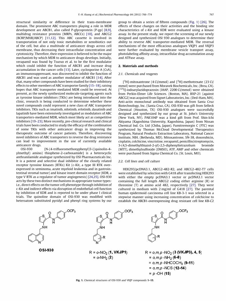
OSI-930 {N-(4-trifluoromethoxyphenyl)3-((quinolin-4-ylmethyl) amino) thiophene-2-carboxamide} is a heterocyclicanthranilamide analogue synthesized by OSI Pharmaceuticals Inc.It is a potent and selective dual inhibitor of the closely relatedreceptor tyrosine kinases (RTKs) Kit (c-Kit, a type III RTK over-expressed in seminoma, acute myeloid leukemia and in gastroin-testinal stromal tumor) and kinase insert domain receptor (KDR, atype V RTK as a regulator of tumor angiogenesis) [24,25]. OSI-930acts by these two distinct mechanisms in appropriate tumor types:i.e., direct effects on the tumor cell phenotype through inhibition ofc-Kit and indirect effects via disruption of endothelial cell functionby inhibition of KDR and is reported to be under phase I clinicaltrials. The quinoline domain of OSI-930 was modified withheteroatom substituted pyridyl and phenyl ring systems by our
Fig. 1. Chemical structures of OSI-9
group to obtain a series of fifteen compounds (Fig. 1) [26]. Theeffects of these changes on their activities and the binding sitecharacteristics of c-Kit and KDR were evaluated using a kinaseassay. In the present study, we report the screening of our newlydesigned and synthesized OSI 930 analogues to determine theirability to reverse ABC transporter-mediated MDR. The reversalmechanisms of the most efficacious analogues VKJP1 and VKJP3were further evaluated by membrane vesicle transport assay,photolabeling affinity assay, intracellular drug accumulation assayand ATPase assay.
2. Materials and methods
2.1. Chemicals and reagents
[3H]-mitoxantrone (4 Ci/mmol) and [3H]-methotrexate (23 Ci/mmol) were purchased from Moravek Biochemicals, Inc. (Brea, CA).[125I]-iodoarylazidoprazosin (IAAP, 2200 Ci/mmol) were obtainedfrom Perkin-Elmer Life Sciences. (Boston, MA). BXP-21 (againstABCG2) was acquired from Signet Laboratories, Inc. (Ded-Ham, MA).Anti-actin monoclonal antibody was obtained from Santa CruzBiotechnology, Inc. (Santa Cruz, CA). OSI-930 was gift from SelleckChemical (Houston, TX) OSI-930 analogues were successfullydesigned and synthesized by our group at St. John’s University(New York, NY). PAK104P was a kind gift from Prof. Shin-IchiAkiyama (Kagoshima University, Kagoshima, Japan) from NissanChemical Ind. Co. Ltd (Chiba, Japan). Fumitremorgin C (FTC) wassynthesized by Thomas McCloud Developmental TherapeuticsProgram, Natural Products Extraction Laboratory, National CancerInstitute, NIH. (Bethesda, MD). Mitoxantrone, SN-38, doxorubicin,cisplatin, colchicine, vincristine, verapamil, penicillin/streptomycin,3-(4,5-dimethylthiazol-2-yl)-2,5-diphenyltetrazolium bromide(MTT), dimethylsulfoxide (DMSO), ATP, AMP and other chemicalswere purchased from Sigma Chemical Co. (St. Louis, MO).
2.2. Cell lines and cell culture
HEK293/pcDNA3.1, ABCG2-482-R2, and ABCG2-482-T7 cellswere established by selection with G418 after transfecting HEK293with either the empty pcDNA3.1 vector or pcDNA3.1 vectorcontaining the full length ABCG2 coding either arginine (R) orthreonine (T) at amino acid 482, respectively [27]. They werecultured in medium with 2 mg/ml of G418 [27]. The parentalhuman epidermoid carcinoma cell line KB-3-1 was selected in astepwise manner using increasing concentration of colchicine toestablish the ABCB1-overexpressing drug resistant cell line KB-C2
30 and VKJP compounds 1–15.
Y.-H. Kuang et al. / Biochemical Pharmacology 84 (2012) 766–774768
and was cultured in the medium with 2 mg/ml of colchicine [28]. AnABCC1-overexpressing MDR cell line, KB-CV60, was also cloned fromKB-3-1 cells and was maintained in medium with 1 mg/mlcepharanthine and 60 ng/ml vincristine [29]. All the cell lines weregrown in Dulbecco’s modified Eagle’s medium (DMEM) supplemen-ted with 10% bovine serum, 100 units/ml penicillin, and 100 mg/mlstreptomycin in a humidified incubator containing 5% CO2 at 37 8C.
2.3. Cell sensitivity to drugs by MTT assay
Cell sensitivity to drugs was analyzed using an MTT colorimetricassay [19]. Cells were harvested with trypsin and resuspended in afinal concentration 8 � 103 cells/ml for KB-C2 and 5 � 103 for all theother cell lines. Cells were seeded evenly into (160 ml/well) 96-wellmultiplates. After 24 h of incubation, the test VKJP compound (20 ml/well) was added 1 h prior to different concentrations of chemother-apeutic drugs (20 ml/well) into designated wells. FTC, verapamil orPAK-104 (20 ml/well) was added as positive control inhibitors ofABCG2, ABCB1 or ABCC1, respectively. After 72 h of incubation, 20 mlof MTT solution (4 mg/ml) was added to each well, further incubatedfor 4 h, the medium was discarded, and 100 ml of DMSO was addedinto each well to dissolve the formazan crystals. The absorbance wasdetermined at 570 nm by an OPSYS microplate Reader from DYNEXTechnologies, Inc. (Chantilly, VA). The concentrations required toinhibit growth by 50% (IC50) were calculated from survival curves.The degree of resistance was calculated by dividing the IC50 of theMDR cells by that of the parental sensitive cells.
2.4. Preparation of cell lysates
Cells in T-25 flask treated with 10 mM VKJP1 or VKJP3 fordifferent time periods (0, 36, 72 h), then were harvested and rinsedtwice with cold PBS. The cell extracts were prepared by incubatingthe cells with the radioimmunoprecipitation assay (RIPA) buffer[1� PBS, 1% Nonidet P-40, 0.5% sodium deoxycholate, 0.1% SDS,100 mM p-aminophenylmethylsulfonyl fluoride (p-APMSF),10 mM leupeptin, and 10 mM aprotinin] for 30 min on ice withoccasional rocking, followed by centrifugation at 12,000 � g at 4 8Cfor 15 min. The supernatant containing total cell lysates werecollected and stored at �80 8C until future experiments. Theprotein concentration was determined by bicinchoninic acid(BCATM)-based protein assay (Thermo Scientific, Rockford, IL).
2.5. Western blotting
Equal amounts of total cell lysates (40 mg) were resolved by 4–12% sodium dodecyl sulfate polyacrylamide gel electrophoresis(SDS–PAGE) and electrophoretically transferred onto PVDF mem-brane, then immersed in blocking solution (5% skim milk in Tris-buffered saline (TBST: 0.3% Tris, 0.8% NaCl, 0.02% KCl, 0.05% Tween20) for 1 h at room temperature. The membrane was thenimmunoblotted overnight with monoclonal anti-ABCG2 antibody(BXP-21) at 1:500 dilution at 4 8C. The following day, themembrane was washed with TBST buffer three times and followedby 3 h incubation with horseradish peroxide (HRP)-conjugatedsecondary anti-mouse IgG at 1:1000 for 3 h. Proteins weredetected by enhanced chemiluminescence detection system(Amersham, NJ). b-Actin was used to confirm equal loading ineach lane in the samples prepared from cell lysates.
2.6. [3H]-mitoxantrone accumulation
The effect of VKJP1 or VKJP3 on the intracellular accumulation of[3H]-mitoxantrone in HEK293/pcDNA3.1, ABCG2-482-R2, andABCG2-482-T7 cells was determined as previously described [30].Confluent cells in 24-well plates were preincubated with or without
VKJP1, VKJP3 (FTC as positive control) for 1 h at 37 8C. Intracellulardrug accumulation was measured by incubating cells with 0.2 mM[3H]-mitoxantrone for another 2 h. The cells were washed threetimes with ice-cold PBS, trypsinized and lysed in 10 mM lysis buffer(pH 7.4, containing 1% Triton X-100 and 0.2% SDS). Each sample wasplaced in scintillation fluid and radioactivity was measured in aPackard TRI-CARB1 1900CA liquid scintillation analyzer fromPackard Instrument Company, Inc (Downers Grove, IL).
2.7. Preparation of membrane vesicles and in vitro transport assays
Membrane vesicles were prepared by using the nitrogencavitation method [31]. Cells were rinsed twice with PBS and thenscraped into PBS containing 1% aprotinin. Cells were then washed at4 8C in PBS, collected by centrifugation (4000 � g for 10 min),suspended in buffer A (10 mM Tris–HCl, pH 7.4, 0.25 M sucrose,1 mM p-APMSF, and 0.2 mM CaCl2) and equilibrated at 4 8C for15 min under a nitrogen pressure of 500 psi. EDTA was added to thesuspension of lysed cells to make a final concentration of 1 mM, andthe suspension was then diluted 1:4 with buffer B (10 mM Tris–HCl,pH 7.4, 0.25 M sucrose, and 1 mM p-APMSF) and centrifuged at4000 � g for 10 min at 4 8C to remove nuclei and unlysed cells. Thesupernatant was layered onto a sucrose cushion (35% sucrose,10 mM Tris–HCl, pH 7.4, and 1 mM EDTA) and centrifuged for 30 minat 16,000 � g at 4 8C. The interface was collected and centrifuged for45 min at 100,000 � g at 4 8C. The pellet was resuspended in buffer Bby repeated passage through a 25-gauge needle.
Transport assays were performed using the rapid filtrationmethod as previously described [19]. Membrane vesicles wereincubated with VKJP1 or VKJP3 (FTC as positive control) for 1 h onice, and then transport experiments were carried out at 37 8C for10 min in a total volume of 50 ml medium (membrane vesicles10 mg, 0.25 M sucrose, 10 mM Tris–HCl, pH 7.4, 10 mM MgCl2,4 mM ATP or 4 mM AMP, 10 mM phosphocreatine, 100 mg/mlcreatine phosphokinase, and 0.5 mM [3H]-methotrexate). Reac-tions were stopped by the addition of 3 ml of ice-cold stop solution(0.25 M sucrose, 100 mM NaCl, and 10 mM Tris–HCl, pH 7.4).During the rapid filtration step, samples were passed through0.22 mM GVWP filters (Millipore Corporation, Billerica, MA) thatwere presoaked in the stop solution. The filters were washed threetimes with 3 ml of ice-cold stop solution. Radioactivity wasmeasured by the use of a liquid scintillation analyzer from PackardInstrument Company, Inc (Downers Grove, IL).
2.8. Effect of VKJP1 and VKJP3 on ATPase activity of ABCG2
ATPase activities of ABCG2 in High Five insect cell crudemembranes were measured by endpoint inorganic phosphate Pi
assay as described previously with minor modifications [32].ABCG2-specific ATPase activity was recorded as BeFx-sensitiveATPase activity. Briefly, The membrane vesicles (100 mg protein/ml) were incubated in ATPase assay buffer (50 mM MES–Tris, pH6.8, 50 mM KCl, 5 mM NaN3, 1 mM EGTA, 1 mM ouabain, 2 mMdithiothreitol and 10 mM MgCl2) for 20 min at 37 8C in the absenceor presence of 0.2 mM beryllium sulfate and 2.5 mM sodiumfluoride (BeFX), then incubated with different concentrations ofVKJP1 or VKJP3 at 37 8C for 3 min. The reaction was initiated by theaddition of 5 mM ATP and terminated by the addition of SDS (2.5%final concentration). The amount of Pi released was quantifiedusing a colorimetric method.
2.9. Effect of VKJP1 and VKJP3 on photoaffinity labeling with [125I]-
iodoarylazidoprazosin (IAAP) of ABCG2
Crude membranes of High Five insect cells (250 mg protein/ml)were incubated with increasing concentrations of VKJP1 or VKJP3
Y.-H. Kuang et al. / Biochemical Pharmacology 84 (2012) 766–774 769
at room temperature in 50 mM Tris–HCl, pH 7.5, for 3 min. [125I]-IAAP (3–6 nM) was added and further incubated for additional5 min under subdued light. The samples were exposed to a UVlamp (365 nm) for 10 min at room temperature and processed asdescribed previously [33]. The samples were separated on a 7%Tris–acetate gel at a constant voltage. Gel was dried and exposed toBio-Max MR film (Eastman Kodak, Rochester, NY) for 9 h at �80 8C.The incorporation of [125I]-IAAP into ABCG2 band was quantifiedby estimating the radioactivity of this band using the STORM 860
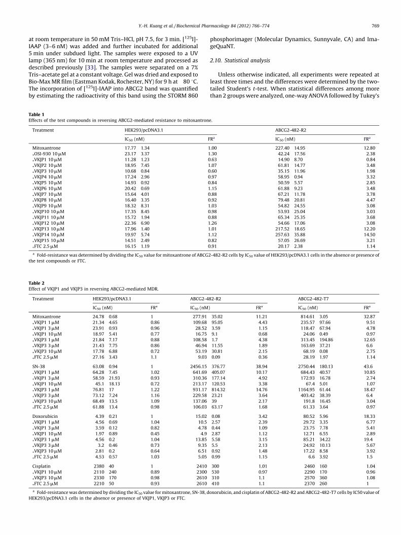
Table 1Effects of the test compounds in reversing ABCG2-mediated resistance to mitoxantron
Treatment HEK293/pcDNA3.1
IC50 (nM) F
Mitoxantrone 17.77 � 1.34 1
+OSI-930 10 mM 23.17 � 3.37 1
+VKJP1 10 mM 11.28 � 1.23 0
+VKJP2 10 mM 18.95 � 7.45 1
+VKJP3 10 mM 10.68 � 0.84 0
+VKJP4 10 mM 17.24 � 2.96 0
+VKJP5 10 mM 14.93 � 0.92 0
+VKJP6 10 mM 20.42 � 0.69 1
+VKJP7 10 mM 15.64 � 4.01 0
+VKJP8 10 mM 16.40 � 3.35 0
+VKJP9 10 mM 18.32 � 8.31 1
+VKJP10 10 mM 17.35 � 8.45 0
+VKJP11 10 mM 15.72 � 1.94 0
+VKJP12 10 mM 22.36 � 6.90 1
+VKJP13 10 mM 17.96 � 1.40 1
+VKJP14 10 mM 19.97 � 5.74 1
+VKJP15 10 mM 14.51 � 2.49 0
+FTC 2.5 mM 16.15 � 1.19 0
a Fold-resistance was determined by dividing the IC50 value for mitoxantrone of ABCG2
the test compounds or FTC.
Table 2Effect of VKJP1 and VKJP3 in reversing ABCG2-mediated MDR.
Treatment HEK293/pcDNA3.1 ABCG2-4
IC50 (nM) FRa IC50 (nM)
Mitoxantrone 24.78 � 0.68 1 277.91 �+VKJP1 1 mM 21.34 � 4.65 0.86 109.68 �+VKJP1 3 mM 23.91 � 0.93 0.96 28.52 �+VKJP1 10 mM 18.97 � 5.41 0.77 16.75 �+VKJP3 1 mM 21.84 � 7.17 0.88 108.58 �+VKJP3 3 mM 21.43 � 7.75 0.86 46.94 �+VKJP3 10 mM 17.78 � 6.88 0.72 53.19 �+FTC 2.5 mM 27.16 � 3.43 1.1 9.03 �
SN-38 63.08 � 0.94 1 2456.15 �+VKJP1 1 mM 64.28 � 7.45 1.02 641.69 �+VKJP1 3 mM 58.59 � 21.93 0.93 310.36 �+VKJP1 10 mM 45.1 � 18.13 0.72 213.17 �+VKJP3 1 mM 76.81 � 17 1.22 931.17 �+VKJP3 3 mM 73.12 � 7.24 1.16 229.58 �+VKJP3 10 mM 68.49 � 13.5 1.09 137.06 �+FTC 2.5 mM 61.88 � 13.4 0.98 106.03 �
Doxorubicin 4.39 � 0.21 1 15.02 �+VKJP1 1 mM 4.56 � 0.69 1.04 10.5 �+VKJP1 3 mM 3.59 � 0.12 0.82 4.78 �+VKJP1 10 mM 1.97 � 0.89 0.45 4.9 �+VKJP3 1 mM 4.56 � 0.2 1.04 13.85 �+VKJP3 3 mM 3.2 � 0.46 0.73 9.35 �+VKJP3 10 mM 2.81 � 0.2 0.64 6.51 �+FTC 2.5 mM 4.53 � 0.57 1.03 5.05 �
Cisplatin 2380 � 40 1 2410 �+VKJP1 10 mM 2110 � 240 0.89 2300 �+VKJP3 10 mM 2330 � 170 0.98 2610 �+FTC 2.5 mM 2210 � 50 0.93 2610 �a Fold-resistance was determined by dividing the IC50 value for mitoxantrone, SN-38, d
HEK293/pcDNA3.1 cells in the absence or presence of VKJP1, VKJP3 or FTC.
phosphorimager (Molecular Dynamics, Sunnyvale, CA) and Ima-geQuaNT.
2.10. Statistical analysis
Unless otherwise indicated, all experiments were repeated atleast three times and the differences were determined by the two-tailed Student’s t-test. When statistical differences among morethan 2 groups were analyzed, one-way ANOVA followed by Tukey’s
e.
ABCG2-482-R2
Ra IC50 (nM) FRa
.00 227.40 � 14.95 12.80
.30 42.24 � 17.56 2.38
.63 14.90 � 8.70 0.84
.07 61.81 � 14.77 3.48
.60 35.15 � 11.96 1.98
.97 58.95 � 0.94 3.32
.84 50.59 � 5.57 2.85
.15 61.88 � 9.23 3.48
.88 67.21 � 11.78 3.78
.92 79.48 � 20.81 4.47
.03 54.82 � 24.55 3.08
.98 53.93 � 25.04 3.03
.88 65.34 � 25.35 3.68
.26 54.66 � 17.06 3.08
.01 217.52 � 18.65 12.20
.12 257.63 � 35.88 14.50
.82 57.05 � 26.69 3.21
.91 20.17 � 2.38 1.14
-482-R2 cells by IC50 value of HEK293/pcDNA3.1 cells in the absence or presence of
82-R2 ABCG2-482-T7
FRa IC50 (nM) FRa
35.02 11.21 814.61 � 3.05 32.87
95.05 4.43 235.57 � 97.66 9.51
3.59 1.15 118.47 � 67.94 4.78
9.1 0.68 24.06 � 0.49 0.97
1.7 4.38 313.45 � 194.86 12.65
11.55 1.89 163.69 � 37.21 6.6
30.81 2.15 68.19 � 0.08 2.75
0.09 0.36 28.19 � 1.97 1.14
376.77 38.94 2750.44 � 180.13 43.6
405.07 10.17 684.43 � 40.57 10.85
177.14 4.92 172.93 � 16.78 2.74
120.53 3.38 67.4 � 5.01 1.07
814.32 14.76 1164.95 � 61.44 18.47
23.21 3.64 403.42 � 38.39 6.4
39 2.17 191.8 � 16.45 3.04
63.17 1.68 61.33 � 3.64 0.97
0.08 3.42 80.52 � 5.96 18.33
2.57 2.39 29.72 � 3.35 6.77
0.44 1.09 23.75 � 7.78 5.41
2.87 1.12 12.71 � 6.55 2.89
5.58 3.15 85.21 � 34.22 19.4
5.5 2.13 24.92 � 10.13 5.67
0.92 1.48 17.22 � 8.58 3.92
0.99 1.15 6.6 � 3.92 1.5
300 1.01 2460 � 160 1.04
530 0.97 2290 � 170 0.96
310 1.1 2570 � 360 1.08
410 1.1 2370 � 260 1
oxorubicin, and cisplatin of ABCG2-482-R2 and ABCG2-482-T7 cells by IC50 value of
Y.-H. Kuang et al. / Biochemical Pharmacology 84 (2012) 766–774770
multiple comparison tests were performed. Results were pre-sented as mean � standard deviations (SD). The statistical signifi-cance was determined to be P < 0.05.
3. Results
3.1. Structure of VKJP1 and VKJP3
A series of OSI-930 analogues 1–15 (Fig. 1) were successfullydesigned, synthesized, and evaluated for their reversal effect ofABC tranpsorter-mediated MDR.
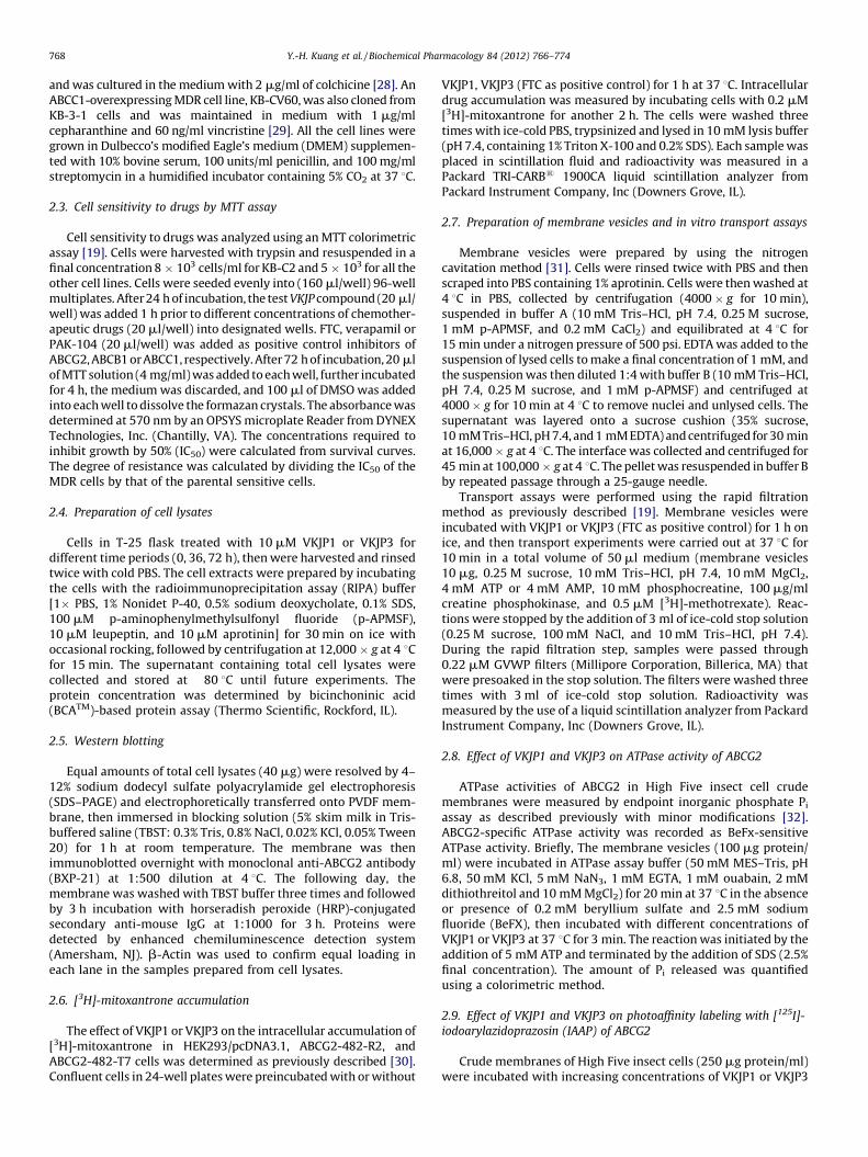
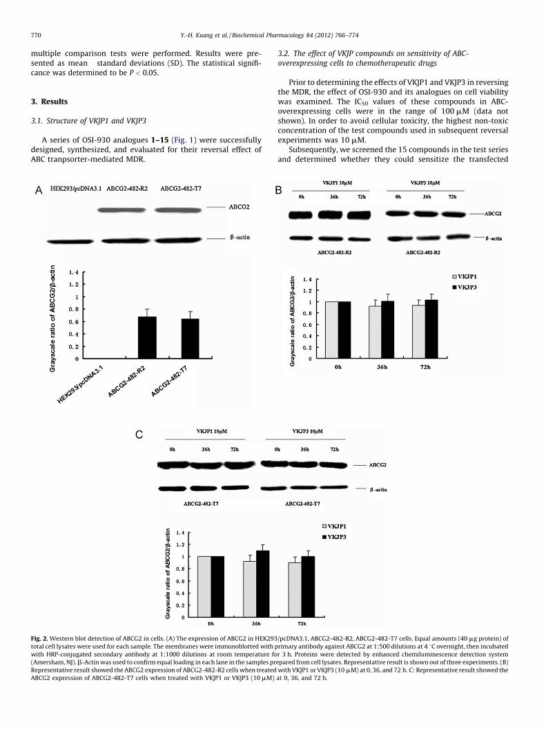
Fig. 2. Western blot detection of ABCG2 in cells. (A) The expression of ABCG2 in HEK293
total cell lysates were used for each sample. The membranes were immunoblotted with p
with HRP-conjugated secondary antibody at 1:1000 dilutions at room temperature fo
(Amersham, NJ). b-Actin was used to confirm equal loading in each lane in the samples pr
Representative result showed the ABCG2 expression of ABCG2-482-R2 cells when treated
ABCG2 expression of ABCG2-482-T7 cells when treated with VKJP1 or VKJP3 (10 mM)
3.2. The effect of VKJP compounds on sensitivity of ABC-
overexpressing cells to chemotherapeutic drugs
Prior to determining the effects of VKJP1 and VKJP3 in reversingthe MDR, the effect of OSI-930 and its analogues on cell viabilitywas examined. The IC50 values of these compounds in ABC-overexpressing cells were in the range of 100 mM (data notshown). In order to avoid cellular toxicity, the highest non-toxicconcentration of the test compounds used in subsequent reversalexperiments was 10 mM.
Subsequently, we screened the 15 compounds in the test seriesand determined whether they could sensitize the transfected
/pcDNA3.1, ABCG2-482-R2, ABCG2-482-T7 cells. Equal amounts (40 mg protein) of
rimary antibody against ABCG2 at 1:500 dilutions at 4 8C overnight, then incubated
r 3 h. Proteins were detected by enhanced chemiluminescence detection system
epared from cell lysates. Representative result is shown out of three experiments. (B)
with VKJP1 or VKJP3 (10 mM) at 0, 36, and 72 h. C: Representative result showed the
at 0, 36, and 72 h.
Y.-H. Kuang et al. / Biochemical Pharmacology 84 (2012) 766–774 771
wild-type ABCG2-overexpressing MDR cells to certain chemo-therapeutic drugs. VKJP1 and VKJP3 were the first two in the rankorder of potencies for increasing the cytotoxicity of mitoxantroneamong all of the members of the test series. For this reason, VKJP1and VKJP3 were chosen for further study. Compound 13 had nosignificant reversing ability, while compound 14 was found toprotect cells from cytotoxicity by mitoxantrone (Table 1). Asshown in Table 2, VKJP1 or VKJP3 at 1, 3 and 10 mM,concentration-dependently decreased the IC50 values of ABCG2substrates including mitoxantrone, SN-38, and doxorubicin of thetransfected wild-type (R2) or mutant (T7) ABCG2-overexpressingcells. VKJP1 was shown to be the most potent inhibitor of ABCG2than VKJP3 at all concentrations. The magnitude of the sensitiza-tion produced by 10 mM VKJP1 was similar to that induced by theknown specific ABCG2 inhibitor FTC at 2.5 mM. In addition, 10 mM
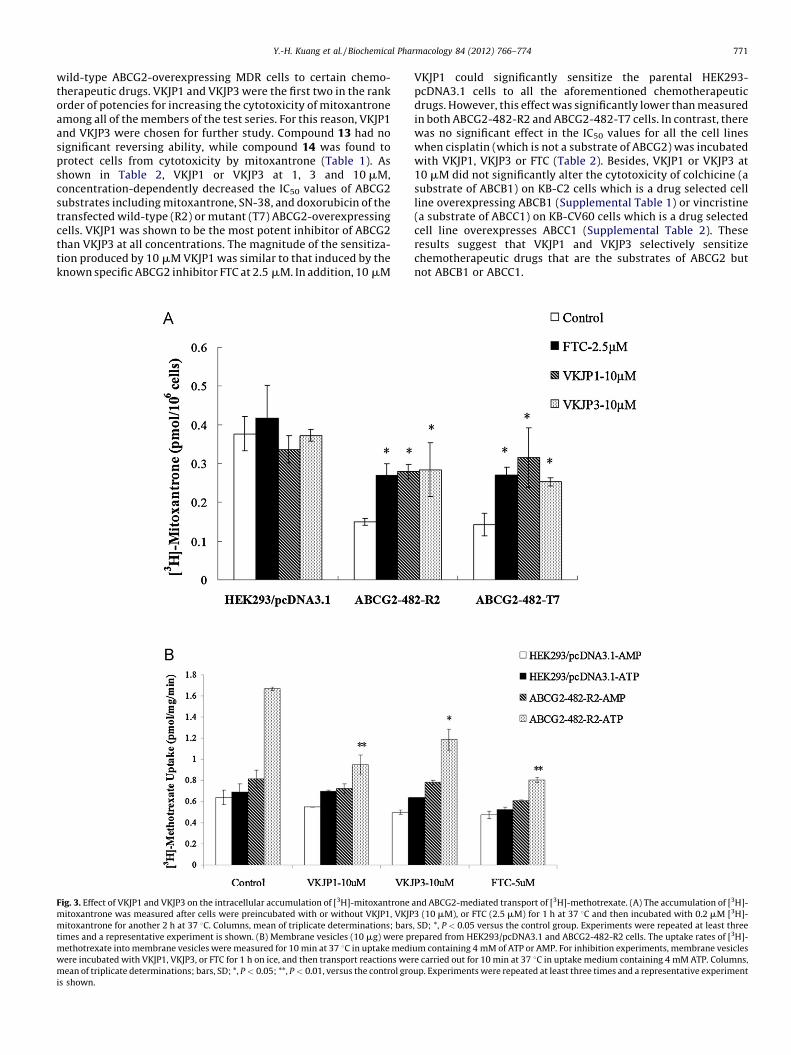
Fig. 3. Effect of VKJP1 and VKJP3 on the intracellular accumulation of [3H]-mitoxantrone
mitoxantrone was measured after cells were preincubated with or without VKJP1, VKJP
mitoxantrone for another 2 h at 37 8C. Columns, mean of triplicate determinations; bars
times and a representative experiment is shown. (B) Membrane vesicles (10 mg) were pr
methotrexate into membrane vesicles were measured for 10 min at 37 8C in uptake medi
were incubated with VKJP1, VKJP3, or FTC for 1 h on ice, and then transport reactions wer
mean of triplicate determinations; bars, SD; *, P < 0.05; **, P < 0.01, versus the control gro
is shown.
VKJP1 could significantly sensitize the parental HEK293-pcDNA3.1 cells to all the aforementioned chemotherapeuticdrugs. However, this effect was significantly lower than measuredin both ABCG2-482-R2 and ABCG2-482-T7 cells. In contrast, therewas no significant effect in the IC50 values for all the cell lineswhen cisplatin (which is not a substrate of ABCG2) was incubatedwith VKJP1, VKJP3 or FTC (Table 2). Besides, VKJP1 or VKJP3 at10 mM did not significantly alter the cytotoxicity of colchicine (asubstrate of ABCB1) on KB-C2 cells which is a drug selected cellline overexpressing ABCB1 (Supplemental Table 1) or vincristine(a substrate of ABCC1) on KB-CV60 cells which is a drug selectedcell line overexpresses ABCC1 (Supplemental Table 2). Theseresults suggest that VKJP1 and VKJP3 selectively sensitizechemotherapeutic drugs that are the substrates of ABCG2 butnot ABCB1 or ABCC1.
and ABCG2-mediated transport of [3H]-methotrexate. (A) The accumulation of [3H]-
3 (10 mM), or FTC (2.5 mM) for 1 h at 37 8C and then incubated with 0.2 mM [3H]-
, SD; *, P < 0.05 versus the control group. Experiments were repeated at least three
epared from HEK293/pcDNA3.1 and ABCG2-482-R2 cells. The uptake rates of [3H]-
um containing 4 mM of ATP or AMP. For inhibition experiments, membrane vesicles
e carried out for 10 min at 37 8C in uptake medium containing 4 mM ATP. Columns,
up. Experiments were repeated at least three times and a representative experiment
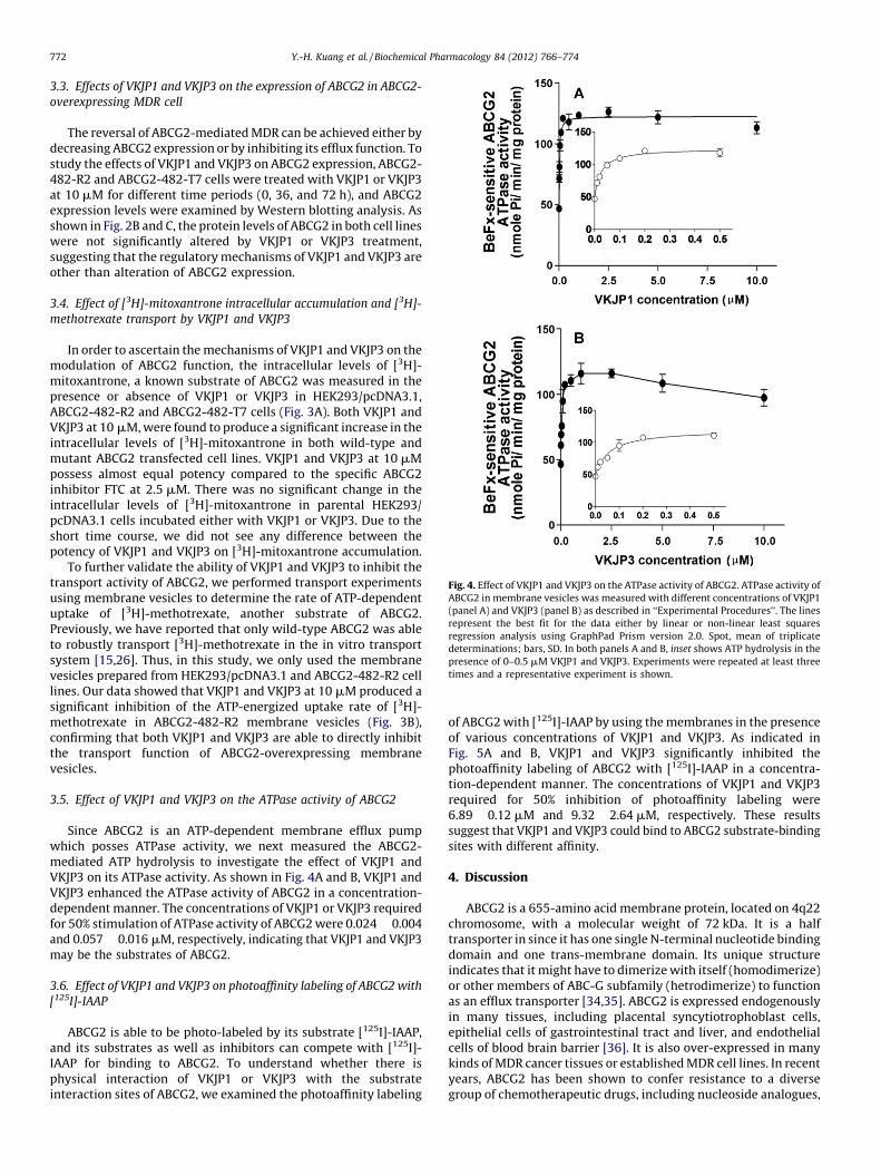
Fig. 4. Effect of VKJP1 and VKJP3 on the ATPase activity of ABCG2. ATPase activity of
ABCG2 in membrane vesicles was measured with different concentrations of VKJP1
(panel A) and VKJP3 (panel B) as described in ‘‘Experimental Procedures’’. The lines
represent the best fit for the data either by linear or non-linear least squares
regression analysis using GraphPad Prism version 2.0. Spot, mean of triplicate
determinations; bars, SD. In both panels A and B, inset shows ATP hydrolysis in the
presence of 0–0.5 mM VKJP1 and VKJP3. Experiments were repeated at least three
times and a representative experiment is shown.
Y.-H. Kuang et al. / Biochemical Pharmacology 84 (2012) 766–774772
3.3. Effects of VKJP1 and VKJP3 on the expression of ABCG2 in ABCG2-
overexpressing MDR cell
The reversal of ABCG2-mediated MDR can be achieved either bydecreasing ABCG2 expression or by inhibiting its efflux function. Tostudy the effects of VKJP1 and VKJP3 on ABCG2 expression, ABCG2-482-R2 and ABCG2-482-T7 cells were treated with VKJP1 or VKJP3at 10 mM for different time periods (0, 36, and 72 h), and ABCG2expression levels were examined by Western blotting analysis. Asshown in Fig. 2B and C, the protein levels of ABCG2 in both cell lineswere not significantly altered by VKJP1 or VKJP3 treatment,suggesting that the regulatory mechanisms of VKJP1 and VKJP3 areother than alteration of ABCG2 expression.
3.4. Effect of [3H]-mitoxantrone intracellular accumulation and [3H]-
methotrexate transport by VKJP1 and VKJP3
In order to ascertain the mechanisms of VKJP1 and VKJP3 on themodulation of ABCG2 function, the intracellular levels of [3H]-mitoxantrone, a known substrate of ABCG2 was measured in thepresence or absence of VKJP1 or VKJP3 in HEK293/pcDNA3.1,ABCG2-482-R2 and ABCG2-482-T7 cells (Fig. 3A). Both VKJP1 andVKJP3 at 10 mM, were found to produce a significant increase in theintracellular levels of [3H]-mitoxantrone in both wild-type andmutant ABCG2 transfected cell lines. VKJP1 and VKJP3 at 10 mMpossess almost equal potency compared to the specific ABCG2inhibitor FTC at 2.5 mM. There was no significant change in theintracellular levels of [3H]-mitoxantrone in parental HEK293/pcDNA3.1 cells incubated either with VKJP1 or VKJP3. Due to theshort time course, we did not see any difference between thepotency of VKJP1 and VKJP3 on [3H]-mitoxantrone accumulation.
To further validate the ability of VKJP1 and VKJP3 to inhibit thetransport activity of ABCG2, we performed transport experimentsusing membrane vesicles to determine the rate of ATP-dependentuptake of [3H]-methotrexate, another substrate of ABCG2.Previously, we have reported that only wild-type ABCG2 was ableto robustly transport [3H]-methotrexate in the in vitro transportsystem [15,26]. Thus, in this study, we only used the membranevesicles prepared from HEK293/pcDNA3.1 and ABCG2-482-R2 celllines. Our data showed that VKJP1 and VKJP3 at 10 mM produced asignificant inhibition of the ATP-energized uptake rate of [3H]-methotrexate in ABCG2-482-R2 membrane vesicles (Fig. 3B),confirming that both VKJP1 and VKJP3 are able to directly inhibitthe transport function of ABCG2-overexpressing membranevesicles.
3.5. Effect of VKJP1 and VKJP3 on the ATPase activity of ABCG2
Since ABCG2 is an ATP-dependent membrane efflux pumpwhich posses ATPase activity, we next measured the ABCG2-mediated ATP hydrolysis to investigate the effect of VKJP1 andVKJP3 on its ATPase activity. As shown in Fig. 4A and B, VKJP1 andVKJP3 enhanced the ATPase activity of ABCG2 in a concentration-dependent manner. The concentrations of VKJP1 or VKJP3 requiredfor 50% stimulation of ATPase activity of ABCG2 were 0.024 � 0.004and 0.057 � 0.016 mM, respectively, indicating that VKJP1 and VKJP3may be the substrates of ABCG2.
3.6. Effect of VKJP1 and VKJP3 on photoaffinity labeling of ABCG2 with
[125I]-IAAP
ABCG2 is able to be photo-labeled by its substrate [125I]-IAAP,and its substrates as well as inhibitors can compete with [125I]-IAAP for binding to ABCG2. To understand whether there isphysical interaction of VKJP1 or VKJP3 with the substrateinteraction sites of ABCG2, we examined the photoaffinity labeling
of ABCG2 with [125I]-IAAP by using the membranes in the presenceof various concentrations of VKJP1 and VKJP3. As indicated inFig. 5A and B, VKJP1 and VKJP3 significantly inhibited thephotoaffinity labeling of ABCG2 with [125I]-IAAP in a concentra-tion-dependent manner. The concentrations of VKJP1 and VKJP3required for 50% inhibition of photoaffinity labeling were6.89 � 0.12 mM and 9.32 � 2.64 mM, respectively. These resultssuggest that VKJP1 and VKJP3 could bind to ABCG2 substrate-bindingsites with different affinity.
4. Discussion
ABCG2 is a 655-amino acid membrane protein, located on 4q22chromosome, with a molecular weight of 72 kDa. It is a halftransporter in since it has one single N-terminal nucleotide bindingdomain and one trans-membrane domain. Its unique structureindicates that it might have to dimerize with itself (homodimerize)or other members of ABC-G subfamily (hetrodimerize) to functionas an efflux transporter [34,35]. ABCG2 is expressed endogenouslyin many tissues, including placental syncytiotrophoblast cells,epithelial cells of gastrointestinal tract and liver, and endothelialcells of blood brain barrier [36]. It is also over-expressed in manykinds of MDR cancer tissues or established MDR cell lines. In recentyears, ABCG2 has been shown to confer resistance to a diversegroup of chemotherapeutic drugs, including nucleoside analogues,
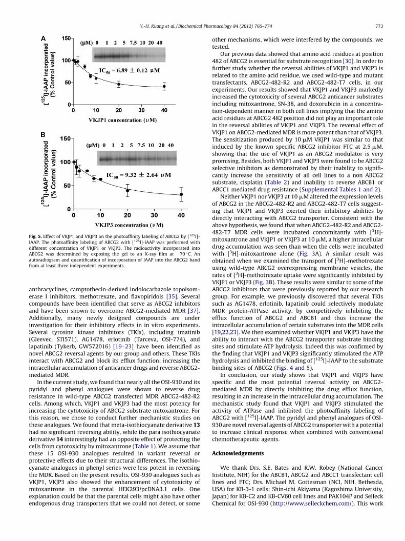
Fig. 5. Effect of VKJP1 and VKJP3 on the photoaffinity labeling of ABCG2 by [125I]-
IAAP. The photoaffinity labeling of ABCG2 with [125I]-IAAP was performed with
different concentration of VKJP1 or VKJP3. The radioactivity incorporated into
ABCG2 was determined by exposing the gel to an X-ray film at �70 8C. An
autoradiogram and quantification of incorporation of IAAP into the ABCG2 band
from at least three independent experiments.
Y.-H. Kuang et al. / Biochemical Pharmacology 84 (2012) 766–774 773
anthracyclines, camptothecin-derived indolocarbazole topoisom-erase I inhibitors, methotrexate, and flavopiridols [35]. Severalcompounds have been identified that serve as ABCG2 inhibitorsand have been shown to overcome ABCG2-mediated MDR [37].Additionally, many newly designed compounds are underinvestigation for their inhibitory effects in in vitro experiments.Several tyrosine kinase inhibitors (TKIs), including imatinib(Gleevec, STI571), AG1478, erlotinib (Tarceva, OSI-774), andlapatinib (Tykerb, GW572016) [19–23] have been identified asnovel ABCG2 reversal agents by our group and others. These TKIsinteract with ABCG2 and block its efflux function; increasing theintracellular accumulation of anticancer drugs and reverse ABCG2-mediated MDR.
In the current study, we found that nearly all the OSI-930 and itspyridyl and phenyl analogues were shown to reverse drugresistance in wild-type ABCG2 transfected MDR ABCG2-482-R2cells. Among which, VKJP1 and VKJP3 had the most potency forincreasing the cytotoxicity of ABCG2 substrate mitoxantrone. Forthis reason, we chose to conduct further mechanistic studies onthese analogues. We found that meta-isothiocyanate derivative 13had no significant reversing ability, while the para isothiocyanatederivative 14 interestingly had an opposite effect of protecting thecells from cytotoxicity by mitoxantrone (Table 1). We assume thatthese 15 OSI-930 analogues resulted in variant reversal orprotective effects due to their structural differences. The isothio-cyanate analogues in phenyl series were less potent in reversingthe MDR. Based on the present results, OSI-930 analogues such asVKJP1, VKJP3 also showed the enhancement of cytotoxicity ofmitoxantrone in the parental HEK293/pcDNA3.1 cells. Oneexplanation could be that the parental cells might also have otherendogenous drug transporters that we could not detect, or some
other mechanisms, which were interfered by the compounds, wetested.
Our previous data showed that amino acid residues at position482 of ABCG2 is essential for substrate recognition [30]. In order tofurther study whether the reversal abilities of VKJP1 and VKJP3 isrelated to the amino acid residue, we used wild-type and mutanttransfectants, ABCG2-482-R2 and ABCG2-482-T7 cells, in ourexperiments. Our results showed that VKJP1 and VKJP3 markedlyincreased the cytotoxicity of several ABCG2 anticancer substratesincluding mitoxantrone, SN-38, and doxorubicin in a concentra-tion-dependent manner in both cell lines implying that the aminoacid residues at ABCG2 482 position did not play an important rolein the reversal abilities of VKJP1 and VKJP3. The reversal effect ofVKJP1 on ABCG2-mediated MDR is more potent than that of VKJP3.The sensitization produced by 10 mM VKJP1 was similar to thatinduced by the known specific ABCG2 inhibitor FTC at 2.5 mM,showing that the use of VKJP1 as an ABCG2 modulator is verypromising. Besides, both VKJP1 and VKJP3 were found to be ABCG2selective inhibitors as demonstrated by their inability to signifi-cantly increase the sensitivity of all cell lines to a non ABCG2substrate, cisplatin (Table 2) and inability to reverse ABCB1 orABCC1 mediated drug resistance (Supplemental Tables 1 and 2).
Neither VKJP1 nor VKJP3 at 10 mM altered the expression levelsof ABCG2 in the ABCG2-482-R2 and ABCG2-482-T7 cells suggest-ing that VKJP1 and VKJP3 exerted their inhibitory abilities bydirectly interacting with ABCG2 transporter. Consistent with theabove hypothesis, we found that when ABCG2-482-R2 and ABCG2-482-T7 MDR cells were incubated concomitantly with [3H]-mitoxantrone and VKJP1 or VKJP3 at 10 mM, a higher intracellulardrug accumulation was seen than when the cells were incubatedwith [3H]-mitoxantrone alone (Fig. 3A). A similar result wasobtained when we examined the transport of [3H]-methotrexateusing wild-type ABCG2 overexpressing membrane vesicles, therates of [3H]-methotrexate uptake were significantly inhibited byVKJP1 or VKJP3 (Fig. 3B). These results were similar to some of theABCG2 inhibitors that were previously reported by our researchgroup. For example, we previously discovered that several TKIssuch as AG1478, erlotinib, lapatinib could selectively modulateMDR protein-ATPase activity, by competitively inhibiting theefflux function of ABCG2 and ABCB1 and thus increase theintracellular accumulation of certain substrates into the MDR cells[19,22,23]. We then examined whether VKJP1 and VKJP3 have theability to interact with the ABCG2 transporter substrate bindingsites and stimulate ATP hydrolysis. Indeed this was confirmed bythe finding that VKJP1 and VKJP3 significantly stimulated the ATPhydrolysis and inhibited the binding of [125I]-IAAP to the substratebinding sites of ABCG2 (Figs. 4 and 5).
In conclusion, our study shows that VKJP1 and VKJP3 havespecific and the most potential reversal activity on ABCG2-mediated MDR by directly inhibiting the drug efflux function,resulting in an increase in the intracellular drug accumulation. Themechanistic study found that VKJP1 and VKJP3 stimulated theactivity of ATPase and inhibited the photoaffinity labeling ofABCG2 with [125I]-IAAP. The pyridyl and phenyl analogues of OSI-930 are novel reversal agents of ABCG2 transporter with a potentialto increase clinical response when combined with conventionalchemotherapeutic agents.
Acknowledgements
We thank Drs. S.E. Bates and R.W. Robey (National CancerInstitute, NIH) for the ABCB1, ABCG2 and ABCC1 transfectant celllines and FTC; Drs. Michael M. Gottesman (NCI, NIH, Bethesda,USA) for KB-3-1 cells; Shin-ichi Akiyama (Kagoshima University,Japan) for KB-C2 and KB-CV60 cell lines and PAK104P and SelleckChemical for OSI-930 (http://www.selleckchem.com/). This work
Y.-H. Kuang et al. / Biochemical Pharmacology 84 (2012) 766–774774
was supported by funds from NIH R15 No. 1R15CA143701 (Z.S.Chen), St. John’s University Seed Grant No. 579-1110, (Z.S. Chen),the National Natural Science Foundation of China No. 81000690(Y.H. Kuang), the Millions of Strategic Project of Xiang Ya Hospital(Y.H. Kuang), the Freedom Explore Program of Central SouthUniversity (Y.H. Kuang).
Appendix A. Supplementary data
Supplementary data associated with this article can be found, inthe online version, at http://dx.doi.org/10.1016/j.bcp.2012.06.019.
References
[1] Pratt WB, Ruddon RW, editors. The anticancer drugs. New York: OxfordUniversity Press; 1979.
[2] Zhou J. Multidrug resistance in cancer. Humana Press; 2010.[3] Gottesman MM. Mechanisms of cancer drug resistance. Annu Rev Med
2002;53:615–27.[4] Beck WT. Mechanisms of multidrug resistance in human tumor cells. The roles
of P-glycoprotein, DNA topoisomerase II, and other factors. Cancer Treat Rev1990;17(Suppl A):11–20.
[5] Gottesman MM, Fojo T, Bates SE. Multidrug resistance in cancer: role of ATP-dependent transporters. Nat Rev Cancer 2002;2:48–58.
[6] Wu CP, Hsieh CH, Wu YS. The emergence of drug transporter-mediatedmultidrug resistance to cancer chemotherapy. Mol Pharmacol 2011;8:1996–2011.
[7] Dean M, Annilo T. Evolution of the ATP-binding cassette (ABC) transportersuperfamily in vertebrates. Annu Rev Genomics Hum Genet 2005;6:123–42.
[8] Juliano RL, Ling V. A surface glycoprotein modulating drug permeability inChinese hamster ovary cell mutants. Biochim Biophys Acta 1976;455:152–62.
[9] Dean M, Rzhetsky A, Allikmets R. The human ATP-binding cassette (ABC)transporter superfamily. Genome Res 2001;11(July):1156–66.
[10] Kruh GD, Belinsky MG. The MRP family of drug efflux pumps. Oncogene2003;22:7537–52.
[11] Doyle LA, Yang W, Abruzzo LV, Krogmann T, Gao Y, Rishi AK, et al. A multidrugresistance transporter from human MCF-7 breast cancer cells. Proc Natl AcadSci USA 1998;95:15665–70.
[12] Maliepaard M, van Gastelen MA, de Jong LA, Pluim D, van Waardenburg RC,Ruevekamp-Helmers MC, et al. Overexpression of the BCRP/MXR/ABCP gene ina topotecan-selected ovarian tumor cell line. Cancer Res 1999;59:4559–63.
[13] Tsuruo T, Iida H, Tsukagoshi S, Sakurai Y. Overcoming of vincristine resistancein P388 leukemia in vivo and in vitro through enhanced cytotoxicity ofvincristine and vinblastine by verapamil. Cancer Res 1981;41:1967–72.
[14] Schinkel AH, Jonker JW. Mammalian drug efflux transporters of the ATPbinding cassette (ABC) family: an overview. Adv Drug Deliv Rev 2003;55:3–29.
[15] Tan B, Piwnica-Worms D, Ratner L. Multidrug resistance transporters andmodulation. Curr Opin Oncol 2000;12:450–8.
[16] Robert J, Jarry C. Multidrug resistance reversal agents. J Med Chem 2003;6(No-vember (46)):4805–17.
[17] Dantzig AH, de Alwis DP, Burgess M. Considerations in the design anddevelopment of transport inhibitors as adjuncts to drug therapy. Adv DrugDeliv Rev 2003;55:133–50.
[18] Chen ZS, Kawabe T, Ono M, Aoki S, Sumizawa T, Furukawa T, et al. Effect ofmultidrug resistance-reversing agents on transporting activity of human
canalicular multispecific organic anion transporter. Mol Pharmacol 1999;56:1219–28.
[19] Shi Z, Peng XX, Kim IW, Shukla S, Si QS, Robey RW, et al. Erlotinib (Tarceva, OSI-774) antagonizes ATP-binding cassette subfamily B member 1 and ATP-binding cassette subfamily G member 2-mediated drug resistance. CancerRes 2007;67:11012–20.
[20] Liu W, Baer MR, Bowman MJ, Pera P, Zheng X, Morgan J, et al. The tyrosinekinase inhibitor imatinib mesylate enhances the efficacy of photodynamictherapy by inhibiting ABCG2. Clin Cancer Res 2007;13:2463–70.
[21] Houghton PJ, Germain GS, Harwood FC, Schuetz JD, Stewart CF, Buchdunger E,et al. Imatinib mesylate is a potent inhibitor of the ABCG2 (BCRP) transporter andreverses resistance to topotecan and SN-38 in vitro. Cancer Res 2004;64:2333–7.
[22] Shi Z, Tiwari AK, Shukla S, Robey RW, Kim IW, Parmar S, et al. Inhibiting thefunction of ABCB1 and ABCG2 by the EGFR tyrosine kinase inhibitor AG1478.Biochem Pharmacol 2009;77:781–93.
[23] Dai CL, Tiwari AK, Wu CP, Su XD, Wang SR, Liu DG, et al. Lapatinib (Tykerb,GW572016) reverses multidrug resistance in cancer cells by inhibiting theactivity of ATP-binding cassette subfamily B member 1 and G member 2.Cancer Res 2008;68:7905–14.
[24] Garton AJ, Crew AP, Franklin M, Cooke AR, Wynne GM, Castaldo L, et al. OSI-930: a novel selective inhibitor of Kit and kinase insert domain receptortyrosine kinases with antitumor activity in mouse xenograft models. CancerRes 2006;66:1015–24.
[25] Petti F, Thelemann A, Kahler J, McCormack S, Castaldo L, Hunt T, et al. Temporalquantitation of mutant Kit tyrosine kinase signaling attenuated by a novelthiophene kinase inhibitor OSI-930. Mol Cancer Ther 2005;4:1186–97.
[26] Patel JP, Kuang YH, Chen ZS, Korlipara VL. Inhibition of c-Kit, VEGFR-2 (KDR),and ABCG2 by analogues of OSI-930. Bioorg Med Chem Lett 2011;21(21):6495–9.
[27] Robey RW, Honjo Y, Morisaki K, Nadjem TA, Runge S, Risbood M, et al.Mutations at amino-acid 482 in the ABCG2 gene affect substrate and antago-nist specificity. Br J Cancer 2003;89:1971–8.
[28] Akiyama S, Fojo A, Hanover JA, Pastan I, Gottesman MM. Isolation and geneticcharacterization of human KB cell lines resistant to multiple drugs. Somat CellMol Genet 1985;11:117–26.
[29] Nagayama S, Chen ZS, Kitazono M, Takebayashi Y, Niwa K, Yamada K, et al.Increased sensitivity to vincristine of MDR cells by the leukotriene D4 receptorantagonist, ONO-1078. Cancer Lett 1998;130:175–82.
[30] Chen ZS, Robey RW, Belinsky MG, Shchaveleva I, Ren XQ, Sugimoto Y. Trans-port of methotrexate, methotrexate polyglutamates, and 17beta-estradiol 17-(beta-D-glucuronide) by ABCG2: effects of acquired mutations at R482 onmethotrexate transport. Cancer Res 2003;63:4048–54.
[31] Cornwell MM, Gottesman MM, Pastan IH. Increased vinblastine binding tomembrane vesicles from multidrug-resistant KB cells. J Biol Chem 1986;261:7921–8.
[32] Ambudkar SV. Drug-stimulatable ATPase activity in crude membranes of humanMDR1-transfected mammalian cells. Methods Enzymol 1998;292:504–14.
[33] Sauna ZE, Ambudkar SV. Evidence for a requirement for ATP hydrolysis at twodistinct steps during a single turnover of the catalytic cycle of human P-glycoprotein. Proc Natl Acad Sci USA 2000;97:2515–20.
[34] Ejendal KF, Hrycyna CA. Multidrug resistance and cancer: the role of thehuman ABC transporter ABCG2. Curr Protein Pept Sci 2002;503:503–11.
[35] Mao Q, Unadkat JD. Role of the breast cancer resistance protein (ABCG2) indrug transport. AAPS J 2005;7:E118–33.
[36] Deeley RG, Westlake C, Cole SP. Transmembrane transport of endo- andxenobiotics by mammalian ATP-binding cassette multidrug resistance pro-teins. Physiol Rev 2006;86:849–99.
[37] Allen JD, van Loevezijn A, Lakhai JM, van der Valk M, van Tellingen O, Reid G,et al. Potent and specific inhibition of the breast cancer resistance proteinmultidrug transporter in vitro and in mouse intestine by a novel analogue offumitremorgin C. Mol Cancer Ther 2002;1:417–25.