Orthopedic Trauma - - RN.org® · Orthopedic Trauma ® Reviewed February, 2018, Expires February,...
32
Orthopedic Trauma WWW.RN.ORG® Reviewed February, 2018, Expires February, 2020 Provider Information and Specifics available on our Website Unauthorized Distribution Prohibited ©2018 RN.ORG®, S.A., RN.ORG®, LLC By Wanda Lockwood, RN, BA, MA The purpose of this course is to outline different types of injuries associated with orthopedic trauma, diagnosis, treatments, as well as common complications. Upon completion of this course, the healthcare providers should be able to: • Describe 5 types of bones and give examples. • Diffentiate between ligaments and tendons. • Discuss the 3 degrees of sprains and treatments. • Discuss the 3 degrees of strains and treatments. • Discuss symptoms and treatment of bursitis. • Discuss 4 common dislocations and specific concerns. • Identify 9 types of fractures. • Discuss 4 types of spinal fractures. • Discuss 4 types of skull fractures. • Discuss the 3 grades of open fractures. • Discuss 5 types of alignment. • Discuss diagnosis of fractures. • Explain the Ottawa rules for x-rays of the ankle, foot, and knee. • Discuss blood loss associated with fracture of at least 5 major bones. • Discuss fat embolism syndrome, including symptoms and treatment. • Explain 3 types of compartment syndrome. • Describe the 5Ps associated with compartment syndrome. • Explain methods of assessing neurological function in the radial, ulnar, tibial, and peroneal nerves. • Explain the 3 most common types of infection associated with fractures. • Discuss symptoms of pulmonary emboli. • Discuss avascular necrosis. Purpose Goals
Transcript of Orthopedic Trauma - - RN.org® · Orthopedic Trauma ® Reviewed February, 2018, Expires February,...

Orthopedic Trauma WWW.RN.ORG®
Reviewed February, 2018, Expires February, 2020 Provider Information and Specifics available on our Website
Unauthorized Distribution Prohibited
©2018 RN.ORG®, S.A., RN.ORG®, LLC By Wanda Lockwood, RN, BA, MA
The purpose of this course is to outline different types of injuries associated with orthopedic trauma, diagnosis,
treatments, as well as common complications.
Upon completion of this course, the healthcare providers should be able to:
• Describe 5 types of bones and give examples.
• Diffentiate between ligaments and tendons. • Discuss the 3 degrees of sprains and treatments.
• Discuss the 3 degrees of strains and treatments.
• Discuss symptoms and treatment of bursitis. • Discuss 4 common dislocations and specific concerns.
• Identify 9 types of fractures. • Discuss 4 types of spinal fractures.
• Discuss 4 types of skull fractures. • Discuss the 3 grades of open fractures.
• Discuss 5 types of alignment. • Discuss diagnosis of fractures.
• Explain the Ottawa rules for x-rays of the ankle, foot, and knee. • Discuss blood loss associated with fracture of at least 5 major
bones. • Discuss fat embolism syndrome, including symptoms and
treatment. • Explain 3 types of compartment syndrome.
• Describe the 5Ps associated with compartment syndrome.
• Explain methods of assessing neurological function in the radial, ulnar, tibial, and peroneal nerves.
• Explain the 3 most common types of infection associated with fractures.
• Discuss symptoms of pulmonary emboli. • Discuss avascular necrosis.
Purpose
Goals

Introduction The number of bones in the human body changes with age. Infants are
born with over 270 bones, but some of these fuse over time, so adults have about 206.
There are 5 types of bones:
• Long bones: Body of the bone is longer than the width, and there are growth plates at both ends (includes femur, humerus,
tibia, fibula, radius, metacarpals, metatarsals, and phalanges). • Flat bones: Flat plates that protect organs and provide
attachment for muscles (includes scapulae, sternum, pelvis, cranium, and ribs. In adults, flat bones produce the largest
number of red blood cells.
• Short bones: Length and width are similar. They provide support and stability (incudes carpals and tarsals).
• Irregular bones: Non-uniform bones (includes vertebrae, sacrum, and mandible).
• Sesamoid bones: Short irregular bones imbedded in a tendon (includes the patella and some small bones in the hands and
feet).
All of these bones are susceptible to trauma as are supporting structures, such as muscles, bursa, ligaments, and tendons. Each type
of orthopedic injury presents different signs and symptoms and challenges for those providing care. Musculoskeletal injuries are one of
the most common, bringing may people to emergency rooms. Most injuries are not life threatening although they certainly can be.
Although treatment may vary widely—splints, casts, surgical repair—depending on the location and type of injury, a number of basic
principles must be considered when caring for those with orthopedic trauma.


Orthopedic trauma is almost always associated with contusions, which are soft tissue injuries that result in hemorrhage into the tissue
because of injury to small vessels, producing ecchymosis and swelling.

Sprains, strains, bursitis, & dislocations Bones are not the only concern with orthopedic trauma. Orthopedic trauma may result in injury to a number of other different injuries.
Ligaments connect bone to bone Tendons connect muscles to
bone
Ligaments may become stretched, strained, torn, or
ruptured. Damage to a ligament is referred to as a sprain.
A sprain results from a traumatic injury that damages a joint and
results in a partial or complete rupture of supporting ligaments. The most common cause is a fall that involves twisting or wrenching of the
joint. In addition to injury to the ligament, surrounding soft tissue is
often damaged with torn blood vessels causing bleeding into the tissue, bruising, and edema.
Sprains

Other typical signs and symptoms of a sprain
include joint tenderness as well as pain on
movement or unstable joint. Pain usually
increased two to three hours following injury.
Sprains may be associated with
fractures, and a torn ligament may pull fragments of bone out of place, so an x-ray of the
joint to rule out fracture is necessary.
Classification of sprains
First
degree
This results from minor stretching of a ligament, so pain is
usually mild and range of motion remains intact with fairly
good stability of the joint. Edema is usually mild but may be more pronounced, depending on associated injury to vessels.
Hemorrhage is usually minimal.
Second
degree
This results from partial rupture of the ligaments, so
symptoms may vary widely depending on the degree of associated injuries. Pain is more pronounced and range of
motion is limited because of pain. Edema is usually present along with bruising. The joint may be somewhat unstable.
Third
degree
This results from complete rupture of the ligament. Pain is
usually severe although, if nerves are severed, localized pain may not be evident because of numbness. Range of motion
is decreased. Edema and bruising are generally evident in varying degrees, and the affected joint is usually unstable.
Because ligaments are relatively avascular, any injury to a ligament
tends to be slow healing. If larger ligaments are injured, they must be
protected until the scar maturates, and this usually requires 8 to 16 weeks.
Treatment varies according to the muscles and/or tendons affected
and the degree of injury, but commonly includes: • RICE (rest, compress, ice, elevate) therapy immediately (ice
applied 20 minutes out of every hour) to reduce edema for the first 24 hours followed by contrast baths for 1-2 weeks:

o Warm water (100 to 105F for 4 minutes to promote
circulation followed by cold water (45 to 60 F) for one
minute to prevent increased swelling. This alternation is continued for 15 minutes two to four times daily.
• Avoidance of weight bearing until pain and edema decreases. • NSAIDs to relieve inflammation and discomfort.
• 2nd degree sprains may require splint to stabilize and protect the joint.
• 3rd degree sprains may require cast immobilization for 2-3 weeks or surgical repair in severe cases.
• Physical therapy for strengthening, ROM, and balance exercises. • Ultrasound and laser treatment may be used to reduce
inflammation.
A strain is often referred to as a “pulled muscle” although it can result from tiny tears in the muscle or tendon (most
common), often related to excess stress or overuse but
may also result from blunt trauma. Sport activities, such as football and soccer, may result in overstretching that causes strain, as well as
failing to adequately warm up prior to exercise. Strains are fairly common in the back, ankle, neck, and hamstrings. Because the
ligaments provide stability and mobility, a torn ligament results in loss of functional ability.
Trauma to a tendon may result in
tendonitis, tear, or rupture.
Strains
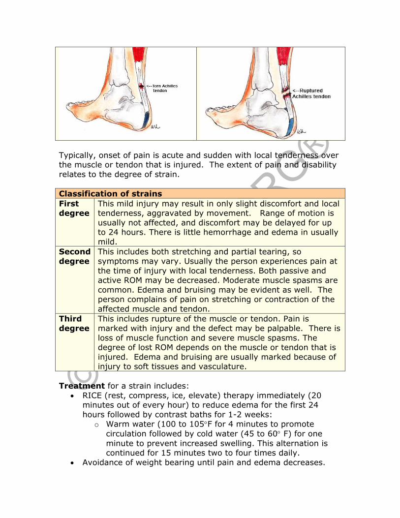
Typically, onset of pain is acute and sudden with local tenderness over the muscle or tendon that is injured. The extent of pain and disability
relates to the degree of strain.
Classification of strains
First degree
This mild injury may result in only slight discomfort and local tenderness, aggravated by movement. Range of motion is
usually not affected, and discomfort may be delayed for up to 24 hours. There is little hemorrhage and edema in usually
mild.
Second degree
This includes both stretching and partial tearing, so symptoms may vary. Usually the person experiences pain at
the time of injury with local tenderness. Both passive and active ROM may be decreased. Moderate muscle spasms are
common. Edema and bruising may be evident as well. The person complains of pain on stretching or contraction of the
affected muscle and tendon.
Third
degree
This includes rupture of the muscle or tendon. Pain is
marked with injury and the defect may be palpable. There is
loss of muscle function and severe muscle spasms. The degree of lost ROM depends on the muscle or tendon that is
injured. Edema and bruising are usually marked because of injury to soft tissues and vasculature.
Treatment for a strain includes:
• RICE (rest, compress, ice, elevate) therapy immediately (20 minutes out of every hour) to reduce edema for the first 24
hours followed by contrast baths for 1-2 weeks: o Warm water (100 to 105F for 4 minutes to promote
circulation followed by cold water (45 to 60 F) for one
minute to prevent increased swelling. This alternation is continued for 15 minutes two to four times daily.
• Avoidance of weight bearing until pain and edema decreases.
• NSAIDs to relieve inflammation and discomfort. • Compression (ACE bandage) applied gently to provide support
and reduce swelling. • Second and third degree injuries may require physical therapy.
• Rupture may require surgical repair.
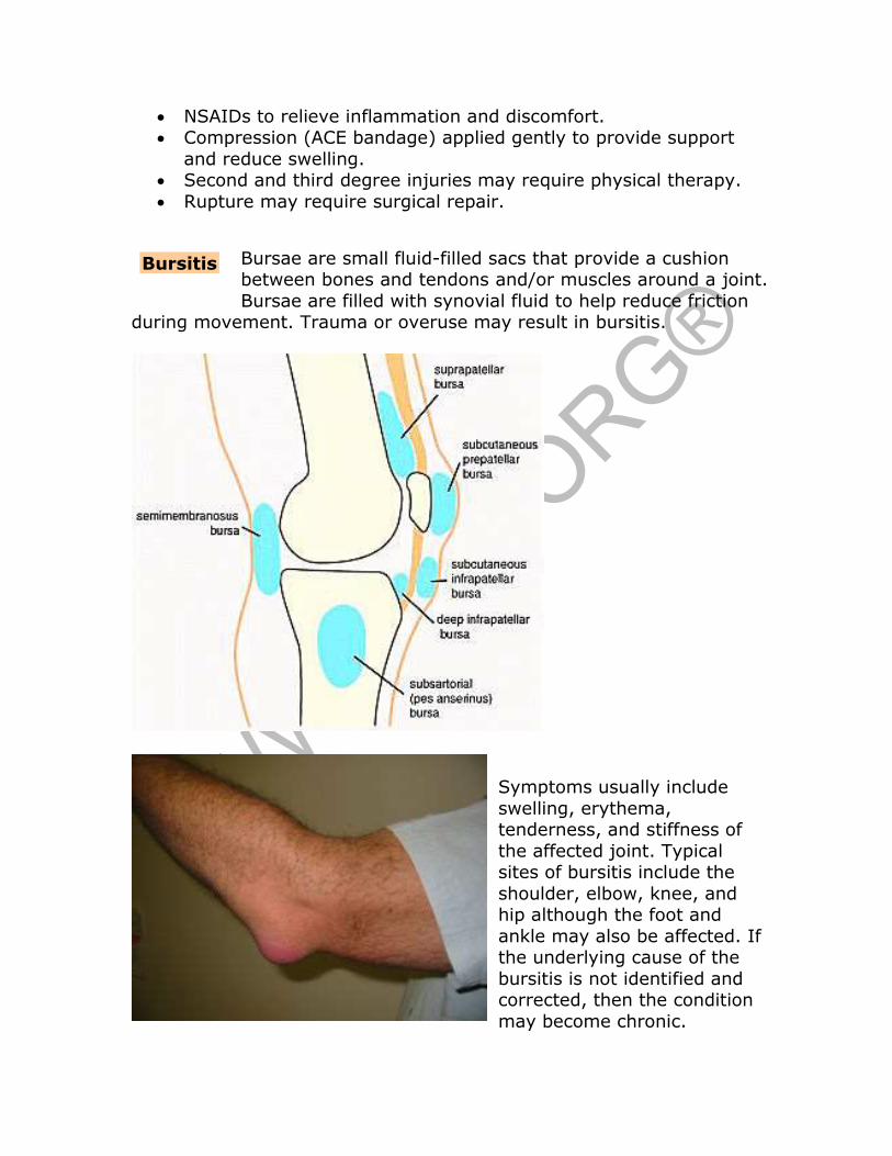
Bursae are small fluid-filled sacs that provide a cushion between bones and tendons and/or muscles around a joint.
Bursae are filled with synovial fluid to help reduce friction during movement. Trauma or overuse may result in bursitis.
Symptoms usually include
swelling, erythema, tenderness, and stiffness of
the affected joint. Typical sites of bursitis include the
shoulder, elbow, knee, and hip although the foot and
ankle may also be affected. If the underlying cause of the
bursitis is not identified and corrected, then the condition
may become chronic.
Bursitis

Treatment includes: • Rest and/or temporary immobilization of affected joint.
• NSAIDS. • If the condition does not resolve or there is a concern about
bacterial bursitis, fluid may be aspirated from the joint. • Corticosteroid injection may be also be used if conservative
treatment is ineffective. Frequent corticosteroid injections may result in injury to tendons although often symptoms resolve
rapidly after a single injection if the condition is not chronic. • Surgical removal of a bursa is rare.
• Antibiotics are indicated only if infection is present. • In some cases, physical therapy may be ordered to strengthen
surrounding muscles to prevent recurrence.
Dislocations When a very strong force is applied to a joint, it may
cause a disruption of the soft tissue and ligamentous structures surrounding the joint, causing the articular surfaces of the joint to
separate in a complete displacement. This displacement is called a dislocation. A subluxation is a partial displacement.
The most commonly affected joints include the thumb, elbow, shoulder,
and hip. The patella may also dislocate.

Dislocations are often obvious because of
asymmetry. For example, a dislocated hip results in
a shortening on the affected side while a
dislocated shoulder results in apparent
lengthening as the elbow is lower on the affected
side.
Symptoms include severe pain and tenderness as
well as loss of function
and edema and bruising of soft tissue. Complications can include intraarticular fractures and damage to neurovascular tissue. Injury to
vasculature can result in avascular necrosis, so evaluation of neurovascular status and prompt treatment is critical. The hip joint is
particularly susceptible to avascular necrosis.
Common dislocations and specific concerns
Shoulder This is the most common type and most often occur as during athletic activities. The primary concern is that
these locations often become chronic and may require surgical repair.
Elbow This dislocation also often results from athletic activities and may cause severe damage to nerves and vessels.
Hip Hip dislocations are commonly associated with vehicle
accidents in which the person’s knees hit the dashboard. This injury may result in avascular necrosis and damage
to the sciatic nerve.
Knee This may causes severe damage to the popliteal artery
and result in amputation, so arteriograms and emergent repair are required.
Treatment includes closed reduction—usually under local anesthesia, general anesthesia, or conscious sedation—and, in some cases, open
surgical reduction. Before reduction, the joint is generally x-rayed to determine how much shifting of internal structures has occurred. The
joint may be aspirated to determine if blood or fat cells are present. The presence of fat cells is associated with intraarticular fractures.
Following reduction, the joint is generally immobilized by taping or application of a sling to allow time for the ligaments and tissue to heal.

Physical therapy may be indicated, often beginning with gentle ROM to avoid stretching the joint. Once a joint has dislocated, the patient is at
risk for repeated dislocation and may be advised to restrict activities that can result in dislocation.
Fractures A fracture is a break in the continuity of a bone. Fractures can range
from simple linear fractures to severe crushing injuries or compound fractures. Most fractures result from traumatic injuries. Symptoms
vary widely depending on the area of the fracture, but general indications of a fracture includes:
• Pain • Impaired function.
• Deformity.
• Abnormal mobility. • Crepitus.
• Edema. • Ecchymosis.
Fractures are classified in a number of different ways.
Type
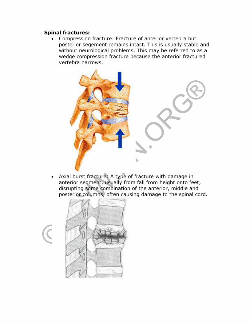
Spinal fractures: • Compression fracture: Fracture of anterior vertebra but
posterior segement remains intact. This is usually stable and without neurological problems. This may be referred to as a
wedge compression fracture because the anterior fractured vertebra narrows.
• Axial burst fracture: A type of fracture with damage in
anterior segment, usually from fall from height onto feet,
disrupting some combination of the anterior, middle and posterior columns, often causing damage to the spinal cord.

• Transverse process fracture: (Rare) Fracture results from extreme lateral bending or rotation and usually does not
affect stability. • Fracture dislocation: This rotation injury results in unstable
fracture/dislocation that can cause spinal cord injury.
Skull fractures:
• Linear (most common): This fracture forms a line.
• Depressed (usually comminuted): Fragments of bone are
displaced, pressing inward.

• Diastatic: Most common in children <3, this causes a widening of
the sutures. • Basilar: This affects the bones at the base of the skull.
Specific bone and anatomical location: Cervical 5 (C5),
distal fibula, etc.
• Open (also called compound): Part of the bone protrudes through the skin to the external environment.
• Grading of open fractures
• Grade I • Clean wound, <1 cm long.
• Grade II • Larger wound with
extensive soft tissue damage.
• Grade III • Large contaminated wound with extensive soft tissue
damage.
An open fracture may result from fragments of the fracture
penetrating the skin or an outside force, such as a knife, penetrating through the skin and into the bone. Open fractures
carry a high risk of infection.
• Closed: Skin is intact.
Location
Relationship to external environment

• Stable: Piece of periosteum is intact and fixation has rendered
fragments stationary (usually transverse, spiral, or greenstick).
• Unstable: Displaced with poor site for fixation.
• Complete: The bones separate into two or more parts. The proximal fragment is referred to as
the uncontrollable fragment because it cannot be
moved or manipulated during reduction. The distal fragment is referred to as the controllable fragment because it can generally
be moved or manipulated to bring about alignment. •
• Incomplete: Only part of the bone is fractured, but the bone remains in one piece.
Diagnosis Diagnosis of orthopedic injuries is based on history, physical examination, observation, and X-ray and sometimes CT or MRI. X-rays
are usually adequate for most fractures, but may not show subtle
Alignment
Stability
Completeness

differences. CT and MRI are better for evaluating soft tissue. CTs are often used to diagnose or evaluate spinal fractures.
In some cases, obvious deformity may be present, as with this Colles
fracture of the wrist:
In other cases, differentiating between a sprain, strain, and fracture
may be less clear, such as with this fibular fracture:
In fact, only about 15% of x-rays of the foot and ankle for injuries prove to be positive for fractures. Because of this, many institutions
and physicians now use the Ottawa rules for ankle, foot, and knee
fractures to determine if x-rays are necessary. Any of the following findings indicates a need for x-rays.

Ottawa rules
Ankle • Bone tenderness at posterior edge or tip of lateral
malleolus. • Bone tenderness at posterior edge or tip of medial
malleolus.
• Inability to bear weight both immediately and in ED.
Foot • Bone tenderness at base of 5th metatarsal.
• Bone tenderness at navicular.
• Inability to bear weight both immediately and in the
ED.
Knee • Age 55.
• Isolated tenderness of the patella.

• Tenderness at the head of the fibula.
• Inability to flex to 90 degrees.
• Inability to bear weight both immediately and in the
ED, evaluated by inability to take 4 steps (transferring weight twice onto each lower limb), regardless of
limp.
Open, or compound, fractures are more easily identified, and fractures should always be suspected with major soft tissue trauma or open
wounds:

Complications There are both immediate and delayed complications related to fractures, but the immediate complications are of most concern during
initial treatment because delayed complications (malunion, delayed union, nonunion, avascular necrosis, and chronic osteomyelitis) are
usually related to orthopedic reconstruction. Immediate complications may be evident on admission to the ED or
may occur within the first few hours after traumatic injury.
Blood loss commonly occurs with fractures and
surgical repair. Most people lose blood during the time of trauma, sometimes in extensive amounts,
and may develop further hemorrhage if a blood vessel is not properly cauterized or sutured or if a drainage tube erodes a vessel. Bones are
very vascular, especially the long bones, pelvis, and vertebrae.
People with clotting abnormalities or those taking antiinflammatory drugs or anticoagulants are at increased risk for hemorrhage.
Fractures that are of special concern are pelvic and femoral fractures. With fractures of the pelvis or long bones, immediately inserting an
intravenous line, administering fluids, and providing oxygen may help to prevent shock.
Estimated blood loss with various types of
fractures (in liters)
Ankle 0.5 to 1.5
Elbow 0.5 to 1.5
Femur 1 to 2
Hip 1.5 to 2.5
Humerus 1 to 2
Knee 1 to 1.5
Pelvis 1.5 to 4.5
Tibia 0.5 to 1.5
Shock can result from the trauma or hypovolemia with loss
of blood or loss of intravascular volume into interstitial spaces, especially into damaged tissues. Fluid and/or blood
replacement is critical with reduced blood volume. Other management includes relieving pain and providing adequate splinting of the fracture.
FES is a primary cause of morbidity and mortality in trauma patients. Fat emboli
are most common with comminuted
Hemorrhage
Shock
Fat embolism syndrome

fractures of the long bones (especially the tibia and femur), ribs, and pelvis, with onset of symptoms usually 12 to 48 hours after trauma or
surgery. There are two theories about the origins of fat emboli: • Fat is released from bone marrow or damaged bone and enters
the circulation, traveling to the pulmonary capillaries where it lodges.
• Catecholamines released at the time of trauma mobilize free fatty acids from fat tissue, resulting in formation of large fat
globules that enter circulation, also traveling to the pulmonary capillaries.
Once the fat emboli are lodged in the lungs, platelets, red blood cells,
and fibrin adhere to the plaques, resulting in lipolysis and chemical pneumonitis, which can lead to respiratory distress syndrome. The
emboli may migrate out of the lungs through pulmonary circulation
and go to the skin where they cause petechiae or into other organs or the brain, causing nervous system changes.
Increasing pulmonary pressure caused by occlusion of large numbers
of small vessels in the lungs may result in right-sided heart failure. Edema and hemorrhage occuring in the alveoli lead to hypoxia.
Indications of FES include abnormal changes in behavior or mentation, chest pain, tachypnea, dyspnea, pallor, widespread petechiae (neck,
anterior chest, axillae, buccal membranes, and conjunctiva), or collapse.
Diagnostic findings associated with FES include:
• Decreasing hematocrit • Arterial blood gases show hypoxia and hypocapnia.
• Chest x-ray shows unevenly distributed areas of congestion.
• Free fat in urine (if fat emboli are in kidneys).

• Increased serum lipase (between 3 to 5 days). • ECG changes indicating myocardial ischemia and right
ventricular strain.
Treatment is primarily supportive, so prevention is critical. This includes splinting and stabilizing fractures (especially of the long
bones) as soon as possible. Treatment options include: • Placing patient in high Fowler’s position.
• Administering oxygen at high flow. • Positive-end-expiratory pressure (PEEP) ventilation to reach
PaO2 of 60 mm Hg. • Hydrocortisone 1 to 1.5 g/day for the first 2 days to reduce
inflammation in the lung and reduce cerebral edema.
An anatomic compartment is an area of
the body enclosed by bone and/or fascia. The human body contains 48
compartments with 36 of these in the extremities. CS occurs when pressure builds up within a confined fascial space (compartment) and
compromises circulation and tissue function.
Fascia is very inelastic, so when pressure increases, it cannot dissipate. Prolonged increased pressure may result in myoneural necrosis,
permanent disability with loss of function and contractures. The 4 compartments in the lower leg are most commonly associated with CS,
but CS can also occur in the arm (upper and lower), hand, thigh, and buttocks.
Compartments in the lower leg:
Compartment syndrome

There are 3 types of CS:
• Acute: Abrupt and severe decrease in circulation distal to an injury that causes ischemic necrosis without immediate
intervention. • Chronic: Increase in muscle volume causing stretching and
inflammation of the fascia, usually resulting from inordinate stress or excess exercise.
• Crush: Results from severe external compression, such as if a very heavy object (such as a vehicle) falls on person with major
crush injuries to tissue and bone.
CS associated with traumatic injuries is most often acute, occcuring when the size of the muscle compartment is too tight or a cast or
splint to constrictive or the contents of the muscle compartment
enlarge because of edema or bleeding.
5Ps associated with CS
Pain deep, throbbing, increasing with passive
stretching.
Paralysis Weakness results from nerve ischemia while paralysis indicates damage to the nerve.
Paresthesias Burning, tingling, numbness, hypoesthesia.
Pallor Cyanosis indicates venous congestion but pallor and dusky and cold digits and prolonged capillary
refill time indicate reduced arterial perfusion.
Pulselessness Severe edema may impact ability to determine
arterial pulsations, so pulse should be verified by Doppler ultrasonography. Complete
pulselessness is a late sign indicating lack of
tissue perfusion.
With CS, the muscle compartment becomes very hard although the clinical presentation may vary and in some cases can be overlooked. If
CS is suspected, then intracompartmental pressure should be measured with an intracompartmental pressure monitor.
• Normal pressure: 8 mm Hg.
• Prolonged pressure >30 mm Hg may cause damage to
microcirculation. • Indication for fasciotomy: 0-45 mm Hg (with diastolic BP of 70
mm Hg)

Treatment includes:
• Elevation of affected extremity above level of the heart. • Release of restrictive devices.
• Fasciotomy (if conservative methods are ineffective within an
hour) or circulatory impairment is severe).
The wound is left open for several days to ensure decompression to although this increases the chance of a wound infection. In severe
cases or with delayed treatment, amputation of a limb may be required.
Neurological impairment may occur with fractures, especially those involving the long bones. Most
injuries are related to stress instead of laceration, so rapid reduction of a fracture can reduce the chance
of nerve damage. Common fractures associated with neurological impairment include:
• Humerus (especially displaced fractures involving the distal third of the bone): Damage to the radial nerve.
• Fracture dislocations of the hip: Damage to the sciatic nerve,
primarily the perineal branch, caused by fragments of bone. • Fractures of the tibia and fibula (especially the proximal fibula):
Damage to the sciatic nerve, perineal branch.
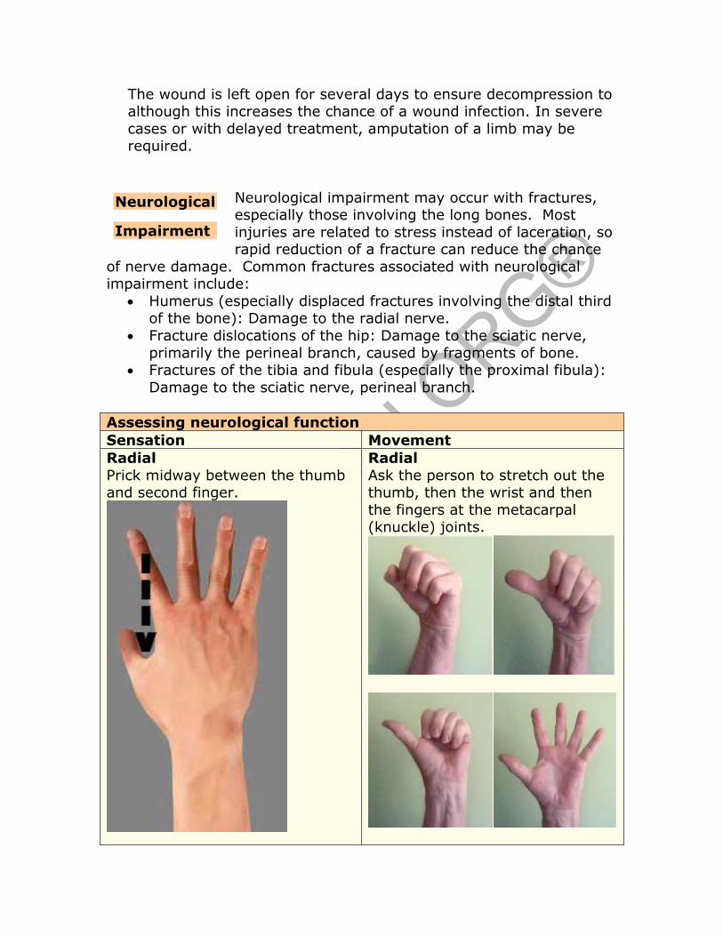
Assessing neurological function
Sensation Movement
Radial
Prick midway between the thumb and second finger.
Radial
Ask the person to stretch out the thumb, then the wrist and then
the fingers at the metacarpal (knuckle) joints.
Neurological
Impairment

Ulnar Prick the distal fad pad of the
small finger.
Ulnar Ask the person to abduct all
fingers.
Tibial
Prick the sole of the foot, medial and lateral surfaces.
Tibial
Ask the person to plantar flex ankle and toes.

Peroneal Prick midway between the great
and second toe.
Peroneal Ask the person to dorsiflex the
ankle and extend the toes.
An infection can occur with any type of fracture, but
open fractures are especially vulnerable, especially traumatic injuries that are grossly contaminated.
Damaged and contaminated tissue provides a good medium for pathogens, including anaerobic bacilli.
Open wounds should be cleansed with jet pulsed lavage and
devitalized and contaminated tissue (including bone fragments) debrided. The wound may be left open to heal by secondary intention.
Closed suction drainage may be required and, in some cases, later skin
grafts to close a wound. Cultures should be taken of the wound at the time of surgery.
Usually open wounds are irrigated with antibiotic solution during
surgery and antibiotic-impregnated beads may be placed within the surgical site. Post-operative antibiotics are generally given IV for up to
a week.
If wounds are near joints, a saline arthrogram is indicated. Saline is injected into the joint space (distant from the wound) and the wound
is observed carefully to determine whether the saline instilled into the joint leaks out of the wound, indicating joint involvement.
Infection

Common infections
Clostridium
tetani
(Tetanus):
Most common in crushing or penetrating wounds
with symptoms occuring 4-21 days after exposure.
Preventive treatment includes wound irrigation and debridement, tetanus toxoid booster for those
previously immunized, or separate tetanus toxoid and tetanus immune globulin (TIG) for those not
previously immunized. Treatment includes penicillin G and TIG.
Gas gangrene Most often caused by Clostridium perfringens, an anaerobic, spore-forming organisms (usually found
in the intestinal tract) produce exotoxins that destroy tissue and are usually found in deep wounds
where oxygen supply is reduced. These bacteria are
most commonly found in thigh wounds or amputations, especially if the patient is incontinent
of stool. Preventive treatment includes thorough debridement of all devitalized and infected tissue
and IV penicillin G. Treatment includes hyperbaric oxygen.
Osteomyelitis
Inflammation of the bone, bone marrow and surrounding tissues by pyogenic bacteria, most often
caused by Staphylococcus aureus followed by
hemolytic Streptococcus. Other causative organisms include Escherichia coli, Neisseria gonorrhea,
Hemophilus influenza, Proteus, and Pseudomonas.

Infection may result from direct invasion of organisms through an open wound, from distant
infections through the bloodstream, or from adjacent soft tissue infections. Infection by direct penetration
is most common, and infection may result from surgical repair, especially without preventive
antibiotics.
Indications of osteomyelitis include acute onset of severe pain at the site of infection, tenderness over
bone, heat, erythema, edema, pain on movement,
and restricted movement. Muscle spasms may occur, causing the patient to hold the affected extremity in
flexion.
Accompanying symptoms may include fever, chills, headache, nausea, diaphoresis, elevated ESR and
positive blood culture. Evidence of infection is usually evident on x-ray about 2 weeks after onset
of infection.
If an abscess forms, incision and drainage may be required and internal fixation devices or prostheses
may need to be removed and antibiotics instilled into the bone cavity.
Complications of osteomyelitis can include septicemia, acute bacterial arthritis, pathological
fractures, and chronic osteomyelitis.
Osteomyelitis is extremely difficult to treat so prevention, including thorough cleansing of wound
and prophylactic antibiotics, is critical.
PE is the most common cause of death in
the immediate postoperative period for those with reconstructive procedures on the
lower extremities and is a significant risk after fractures of the pelvis, hip, and femur. PE may occur within 2 to 3 days or be delayed for
several weeks.
Most PE in orthopedic patients is associated with thrombus or thrombi formation somewhere in the venous system. The thrombi dislodge,
travel through the blood to the lungs, and become lodged in one or
Pulmonary embolism

more of the pulmonary arteries. About 95% of thrombi originate from the deep veins proximal to or at the popliteal vein although they may
also originate in the pelvic veins or right atrium of the heart.
While classic signs include sudden onset of dyspnea and chest pain
(usually pleuritic), symptoms may vary according to the extent of obstruction, the number of thrombi, and the site of obstruction:
• Bifurcation of pulmonary artery: Dyspnea, acute substernal pain, tachycardia, thready pulse, shock, syncope, and sudden death.
• Branch or branches of the right or left pulmonary artery: Dyspnea, mild substernal pain, anxiety, weakness, and
tachycardia. Fever, cough, and hemoptysis may also be present. • Terminal pulmonary arteries: Pleuritic-type pain, cough, and
hemoptysis. • Multiple sites: Symptoms similar to bronchopneumonia.
Diagnostic studies include arterial blood gases, chest x-ray, ECT,
pulmonary angiography, ventilation-perfusion scan, and tests for deep vein thrombosis.
Preventive treatment includes adequate hydration, active and passive exercises, early ambulation, elastic stockings, and external pneumatic
compression leggings.
Treatment includes anticoagulation therapy and thrombolytic therapy. In some cases, such as for those who cannot tolerate anticoagulation,

pulmonary embolectomy or insertion of inferior vena caval filter may be indicated.
AVN may result from infection or disruption of
blood, such as may occur with dislocation or fractures, especially those involving the head
and neck of the femur. Incidence of AVN after hip dislocation ranges from 6 to 40%. Delaying reduction more than 12 hours triples the risk
of developing AVN, so early reduction is especially important.
When AVN is present, x-rays usually show a loss of mineralized matrix and structural collapse of the bone.
If AVN occurs, healing can still take place as the necrotic tissue will be
reabsorbed and new bone will form; however, the patient usually develops painful arthritis as a result. Thus, when fractures involve the
head or neck of the femur, a prosthesis is usually inserted. Other treatment may include bone grafts or arthrodesis (fusion).
Conclusion Prompt treatment is especially important in orthopedic trauma in order to reduce stress on surrounding tissues and reduce the chance of
complications. Fractures are of special concern as they take weeks or months to heal. The duration of time required for healing various with
the type of fracture. For example, flat bones (pelvis, scapulae) tend to heal fairly rapidly. Fractures at the vascular ends of long bones heal
more rapidly than the more dense and less vascular midshaft areas.
Avascular necrosis

Factors that enhance healing of fractures include ensuring immobilization of fracture fragments and maximum contact between
bone fragments as well as adequate bone supply and proper nutrition and exercise.
Factors that can inhibit fracture healing include extensive local trauma,
loss of bone, inadequate immobilization, space or tissue left between bone fragments during reduction, infection, and avascular necrosis.
Elderly patients tend to heal more slowly than younger, and concurrent diseases may impact healing as well. Corticosteroids inhibit healing.
References
• Buckley, R. (2010, January 15). General principles of fracture
care. Medscape Reference. Retrieved October 20, 2011, from http://emedicine.medscape.com/article/1270717-overview
• Compartment syndrome. (2010, July 28). PubMed Health. Retrieved October 20, 2011, from
http://www.ncbi.nlm.nih.gov/pubmedhealth/PMH0002204/ • Fractures of the thoracic and lumbar spine. (2010, February).
AAOS. Retrieved October 20, 2011, from http://orthoinfo.aaos.org/topic.cfm?topic=a00368#Types%20of
%20Spinal%20Fractures • Lotfipour, S. (2005, August 10). Muscle strain. Retrieved October
19, 2011, from http://www.emedicinehealth.com/muscle_strain/article_em.htm
• Mayo Clinic Staff. (2010, January 29). Avascular necrosis. Retrieved October 20, 2011, from
http://www.mayoclinic.com/health/avascular-necrosis/DS00650
• McPhee, SM, & Papadakis, MA. (2009). Current Medical Diagnosis & Treatment. San Francisco: McGraw Hill Medical.
• Mitchell, EL, & Medson, R. (2005). Introduction to Emergency Medicine. Philadelphia: Lippincott Williams & Wilkins.
• Ottawa rules for x-ray for knee, ankle, and foot. (2009). GP-training.net. Retrieved October 20, 2011, from http://www.gp-
training.net/rheum/ottawa.htm • Schoen, DC. (2000). Adult Orthopedic Nursing. Philadelphia:
Lippincott. • Smeltzer, SC, Bare, BG, Hinkle, JL, & Cheever, KH. (2008).
Brunner & Suddarth’s Textbook of Medical-Surgical Nursing, 11 ed., Philadelphia: Wolters Kluwer/Lippincott, Williams, & Wilkins.